Mechanical properties, volumetric shrinkage and depth of ...
7
INTRODUCTION The use of direct chair-side application of resin composite for restoring cavities in stress-bearing posterior teeth has increased rapidly in recent years 1,2) . Beside the ability to bond to hard tooth tissues, facilitated by adhesive systems, resin composites have the advantages of being less invasive and more economical when compared with cast gold and ceramic inlay restorations 3) . However, inadequate material properties, including mechanical deficiencies, polymerization shrinkage and susceptibility to degradation in the oral environment, have limited the success of resin composite restorations in high stress bearing areas 4) . Fracture within the body and marginal closure areas of restorations, and polymerization shrinkage have been cited as major problems leading to the failure of posterior resin composite restorations 5-7) . Fractures related to mechanical properties have usually been evaluated by the determination of material parameters such as fracture toughness, flexural strength, and elastic modulus 8) . Fracture toughness values and flexural properties are dependent on the physical properties and chemical composition of the individual components of a restorative material 9) . A material which has high fracture toughness and flexural properties has enhanced abilities to resist crack initiation and propagation 10) . Consequently, the properties of fracture toughness and flexural properties become important criteria for a dental material’s longevity 11) . Depending on the study, the polymerization shrinkage of resin composites averages from 1.5 to 6% 12) . Such polymerization shrinkage induces contraction stress at the interface between the resin composite and the walls of the cavity, leading to gap- formation, and predisposition to secondary caries. Many factors affect the polymerization shrinkage of resin composites, including resin matrix composition, filler content, and the polymerization method 13,14) . Since there have been no significant advances in improving the properties of polymer matrix materials, recent improvements in resin composite properties are due primarily to advances in filler technology 15) . Generally, particles (random orientation), whiskers (single or multi-layer) and fibers (long or short fibers in various orientations) have been used to reinforce resin composite materials 16-19) . Fiber-reinforced resin composites have been shown to possess a suitable flexural strength and elastic modulus to function successfully in oral cavities 20) . Improvements in handling properties and preimpregnation with light-polymerizable resins have extended the use of fiber-reinforced resin composites in direct chair-side placement of posterior restorations. Recently, a short fiber reinforced resin composite (SFRC) with similar mechanical properties to dentin was introduced for use in restorative dentistry 21) . This resin composite is intended to be used as a base material in high stress bearing areas, especially for large restorations in posterior teeth. It consists of a combination of a resin matrix, which contains Bis-GMA, TEGDMA and PMMA forming a matrix called a semi-Interpenetrating Polymer Network (semi-IPN), which provides good adhesive bonding properties, E-glass fibers, which improve the toughness of the polymer matrix, and an inorganic particulate filler 18,21) . Therefore, the characteristics of SFRC are different from those of conventional glass/ ceramic-filled resin composite (CGRC) due to the fibers. However, there is minimal research available on the mechanical properties of this newly developed SFRC. A problem associated with using light cured resin composite directly in the posterior region is the decrease in light intensity as the depth of material increases in the proximal areas 22,23) . Thus, several manufacturers Mechanical properties, volumetric shrinkage and depth of cure of short fiber- reinforced resin composite Akimasa TSUJIMOTO 1,2 , Wayne W. BARKMEIER 2 , Toshiki TAKAMIZAWA 1 , Mark A. LATTA 2 and Masashi MIYAZAKI 1 1 Department of Operative Dentistry, Nihon University School of Dentistry, 1-8-13 Kandasurugadai, Chiyoda-Ku, Tokyo 101-8310, Japan 2 Department of General Dentistry, Creighton University School of Dentistry, 2500 California Plaza, Omaha, NE 68178, USA Corresponding author, Akimasa TSUJIMOTO; E-mail: [email protected] The mechanical properties, volumetric shrinkage and depth of cure of a short fiber-reinforced resin composite (SFRC) were investigated in this study and compared to both a bulk fill resin composite (BFRC) and conventional glass/ceramic-filled resin composite (CGRC). Fracture toughness, flexural properties, volumetric shrinkage and depth of cure of the SFRC, BFRC and CGRC were measured. SFRC had significantly higher fracture toughness than BFRCs and CGRCs. The flexural properties of SFRC were comparable with BFRCs and CGRCs. SFRC showed significantly lower volumetric shrinkage than the other tested resin composites. The depth of cure of the SFRC was similar to BFRCs and higher than CGRCs. The data from this laboratory investigation suggests that SFRC exhibits improvements in fracture toughness, volumetric shrinkage and depth of cure when compared with CGRC, but depth of cure of SFRC was similar to BFRC. Keywords: Short fiber-reinforced resin composite, Mechanical properties, Volumetric shrinkage, Depth of cure Received Aug 26, 2015: Accepted Dec 25, 2015 doi:10.4012/dmj.2015-280 JOI JST.JSTAGE/dmj/2015-280 Dental Materials Journal 2016; 35(3): 418–424
Transcript of Mechanical properties, volumetric shrinkage and depth of ...

INTRODUCTION
The use of direct chair-side application of resin composite for restoring cavities in stress-bearing posterior teeth has increased rapidly in recent years1,2). Beside the ability to bond to hard tooth tissues, facilitated by adhesive systems, resin composites have the advantages of being less invasive and more economical when compared with cast gold and ceramic inlay restorations3). However, inadequate material properties, including mechanical deficiencies, polymerization shrinkage and susceptibility to degradation in the oral environment, have limited the success of resin composite restorations in high stress bearing areas4).
Fracture within the body and marginal closure areas of restorations, and polymerization shrinkage have been cited as major problems leading to the failure of posterior resin composite restorations5-7). Fractures related to mechanical properties have usually been evaluated by the determination of material parameters such as fracture toughness, flexural strength, and elastic modulus8). Fracture toughness values and flexural properties are dependent on the physical properties and chemical composition of the individual components of a restorative material9). A material which has high fracture toughness and flexural properties has enhanced abilities to resist crack initiation and propagation10). Consequently, the properties of fracture toughness and flexural properties become important criteria for a dental material’s longevity11). Depending on the study, the polymerization shrinkage of resin composites averages from 1.5 to 6%12). Such polymerization shrinkage induces contraction stress at the interface between the resin composite and the walls of the cavity, leading to gap-formation, and predisposition to secondary caries. Many factors affect the polymerization shrinkage of resin
composites, including resin matrix composition, filler content, and the polymerization method13,14).
Since there have been no significant advances in improving the properties of polymer matrix materials, recent improvements in resin composite properties are due primarily to advances in filler technology15). Generally, particles (random orientation), whiskers (single or multi-layer) and fibers (long or short fibers in various orientations) have been used to reinforce resin composite materials16-19). Fiber-reinforced resin composites have been shown to possess a suitable flexural strength and elastic modulus to function successfully in oral cavities20). Improvements in handling properties and preimpregnation with light-polymerizable resins have extended the use of fiber-reinforced resin composites in direct chair-side placement of posterior restorations.
Recently, a short fiber reinforced resin composite (SFRC) with similar mechanical properties to dentin was introduced for use in restorative dentistry21). This resin composite is intended to be used as a base material in high stress bearing areas, especially for large restorations in posterior teeth. It consists of a combination of a resin matrix, which contains Bis-GMA, TEGDMA and PMMA forming a matrix called a semi-Interpenetrating Polymer Network (semi-IPN), which provides good adhesive bonding properties, E-glass fibers, which improve the toughness of the polymer matrix, and an inorganic particulate filler18,21). Therefore, the characteristics of SFRC are different from those of conventional glass/ceramic-filled resin composite (CGRC) due to the fibers. However, there is minimal research available on the mechanical properties of this newly developed SFRC.
A problem associated with using light cured resin composite directly in the posterior region is the decrease in light intensity as the depth of material increases in the proximal areas22,23). Thus, several manufacturers
Mechanical properties, volumetric shrinkage and depth of cure of short fiber-reinforced resin compositeAkimasa TSUJIMOTO1,2, Wayne W. BARKMEIER2, Toshiki TAKAMIZAWA1, Mark A. LATTA2 and Masashi MIYAZAKI1
1 Department of Operative Dentistry, Nihon University School of Dentistry, 1-8-13 Kandasurugadai, Chiyoda-Ku, Tokyo 101-8310, Japan2 Department of General Dentistry, Creighton University School of Dentistry, 2500 California Plaza, Omaha, NE 68178, USACorresponding author, Akimasa TSUJIMOTO; E-mail: [email protected]
The mechanical properties, volumetric shrinkage and depth of cure of a short fiber-reinforced resin composite (SFRC) were investigated in this study and compared to both a bulk fill resin composite (BFRC) and conventional glass/ceramic-filled resin composite (CGRC). Fracture toughness, flexural properties, volumetric shrinkage and depth of cure of the SFRC, BFRC and CGRC were measured. SFRC had significantly higher fracture toughness than BFRCs and CGRCs. The flexural properties of SFRC were comparable with BFRCs and CGRCs. SFRC showed significantly lower volumetric shrinkage than the other tested resin composites. The depth of cure of the SFRC was similar to BFRCs and higher than CGRCs. The data from this laboratory investigation suggests that SFRC exhibits improvements in fracture toughness, volumetric shrinkage and depth of cure when compared with CGRC, but depth of cure of SFRC was similar to BFRC.
Keywords: Short fiber-reinforced resin composite, Mechanical properties, Volumetric shrinkage, Depth of cure
Received Aug 26, 2015: Accepted Dec 25, 2015doi:10.4012/dmj.2015-280 JOI JST.JSTAGE/dmj/2015-280
Dental Materials Journal 2016; 35(3): 418–424

have developed bulk fill resin composite (BFRC), which purportedly can be applied to prepared cavities to a depth of 4 mm, with enhanced curing and mechanical properties, and reduced polymerization shrinkage24). The light intensity at a given depth and for a specified irradiance period is a critical factor in determining the extent of monomer conversion into polymer, and directly relates to values of mechanical properties, biocompatibility, and color stability25). In addition, irradiance would be expected to be associated with the clinical success of a restoration. Therefore, it is important to achieve sufficient irradiance on the bottom surface of each of the incremental layers used in building up a restoration. The concept of the point of sufficiency in this respect is known as depth of cure. Inadequate polymerization throughout the bulk of a restoration can lead to undesirable effects, such as marginal gap formation, marginal leakage, recurrent caries, adverse pulpal effects, and the ultimate failure of the restoration. In the overall context of successful restorative treatment, the relationship between mechanical properties and polymerization properties is important.
The purpose of this laboratory study was to investigate the fracture toughness, flexural strength, elastic modulus, polymerization shrinkage and depth of cure of SFRC, with comparison to BFRC and CGRC.
MATERIALS AND METHODS
Study materialsSix resin composites were used in this study: 1) SFRC: everX Posterior (Shade: Transparent, EP, GC, Tokyo, Japan); and BFRCs: 2) TetricEvoCeram Bulk Fill (Shade: IVA, TB, Ivoclar Vivadent, Schaan, Liechtenstein) and 3) SureFil SDR Flow (Shade: A2, SD, DENTSPLY Caulk, Milford, DE, USA); and CGRCs: 4) Z100 Restorative (Shade: A2, Z1, 3M ESPE, St. Paul, MN, USA), 5) Tetric EvoCeram (Shade: A2, TC, Ivoclar Vivadent) and 6) Clearfil AP-X (Shade: A2, AP, Kuraray Noritake Dental, Tokyo, Japan).
Fracture toughness measurementFracture toughness was determined on single-edge notch specimens using the three-point bending method according to the procedure outlined in ASTM E1820-1326). The knife edge notch samples (25×2.5×5 mm; 0.5 mm notch width and 2.5 mm depth) were prepared in a stainless steel split mold; a sharp blade was used to make the notch. Resin composites were condensed into the mold between two sheets of strips and pressed with a glass plate with a 5 N load. The exit window of the quartz-tungsten halogen unit (Optilux 501, Demetron, Kerr, Danbury, CT, USA) was placed against the glass plate at the center of the specimen, which was exposed to light irradiation for 30 s set at a light irradiance average of 600 mW/cm2. The power density (above 600 mW/cm2) of quartz-tungsten halogen unit was checked using a dental radiometer (model 100, Demetron, Danbury, CT, USA) before preparing the specimens. Following this, the exit window was moved to a section next to the center,
overlapping the previous section. Light irradiation was performed by sequentially curing at three overlapping points on each upper and lower side until the entire sample surface had been irradiated. Ten min after light exposure, the hardened specimens were carefully removed from the mold and stored in 37°C distilled water for 24 h. Immediately after storage, the specimens were subjected to fracture toughness measurements. Fifteen specimens for each material were prepared and tested. A three point bending test was performed with a universal testing machine (Type 5500R, Instron, Norwood, MA, USA) at a crosshead speed of 1.0 mm/min until specimen fracture. The fracture toughness, KІC
(MPa/m), was calculated from the following equation:KІC=(PQ•S)/(B•W3/2)•f (a/W)PQ=peak load (N), S=span (m), B=specimen thickness (m),W=specimen width (m), and a=crack length.
Because it is difficult to measure crack length precisely, the crack length was taken to be the distance from the base of the notch to the opposing surface of the specimens (2.5 mm). Here f (a/W) is a function of a/W and is calculated according to ASTM E-399-90 as follows:
f(a/W)=3(a/W)1/2[1.99−(a/W)•(1−a/W)•(2.15−3.93a/W+2.7a2/W2]/2(1+2a/W)(1−a/W)3/2
The fractured surface of representative specimens after fracture toughness measurements were sputter coated with Au and Pd (Emitech SC7620 Mini Sputter Coater, Quorum Technologies, Ashford, UK). The coated specimens were then examined with a TM3000 Tabletop Microscope (Hitachi-High Technologies, Tokyo, Japan) using an accelerating voltage of 15 kV.
Flexural strength measurement For preparing specimens for flexural strength measurement, a 25×2×2 mm stainless steel mold was used in accordance with ISO 404927). These specimens were made, stored, and tested in the same manner as those for the fracture toughness tests. Fifteen specimens for each material were prepared and the maximum loads applied to the specimens were recorded. Flexural strength (σf) in MPa was calculated as follows:
σf=3W•l/2b•d2
W= maximum load, l=distance between the supports (=20.0 mm),
b=width, and d=depth of the specimen.In addition, elastic modulus in GPa was determined from the stress-strain curve using computer software (Bluehill 2 Ver. 2.5, Instron) linked to the testing machine.
Volumetric shrinkage measurementThe test apparatus consisted of a water-filled dilatometer with a capillary tube of uniform 0.5 mm diameter and a length of approximately 130 mm. It was attached to a 25 cm3, brass-bottom density bottle by means of a ground glass joint. The density bottle was filled with distilled water. During the test, liquid temperature was maintained by placing the density bottle on a thermostatically controlled plate.
Resin pastes were placed into a Teflon mold with
419Dent Mater J 2016; 35(3): 418–424
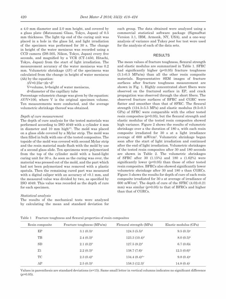
Table 1 Fracture toughness and flexural properties of resin composites
Resin composite Fracture toughness (MPa/m) Flexural strength (MPa) Elastic modulus (GPa)
EP 3.1 (0.3)a 124.3 (5.5)d 9.5 (0.3)g
TB 2.4 (0.3)b 123.3 (10.4)d 8.0 (0.5)h
SD 2.1 (0.2)b 127.5 (8.2)d 6.7 (0.6)i
Z1 2.2 (0.3)b 138.7 (7.6)e 12.5 (0.6)h
TC 2.3 (0.4)b 134.4 (8.4)d,e 9.8 (0.4)g
AP 2.5 (0.3)b 158.3 (12.3)f 14.8 (0.4)j
Values in parenthesis are standard deviations (n=15). Same small letter in vertical columns indicates no significant difference (p>0.05).
a 4.0 mm diameter and 2.0 mm height, and covered by a glass plate (Matsunami Glass, Tokyo, Japan) of 0.5 mm thickness. The light tip end of the curing unit was placed in a hole in the glass lid, and light irradiation of the specimen was performed for 30 s. The change in height of the water meniscus was recorded using a CCD camera (DS-505, Nikon, Tokyo, Japan) every five seconds, and magnified by a VCR (CT-1450, Hitachi, Tokyo, Japan) from the start of light irradiation. The measurement accuracy of the water meniscus was 0.2 mm. Volumetric shrinkage (ΔV) of the specimens was calculated from the change in height of water meniscus (Δh) by the equation:
ΔV=0.25π•Δh•d2
V=volume, h=height of water meniscus, d=diameter of the capillary tube
Percentage volumetric change was given by the equation: ΔV/V×100, where V is the original specimen volume. Ten measurements were conducted, and the average volumetric shrinkage thereof was obtained.
Depth of cure measurementThe depth of cure analysis for the tested materials was performed according to ISO 4049 with a cylinder 4 mm in diameter and 10 mm high27). The mold was placed on a glass slide covered by a Mylar strip. The mold was then filled in bulk with one of the tested composites. The topside of the mold was covered with second Mylar strip and the resin material made flush with the mold by use of a second glass slide. Ten specimens were polymerized from the top of the cylinder mold with a hand-light curing unit for 30 s. As soon as the curing was over, the material was pressed out of the mold, and the part which had not been polymerized was removed with a plastic spatula. Then the remaining cured part was measured with a digital caliper with an accuracy of ±0.1 mm, and the measured value was divided by two, as specified by ISO 4049. This value was recorded as the depth of cure for each specimen.
Statistical analysisThe results of the mechanical tests were analyzed by calculating the mean and standard deviation for
each group. The data obtained were analyzed using a commercial statistical software package (SigmaStat Version 3.1, IBM, Armonk, NY, USA), and a one-way analysis of variance and Tukey post hoc test were used for the analysis of each of the data sets.
RESULTS
The mean values of fracture toughness, flexural strength and elastic modulus are summarized in Table 1. SFRC had significantly higher (p<0.05) fracture toughness (3.1±0.3 MPa/m) than all the other resin composite materials. Representative SEM images of fracture surfaces after fracture toughness measurement are shown in Fig. 1. Highly concentrated short fibers were observed on the fractured surface in EP, and crack propagation was observed through the short fibers. The observed fracture surfaces of BFRC and CGRC were flatter and smoother than that of SFRC. The flexural strength (124.3±5.5 MPa) and elastic modulus (9.5±0.3 GPa) of SFRC were comparable with the other tested resin composites (p>0.05), but the flexural strength and elastic modulus of the tested resin composites showed high variance. Figure 2 shows the results of volumetric shrinkage over a the duration of 180 s, with each resin composite irradiated for 30 s at a light irradiance average of 600 mW/cm2. Volumetric shrinkage began soon after the start of light irradiation and continued after the end of light irradiation. Volumetric shrinkages of the tested resin composites after 30 and 180 seconds are shown in Table 2. The volumetric shrinkages of SFRC after 30 (1.15%) and 180 s (1.62%) were significantly lower (p<0.05) than those of other tested resin composites. BFRCs also showed significantly lower volumetric shrinkage after 30 and 180 s than CGRCs. Figure 3 shows the results for depth of cure of each resin composite irradiated for 30 s at average of irradiance of 600 mW/cm2. The depth of cure of the SFRC (4.02±0.21 mm) was similar (p>0.05) to that of BFRCs and higher than that of CGRCs.
420 Dent Mater J 2016; 35(3): 418–424

Fig. 1 Representative SEM images of fracture surface after fracture toughness measurement of 200× magnification (a) and 1,000× magnification (b).
Highly concentrated short fibers were observed on the fractured surface in EP, and crack propagation was observed through the short fibers. The observed fracture surfaces of BFRC and CGRC were flatter and smoother than that of SFRC.
Fig. 2 Volumetric shrinkage for each resin composite irradiated for 30 s at average irradiance of 600 mW/cm2
Fig. 3 Depth of cure of each resin composite irradiated for 30 s at average irradiance of 600 mW/cm2
Table 2 Volumetric shrinkage of resin composites
Resin composite Shrinkage at the end of light irradiation (%) Shrinkage at 180 s (%)
EP 1.15 (0.04)a 1.62 (0.08)g
TB 1.78 (0.05)b 2.34 (0.12)h
SD 1.33 (0.04)c 2.07 (0.10)i
Z1 2.34 (0.03)d 3.34 (0.16)j
TC 1.90 (0.04)e 3.01 (0.14)k
AP 2.45 (0.03)f 3.54 (0.23)j
Values in parenthesis are standard deviations (n=10).Same small letter in vertical columns indicates no significant difference (p>0.05).
421Dent Mater J 2016; 35(3): 418–424

Table 3 Resin composites used in this study
Resin composite (Shade)
Type of resin composite (Code)
Resin matrix composition
Inorganic filler (Content)Manufacturer
(Lot No.)
everX Posterior (Transparent)
Short fiber-reinforced (EP)
Bis-GMA, PMMA, TEGDMA
Short E-glass fiber, barium glass (74.2 wt%, 53.6 vol%)
GC, Tokyo, Japan (13312271)
Tetric EvoCeram Bulk Fill (IVA)
Bulk fill (TB)
Bis-GMA, Bis-EMA, UDMA, Dimethacrylate
co-monomers
Barium alumino silicate glass filler, prepolymer filler
(80.0 wt%, 60.0 vol%)
Ivoclar Vivadent, Schaan, Liechtenstein
(P63316)
SureFil SDR Flow (A2)
Bulk fill (SD)
TEGDMA, EBADMA,
Modified urethane dimethacrylate
Barium alumino fluoro boron silicate glass, Strontium
alumino fluoro silicate glass (68.0 wt%, 44.0 vol%)
DENTSPLY Caulk, Milford, DE, USA
(100924)
Z100 Restorative (A2)
Conventional glass/ceramic filled (Z1)
Bis-GMATEGDMA
Silane treated ceramics (84.0 wt%, 66.0 vol%)
3M ESPE St Paul, MN, USA (N554909)
Tetric EvoCeram (A2)
Conventional glass/ceramic filled (TC)
Bis-GMA, Bis-EMA, UDMA
Barium alumino silicate glass filler, prepolymer filler
(76.0 wt%, 54.0 vol%)
Ivoclar Vivadent (P48871)
Clearfil AP-X (A2)
Conventional glass/ceramic filled (AP)
Bis-GMATEGDMA
Barium glass filler, Silanated colloidal silica
filler (85.0 wt%, 70.0 vol%)
Kuraray Noritake Dental, Tokyo, Japan
(9B0035)
Bis-GMA: 2,2-bis[4-(2-hydroxyl-3-methacryloyloxypropoxy)phenyl]propane, PMMA: polymethylmethacrylate, TEGDMA: triethyleneglycol dimethacrylate, Bis-EMA: ethoxylated bisphenol-A-dimethacrylate, UDMA: urethane dimethacrylate,EBADMA: ethoxylated bisphenol-A-dimethacrylate, DMDMA: decamethylendimethacrylate, wt%, weight percentage; vol%, volume percentage.
DISCUSSION
SFRC, BFRCs, and CGRCs, which are commonly used for restoring stress-bearing posterior teeth, were evaluated in this laboratory study. A large variation in the loading and constitution of filler particles can be seen in the tested resin composites (Table 3).
In the present study, the SFRC showed significantly higher (p<0.05) fracture toughness than the other tested resin composites. It is reported that the fracture toughness of polymer based materials is improved when they are reinforced with glass fiber28). Thus it is not surprising that short fiber inclusion in a semi-IPN resin matrix led to substantial improvements in this mechanical property. The reinforcing effect of the fibers is based on the behavior of individual fibers as crack stoppers and stronger elements within the matrix18). In addition, the SEM image of the fracture surface of SFRC after fracture toughness measurement indicate the possibility that short E-glass fibers retard crack propagation along the fracture line. If a minor crack propagates through this kind of material, it encounters a fiber and cannot grow further. On the other hand, in BFRC and CGRC with tiny particle fillers, there is nothing to stop the fracture propagating through the whole material and weakening it after the restoration has been placed. In addition, fiber reinforced resin
composites can mitigate damage and dissipate energy, which greatly improves their mechanical performance by preventing brittle failure and avoiding the loss of structural integrity29). In addition to the toughening mechanism of the fibers, the linear polymer chains of PMMA in the cross-linked matrix of Bis-GMA and TEGDMA plasticize the polymer matrix to some extent and increases the fracture toughness of the resin composite21). Therefore, the random fiber orientation and the semi-IPN structure of the polymer matrix likely had a significant role in improving mechanical properties.
The flexural strength and elastic modulus of SFRC were comparable with the other tested resin composites, but the flexural strength and elastic modulus of the tested resin composites show high variance. In theory, the reinforcing effect of the fiber fillers is based on stress transfer from the polymer matrix to fibers30). This is achieved by having fiber lengths equal or greater than the so-called critical fiber length. The critical fiber length of E-glass with Bis-GMA polymer matrix varies between 0.5 and 1.6 mm, as measured using a fiber fragmentation test31). It is reported that the SFRC used in this study had fiber lengths between 1 and 2 mm, thus exceeding the critical fiber length21). Therefore, the fibers of SFRC, being longer than the critical fiber length of E-glass with the Bis-GMA polymer matrix, allow for stress transmission from matrix to fibers, thus producing
422 Dent Mater J 2016; 35(3): 418–424

effective reinforcement. However, including fiber in resin composite results in a decrease in the filler content of particle filler in the resin composite. Previous studies found a positive correlation between filler loading and flexural properties32). Therefore, the fiber reinforcing effect for flexural properties may cancel out decreasing filler content, resulting in the similar flexural properties observed in the present study.
The magnitude of volumetric shrinkage and the accompanying stress generated by the polymerization reaction of the resin composite material are the main factors for in vivo problems like poor marginal adaptation, postoperative pain, and recurrent carries12). The present study showed that volumetric shrinkage began soon after the start of light irradiation and continued after the end of light irradiation. The shrinkage of tested resin composites noted after removal of the light source might be partially attributed to post polymerization reaction of residual monomers. SFRC showed significantly lower volumetric shrinkage than the other tested resin composites. The results are consistent with those of past studies that compared SFRC with CGRCs21,33). Volumetric shrinkage of resin composite depends on factors such as filler load, type of filler and size of fill34). Therefore using short E-glass fibers with a semi-IPN-resin matrix may be one of the reasons why the volumetric shrinkage of SFRC is reduced. Other factors that affect shrinkage are type of resin matrix, monomer concentration, and polymerization initiator systems because they determine the polymer structure of the resin composites35). Unfortunately, the information about the polymerization methods from the manufacturers is so modest that, in this respect, no further comparison can be made.
BFRC also showed significantly lower volumetric shrinkage than CGRC. The tested BFRCs contain a polymerization modulator, which has a high molecular weight, to lower polymerization shrinkage36,37). This unique molecular structure contributes to the delay of the gel point, which represents an increase of viscosity through network formation, and could allow more time to compensate for the shrinkage; consequently, this may be why volumetric shrinkage is reduced.
In some clinical situations the light guide tip cannot be placed in close contact with the restoration surface. Therefore, any increase in the depth of cure should be considered important for daily clinical practice. The depth of cure of the SFRC evaluated in this study was similar to that of BFRCs and higher than that of CGRC. The translucency of SFRC is relatively higher than the other tested resin composites. Therefore, this may be why SFRC shows higher depth of cure than CGRC, and similar to that of BFRCs.
CONCLUSION
SFRC showed improvements in fracture toughness, volumetric shrinkage and depth of cure compared with CGRC, and the depth of cure of SFRC was similar to BFRC. The enhanced mechanical properties of the
SFRC suggest that the SFRC might perform better in high stress-bearing restorative situations.
ACKNOWLEDGMENTS
This work was supported, in part, by a Grant-in-Aid for Young Scientists (B) 10608409 from the Japan Society for the Promotion of Science. This project was also supported, in part, by the Sato Fund and by a grant from the Dental Research Center of the Nihon University School of Dentistry, Japan.
REFERENCES
1) Ferracane JL. Resin composite —state of the art. Dent Mater 2010; 27: 29-38.
2) Jandt KD, Sigusch BW. Future perspectives of resin-based dental materials. Dent Mater 2009; 25: 1001-1006.
3) Burke FJ. Amalgam to tooth-coloured materials-implications for clinical practice and dental education: governmental restrictions and amalgam-usage survey results. J Dent 2004; 32: 343-350.
4) Mitra SB, Wu D, Holmes BN. An application of nanotechnology in advanced dental materials. J Am Dent Assoc 2003; 134: 1382-1390.
5) Sarrett DC. Clinical challenges and the relevance of materials testing for posterior composite restorations. Dent Mater 2005; 21: 9-20.
6) Da Rosa Rodolpho PA, Donassollo TA, Cenci MS, Loguércio AD, Moraes RR, Bronkhorst EM, Opdam NJ, Demarco FF. 22-year clinical evaluation of the performance of two posterior composites with different filler characteristics. Dent Mater 2011; 27: 955-963.
7) Baracco B, Perdigão J, Cabrera E, Ceballos L. Two-year clinical performance of a low-shrinkage composite in posterior restorations. Oper Dent 2013; 38: 591-600.
8) Ferracane JL. Resin-based composite performance: are there some things we can’t predict? Dent Mater 2013; 29: 51-58.
9) Lohbauer U, Belli R, Ferracane. Factors involved in mechanical fatigue degradation of dental resin composites. J Dent Res 2013; 92: 584-591.
10) Drummond JL, Lin L, Al-Turki LA, Herley RK. Fatigue behaviour of dental composite materials. J Dent 2009; 37: 321-330.
11) Thomaidis S, Kakaboura A, Mueller WD, Zinelis S. Mechanical properties of contemporary composite resins and their interrelations. Dent Mater 2013; 29: 132-141.
12) Ferracane JL. Buonocore lecture. Placing dental composites —a stressful experience. Oper Dent 2008; 33: 247-257.
13) Davidson CL, Feilzer AJ. Polymerization shrinkage and polymerization shrinkage stress in polymer-based restoratives. J Dent 1997; 25: 435-440.
14) Watts DC. Reaction kinetics and mechanics in photo-polymerised networks. Dent Mater 2005; 21: 27-35.
15) Ruddell DE, Maloney MM, Thompson JY. Effect of novel filler particles on the mechanical and wear properties of dental composites. Dent Mater 2002; 18: 72-80.
16) Xu HHK, Quinn JB, Smith DT, Giuseppetti AA, Eichmiller FC. Effect of different whiskers on the reinforcement of dental resin composites. Dent Mater 2003; 19: 359-367.
17) Zandinehad AA, Atai M, Pahlevan A. Effect of ceramic and porous filers on the mechanical properties of experimental dental composites. Dent Mater 2006; 22: 382-387.
18) Garoushi S, Vallittu PK, Lassila LVJ. Short glass fiber reinforced restorative composite resin with semi-interpenetrating polymer network matrix. Dent Mater 2007; 23: 1356-1362.
423Dent Mater J 2016; 35(3): 418–424

19) Kondo Y, Takagaki T, Okuda M, Ikeda M, Kadoma Y, Yamauchi J, Okada K, Sadr A, Nikaido T, Tagami J. Effect of PMMA filler particles addition on the physical properties of resin composite. Dent Mater J 2010; 29: 596-601.
20) Xu HH, Schumacher GE, Eichmiller FC, Peterson RC, Antonucci JM, Mueller HJ. Continuous-fiber preform reinforcement of dental resin composite restorations. Dent Mater 2003; 19: 523-530.
21) Garoushi S, Säilynoja E, Vallittu PK, Lassila L. Physical properties and depth of cure of a new short fiber reinforced composite. Dent Mater 2013; 29: 835-841.
22) Shimizu Y, Tsujimoto A, Furuichi T, Suzuki T, Tsubota K, Miyazaki M, Platt JA. Influence of light intensity on surface free energy and dentin bond strength of core build-up resins. Oper Dent 2015; 40: 87-95.
23) Nojiri K, Tsujimoto A, Suzuki T, Shibasaki S, Matsuyoshi S, Takamizawa T, Miyazaki M. Influence of light intensity on surface-free energy and dentin bond strength of single-step self-etch adhesives. Dent Mater J 2015; 34: 611-617.
24) Czasch P, Ilie N. In vitro comparison of mechanical properties and degree of cure of bulk fill composites. Clin Oral Investig 2013; 17: 227-235.
25) Musanje L, Darvell BW. Curing-light attenuation in filled-resin restorative materials. Dent Mater 2006; 22: 804-817.
26) ASTM E1820-13. Standard test method for measurement for fracture toughness. American Society of Testing and Materials, 2013.
27) ISO 4049: 2009. Dentistry —Polymer-based restorative materials. International Organization for Standardization, 2009.
28) Kim SH, Watts DC. Effect of glass-fiber reinforcement and water storage on fracture toughness (KІC) of polymer-based
provisional crown and FPD materials. Int J Prosthodont 2004; 17: 318-322.
29) Choi WC, Yun HD. Effect of expansive admixtures on the shrinkage and mechanical properties of high-performance fiber-reinforced cement composites. Sci World J 2013; 24: 1-11.
30) Petersen RC. Discontinuous fiber-reinforced composites above critical length. J Dent Res 2005; 84: 365-370.
31) Cheng TH, Jones FR, Wang D. Effect of fiber conditioning on the interfacial shear strength of glass-fiber composite. Compos Sci Technol 1993; 48: 89-96.
32) Kim KH, Ong Jl, Okuno O. The effect of filler loading and morphology on the mechanical properties of contemporary composites. J Prosthet Dent 2002; 87: 642-649.
33) Garoushi S, Vallittu PK, Watts DC, Lassila LV. Polymerization shrinkage of experimental short glass fiber-reinforced resin composite with semi-inter penetrating polymer network matrix. Dent Mater 2008; 24: 211-215.
34) Lu H, Lee YK, Oguri M, Powers JM. Properties of a dental resin composite with a spherical inorganic filler. Oper Dent 2006; 31: 734-740.
35) Ellakwa A, Cho N, Lee IB. The effect of resin matrix composition on the polymerization shrinkage and rheological properties of experimental dental composites. Dent Mater 2007; 23: 1229-1235.
36) Ilie N, Bucuta S, Draenert M. Bulk-fill resin-based composites: an in vitro assessment of their mechanical performance. Oper Dent 2013; 38: 618-625.
37) Zorzin J, Maier E, Harre S, Fey T, Belli R, Lohbauer U, Petschelt A, Taschener M. Bulk-fill resin composites: polymerization properties and extended light curing. Dent Mater 2015; 31: 293-301.
424 Dent Mater J 2016; 35(3): 418–424