Mechanical Design of the Third FnIII Domain of Tenascin-C
16
Mechanical Design of the Third FnIII Domain of Tenascin-C Qing Peng, Shulin Zhuang, Meijia Wang, Yi Cao, Yuanai Khor and Hongbin Li⁎ Department of Chemistry, The University of British Columbia, Vancouver, Canada BC V6T 1Z1 Received 24 October 2008; received in revised form 9 January 2009; accepted 14 January 2009 Available online 22 January 2009 By combining single-molecule atomic force microscopy (AFM), proline mutagenesis and steered molecular dynamics (SMD) simulations, we investigated the mechanical unfolding dynamics and mechanical design of the third fibronectin type III domain of tenascin-C (TNfn3) in detail. We found that the mechanical stability of TNfn3 is similar to that of other constituting FnIII domains of tenascin-C, and the unfolding process of TNfn3 is an apparent two-state process. By employing proline mutagenesis to block the formation of backbone hydrogen bonds and introduce structural disruption in β sheet, we revealed that in addition to the important roles played by hydrophobic core packing, backbone hydrogen bonds in β hairpins are also responsible for the overall mechanical stability of TNfn3. Furthermore, proline mutagenesis revealed that the mechanical design of TNfn3 is robust and the mechanical stability of TNfn3 is very resistant to structural disruptions caused by proline substitutions in β sheets. Proline mutant F88P is one exception, as the proline mutation at position 88 reduced the mechanical stability of TNfn3 significantly and led to unfolding forces of b 20 pN. This result suggests that Phe88 is a weak point of the mechanical resistance for TNfn3. We used SMD simulations to understand the molecular details underlying the mechanical unfolding of TNfn3. The comparison between the AFM results and SMD simulations revealed similarities and discrepancies between the two. We compared the mechanical unfolding and design of TNfn3 and its structural homologue, the tenth FnIII domain from fibronectin. These results revealed the complexity underlying the mechan- ical design of FnIII domains and will serve as a starting point for systematically analyzing the mechanical architecture of other FnIII domains in tenascins-C, and will help to gain a better understanding of some of the complex features observed for the stretching of native tenascin-C. © 2009 Elsevier Ltd. All rights reserved. Edited by C. R. Matthews Keywords: mechanical unfolding; single molecule force spectroscopy; steered molecular dynamics simulation; unfolding pathway; tenascin Introduction It is well recognized that mechanotransduction is a mechanism in which mechanical force acts on cells to serve as a physiological signal that triggers a variety of biological processes. 1–3 In living tissues, mechanical force is transmitted to cells through the extracellular matrix (ECM), which serves as a mechanical scaffold for cells to adhere, migrate and differentiate. 4 ECM is linked to the intracellular cytoskeleton through cell membrane receptors integrins and establishes a mechanical continuum that allows the mechanical force be transmitted as a physiological signal between the interior and exter- ior of cells. A wide variety of ECM proteins are subject to mechanical tension in biological environ- ments and many of them share a similar tandem modular architecture. 4 Mechanical tension alters the conformational states of such mechanical proteins, and may modulate biological functions via force- modulated conformational changes. Therefore, understanding the relationship between the struc- *Corresponding author. E-mail address: [email protected]. Abbreviations used: ECM, extracellular matrix; FnIII, fibronectin type III; AFM, atomic force microscopy; TNfn3, the third FnIII domain of tenascin-C; SMD, steered molecular dynamics; WLC, worm-like chain. doi:10.1016/j.jmb.2009.01.019 J. Mol. Biol. (2009) 386, 1327–1342 Available online at www.sciencedirect.com 0022-2836/$ - see front matter © 2009 Elsevier Ltd. All rights reserved.
Transcript of Mechanical Design of the Third FnIII Domain of Tenascin-C

doi:10.1016/j.jmb.2009.01.019 J. Mol. Biol. (2009) 386, 1327–1342
Available online at www.sciencedirect.com
Mechanical Design of the Third FnIII Domainof Tenascin-C
Qing Peng, Shulin Zhuang, Meijia Wang, Yi Cao, Yuanai Khorand Hongbin Li⁎
Department of Chemistry,The University of BritishColumbia, Vancouver,Canada BC V6T 1Z1
Received 24 October 2008;received in revised form9 January 2009;accepted 14 January 2009Available online22 January 2009
*Corresponding author. E-mail [email protected] used: ECM, extrace
fibronectin type III; AFM, atomic foTNfn3, the third FnIII domain of tenmolecular dynamics; WLC, worm-li
0022-2836/$ - see front matter © 2009 E
By combining single-molecule atomic force microscopy (AFM), prolinemutagenesis and steered molecular dynamics (SMD) simulations, weinvestigated the mechanical unfolding dynamics and mechanical design ofthe third fibronectin type III domain of tenascin-C (TNfn3) in detail. Wefound that the mechanical stability of TNfn3 is similar to that of otherconstituting FnIII domains of tenascin-C, and the unfolding process ofTNfn3 is an apparent two-state process. By employing proline mutagenesisto block the formation of backbone hydrogen bonds and introduce structuraldisruption in β sheet, we revealed that in addition to the important rolesplayed by hydrophobic core packing, backbone hydrogen bonds in βhairpins are also responsible for the overall mechanical stability of TNfn3.Furthermore, proline mutagenesis revealed that the mechanical design ofTNfn3 is robust and the mechanical stability of TNfn3 is very resistant tostructural disruptions caused by proline substitutions in β sheets. Prolinemutant F88P is one exception, as the proline mutation at position 88 reducedthe mechanical stability of TNfn3 significantly and led to unfolding forces ofb20 pN. This result suggests that Phe88 is a weak point of the mechanicalresistance for TNfn3.We used SMD simulations to understand themoleculardetails underlying the mechanical unfolding of TNfn3. The comparisonbetween the AFM results and SMD simulations revealed similarities anddiscrepancies between the two. We compared the mechanical unfolding anddesign of TNfn3 and its structural homologue, the tenth FnIII domain fromfibronectin. These results revealed the complexity underlying the mechan-ical design of FnIII domains and will serve as a starting point forsystematically analyzing the mechanical architecture of other FnIII domainsin tenascins-C, and will help to gain a better understanding of some of thecomplex features observed for the stretching of native tenascin-C.
© 2009 Elsevier Ltd. All rights reserved.
Keywords: mechanical unfolding; single molecule force spectroscopy;steered molecular dynamics simulation; unfolding pathway; tenascin
Edited by C. R. MatthewsIntroduction
It is well recognized that mechanotransduction isa mechanism in which mechanical force acts on cellsto serve as a physiological signal that triggers avariety of biological processes.1–3 In living tissues,mechanical force is transmitted to cells through the
ess:
llular matrix; FnIII,rce microscopy;ascin-C; SMD, steeredke chain.
lsevier Ltd. All rights reserve
extracellular matrix (ECM), which serves as amechanical scaffold for cells to adhere, migrateand differentiate.4 ECM is linked to the intracellularcytoskeleton through cell membrane receptorsintegrins and establishes a mechanical continuumthat allows the mechanical force be transmitted as aphysiological signal between the interior and exter-ior of cells. A wide variety of ECM proteins aresubject to mechanical tension in biological environ-ments and many of them share a similar tandemmodular architecture.4 Mechanical tension alters theconformational states of such mechanical proteins,and may modulate biological functions via force-modulated conformational changes. Therefore,understanding the relationship between the struc-
d.
1328 Mechanical Design of TNfn3 Domain
ture andmechanical properties of these proteins is ofimportant biological significance. Tenascin-C, ahighly conserved oligomeric ECM glycoprotein,5–8
is one of the model systems for such studies.Tenascin-C is an extracellular matrix protein with
important roles in regulating the cell-matrix inter-actions.7 Tenascin-C is a tandemmodular protein andconsists of a tenascin-assembly domain, a stretch ofepidermal growth factor-like repeats, a fibronectintype III (FnIII) domain region that is composed of aseries of FnIII domains, and a terminal knob domainthat is homologous to the globular domain of fibri-nogen. Tenascins are expressedmainly in regions thatare subject to heavy tensile load9 or in tissues thatundergo extensive structural re-modeling duringprocesses such as tissue injury and tumorigenesis.10–12As tenascins are subject to mechanical stretchingforces under physiological conditions, it is possiblethat the force-induced unfolding/refolding reactionsof FnIII domains may be an important part oftenascins dynamics in vivo.13
Single-molecule atomic force microscopy (AFM)studies have provided insights into the mechanicaldesign and functions of tenascin-C. It was revealedthat tenascin-C is an elastic protein that can extendto several times its resting length via force-inducedunfolding of FnIII domains.13,14 It has beensuggested that the mechanical unfolding of FnIIIdomains serves as a shock-absorber to prolong thelifetime of the tenascin-ligand bond.13 Recentstudies revealed that some FnIII domains displayweakly populated folded microstates in addition totheir native states, which may entail a possiblemechanism for these FnIII domains to recover theirmechanical resistance more rapidly after mechan-ical unfolding.15 Since these studies were carriedout on native fragments of tenascin-C, the inherentheterogeneity of the constituting FnIII domains innative fragments makes it difficult to assignmechanical features observed on native tenascinfragments to specific FnIII domains, and makesdetailed molecular interpretation of the experimen-tal results difficult.To overcome these difficulties, detailed studies of
the mechanical unfolding dynamics of individualFnIII domains become necessary. Using polypro-teins made of identical tandem repeats of the proteinof interest has become the standard approach tostudy their mechanical properties using single-molecule AFM.16,17 Recently, mechanical ϕ valueanalysis has been done on the third FnIII domain oftenascin-C (TNfn3) via single-molecule AFM andmolecular dynamics simulation using an implicitwater model.18 However, this study was focused onprobing the role of hydrophobic core in themechanical unfolding of TNfn3. The role of back-bone hydrogen bonds and β sheet stability, twoimportant factors for mechanical stability of pro-teins, were not probed. Here, in order to address theimportance of backbone hydrogen bonds as well asβ sheet stability in the mechanical unfolding ofTNfn3, we combine single-molecule AFM, prolinemutagenesis and steeredmolecular dynamics (SMD)
simulations to investigate the mechanical unfoldingdynamics and mechanical design of TNfn3 in detail.Our results revealed that the mechanical stability ofTNfn3 is similar to that of other FnIII domains oftenascin-C,13,15 and the unfolding process of TNfn3is an apparent two-state process. To probe the me-chanical design of TNfn3, we used proline mutagen-esis to block the formation of backbone hydrogenbonds and introduce structural disruption in β sheetin order to affect its mechanical stability and unfold-ing kinetics. Our results revealed that hydrophobiccore packing is not the only factor that is importantfor determining the mechanical stability of TNfn3,and backbone hydrogen bonds in β hairpins are alsoresponsible for the overall mechanical stability ofTNfn3. Furthermore, proline mutagenesis revealedthe robust mechanical design of TNfn3, as themechanical stability of TNfn3 is resistant to dis-ruptive proline mutations in β sheets of TNfn3. Weidentified that residue Phe88 is a weak point inTNfn3, and a single substitution of Phe88 withproline results in the unfolding of TNfn3 at forcesthat are lower than the detection limit in AFM. Wecompared the AFM results with SMD simulations tounderstand the molecular details underlying themechanical unfolding of TNfn3. A comparisonbetween the mechanical features of TNfn3 with thetenth FnIII domain from fibronectin revealed thesignificant differences in mechanical unfolding anddesign of these two structurally homologous FnIIIdomains. These results pave the way for systematicanalysis of the mechanical architecture of other FnIIIdomains in tenascin-C and will help us to obtain abetter understanding of some of the complexfeatures observed for the stretching of nativetenascin-C.
Results
Themechanical unfolding of TNfn3 is an apparenttwo-state process
TNfn3 is an all-β protein of 90 amino acidresidues. It has a typical immunoglobulin-like β-sandwich structure, in which the two β sheets ofTNfn3 pack against each other (Fig. 1).19 The topsheet consists of strands A-B-E and the bottom sheetconsists of C-C′-F-G. The two force-bearing terminalβ strands A and G are parallel with each other, andthe N- and C-termini point in opposite directions.Such an arrangement of the two terminal strandsforms a shear topology upon stretching, which is acommon feature among proteins that are mechani-cally stable.16,17,20–26 It is of note that the two terminalforce-bearing strands of TNfn3 are not bondeddirectly by backbone hydrogen bonds like otherelastomeric proteins, such as I27 of the muscleprotein titin.17
Using protein engineering techniques, we engi-neered a polyprotein (TNfn3)8, which consists ofeight identical tandem repeats of TNfn3. We then

Fig. 1. The three-dimensionalstructure of the third fibronectintype III domain of tenascin-C(TNfn3). (a) TNfn3 has a typical β-sandwich structure. The two force-bearing β strands are parallel witheach other and are pointing in oppo-site directions. The backbone hydro-gen bonds associated with the twoforce-bearingβ strands are indicatedby black bars. (b) The location ofamino acids that are substituted byproline residues in this work.
Fig. 2. Typical force-extension curves of polyprotein(TNfn3)8. Stretching polyprotein (TNfn3)8 results in force-extension curves with the characteristic saw-tooth pattern.The equally spaced force peaks result from the mechanicalunfolding of the individual TNfn3 domains in thepolyprotein chain. The last peak in force-extension curvescorresponds to the detachment of the protein from eitherthe AFM tip or the substrate. WLC fits (thin lines) to theconsecutive unfolding force peaks measure a contourlength increment ΔLc of ∼29.0±0.8 nm.
1329Mechanical Design of TNfn3 Domain
used single-molecule AFM to stretch polyprotein(TNfn3)8 and characterize its mechanical unfoldingbehaviors. Stretching polyprotein (TNfn3)8 resultedin force–extension relationships of the characteristicsawtooth pattern appearance, where the individualsawtooth peaks correspond to the mechanical un-folding of each individual TNfn3 domain in thepolyprotein chain. The force–extension relationshipsof (TNfn3)8 can be well described by the worm-likechain (WLC) model of polymer elasticity,27 and thereis no apparent deviation from the WLC fits (Fig. 2).WLC fits to consecutive unfolding force peaks mea-sure an average contour length increment (ΔLc) of29.0±0.8 nm (average±standard deviation). TNfn3 is90 residues long and the distance between its N-, andC- termini is 3.1 nm in the folded state.19 The contourlength of the unfolded and fully stretched TNfn3 is32.4 nm (90 residues×0.36 nm/residue). Hence, acomplete unraveling of a TNfn3 domain shouldresult in a contour length incrementΔLc of∼29.3 nm,which is in excellent agreement with the experimen-tally determined ΔLc. This result suggests that themechanical unfolding of TNfn3 corresponds to thecomplete unfolding of TNfn3 in an apparent two-state fashion, and there is no visible intermediatestate along its mechanical unfolding pathway.The amplitude of the unfolding force peaks varies
around ∼120 pN. A histogram of the unfoldingforces compiled from ∼4000 unfolding events at apulling speed of 400 nm/s measured an averageunfolding force of 125±14 pN (n=4198, Fig. 3a),which is in agreement with previous measurementson a similar TNfn3 polyprotein.18 The measuredmechanical stability of TNfn3 is similar to theaverage mechanical unfolding force of all the 15FnIII domains measured from a recombinant frag-ment of tenascin-C containing all the 15 FnIII do-mains, consistent with the previous conclusion thatall the FnIII domains of tenascin-C have similarlevels of mechanical stability.13,28
It was shown that extending the C-terminus ofTNfn3 by two residues can increase the thermody-namic stability of TNfn3 significantly.29 However, wefound that such an extension of TNfn3 does not affectthemechanical stability of TNfn3 in anyway (data notshown), indicating that the two additional residues atthe C-terminus of TNfn3 are already detached fromthe folded TNfn3 before TNfn3 reaches the mechan-ical unfolding transition state.
The mechanical unfolding of TNfn3 ischaracterized by a long unfolding distance fromthe native state to the transition state
To further characterize the mechanical unfoldingof TNfn3 in detail, we measured the pulling speed-dependence of the unfolding forces of TNfn3 bystretching (TNfn3)8 at different pulling speeds.Similar to other elastomeric proteins, the mechanicalunfolding of TNfn3 is a non-equilibrium process andits unfolding force depends on the pulling speed: thehigher the pulling speed is, the bigger the unfoldingforce (Fig. 3b). It is of note that, as compared with Igdomains from the muscle protein titin,17,30,31 thepulling speed-dependence of the unfolding force ofTNfn3 is relatively weak: the unfolding force ofTNfn3 increases from 109 pN at a pulling speed of50 nm/s to 154 pN at a pulling speed of 2700 nm/s.To estimate the spontaneous unfolding rate constantat zero force (α0) and the unfolding distance (Δxu)between the folded state and the transition state

Fig. 3. Unfolding force of TNfn3 and its dependence onthe pulling speed. (a) Histogram of unfolding forces forTNfn3. The unfolding force histogram spans a range of∼100 pN (60 pN∼160 pN) with an average value of 125±14 pN (n=4198). The red line corresponds to Monte Carlosimulation of the mechanical unfolding of TNfn3 using α0of 1.5×10⁻⁴ s−1 and Δxu of 0.42 nm. The pulling speed is400 nm/s. (b) The pulling speed dependence of theunfolding forces of TNfn3 (symbols). The pulling speeddependence of the unfolding forces of TNfn3 can be re-produced adequately byMonte Carlo simulations using α0of 1.5×10⁻⁴ s−1 and Δxu of 0.42 nm (red line).
1330 Mechanical Design of TNfn3 Domain
along the reaction coordinate, two important para-meters characterizing the mechanical unfolding freeenergy diagram, we carried out Monte Carlosimulations to reproduce the force-extension rela-tionships of (TNfn3)8. In the Monte Carlo simula-tion, we assumed that the unfolding of TNfn3 is atwo-state process and the force-dependent unfold-ing rate constant follows the classical Bell modela Fð Þ = a0exp FDxu
kBT
� �, where kB is the Boltzmann cons-
tant and T is absolute temperature (in K). We foundthat both the unfolding force histogram (Fig. 3a)and the pulling speed-dependence of the unfoldingforces (Fig. 3b) are well described using an α0 of1.5×10−4 s−1 and a Δxu of 0.42 nm. This resultsuggests that the mechanical resistance to unfoldingis distributed over a distance of 0.42 nm. Thisunfolding distance is notably longer than that forother typical elastomeric proteins, such as I27,17
ubiquitin,22 and GB1 domains,23 suggesting thatthe mechanical resistance of TNfn3 is distributedalong a longer distance, which is in contrast with thehighly localized mechanical resistance for otherelastomeric proteins. The molecular basis for theobserved long unfolding distance Δxu will be add-ressed in Discussion. The measured α0 and Δxu arecomparable to those measured in previous studieson tenascin-C fragment and TNfn3 polyprotein.13,18
The SMD simulations of themechanical unfoldingof TNfn3
The single-molecule AFM results suggest that themechanical unfolding of TNfn3 is an apparent two-state process. In order to understand the molecularevents leading to the mechanical unfolding ofTNfn3, we carried out SMD simulations of themechanical unfolding of TNfn3. In contrast toprevious molecular dynamics simulation workusing an implicit water model,18 we used explicitwater model TIP3P in our simulation to explicitlyaddress the potential role of the solvent watermolecules during the mechanical unfolding processof TNfn3. This strategy has been used extensively tosimulate the mechanical unfolding of proteins,including FnIII domains from fibronectin andTNfn3.32,33In our SMD simulations, we simulated the
mechanical unfolding of TNfn3 using both constantvelocity and constant force protocols. Starting from1 ns and 1.5 ns equilibrated conformations, TNfn3was pulled at a constant force (500 pN) or at aconstant velocity (0.05 Å/ps). In total, we performed16 constant force SMD and 17 constant velocitySMD simulations with a total simulation time of57 ns. Both constant velocity and constant forcetrajectories revealed similar features of the mechan-ical unfolding of TNfn3.In constant force SMD simulations, TNfn3 was
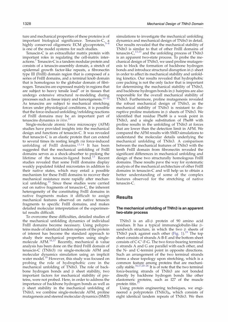
stretched at a constant force of 500 pN from its N-and C-termini, and the distance between the twotermini (RNC) was monitored as a function of time.Representative RNC versus time curves from con-stant force SMD simulations are shown in Fig. 4a.The presence of multiple plateaus in RNC versus timeprofiles is clearly visible, indicating the presence ofstable intermediates populated along the unfoldingtrajectories. From the native state with RNC of∼34 Å, TNfn3 elongates by ∼6 Å via straighteningthe disordered N-terminal end of the protein andenters into a stable intermediate state I1, in whichthe tertiary structure of TNfn3 remains largelyintact. It is of note that the two backbone hydrogenbonds between Ser6 and Phe23 are relatively weakand break in state I1 during most of the trajectories.The rupture of the backbone hydrogen bondsbetween Ser6 and Phe23 leads to the detachmentof the first six residues of strand A from the foldedstructure of TNfn3. In the second stage, TNfn3elongates further by∼10 Å to reach the intermediatestate I2. During this process, the two β sheets, onecontaining strands A–B –E (colored red) and the
Fig. 4. Constant force and con-stant velocity SMD simulations ofthe mechanical unfolding of TNfn3.(a) Representative RNC versus timeprofiles from constant force SMDsimulations at a stretching force of500 pN. The presence of threeplateaus indicated kinetic inter-mediates I1, I2 and I3. Trajectories1–3 correspond to the A-strandseparates first pathway, and trajec-tory 4 corresponds to the A-Gstrands separate simultaneouslypathway. (b) Representative Forceversus RNC curves from constantvelocity SMD simulations. The pull-ing velocity is 0.05 Å/ps. The firstforce peak occurs at an RNC of∼40 Å, corresponding to the transi-tion from I1 to I2; the second peakoccurs at an RNC of ∼50 Å andcorresponds to the unraveling ofintermediate I2.
1331Mechanical Design of TNfn3 Domain
other containing strands C′–C–F–G (colored blue),rotate relative to each other and both align with thepulling force (Fig. 5), leading to the so-called alignedβ sandwich intermediate I2. This alignment resultedin partial solvation of the periphery of the hydro-phobic core. In intermediate I2, the remainingbackbone hydrogen bonds in strands A-B betweenGlu9–Trp21, Lys11–Leu19, Asp12–Leu19, andThr14–Thr17 remained intact, so do the backbonehydrogen bonds in hairpin F-G.Immediately following the transition from I1 to I2,
TNfn3 elongated further following two distinctunfolding pathways (Fig. 5). The first pathway ischaracterized by the presence of the partially un-folded intermediate state I3 with RNC of ∼130 Å.Along this pathway (the “A-strand separates first”pathway), strand A of TNfn3 separates first from thefolded structure, followed by the subsequent unra-veling of strands B and E. However, the overallstructure of the β sheet containing strands C′-C-F-Gremains largely intact. This unfolding pathwaywas observed in ∼70% of the constant force SMDsimulation trajectories (11 out of 16) and threerepresentative trajectories of this type are shown inFig. 4a (trajectories 1–3). The second pathway ischaracterized by simultaneous detachment ofstrands A and G from the folded structure and istermed “A-G strands separate simultaneously”.After the simultaneous detachment of strands A
and G, TNfn3 unfolds readily without significantbarrier and hence intermediate state I3 is notdetected in this pathway. This type of pathwaywas observed in ∼30% of the constant force SMDsimulations (5 out of 16) and trajectory 4 in Fig. 4a isone example.The dwell time of a given state along the unfolding
trajectory is a measure of the stability of the givenstate. Constant force simulations revealed that thedwell time of intermediate I1 is, on average, longerthan that of I2 and I3, as well as that of the nativestate, suggesting that intermediate state I1 is themost stable one at a force of 500 pN. This resultsuggests that under a stretching force of 500 pN, thenative state of TNfn3 is transformed rapidly intointermediate state I1, and the mechanical unfoldingof TNfn3 is not directly from its native state. Thus,the intermediate I1 is the pseudo-native state for themechanical unfolding of TNfn3. It is important topoint out that the dwell time of individual inter-mediate states does vary from trajectory to trajectory.To reveal a more detailed picture of the transition
from mechanical unfolding of TNfn3, we monitoredthe breakage of backbone hydrogen bonds in hair-pins A-B and F-G by calculating hydrogen bondenergies along the SMD trajectories. Five backbonehydrogen bonds between strands A and B,Glu9(H)–Thr21(O), Glu9(O)–Thr21(H), Lys11(H)–Leu19(O),Asp12(O)–Leu19(H), Thr14(H)–Thr17(O) and

Fig. 5. Snapshots of TNfn3 during its simulated mechanical unfolding. For all the snapshots, the N terminus (red ball) wasfixed and the C terminus (blue ball) was pulled during these simulations. From the native state, TNfn3 elongates bystraightening theN-terminus and enters into the so-called twist intermediate state I1; then the twoβ sheets rotate relative to eachother and alignwith the stretching force vector, leading to the so-called aligned intermediate state I2. After I2, TNfn3 unfolds viatwo distinct pathways: the first is by separating A strand first from the folded structure, followed by subsequent unfolding of Band E strands, leading to a partially unfolded intermediate state I3; the second pathway is via simultaneous unfolding anddetachment of A and G strands from the folded structure. After this event, TNfn3 unfolds readily and the existence ofintermediate state I3 is not detected. For the snapshot of I3, the N terminus is artificially shortened to fit into the figure.
1332 Mechanical Design of TNfn3 Domain
another five backbone hydrogen bonds betweenstrands F and G, Phe88(H)–Tyr68(O), Glu86(O)–Val70(H), Glu86(H)–Val70(O), Ala84(O)–Leu72(H),Ala 84(H)–Thr72(O) were selected for the hydrogenbond energy calculation. Figure 6 shows the hydro-gen bond energy as a function of time for these tenbackbone hydrogen bonds for the trajectories 2 and4 shown in Fig. 4a. At the beginning of the unfoldingtrajectories, these backbone hydrogen bonds arestable and the hydrogen bond energies fluctuatebetween approximately –5 kcal/mol and –3 kcal/mol with an average value of approximately–4.3 kcal/mol. Upon further stretching, β strandswill begin to separate and the strength of itshydrogen bonds become weaker; accordingly, thehydrogen bond energies gradually increase to zero,at which point the hydrogen bonds are alreadybroken. In the “A-strand separates first” pathway(Fig. 6a and b), after ∼1.8 ns, the hydrogen bondenergy of Glu9(H)–Thr21(O), Glu9(O)–Thr21(H),Lys11(H)–Leu19(O), Asp12(O)–Leu19(H), Thr14(H)–Thr17(O)increase to zero fairly rapidly accompany-ing the transition from intermediate state I2 to I3,indicating that these five backbone hydrogen bondsin strands A and B break concurrently during thisprocess. In contrast, the hydrogen bonds in strands Fand G (Phe88(H) – Tyr68(O), Glu86(O) –Val70(H),Glu86(H) –Val70(O), Ala84(O) – Leu72(H), Ala 84(H)– Thr72(O)) remain steady during this process. After2.6 ns, three hydrogen bonds, Phe88(H) – Tyr68(O),
Glu86(O) –Val70(H) and Glu86(H) –Val70(O), start tobreak.In the “A-G strands separate simultaneously”
pathway, the hydrogen bond energy for Glu9(H) –Thr21(O), Glu9(O) –Thr21(H), Lys11(H)–Leu19(O),Asp12(O) – Leu19(H), Thr14(H) – Thr17(O), Phe88(H) – Tyr68(O), Glu86(O) –Val70(H), Glu86(H) –Val70(O), Ala84(O) – Leu72(H), and Ala 84(H) –Thr72(O) increases simultaneously after 2.7 ns,indicating that these ten backbone hydrogen bondsbreak concurrently during the “A-G strands sepa-rate simultaneously” pathway (Fig. 6c and d).
Constant velocity SMD simulations
We also carried out constant velocity SMD simula-tions of the mechanical unfolding of TNfn3 (17trajectories in total). Figure 4b shows representativeforce versus RNC curves. It is evident that there aremultiple force peaks along the unfolding pathway:the first force peak occurs at an RNC of ∼40 Å, whichcorresponds to the transition from I1 to I2; the secondpeak occurs at an RNC of ∼50 Å and corresponds tothe unraveling of intermediate state I2. In all thetrajectories, the amplitudes of the first and secondpeak show slight difference: in 13 trajectories, the firstunfolding force peak is higher than the second one(∼1300 pN versus ∼1050 pN), while in four tra-jectories, the second unfolding force peak in higherthan the first one (∼1200 pN versus 1100 pN). This

Fig. 6. Profiles of hydrogen bondenergy of inter-strand hydrogenbond in A-B and F-G strands versustime in two representative SMDunfolding trajectories of TNfn3. (aand b) The energy change of thehydrogen bonds as a function of timein the trajectory following the A-strand separates first pathway. Theenergywas calculated from trajectory2 in Fig. 4a. (c and d) The energychange of the hydrogen bonds as afunction of time in the trajectoryfollowing the A-G strands separatesimultaneously pathway. The energywas calculated from trajectory 4 inFig. 4a. The sudden increase inhydrogen bond energy indicates thebreaking of hydrogen bonds. In bothunfolding pathways, the hydrogenbonds in A-B and F-G break duringthe unfolding of the intermediatestate I2.
1333Mechanical Design of TNfn3 Domain
observation is consistent with the observed variationin dwell time of the intermediates.
Using site-directed mutagenesis to probe thenature of unfolding transition state observed insingle-molecule AFM
SMD simulations on TNfn3 revealed that themechanical unfolding of TNfn3 proceeds via severalintermediate states. However, single-molecule AFM
experiments indicate that the mechanical unfoldingof TNfn3 is an apparent two-state process. Thediscrepancy between SMD simulations and AFMexperiments suggests that some of the intermediatestates observed in SMD simulations do not populateon the time-scale of single-molecule AFM experi-ments. Considering the large RNC of the inter-mediate I3, we can easily rule out the possibilitythat the intermediate I3 is a stable intermediatestate. Previous SMD simulations and mechanical

1334 Mechanical Design of TNfn3 Domain
ϕ-value analysis suggested that the transition fromthe twist intermediate I1 to the aligned intermedi-ate I2 is most likely to be the rate-limiting step forthe mechanical unfolding observed in AFM, andthat the energy barrier for the transition fromintermediate I2 to I3 is too low to be observedexperimentally.18,33 Our SMD simulation resultsreveal that in majority of the unfolding trajec-tories, the plateau of intermediate state I1 is thelongest in constant force SMD and the unfoldingforce peak for the transition from I1 to I2 is thehighest in constant velocity SMD, supporting theview that the transition from I1 to I2 is the rate-limiting step, which corresponds to the mechanicalunfolding force peak observed in single-moleculeAFM experiments.If this view is correct, it means that the rupture
events of the backbone hydrogen bonds (Fig. 5)occur after the rate-limiting step. Thus, it is temptingto conclude that the hydrogen bonds are not criticalfor the mechanical stability of TNfn3. For example, aprevious single-molecule AFM study on TNfn3singled out the importance of hydrophobic interac-tions to the mechanical unfolding of TNfn3.18,34
However, the backbone hydrogen bonds are impor-tant in protecting the hydrophobic core from theattack by water molecules, especially the backbone
Fig. 7. Mechanical unfolding of proline mutants of TNfnTNfn3⁎)4. TNfn3⁎ denotes the proline mutant of TNfn3. (bchimera (GB1-TNfn3⁎)4 for each proline mutant. b, The forcestrand were substituted by proline. (c) The force-extension curvThe mechanical unfolding events of the well characterized GB∼18 nm and served as fingerprints to identify single-moleculeshow clear mechanical unfolding events with ΔLc of ∼29 nm (90∼120 pN, which is lower than that of wt TNfn3. Dotted linesproline mutants, the unfolding of F88P does not result in clespacer is typically observed before the mechanical unfoldingdomains unfold at forces that are below our detection limit (∼unfolding events, as the one shown in curve b.
hydrogen bonds in hairpins A-B and F-G. It can beimagined that destabilization of strands A-B or F-Gby deleting backbone hydrogen bonds and intro-duction of a bulge would facilitate the attack of thehydrophobic core by water molecules and lead toreduced mechanical stability. To further exploresuch scenarios, we used proline mutagenesis toselectively disrupt strands A-B and F-G to directlyprobe the role of backbone hydrogen bonds and βsheet stability on the mechanical stability of TNfn3.
The design of proline mutants of TNfn3
It is well known that proline substitution in a βsheet region can block the formation of backbonehydrogen bonds, cause a bulge in the β strand anddisrupt hydrophobic packing.35 These combineddisruptive effects by proline substitution result inthe discontinuity of the β strand and lead to theselective disruption of local β sheet structure. Such arelatively large structural perturbation is ideal forprobing the mechanical unfolding pathway ofproteins, as such a large perturbation of the proteinstructure generally cannot be compensated easily bythe structural rearrangement of the protein. There-fore, the structural perturbation caused by prolinesubstitution can be located easily and its effect on
3. (a) An illustration of the polyprotein chimera (GB1-and c) Typical force-extension curves of the polyprotein-extension curves of mutants in which residues in the Aes of mutants involving proline mutations in the G-strand.1 domains (colored red) occurred at ∼180 pN with ΔLcofstretching events. Except for F88P, all the proline mutantscolored green), and their unfolding forces are in the rangeare WLC fits to the experimental data. In contrast to otherar mechanical unfolding events. Instead, long featurelessevents of GB1 domains (curve a), suggesting that F88P20 pN). Only a small fraction of F88P domains show clear

1335Mechanical Design of TNfn3 Domain
mechanical unfolding pathway can be identifiedreadily.36,37
Since breaking the hydrogen bond between S6and F23 occurs in intermediate state I1 and is thefirst event during the mechanical unfolding processof TNfn3, we engineered the S6P mutant tospecifically probe the mechanical unfolding inter-mediate state I1. To probe the effect of backbonehydrogen bonds in hairpin A-B during the mechan-ical unfolding, we engineered mutants E9P, K11Pand T14P. Similarly, we introduced proline muta-tions in strand G. To investigate the importance ofthe C-terminus on the mechanical stability of TNfn3,we engineered mutant T90P. We engineered prolinemutants A84P, E86P and F88P to further probe theimportance of strand G in the mechanical unfoldingof TNfn3. The locations of these proline probes inTNfn3 are highlighted in Fig. 1b.
Phenotypic effects of proline mutations onstrand A of TNfn3
To investigate the phenotypic effects of prolinemutations in the region of strand A, we constructedthe four proline mutants S6P, E9P, K11P, and T14P. In
order to characterize unambiguously the mechan-ical unfolding of proline mutants using single-molecule AFM, we constructed heteropolyprotein(GB1-ProlineMutant)4, in which TNfn3 mutantsalternate with GB1 domains (Fig. 7a). In thepolyprotein chimera, the well-characterized GB1domains serve as fingerprints for identifying single-molecule stretching events and discerning thesignatures of the mechanical unfolding of TNfn3proline mutants.38–40 The mechanical unfolding ofGB1 is characterized by contour length incrementΔLc of ∼18 nm and unfolding force of ∼180 pN at apulling speed of ∼400 nm/s.15,23 Typical force-extension curves of the four heteropolyproteinsinvolving the proline mutations in strand A areshown in Fig. 7b. Since TNfn3 alternates with GB1domains in the heteropolyprotein, if we observed Nunfolding events of GB1 in a given force-extensioncurve, we are certain that the force-extension curvemust contain the signature of the stretching andunfolding of at least N–1 TNfn3 mutant domains.Indeed, in the force-extension curves shown in Fig.7b, the GB1 unfolding events withΔLc of∼18 nm (inred) are preceded by the low force unfolding eventsat ∼100-120 pN (colored green). It is evident that
Fig. 8. The unfolding force his-tograms of TNfn3 proline mutantsas well as F88A. All the prolinemutants, except F88P, show welldefined mechanical unfoldingforces in the range 90 pN∼120 pN.Most of F88P unfold at forces below20 pN, and a small population ofF88P show clear unfolding events atsignificant forces.

1336 Mechanical Design of TNfn3 Domain
these low force unfolding events correspond to themechanical unfolding of the proline mutants (S6P,E9P, K11P, and T14P) in their respective hetero-polyprotein. Indeed, WLC fits to these unfoldingevents measure ΔLc of ∼28 nm, corroborating thatthese events indeed correspond to the completemechanical unfolding of TNfn3 proline mutants.The average ΔLc is 28.9 nm, 28.8 nm, 28.6 nm, and28.8 nm for S6P, E9P, K11P, and T14P, respectively,which is identical with that of wild type TNfn3within the resolution of our experiments.It is important to note that in some force-extension
curves, it appears that the unfolding events of someTNfn3 mutant domains are “missing”. For example,in the curve shown in Fig. 1S (Supplementary Data),there are four GB1 unfolding events but only oneE9P unfolding event. The absence of at least twoadditional E9P domains in this particular curvesuggests that at least two additional E9P domainsunfold at forces of less than 20 pN. This is a generalfeature for all the proline mutants investigated here,suggesting that a minute population of TNfn3mutants are already “unfolded” before the stretch-ing and there might be conformational heterogene-ity in the native conformation of the prolinemutants. Nonetheless, the origin of this observationwill be investigated in detail elsewhere.The unfolding force histograms of S6P, E9P, K11P,
and T14P are shown in Fig. 8a. It is noticeable that theaverage unfolding force of S6P is ∼127 pN, almostidentical with that of wild type TNfn3, despite thesignificant local structure disruption. This result is ingood agreement with the SMD simulation result.SMD simulations showed that the first six residuesare detached from the rest of TNfn3 in the twistintermediate state I1. Since the first six residues arealready disordered in the pseudo native state ofTNfn3, disrupting the interactions in this regionshould not have any effect on the mechanicalunfolding kinetics as well as mechanical unfoldingforce. Indeed, our results on S6P are consistent withthis picture.In contrast, mutations E9P, K11Pm and T14P
reduce the average unfolding force of TNfn3 by anaverage of ∼30 pN (see Table 1), reflecting the effectof structural disruption on the mechanical stabilityof TNfn3. Compared with the destabilization effect
Table 1. Unfolding force and kinetic parameters for the mech
Strand Mutant Unfolding force (pN)a
Wt 125±14A S6P 127±22
E9P 96±21K11P 103±16T14P 94±18
G A84P 118±20E86P 98±20F88P b20 pNF88A 115±23T90P 110±17
a The data are given as average±standard deviation, and n indicatmeasured at a pulling speed of 400 nm/s.
of more subtle alanine mutations in the same region,for example L8A,18 proline mutations in the Astrand causes slightly bigger destabilization effects.Considering that E9P, K11P, and T14P are mutationsfrom polar or charged residues to proline, theseresults suggest that in addition to the hydrophobicinteraction, the overall stability of the β hairpinplays important role in determining the mechanicalstability of TNfn3.
Phenotypic effects of prolinemutations on strandG of TNfn3: F88 is the Achilles heel of TNfn3
Using similar strategies, we constructed fourproline mutants in the strand G region to weakenhairpin F-G to investigate their effects on themechanical unfolding of TNfn3. Fig. 7c shows thetypical force-extension curves of the heteropolypro-teins containing proline mutants. The force-exten-sion curves of mutant A84P, E86P, and T90P showclear mechanical unfolding events with ΔLc of∼28 nm (colored green), corresponding to thecomplete mechanical unfolding of these three pro-line mutants. The unfolding force histograms forthese three proline mutants are shown in Fig. 8b. It isevident that the average unfolding force for thesethree proline mutants is slightly lower than that forwild type TNfn3 by 10 – 20 pN, indicating that sucha large perturbation to the β sheet structure ofTNfn3 in strand G has a very mild effect on themechanical stability of TNfn3.Compared with A84P, E86P, and T90P, mutant
F88P exhibits the strongest phenotypic effect in itsmechanical unfolding behavior. In contrast to otherproline mutants, the force-extension curves of (GB1-F88P)4 are characterized by a long featureless spacerfollowed by the GB1 unfolding events (colored red).Clear unfolding events of ΔLc of ∼28 nm wereabsent from the vast majority of the force-extensioncurves of (GB1-F88P)4 (Fig. 7c, force-extension curvea). Since GB1 alternates with F88P in the hetero-polyprotein, the force-extension curves should con-tain roughly the same number of the stretching andunfolding events of the GB1 and F88P domains.Therefore, the long featureless spacer must corre-spond to the stretching and subsequent unfolding ofF88P domains, suggesting that F88P domains unfold
anical unfolding of TNfn3 and its mutants
n ΔLc (nm) α0 (s-1) Δxu (nm)
4198 29.0±0.8 1.5×10⁻⁴ 0.421053 28.9±1.2 1.0×10⁻⁴ 0.441176 28.8±1.4 1.0×10⁻² 0.411499 28.6±1.2 5×10⁻³ 0.42879 28.8±1.4 8×10⁻³ 0.42959 28.7±1.0 7×10⁻⁴ 0.44678 28.0±1.2 4×10⁻² 0.44– – – –
1121 28.9±1.1 1.5×10⁻³ 0.421283 28.0±1.4 7×10⁻⁴ 0.44
es the number of observations. All of these unfolding forces were

1337Mechanical Design of TNfn3 Domain
at forcesb20 pN. This result indicates that mutationF88P causes significant destabilization on TNfn3,such that the mechanical unfolding of TNfn3 occursat very low forces. Occasionally, we observed that asmall number of F88P domains can unfold atsignificant forces. For example, one F88P domainin the force-extension curve b shown in Fig. 7cunfolds at ∼68 pN with ΔLc of 27 nm, while theother three F88P domains in the same chimerapolyprotein did not generate any unfolding event.Unfolding force histogram of such rare unfoldingevents for F88P (Fig. 8b) measures an averageunfolding force of 104±49 pN (n=320). Such highunfolding forces are unlikely, due to the fluctuationsin unfolding forces from those that occurred atb20 pN. Instead, the difference in unfolding forcessuggests that there are two distinct populations ofF88P that have different mechanical stability: themajority of F88P is mechanically weak and unfoldsat forces b20 pN, and a small percentage of F88P canunfold at forces of ∼100 pN. This observation issuggestive of heterogeneity of the native state ofF88P, which deserves further experimental work inthe future to fully characterize the origin of such aconformational heterogeneity of the native states ofF88P.These results indicate that the phenotypic effect of
proline substitution in strand G is context-depen-dent: at the N-terminal end of strand G, prolinesubstitution had little effect on the mechanicalstability. Proline substitutions at the C-terminalend of strand G, with the exception of F88P, haveonly a mild effect on the mechanical stability ofTNfn3. F88P has the strongest destabilization effecton the mechanical stability of TNfn3 and seems to bethe Achilles heel for the mechanical unfolding ofTNfn3. These observations suggest that the C-terminal end of TNfn3 can play important roles inthe mechanical unfolding of TNfn3. It is of note thatsimilar context-dependent phenotypic effect havebeen observed in FNfn10 domain.37 Comparisonbetween the two FnIII domains is discussed inDiscussion.It is of note that the F88P substitution blocks the
formation of backbone hydrogen bond between F88and Y68 and significantly disrupts the hydrophobicpacking interactions of TNfn3 mediated by hydro-phobic residue F88. To distinguish the contributionof a hydrogen bond from that of hydrophobicinteractions to mechanical stability, we constructedan alanine mutant F88A, which affects only hydro-phobic interactions and not the backbone hydrogenbond. Single-molecule AFM experiments on (GB1-F88A)4 shows that the average unfolding force ofF88A is ∼115 pN, indicating that mutant F88A ismuch more stable than F88P. This result suggeststhat the backbone hydrogen bond between residues88 and 68 is critical for the mechanical andthermodynamic stability of TNfn3. Proline mutationF88P will block the original backbone hydrogenbond and introduce a bugle at the C-terminal end ofG-strand, which is likely to open the flood-gate forwater molecules to enter and solvate the hydro-
phobic core of TNfn3, leading to the significantdestabilization of TNfn3.
Proline substitutions do not affect themechanical unfolding distance of TNfn3
To investigate how the proline substitutions affectthe mechanical unfolding distance, we carried outpulling experiments on TNfn3 proline mutants atdifferent pulling speeds. Similar to that of wild typeTNfn3, the average unfolding force for TNfn3proline mutants also exhibits weak dependence onthe pulling speeds at which the polyprotein is beingstretched and unraveled (Fig. 9). The slope of thepulling speed dependence of the unfolding force forTNfn3 proline mutants is similar to that of the wildtype FNfn3, suggesting that proline mutations instrands A and G do not have a significant effect onthe mechanical unfolding distance Δxu of TNfn3.Therefore, the phenotypic effects of proline mutantsresult from lowering the mechanical unfoldingenergy barrier. Using Monte Carlo simulations, wefound that using the parameters given in Table 1 forα0 and Δxu can adequately describe the unfoldingforce histogram and the pulling speed dependenceof the unfolding forces for TNfn3 proline mutants.These results corroborate that the phenotypic effectobserved on TNfn3 proline mutants are due to thereduced mechanical unfolding energy barrier, ratherthan the change in mechanical unfolding distance.
Discussion
Mechanical unfolding of TNfn3: an FnIII domainof a robust mechanical design
Our single-molecule AFM experiments revealedthat TNfn3 is a mechanically stable protein, whichunfolds at an average force of ∼130 pN at a pullingspeed of 400 nm/s. The mechanical unfolding ofTNfn3 is an apparent two-state process withoutany observable intermediate state populating alongits mechanical unfolding pathway. Using prolinemutagenesis, we demonstrated that disrupting theA-B or F-G β hairpin by proline substitution canlead to reduction of the mechanical stability ofTNfn3, and the amplitude of reduction of the me-chanical stability depends on the location of theproline substitution. Recent work downplayed therole of backbone hydrogen bonds in the mechanicalunfolding of FnIII domains and suggested that thehydrophobic packing is critical for the mecha-nical stability of TNfn3,18 and this hypothesis wasthe basis for swapping the hydrophobic coresbetween different FnIII domains in order to inc-rease the mechanical stability of engineered FnIIIdomains.41 Our results with proline mutants clearlydemonstrate that hydrophobic packing is not theonly important factor in determining the me-chanical stability of TNfn3. Backbone hydrogenbonds as well as the structural integrity of force-bearing β hairpins are important structural para-
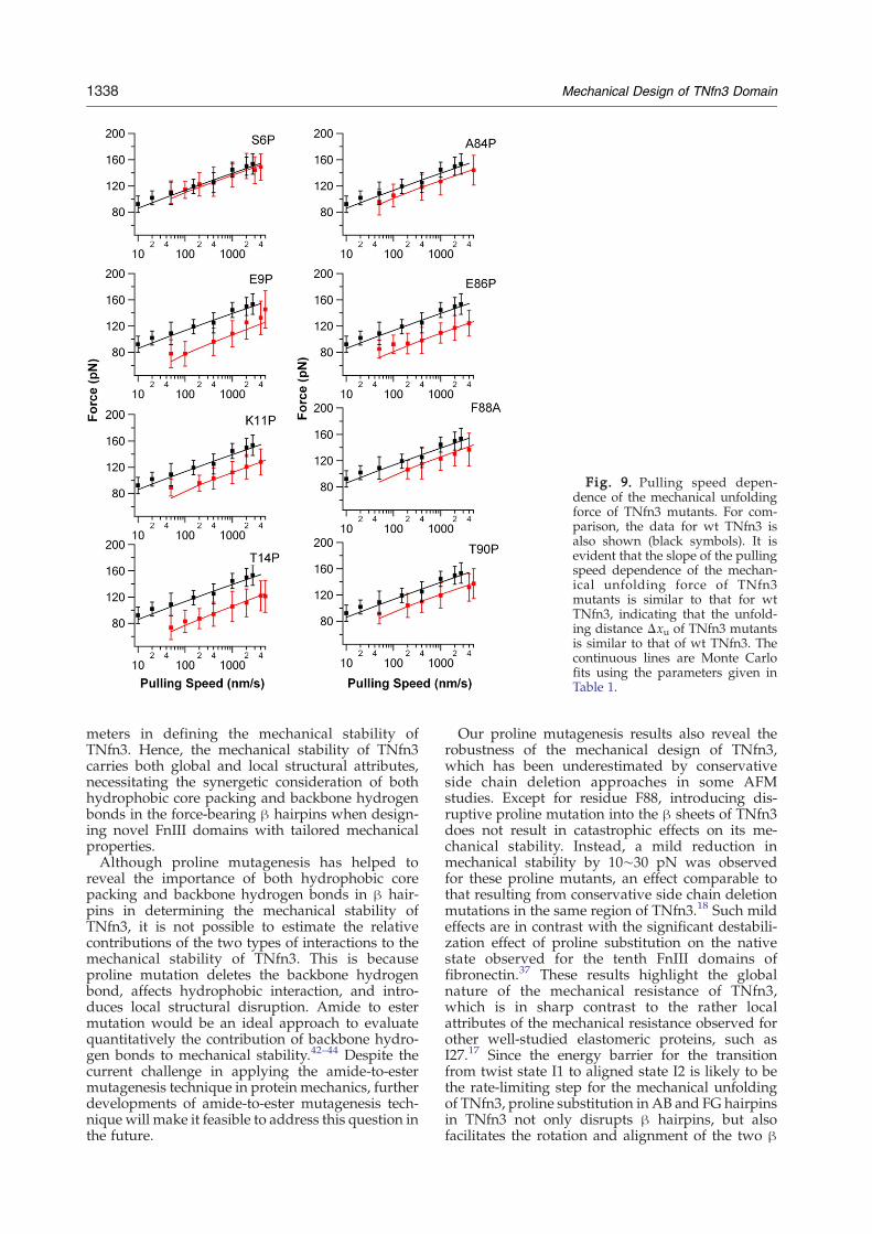
Fig. 9. Pulling speed depen-dence of the mechanical unfoldingforce of TNfn3 mutants. For com-parison, the data for wt TNfn3 isalso shown (black symbols). It isevident that the slope of the pullingspeed dependence of the mechan-ical unfolding force of TNfn3mutants is similar to that for wtTNfn3, indicating that the unfold-ing distance Δxu of TNfn3 mutantsis similar to that of wt TNfn3. Thecontinuous lines are Monte Carlofits using the parameters given inTable 1.
1338 Mechanical Design of TNfn3 Domain
meters in defining the mechanical stability ofTNfn3. Hence, the mechanical stability of TNfn3carries both global and local structural attributes,necessitating the synergetic consideration of bothhydrophobic core packing and backbone hydrogenbonds in the force-bearing β hairpins when design-ing novel FnIII domains with tailored mechanicalproperties.Although proline mutagenesis has helped to
reveal the importance of both hydrophobic corepacking and backbone hydrogen bonds in β hair-pins in determining the mechanical stability ofTNfn3, it is not possible to estimate the relativecontributions of the two types of interactions to themechanical stability of TNfn3. This is becauseproline mutation deletes the backbone hydrogenbond, affects hydrophobic interaction, and intro-duces local structural disruption. Amide to estermutation would be an ideal approach to evaluatequantitatively the contribution of backbone hydro-gen bonds to mechanical stability.42–44 Despite thecurrent challenge in applying the amide-to-estermutagenesis technique in protein mechanics, furtherdevelopments of amide-to-ester mutagenesis tech-nique will make it feasible to address this question inthe future.
Our proline mutagenesis results also reveal therobustness of the mechanical design of TNfn3,which has been underestimated by conservativeside chain deletion approaches in some AFMstudies. Except for residue F88, introducing dis-ruptive proline mutation into the β sheets of TNfn3does not result in catastrophic effects on its me-chanical stability. Instead, a mild reduction inmechanical stability by 10∼30 pN was observedfor these proline mutants, an effect comparable tothat resulting from conservative side chain deletionmutations in the same region of TNfn3.18 Such mildeffects are in contrast with the significant destabili-zation effect of proline substitution on the nativestate observed for the tenth FnIII domains offibronectin.37 These results highlight the globalnature of the mechanical resistance of TNfn3,which is in sharp contrast to the rather localattributes of the mechanical resistance observed forother well-studied elastomeric proteins, such asI27.17 Since the energy barrier for the transitionfrom twist state I1 to aligned state I2 is likely to bethe rate-limiting step for the mechanical unfoldingof TNfn3, proline substitution in AB and FG hairpinsin TNfn3 not only disrupts β hairpins, but alsofacilitates the rotation and alignment of the two β

1339Mechanical Design of TNfn3 Domain
sheets. However, the exact molecular mechanismunderlying such mechanical weakening effect onTNfn3 still needs to be elucidated. Moreover, theobservation of a single weak point in TNfn3, theAchilles heel F88, is quite surprising. It seems thatF88 is the key to protecting the hydrophobic core viathe combination of backbone hydrogen bonds andhydrophobic interactions. Since the mechano-phe-notype is context-dependent, the dramatic pheno-type observed for F88P cannot be extended directlyto its neighboring residues 87 and 89. Since residues87 and 89 do not involve the formation of backbonehydrogen bonds, it will be of interest to examinewhether proline mutation on such residues leads toa similar dramatic destabilization effect. Further-more, since all the FnIII domains of tenascin-C sharesimilar mechanical stability,13,15,28 it will be inter-esting to check whether the features observed herefor the mechanical design of TNfn3 will also applyto other FnIII domains of tenascin-C.
Comparison of the mechanical unfolding ofTNfn3 versus FNfn10: similar structure butdifferent unfolding behaviors
The mechanical unfolding of the tenth FnIIIdomain of fibronectin (FNfn10) has been character-ized in detail using single-molecule AFM and SMDsimulations.32,33,37,45,46 Despite the highly homolo-gous structures of FNfn10 and TNfn3, their me-chanical unfolding behavior shows interestingcommonality as well as differences.SMD simulations show strikingly similar signa-
tures in the mechanical unfolding of these twodomains.32,33,45,46 FNfn10 and TNfn3 follow verysimilar sequences of molecular events: first, the pre-detachment of the first few residues of the N-terminal end of strand A leads to a slightly twistedstate I1; then the two β sheets rotate and align witheach other to enter the aligned state I2; after that, theprotein unfolds in two distinct pathways; e. g.strand A separates first and strands A-G separatesimultaneously, and reach the partially unfoldedintermediate state I3 in the “A-strand separatesfirst” pathway; finally the domain unravels com-pletely. The transition from the twisted state I1 to thealigned state I2 has been indicated as the mainunfolding barrier.Despite the similarity in the unfolding sequences,
a significant difference sets the two FnIII domainsapart: the presence of stable intermediate state I3predicted in SMD simulations for FNfn10 wasobserved experimentally in single-molecule AFMexperiments,37 while the similar intermediate stateI3 was not observed for TNfn3 in AFM experiments.The similarity and difference between the two
FnIII domains reside in their response to prolinemutation. The phenotypic effect of proline muta-tions in strand A is catastrophic for FNfn10, asproline mutations in strand A destabilize theprotein so dramatically that the transition from thealigned state I2 to I3 is abolished;37 and the FNfn10proline mutant unfolds directly from the intermedi-
ate state I3. In contrast, the phenotypic effect ofproline mutations in strand A in TNfn3 is muchmilder and the destabilization facilitates the transi-tion from I1 to I2.Compared with the difference in phenotypic effect
in strand A, proline mutations in strand G havestrikingly similar effects for FNfn10 and TNfn3.Residue 88 seems to be the weak point for bothproteins, as proline substitution at this residue leadsto significant destabilization of both proteins: forFNfn10, destabilization leads to the elimination ofthe transition from I2 to I3, and FNfn10 unfoldsdirectly from intermediate state I3;37 for TNfn3,since the intermediate state I3 is much less stable,destabilization caused by substitution of residue 88with proline leads to the complete unraveling ofTNfn3 at low forces. Proline substitution at the N-terminal end of strand G shows a very similarminute effect on the mechanical unfolding of bothproteins.Understanding the relationship between sequence
variation andmechanical unfolding features of thesetwo proteins will be critical for future efforts toengineer FnIII domains of tailored mechanicalproperties. A recent study has made an encouragingfirst step toward such purposes.41
Single-molecule AFM versus SMD: similaritiesand discrepancies
Combining protein engineering, single-moleculeAFM and SMD simulation techniques, we havecharacterized the mechanical unfolding pathways ofTNfn3 on two vastly different time-scales. Compar-ing the single-molecule AFM results with SMDpredictions, some of the SMD predictions areverified by the single-molecule AFM results, butthere are some important discrepancies between thetwo, and the SMD simulation results cannot fullyexplain the experimental findings in single-moleculeAFM experiments.Similar to previous SMD simulations,18,33 our
SMD simulations of the mechanical unfolding ofTNfn3 using an explicit water model predicted thatthe first step of the mechanical unfolding process ofTNfn3 is the disruption and detachment of the N-terminus (residues 1–6), followed by the alignmentof the two β-sheets. Afterwards, the unfolding of thealigned β-sheets is initiated by the rupture of AB βhairpin followed by a mechanical unfolding inter-mediate state I3 or by the simultaneous separation ofstrands A-G from TNfn3. The unraveling of the I3state will lead to a fully unfolded TNfn3. The threeon-pathway unfolding intermediate states observedin SMD simulation have different stabilities. Analy-sis of both the unfolding force (from the constantvelocity SMD) and dwell time (from the constantforce SMD) of the three intermediate states showedthat I1 is the most stable and I2 is the least stable.Compared with the SMD simulation results, the
single-molecule AFM results confirm that the dis-ruption and detachment of the N-terminus of TNfn3is likely to be the very first step in the mechanical

1340 Mechanical Design of TNfn3 Domain
unfolding of TNfn3, as the S6P mutation does nothave any effect on the mechanical unfolding ofTNfn3. This agreement confirms that the mechanicalunfolding of TNfn3 observed in single-moleculeAFM is not the unraveling of TNfn3 from its nativestate, but from a force-induced pseudo ground state,in which the N-terminal end of strand A is alreadydetached. In addition, SMD simulations predict thatthe transition from twisted intermediate state I1 tothe aligned state I2 is the rate-limiting step for theunfolding of TNfn3. This transition involves therotation and alignment of the two β sheets, and isaccompanied by the extension of the distancebetween the N- and C-termini by ∼10 Å. Therefore,TNfn3 can be deformed over a longer distance beforeit unfolds. This description provides a plausiblemolecular level explanation for the observed longerunfolding distance Δxu for TNfn3. In contrast,unraveling I27-like elastomeric proteins requiressimultaneous rupture of multiple hydrogen bondsholding the two force-bearing β strands together.Therefore, I27-like proteins can be deformed onlyover a shorter distance before they unfold, giving riseto shorter unfolding distances.Despite the agreements between SMD simulation
predictions with single-molecule AFM experiments,some discrepancies exist between the two. Incontrast to the SMD prediction of the existence ofintermediate I3, the single-molecule AFM resultsindicated that the mechanical unfolding of TNfn3 isan apparent two-state process, and no intermediatestate is observed in single-molecule AFM experi-ments. Although intermediate I3 was predicted to bemechanically stable in both constant force andconstant velocity SMD simulations, intermediatestate I3 does not populate on the time-scale of single-molecule AFM experiments. The origin of such adiscrepancy remains to be explored. This situation isin sharp contrast to that for FNfn10, for which a verygood agreement was reached for the similar inter-mediate state I3 between single-molecule AFMexperiments and SMD simulations.32,33,37,46 Despitesuch limitations, it is clear that SMD has offeredvaluable insights into the molecular mechanism ofmechanical unfolding of proteins. Improvements inSMD methodology will continue to further ourunderstanding of the mechanical unfolding andmechanical design of elastomeric proteins at anunprecedented detail.In summary, we have characterized the mechan-
ical unfolding of TNfn3 using a combination ofsingle-molecule AFM, SMD simulations, and prolinemutagenesis techniques. Our results have revealedthe robust mechanical design of TNfn3 that confersthe resistance of the protein to disruptive mutationssuch as proline substitution. Moreover, both localand global structural features are important fordetermining the mechanical resistance of TNfn3. Ithas become evident that the mechanical unfolding ofTNfn3 is much more complex than other typicalelastomeric proteins, such as I27,17,47 due to thestructural flexibility and deformability of TNfn3.Therefore, the TNfn3 and FnIII domains, in general,
present further challenges for protein engineers toenhance the mechanical stability of FnIII domain in asystematic and rational fashion.
Materials and Methods
Protein engineering
The DNA sequence coding TNfn3, flanked with a 5′BamHI restriction site and 3′ BglII, HindIII restrictionsites, was amplified by the polymerase chain reaction(PCR) from the plasmid TNfnALL encoding all of the 15FnIII domains. The plasmid TNfnALL was a generous giftfrom Professor Harold Erickson (Duke Univeristy). Poly-protein (TNfn3)8 was constructed using a consecutiveDNA concatamerization method based on the identity ofthe sticky ends generated by the BamHI and BglII restric-tion enzymes.17
To facilitate the identification of the mechanical unfold-ing signatures of the proline mutants of TNfn3 using AFM,we constructed heteropolyproteins consisting of alternatingGB1 domains and proline mutant TNfn3, where the well-characterized GB1 domains serve as internal fingerprintsfor identifying single-molecule stretching events. Since GB1gene carries a 5′ BamHI restriction site and 3′ BglII, KpnIrestriction sites, we constructed a new version of TNfn3,which carries a 5′ BamHI restriction site and 3′ BglII, KpnIrestriction sites, to facilitate the construction of the hetero-polyprotein. Proline mutants were constructed usingstandard site-directed mutagenesis methods. Genes encod-ing heteropolyproteins were constructed using protocolssimilar to those used for constructing polyprotein (TNfn3)8based on the identity of the sticky ends generated by theBamHI and BglII restriction enzymes.Polyproteins were over-expressed in strain DH5α and
purified from supernatant using Ni2+-affinity chromato-graphy. The polyproteins were kept at 4 °C at a concen-tration of ∼200 μg/mL in PBS.
Single-molecule AFM experiments
Single-molecule AFM experiments were done with acustom-built atomic force microscope, which was con-structed as described.48 All the force-extension measure-ments were carried out in PBS. In a typical experiment,the polyprotein sample (1 μL) was deposited onto a cleanglass coverslip covered by PBS (50 μL), resulting in a 10–20 nm thick layer of protein. The thickness of this proteinlayer depends upon the amount of protein deposited ontothe glass coverslip; i.e., the more protein, the thicker thelayer. The thickness of the protein layer may contribute tothe apparent contour length of the polyprotein, some-times resulting in an apparent contour length that isgreater than the theoretical contour length of the polypro-tein.38 The spring constant of each individual cantilever(Si3N4 cantilevers from Vecco, with a typical spring cons-tant of 40 pN/nm) was calibrated in solution using theequipartition theorem before and after each experiment.
SMD simulation
The crystal structure of TNfn3 (PDB accession code1TEN) was used as the starting conformation of TNfn3 forsimulated equilibration. The protein was solvated in awater box (length 107 Å, width 62 Å, height 53 Å) with a

1341Mechanical Design of TNfn3 Domain
TIP3P water model.49 The whole protein–water systemcontains 33,194 atoms. SMD simulations of the mechanicalunfolding of TNfn3 was done with the program NAMD2.650 and with the CHARMM22 force field,51 asdescribed.39,52 The initial structure of TNfn3 was equili-brated for 1.5 ns at 300 K. Compared to the crystalstructure, the equilibrated structures at 1 ns and 1.5 ns havea backbone RMSD of 0.93 Å and 0.98 Å, respectively, andwere used as the starting conformation for the constantforce and constant velocity SMD simulations. During theSMD simulations, the N-terminal Cα atom was fixed andthe C-terminal Cα atom was pulled. The pulling speed was0.05 Å/ps in constant velocity SMD simulations and apulling force of 500 pNwas used in the constant force SMDsimulations. The simulation time was 57 ns in total. Systemsetup, structural analysis and calculation of hydrogen bondenergies were performed using VMD1.8653 and hydrogenbond energies were calculated using custom written scriptkindly provided by Hui Lu and Morten Kallberg.
Monte Carlo simulation
Monte Carlo simulations of the stretching and unfold-ing of the polyproteins were done as described.20 Theunfolding rate constant at zero force α0 and the distance ofthe native state to the transition state Δxu along thereaction coordinate of the mechanical unfolding reactionwere estimated using Monte Carlo simulation proceduresin a trial-and-error fashion. Such a procedure is necessarydue to the lack of analytical solutions to the unfoldingforce distribution obtained from force-extension measure-ment for polyproteins. The accuracy of the fittingparameters is exemplified in Supplementary Data Fig. 2S,which plots the simulated unfolding force histogram andthe pulling speed dependence of the unfolding forcesusing different sets of unfolding rate constant α0 andunfolding distance Δxu. Typically, α0 is accurate within afactor of 3 and the unfolding distance Δxu is accuratewithin 0.05 nm.
Acknowledgements
We thank Prof. Hui Lu and Dr. Gang Feng for theirgenerous help in SMD simulation, and Dr. Mu Gaofor stimulating discussion. We also thank Prof. HuiLu and Morten Kallberg for their generous scriptand help in hydrogen bond energy calculations. Thiswork is supported by Canadian Institutes of HealthResearch (CIHR) Operating grant MOP-81225,Michael Smith Foundation for Health Research,University of British Columbia Health Research Re-sources Office and Canada Research Chairs Program.H. L. is a Michael Smith Foundation for HealthResearch Career Investigator. Q. P. is supported by aPacific Century Graduate Scholarship from theProvince of British Columbia.
Supplementary Data
Supplementary data associated with this articlecan be found, in the online version, at doi:10.1016/j.jmb.2009.01.019
References
1. Alenghat, F. J. & Ingber, D. E. (2002). Mechanotrans-duction: all signals point to cytoskeleton, matrix, andintegrins. Sci. STKE, 2002, PE6.
2. Kjaer, M. (2004). Role of extracellular matrix inadaptation of tendon and skeletal muscle to mechan-ical loading. Physiol. Rev. 84, 649–698.
3. Chen, C. S., Tan, J. & Tien, J. (2004). Mechanotransduc-tion at cell-matrix and cell-cell contacts. Annu. Rev.Biomed. Eng. 6, 275–302.
4. Jones, F. S. & Jones, P. L. (2000). The tenascin family ofECM glycoproteins: structure, function, and regula-tion during embryonic development and tissueremodeling. Dev. Dyn. 218, 235–259.
5. Erickson, H. P. (1994). Reversible unfolding offibronectin type III and immunoglobulin domainsprovides the structural basis for stretch and elasticityof titin and fibronectin. Proc. Natl Acad. Sci. USA, 91,10114–10118.
6. Chiquet-Ehrismann, R. (1995). Tenascins, a growingfamily of extracellular matrix proteins. Experientia, 51,853–862.
7. Jones, P. L. & Jones, F. S. (2000). Tenascin-C in deve-lopment and disease: gene regulation and cell func-tion. Matrix Biol. 19, 581–596.
8. Hsia, H. C. & Schwarzbauer, J. E. (2005). Meet thetenascins: multifunctional and mysterious. J. Biol.Chem. 280, 26641–26644.
9. Kannus, P., Jozsa, L., Jarvinen, T. A., Jarvinen, T. L.,Kvist, M., Natri, A. & Jarvinen, M. (1998). Locationand distribution of non-collagenous matrix proteinsin musculoskeletal tissues of rat. Histochem. J. 30,799–810.
10. Jarvinen, T. A., Jozsa, L., Kannus, P., Jarvinen, T. L.,Hurme, T., Kvist, M. et al. (2003). Mechanical loadingregulates the expression of tenascin-C in the myoten-dinous junction and tendon but does not induce denovo synthesis in the skeletal muscle. J. Cell Sci. 116,857–866.
11. Jarvinen, T. A., Kannus, P., Jarvinen, T. L., Jozsa, L.,Kalimo, H. & Jarvinen, M. (2000). Tenascin-C in thepathobiology and healing process of musculoskeletaltissue injury. Scand. J. Med. Sci. Sports, 10, 376–382.
12. Chiquet-Ehrismann, R. & Chiquet, M. (2003). Tenas-cins: regulation and putative functions during patho-logical stress. J. Pathol. 200, 488–499.
13. Oberhauser, A. F., Marszalek, P. E., Erickson, H. P. &Fernandez, J. M. (1998). The molecular elasticity of theextracellular matrix protein tenascin. Nature, 393,181–185.
14. Rief, M., Gautel, M., Schemmel, A. & Gaub, H. E.(1998). The mechanical stability of immunoglobulinand fibronectin III domains in the muscle protein titinmeasured by atomic force microscopy. Biophys. J. 75,3008–3014.
15. Cao, Y. & Li, H. (2006). Single molecule force spectro-scopy reveals a weakly populated microstate of theFnIII domains of tenascin. J. Mol. Biol. 361, 372–381.
16. Carrion-Vazquez, M., Oberhauser, A. F., Fisher, T. E.,Marszalek, P. E., Li, H. & Fernandez, J. M. (2000).Mechanical design of proteins studied by single-molecule force spectroscopy and protein engineering.Prog. Biophys. Mol. Biol. 74, 63–91.
17. Carrion-Vazquez, M., Oberhauser, A. F., Fowler, S. B.,Marszalek, P. E., Broedel, S. E., Clarke, J. & Fernandez,J. M. (1999). Mechanical and chemical unfolding of asingle protein: a comparison. Proc. Natl Acad. Sci. USA,96, 3694–3699.

1342 Mechanical Design of TNfn3 Domain
18. Ng, S. P., Rounsevell, R. W., Steward, A., Geierhaas,C. D., Williams, P. M., Paci, E. & Clarke, J. (2005).Mechanical unfolding of TNfn3: the unfolding path-way of a fnIII domain probed by protein engineering,AFM and MD simulation. J. Mol. Biol. 350, 776–789.
19. Leahy, D. J., Hendrickson, W. A., Aukhil, I. &Erickson, H. P. (1992). Structure of a fibronectintype-Iii domain from tenascin phased by MADanalysis of the selenomethionyl protein. Science, 258,987–991.
20. Li, H. (2008). “Mechanical engineering” of elastomericproteins: toward designing new protein building blocksfor biomaterials. Adv. Funct. Mater. 18, 2643–2657.
21. Schwaiger, I., Kardinal, A., Schleicher, M., Noegel,A. A. & Rief, M. (2004). A mechanical unfoldingintermediate in an actin-crosslinking protein. NatureStruct. Mol. Biol. 11, 81–85.
22. Carrion-Vazquez, M., Li, H., Lu, H., Marszalek, P. E.,Oberhauser, A. F. & Fernandez, J. M. (2003). Themechanical stability of ubiquitin is linkage dependent.Nature Struct. Biol. 10, 738–743.
23. Cao, Y., Lam, C., Wang, M. & Li, H. (2006).Nonmechanical protein can have significant mechan-ical stability. Angew Chem. Int. Ed. Engl. 45, 642–645.
24. Oberhauser, A. F., Badilla-Fernandez, C., Carrion-Vazquez, M. & Fernandez, J. M. (2002). The mechan-ical hierarchies of fibronectin observed with single-molecule AFM. J. Mol. Biol. 319, 433–447.
25. Schoenauer, R., Bertoncini, P., Machaidze, G., Aebi, U.,Perriard, J. C., Hegner, M. & Agarkova, I. (2005).Myomesin is a molecular spring with adaptableelasticity. J. Mol. Biol. 349, 367–379.
26. Brockwell, D. J., Beddard, G. S., Paci, E., West, D. K.,Olmsted, P. D., Smith, D. A. & Radford, S. E. (2005).Mechanically unfolding the small, topologically sim-ple protein L. Biophys. J. 89, 506–519.
27. Marko, J. F. & Siggia, E. D. (1995). Stretching DNA.Macromolecules, 28, 8759–8770.
28. Wang, M. J., Cao, Y. & Li, H. B. (2006). The unfoldingand folding dynamics of TNfnALL probed by singlemolecule force-ramp spectroscopy. Polymer, 47,2548–2554.
29. Clarke, J., Hamill, S. J. & Johnson, C. M. (1997).Folding and stability of a fibronectin type III domainof human tenascin. J. Mol. Biol. 270, 771–778.
30. Li, H., Linke, W. A., Oberhauser, A. F., Carrion-Vazquez, M., Kerkvliet, J. G., Lu, H. et al. (2002).Reverse engineering of the giant muscle protein titin.Nature, 418, 998–1002.
31. Li, H. B., Oberhauser, A. F., Fowler, S. B., Clarke, J. &Fernandez, J. M. (2000). Atomic force microscopyreveals the mechanical design of a modular protein.Proc. Natl Acad. Sci. USA, 97, 6527–6531.
32. Gao, M., Craig, D., Vogel, V. & Schulten, K. (2002).Identifying unfolding intermediates of FN-III(10) bysteered molecular dynamics. J. Mol. Biol. 323, 939–950.
33. Craig, D., Gao, M., Schulten, K. & Vogel, V. (2004).Tuning the mechanical stability of fibronectin type IIImodules through sequence variations. Structure, 12,21–30.
34. Billings, K. S., Best, R. B., Rutherford, T. J. & Clarke, J.(2008). Crosstalk between the protein surface andhydrophobic core in a core-swapped fibronectin typeIII domain. J. Mol. Biol. 375, 560–571.
35. Wood, S. J., Wetzel, R., Martin, J. D. & Hurle, M. R.(1995). Prolines and amyloidogenicity in fragments ofthe Alzheimer's peptide beta/A4. Biochemistry, 34,724–730.
36. Li, H., Carrion-Vazquez, M., Oberhauser, A. F.,Marszalek, P. E. & Fernandez, J. M. (2000). Pointmutations alter the mechanical stability of immuno-globulin modules. Nature Struct. Biol. 7, 1117–11120.
37. Li, L., Huang, H. H., Badilla, C. L. & Fernandez, J. M.(2005). Mechanical unfolding intermediates observedby single-molecule force spectroscopy in a fibronectintype III module. J. Mol. Biol. 345, 817–826.
38. Cao, Y. & Li, H. (2007). Polyprotein of GB1 is an idealartificial elastomeric protein. Nature Mater. 6, 109–114.
39. Sharma, D., Perisic, O., Peng, Q., Cao, Y., Lam, C., Lu,H. & Li, H. (2007). Single-molecule force spectroscopyreveals a mechanically stable protein fold and therational tuning of its mechanical stability. Proc. NatlAcad. Sci. USA, 104, 9278–9283.
40. Peng, Q. & Li, H. (2008). Atomic force microscopyreveals parallel mechanical unfolding pathways of T4lysozyme: evidence for a kinetic partitioning mechan-ism. Proc. Natl Acad. Sci. USA, 105, 1885–1890.
41. Ng, S. P., Billings, K. S., Ohashi, T., Allen, M. D., Best,R. B., Randles, L. G. et al. (2007). Designing anextracellular matrix protein with enhanced mechan-ical stability. Proc. Natl Acad. Sci. USA, 104, 9633–9637.
42. Blankenship, J. W., Balambika, R. & Dawson, P. E.(2002). Probing backbone hydrogen bonds in thehydrophobic core of GCN4. Biochemistry, 41,15676–15684.
43. Deechongkit, S., Dawson, P. E. & Kelly, J. W. (2004).Toward assessing the position-dependent contribu-tions of backbone hydrogen bonding to beta-sheetfolding thermodynamics employing amide-to-esterperturbations. J. Am. Chem. Soc. 126, 16762–16771.
44. Deechongkit, S., Nguyen, H., Powers, E. T., Dawson,P. E., Gruebele, M. & Kelly, J. W. (2004). Context-dependent contributions of backbone hydrogen bond-ing to beta-sheet folding energetics.Nature, 430, 101–105.
45. Craig, D., Krammer, A., Schulten, K. & Vogel, V.(2001). Comparison of the early stages of forced un-folding for fibronectin type III modules. Proc. NatlAcad. Sci. USA, 98, 5590–5595.
46. Paci, E. & Karplus, M. (1999). Forced unfolding offibronectin type 3 modules: an analysis by biased mole-cular dynamics simulations. J. Mol. Biol. 288, 441–459.
47. Lu, H. & Schulten, K. (1999). Steered moleculardynamics simulations of force-induced protein domainunfolding. Proteins: Struct. Funct. Genet. 35, 453–463.
48. Fernandez, J. M. & Li, H. (2004). Force-clamp spectro-scopy monitors the folding trajectory of a single pro-tein. Science, 303, 1674–1678.
49. Jorgensen, W. L., Chandrasekhar, J., Madura, J. D.,Impey, R. W. & Klein, M. L. (1983). Comparison ofsimple potential functions for simulating liquid water.J. Chem. Phys. 79, 926–935.
50. Phillips, J. C., Braun, R., Wang, W., Gumbart, J.,Tajkhorshid, E., Villa, E. et al. (2005). Scalable mole-cular dynamics with NAMD. J. Comput. Chem. 26,1781–1802.
51. MacKerell, A. D., Bashford, D., Bellott, M., Dunbrack,R. L., Evanseck, J. D., Field, M. J. et al. (1998). All-atomempirical potential for molecular modeling anddynamics studies of proteins. J. Phys. Chem. B, 102,3586–3616.
52. Lee, E. H., Hsin, J., Mayans, O. & Schulten, K. (2007).Secondary and tertiary structure elasticity of titin Z1Z2and a titin chain model. Biophys. J. 93, 1719–1735.
53. Humphrey, W., Dalke, A. & Schulten, K. (1996). VMD:visual molecular dynamics. J. Mol. Graph. 14, 33-8,27–28.