Mechanical and enzymatic remodeling of collagen ...eprints.gla.ac.uk/119626/14/119626.pdf ·...
47
Zhao, M., Altankov, G., Grabiec, U., Bennett, M., Salmeron-Sanchez, M. , Dehghani, F. and Groth, T. (2016) Molecular composition of GAG-collagen I multilayers affects remodeling of terminal layers and osteogenic differentiation of adipose-derived stem cells. Acta Biomaterialia, 41, pp. 86-99. (doi:10.1016/j.actbio.2016.05.023) This is the author’s final accepted version. There may be differences between this version and the published version. You are advised to consult the publisher’s version if you wish to cite from it. http://eprints.gla.ac.uk/119626/ Deposited on: 26 May 2016 Enlighten – Research publications by members of the University of Glasgow http://eprints.gla.ac.uk
Transcript of Mechanical and enzymatic remodeling of collagen ...eprints.gla.ac.uk/119626/14/119626.pdf ·...

Zhao, M., Altankov, G., Grabiec, U., Bennett, M., Salmeron-Sanchez, M. ,
Dehghani, F. and Groth, T. (2016) Molecular composition of GAG-collagen
I multilayers affects remodeling of terminal layers and osteogenic
differentiation of adipose-derived stem cells. Acta Biomaterialia, 41, pp.
86-99. (doi:10.1016/j.actbio.2016.05.023)
This is the author’s final accepted version.
There may be differences between this version and the published version.
You are advised to consult the publisher’s version if you wish to cite from
it.
http://eprints.gla.ac.uk/119626/
Deposited on: 26 May 2016
Enlighten – Research publications by members of the University of Glasgow
http://eprints.gla.ac.uk

1
Molecular composition of GAG-collagen I
multilayers affects remodeling of terminal layers
and osteogenic differentiation of adipose-derived
stem cells
Mingyan Zhao1,2, George Altankov3,4 , Urszula Grabiec 5, Mark Bennett6,Manuel
Salmeron-Sanchez6, Faramarz Dehghani 5, Thomas Groth2*
1 Stem Cell Research and Cellular Therapy Center, Affiliated Hospital of Guangdong Medical
University, Renmin Dadao Road, Xiashan District, Zhanjiang 524001, China
2Biomedical Materials Group, Martin Luther University Halle-Wittenberg,
Heinrich-Damerow-Strasse 4, Halle (Saale) 06120, Germany
3Institut for Bioengineering of Catalonia, Baldiri Reixac, 10-12, Barcelona 08028, Spain
4ICREA (Institucio Catalana de Recerca i Estudis Avancats), Barcelona 08010, Spain
5 Department of Anatomy and Cell Biology, Faculty of Medicine, Martin Luther University
Halle-Wittenberg, Große Steinstraße 52, Halle (Saale) 06108, Germany
6 Division of Biomedical Engineering, School of Engineering, University of Glasgow,
Glasgow, UK
e-mails for each author:
Mingyan Zhao:[email protected]
George Altankov: [email protected]
Urszula Grabiec: [email protected]

2
Mark Bennett: [email protected]
Manuel Salmeron-Sanchez: [email protected]
Faramarz Dehghani: [email protected]
Thomas Groth: [email protected]
*To whom correspondence should be addressed:
Prof. Dr. Thomas Groth
Biomedical Materials Group
Martin Luther University Halle-Wittenberg
Heinrich-Damerow-Strasse 4
06120 Halle (Saale), Germany
phone: +49 (0345) 55 28460
fax: +49 (0345) 55 27379
e-mail: [email protected]

3
Abstract
The effect of molecular composition of multilayers, by pairing type I collagen (Col I) with
either hyaluronic acid (HA) or chondroitin sulfate (CS) was studied regarding the osteogenic
differentiation of adhering human adipose-derived stem cells (hADSCs). Multilayer (PEM)
formation was based primarily on ion pairing and on additional intrinsic cross-linking through
imine bond formation replacing native by oxidized HA (oHA) or CS (oCS) with Col I.
Significant amounts of Col I fibrils were found on both native and oxidized CS-based PEMs,
resulting in higher water contact angles and surface potential under physiological condition,
while much less organized Col I was detected in either HA-based multilayers, which were
more hydrophilic and negatively charged. An important finding was that hADSCs remodeled
Col I at the terminal layers of PEMs by mechanical reorganization and pericellular proteolytic
degradation, being more pronounced on CS-based PEMs. This was in accordance with the
higher quantity of Col I deposition in this system, accompanied by more cell spreading, focal
adhesions (FA) formation and significant α2β1 integrin recruitment compared to HA-based
PEMs. Both CS-based PEMs caused also an increased fibronectin (FN) secretion and cell
growth. Furthermore, significant calcium phosphate deposition, enhanced ALP, Col I and
Runx2 expression were observed in hADSCs on CS-based PEMs, particularly on
oCS-containing one. Overall, multilayer composition can be used to direct cell-matrix
interactions, and hence stem cell fates showing for the first time that PEMs made of biogenic
polyelectrolytes undergo significant remodeling of terminal protein layers, which enables
cells to form a more adequate extracellular matrix-like environment.
Keywords: Layer-by-layer technique, glycosaminoglycans, collagen reorganization,

4
mesenchymal stem cells, osteogenic differentiation
5
1. Introduction
In native tissues, cells are tightly connected to the extracellular matrix (ECM), which
regulates development, functioning and regeneration of tissues[1, 2]. The ECM acts as
physical support of cells and provides them with environmental signals, which determine
cellular fates[3]. In particular, the molecular composition and specific structure of ECM
regulate cellular processes, like adhesion, proliferation and differentiation[4-6]. Hence, the
ECM plays a fundamental role during development and regeneration. Thereby, the cell-ECM
interactions have a highly dynamic character, since cells continuously remodel the matrix
changing this way the cellular microenvironment, which eventually affects cell behavior[7]. In
this context, it was observed recently that cells remodel adsorbed collagens both by
mechanical reorganization and proteolytic degradation[8-10]. It was also found that
fibronectin (FN) possesses an important role for the mechanical reorganization of
collagen[9-11]. Indeed, the relative amounts and organization of different ECM components
vary from tissue to tissue, reflecting its peculiar functions. Hence, one aim of regenerative
medicine is to design biomaterials that mimic the ECM composition of a specific tissue to
guide cell differentiation and function in the desired lineage type.
Bone is a complex tissue with an intricate and ordered association of organic and inorganic
components in a continuously renewing biological environment[12]. During the in vivo bone
formation, mesenchymal stem cells (MSCs) derived osteoblasts synthesize and excrete type I
collagen (Col I) the main component of the organic matrix of bone, but also other specific
bone matrix components, such as osteocalcin, proteoglycans, etc. The Col I molecular chains
self-assemble in triple helical molecules, fibrils and three dimensional networks and become

6
then mineralized with hydroxyapatite, which is embedded within the interstices and roughly
aligned parallel to the long axis of the fibrils[12]. The spatial organization of Col I fibers in
vivo is believed to play an important role in directing cell behavior and stem cell fate
decisions[13]. Additionaly, Choindroitin sulfate (CS) from proteoglycans is another important
macromolecule that present at the early stage in bone and tooth formation, and found to be an
effective promoter in hydroxyapatite nucleation and growth[14].
MSCs hold great promise for tissue engineering and regenerative medicine owing to their
ability to differentiate into cell types of several mesenchymal tissues[15]. Particularly, human
adipose-derived stem cells (hADSCs) have gained an increasing attention since they are
readily accessible in large amounts and possess the potential to form bone, cartilage, fat and
other tissues[16, 17]. Therefore, the successful application of hADSCs in bone tissue
engineering is highly dependent on the capacity of culturing matrix-like environment to
trigger a specific osteogenic response[18]. Yet, to provide such an environment at
biomaterial’s interface to cell and tissues is a challenging task [19].
Indeed, chemical and physical surface modifications are frequently applied to address
biomaterials to the specific tissue demands by binding ECM components like proteins or
glycosaminoglycans (GAGs) to address specific cell receptors like integrins or growth factor
receptors important for growth and differentiation of cells[20, 21]. Among them,
layer-by-layer (LbL) technique has emerged as a versatile method, which is based on
alternating deposition of biogenic polyelectrolytes such as proteins, polypeptides and GAGs
for formation of bioactive multilayer coatings[22, 23]. Based on their opposite charge under
acidic condition, Col I paired with either hyaluronic acid (HA) or CS represents an interesting

7
system to create multilayers with a composition similar to connective tissue ECM[22].
Unfortunately, some of these biopolymer-based multilayers have been found relatively
unstable under physiological conditions[24, 25]. To enhance their stability, recently an
intrinsic crosslinking between aldehydes of oxidized HA or CS and amino groups of Col I was
introduced, resulting in enhanced stability and biocompatibility[22]. Here we continue these
studies with these multilayer systems looking at the ability of hADSCs to remodel the
ECM-like multilayers during culture and the effect of multilayer composition and intrinsic
cross-linking on differentiation of hADSCs under the effect of osteogenic growth media.
Results are reported herein.
2. Materials and methods
2.1. Preparation of polyelectrolyte solutions
Poly (ethylene imine) (PEI, Mw ~ 750 kDa, Sigma, Steinheim, Germany) was dissolved in a
0.15 M sodium chloride (Roth, Karlsruhe, Germany) solution at a concentration of 5 mg mL-1.
Native chondroitin sulfate (nCS Mw ~ 25 kDa) was obtained from Sigma while native
hyaluronan (nHA Mw ~ 1.3 MDa) was a kind gift from Innovent (Jena, Germany). Oxidized
CS and HA (oCS and oHA) were prepared and characterized as described previously[22].
Native polyanion GAGs (nCS, nHA) and oxidized polyanion GAGs (oCS, oHA) were
adjusted to a concentration of 0.5 mg mL-1. Type I collagen (Col I) from porcine skin
(polycation, Mw ~ 100 kDa, Sichuan Mingrang Bio-Tech, Sichuan, China) was used as
polycation and dissolved in 0.2 M acetic acid (Roth) at a concentration of 2 mg mL-1 at 4°C.
After dissolution, the recovered solution was centrifuged at 9,000 g for 10 min to remove any
insoluble precipitates. A final concentration of 0.5 mg mL-1 was achieved by diluting the stock

8
solution in 0.2 M acetic acid supplement with sodium chloride (final concentration to 0.15 M
sodium chloride). Prior to use, the pH value of all polyelectrolyte solutions was adjusted to
pH 4.0 except for PEI (pH adjusted to 7.4).
2.2. Preparation of polyelectrolyte multilayers (PEMs)
Polyelectrolyte multilayer (PEM) fabrication was performed on glass coverslips (Roth) of size
12 × 12 mm and 15 × 15 mm, respectively. Prior to multilayer coating, glass coverslips were
cleaned with 0.5 M sodium hydroxide (Roth) dissolved in 96% Ethanol (Roth) at room
temperature for 2 h followed by extensive rinsing with ultrapure water and drying under
nitrogen flow. Silicon wafers (Silicon materials, Kaufering, Germany) with a size of
(10×10) mm² was treated with a solution of ammonium hydroxide (25%, Roth), hydrogen
peroxide (35%, Roth) and ultrapure water (1:1:5, v/v/v) at 75°C for 15 min followed by
thoroughly washing with ultrapure water [26].
A first anchoring layer of PEI was formed on cleaned glass and silicon wafer (with its oxide
layer on top) substrates to obtain a positive net surface charge, which was followed then by
alternating adsorption of nGAGs (nCS, nHA) or oGAGs (oCS, oHA) as polyanions and Col I
as polycation. The polyanions were adsorbed for 15 min at pH 4.0, while Col I was adsorbed
for 20 min at the same pH value. Each adsorption step was followed by three times rinsing
with a solution of 0.15 M sodium chloride (pH 4.0) for 5 min. Finally, Col I terminated
multilayer systems with eight total layers (8th) on top of the PEI layer were obtained. The four
different PEM systems were denoted as: nCS-Col I; nHA-Col I; oCS-Col I; and oHA-Col I.
2.3. Characterization of polyelectrolyte multilayers (PEMs)
2.3.1. Water contact angle, zeta potential and ellipsometry measurements

9
The surface wettability of Col I terminated PEMs (8th) was determined by static water contact
angle (WCA) measurements using an OCA15+ device from Dataphysics (Filderstadt,
Germany) and applying the sessile drop method. For each PEMs, three independent samples
were measured with 5 droplets per sample.
Zeta potential of Col I terminated PEM surfaces were measured using a SurPASS device
(Anton Paar, Graz, Austria). Two cover slips, modified with identical multilayers were fixed
on stamps and placed oppositely into the SurPASS flow chamber. A flow rate of 100 to 150
mL min-1 was reached at a maximum pressure of 300 mbar by adjusting the gap of the flow
chamber. One mM potassium chloride (Roth) was utilized as electrolyte while 0.1 M
hydrochloric acid (Roth) was used for pH titration. The zeta potentials measured at pH 7.4
were recorded here.
The thickness of PEMs (four bilayers) immersed in PBS (150 mM NaCl, 5.8 mM Na2PO4,
5.8 mM NaHPO4, pH 7.4) was determined with an M-2000V scanning ellipsometer (J.A.
Woollam Co. Inc. Lincoln, NE, USA), which was equipped with a liquid cell (J.A. Woollam
Co. Inc.) that can be utilized for solution injection. The thickness of the hydrated PEMs was
calculated by fitting the experimental data to an additional Cauchy layer. For calculation, a
refractive index of 1.36 was used, which corresponds to native PEM matrix values[27]. The
experimental data were analyzed using the software of the device (WVase32).
2.3.2. Measurement of type I collagen (Col I) concentration in multilayers
A protein quantification assay (BCA assay) was used to quantify the Col I content in the four
PEM systems. In brief, PEMs coated glass coverslips were placed in 24-well plates (Greiner,
Frickenhausen, Germany), followed by the reaction with 400 µL working reagent of BCA
10
Protein Assay Kit (Pierce, Rockford, USA) at 37 °C for 5 h. Thereafter, 225 µL of supernatant
from each well was carefully transferred to a new 96-well plate (Greiner), followed by
measurement of absorbance at 562 nm with a plate reader (FLUOstar, BMG Lab Tech,
Offenburg, Germany). The amount of Col I was determined from a calibration curve plotted
from a series of Col I solutions with known concentrations.
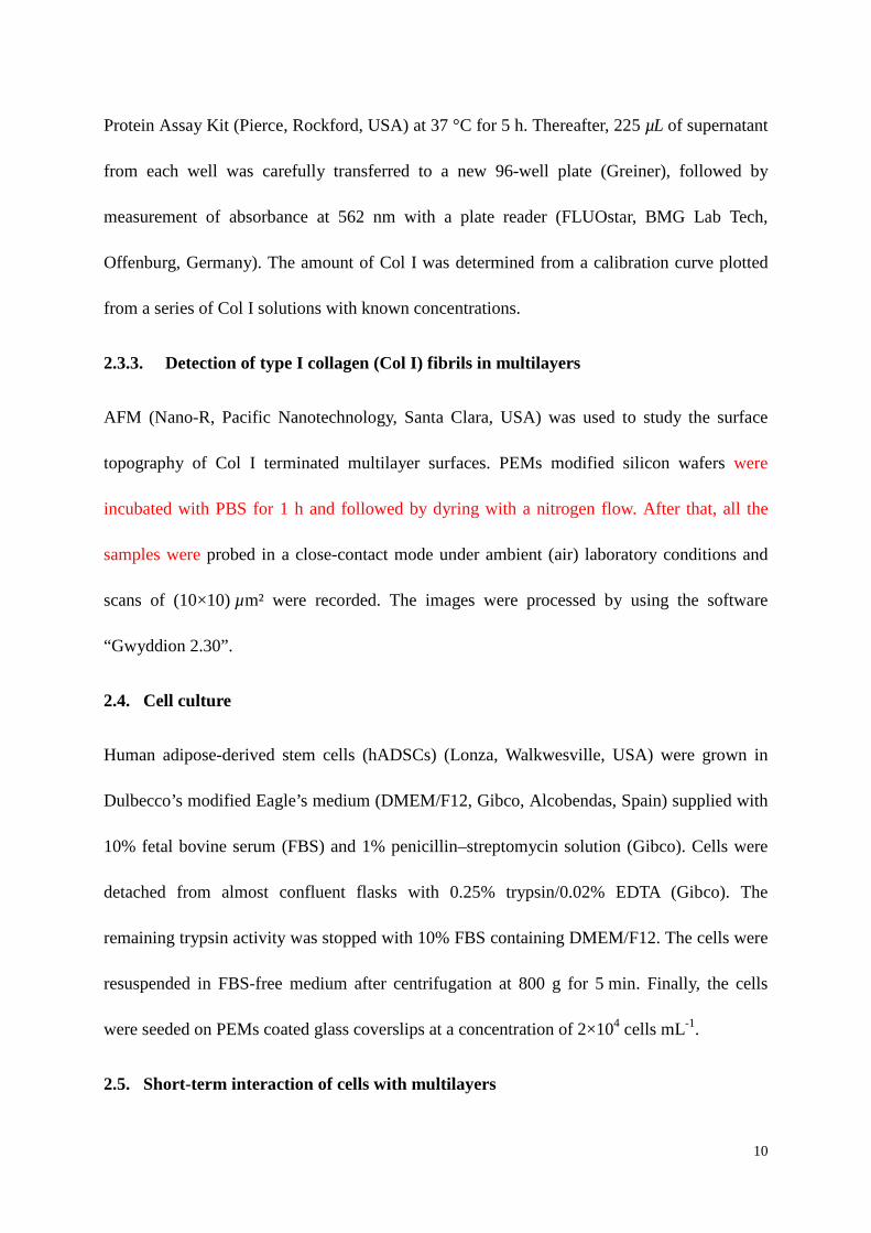
2.3.3. Detection of type I collagen (Col I) fibrils in multilayers
AFM (Nano-R, Pacific Nanotechnology, Santa Clara, USA) was used to study the surface
topography of Col I terminated multilayer surfaces. PEMs modified silicon wafers were
incubated with PBS for 1 h and followed by dyring with a nitrogen flow. After that, all the
samples were probed in a close-contact mode under ambient (air) laboratory conditions and
scans of (10×10) µm² were recorded. The images were processed by using the software
“Gwyddion 2.30”.
2.4. Cell culture
Human adipose-derived stem cells (hADSCs) (Lonza, Walkwesville, USA) were grown in
Dulbecco’s modified Eagle’s medium (DMEM/F12, Gibco, Alcobendas, Spain) supplied with
10% fetal bovine serum (FBS) and 1% penicillin–streptomycin solution (Gibco). Cells were
detached from almost confluent flasks with 0.25% trypsin/0.02% EDTA (Gibco). The
remaining trypsin activity was stopped with 10% FBS containing DMEM/F12. The cells were
resuspended in FBS-free medium after centrifugation at 800 g for 5 min. Finally, the cells
were seeded on PEMs coated glass coverslips at a concentration of 2×10⁴ cells mL-1.
2.5. Short-term interaction of cells with multilayers

11
2.5.1. Quantification of hADSCs adhesion and spreading
500 µL of cell suspension at a concentration of 2×10⁴ cells mL−1 were seeded onto
PEMs-coated glass slides for 1 h in serum free DMEM/F12. 10% FBS was added at the end
of the first hour considering the importance of serum proteins for the remodeling process, and
incubated for another 3 h[28]. After incubation, samples were washed once with PBS and
stained with crystal violet (Roth) (0.5% (w/v)) in methanol (Roth) at room temperature for
30 min. Finally, samples were carefully washed with ultrapure water and air dried. Images
were taken in transmission mode with an Axiovert 100 (Carl Zeiss MicroImaging GmbH,
Göttingen, Germany) equipped with a CCD camera (Sony, MC-3254, AVT-Horn, Aalen,
Germany). The number of adhering cells and the mean cell area were evaluated from five
images per sample using image processing software “ImageJ, NIH, USA”. Three samples
were studied for each type of multilayers.
2.5.2. Focal adhesion (FA) complex formation and actin organization
hADSCs were seeded as described above. After 4 h incubation cells were fixed with 4%
paraformaldehyde (Sigma) (10 min), permeabilized in 0.1 % Triton X-100 in PBS (v/v)
(Sigma) for another 10 min and blocked with PBS containing 1% (w/v) bovine serum albumin
(BSA, Sigma) for 30 min and washed finally with PBS. Focal adhesions (FA) were labelled
using a monoclonal anti-vinculin antibody (1:400, Sigma) diluted in 1% (w/v) BSA in PBS at
room temperature for 30 min followed by AlexaFluor 488 goat anti-mouse (1:400, InVitrogen,
Engene, Oregon, USA) as a secondary antibody. The actin cytoskeleton was visualized at the
same time by parallel incubation with AlexaFluor 555 Phalloidin (1:100, inVitrogen) for 30
min, too. After washing with PBS and distilled water, samples were mounted with Mowiol

12
(Merck, Darmstadt, Germany), examined and photographed with confocal laser scanning
microscopy (CLSM 710, Carl Zeiss Micro-Imaging GmbH, Jena, Germany) using a 63× oil
immersion objective. Images were processed with the ZEN2011 (Carl Zeiss) and ImageJ
software.
Organization of cellular integrins
hADSCs were incubated and fixed as described above. To study the distribution of ±2² 1
integrin, they were labelled by monoclonal anti-human integrin antibody of ±2² 1 (1:100,
Abcam, Cambridge, UK), followed by an incubation with goat-anti-mouse AlexFluor 488 as a
secondary antibody. AlexaFluor 555 Phalloidin was mixed with the secondary antibody to
stain the actin cytoskeleton. The incubation time for all antibodies and phalloidin was 30 min.
Samples were washed, mounted and viewed as described above.
Remodeling of type I collagen (Col I) by hADSCs
hADSCs were seeded and incubated as described above for 4 h in the presence of 10% FBS.
To study the fate of substratum adsorbed Col I, native Col I-containing PEM samples were
processed for immunofluorescence using a monoclonal anti-Col I antibody (1:100, Sigma)
followed by Cy2-conjugated goat anti mouse IgG (1:100, Dianova, Hamburg, Germany) as a
secondary antibody. After washing with PBS and distilled water, samples were mounted and
examined and photographed with CLSM using a 63× oil immersion objective. To assess the
degree of rearrangement of Col I by hADSCs a method was adapted from published
papers[29, 30], with quantification being performed in ImageJ. Briefly, the images were first
normalised to ensure the same total range of pixel values. The background intensity range of
the Col I layer was taken from a defined region (25 ×25 µm) outside of the cell area for each

13
condition. Across the samples the average of the minimum and maximum intensity values
were used as thresholds to define the unaffected Col I layer. The cell area was then used as a
mask over the Col I layer and the intensity values within the defined threshold were measured.
The values obtained provided a % area fraction of the selected area that was within the
threshold values and was thus subtracted from 100 % to indicate the amount of the Col I that
has been rearranged.
To learn more about the fate of Col I layers at the internal part of PEMs, another approach
was used to visualize Col I distribution via direct in-situ labelling with fluorescein
isothiocyanate (FITC) dissolved in DMSO, a procedure which was described elsewhere[31].
Briefly, the FITC-labeled Col I was used for the initial 4 layers and then continuing layer
formation with non-labelled Col I. To view simultaneously the cells, all samples, either native
Col I or FITC-Col I -containing samples were further stained for actin with Alexa Fluor 555
Phalloidin as described above.
2.5.3. Detection of fibronectin matrix and colocalization with type I collagen (Col I)
To study the development of a FN matrix secreted and organized by hADSCs and FN
colocalization with Col I, the cells were cultured for 4 hours on native Col I or FITC-Col I
-containing PEM surfaces. Native Col I was viewed as described above using Cy2-conjugated
goat anti mouse IgG as a secondary antibody, while secreted FN was viewed by a polyclonal
anti-FN antibody (dilution 1:250, Sigma) followed by AlexaFluor 555-labelled anti-rabbit
antibody (dilution 1:400, Invitrogen). All samples were finally mounted with Mowiol (Merck)
and examined with CLSM.
2.5.4. Studies on cellular proteolytic activity

14
Peri-cellular proteolytic activity was quantified in two levels: (i) by measuring the released
fluorescent signal in the medium from FITC-labelled Col I samples (comparing with to
without cells) following a protocol described previously [11] and (ii) measuring the signal
from the extracted PEM- associated Col I (e.g. the protein that might be proteolytically altered
but still remains adsorbed to the PEMs). For that purpose the Col I was labelled directly with
FITC within the PEMs, as explained above, following the protocol described by McSwain et
al.[31].
FITC-Col I containing PEMs prepared as above were seeded with hADSCs (5×10⁴ cells per
well, in 12 well-plates) and incubated overnight (first 1 h in serum-free medium to allow
direct attachment to Col I on multilayers and then 10% serum was added). At the end of
incubation supernatants were collected to measure the FITC-Col I release (first level) by
samples that had been cultured either with or without cells using a fluorescent spectrometer
(Fluoromax-4, HORIBA Scientific, Edison, USA) (488 nm excitation and 530 nm emission).
Conversely, the PEM-associated FITC-Col I (second level) was measured after extraction of
the protein with 0.2 M sodium hydroxide (NaOH, 500 µL per PEMs) for 2 hours at 37°C from
the samples comparing again samples with cells versus those without cells (e.g. control
samples). Here we have to note that in preliminary studies we had observed that protein
degradation by collagenase (20 unit mL-1 collagenase at 37°C for 2 h) greatly increased the
fluorescent yield of FITC-Col I, indicating that the labelled protein was quenched,
presumably as a consequence of being labelled in excess of dye. We therefore speculated that
eventual pericellular proteolysis, will cause de-quenching of FITC-Col I, e.g. will increase its
fluorescence just as collagenase does, accepting the collagenase value as maximal protein
degradation (100%). Thus we obtained a scale varying between zero (sample without cells)
15
and the maximal degradation (i.e. 100% that is achieved by collagenase treatment), in which
we could quantify the cellular proteolytic activity against Col I (samples with cells). More
specifically we measured the fluorescent intensity of NaOH-extracts obtained from a given
FITC labelled PEMs, cultured either with or without cells, and quantified the amount of its
de-quenching (increase of fluorescence) in the scale between zero (no cells) and 100%
(collagenase activity). FITC-Col I samples were treated with collagenase (200 unit mL-1 from
stock solution diluted with fresh medium at a ratio of 1:10) for 2 hours, which was accepted
as positive control as explained above. All experiments were run in triplicates.
2.6. Study of hADSCs proliferation on type I collagen (Col I) terminated multilayers
One mL serum-containing hADSCs suspension (1×10⁴ cells mL-1) was seeded onto each PEM
sample. The quantity of metabolic active cells after 1, 2 and 3 days was measured with a
QBlue cell viability assay kit (BioChain, Newark, USA). Briefly, the medium was carefully
aspirated and cells were washed once with sterile PBS. Then, 300 µL of pre-warmed colorless
DMEM supplemented with QBlue assay reagent (ratio of 10:1) were added to each well,
followed by incubation for another 2 h. Thereafter, 100 µL supernatants were transferred to a
black 96-well plate (Greiner) and fluorescence signals were measured at an excitation
wavelength of 544 nm and emission wavelength of 590 nm with a plate reader.
2.7. Induction of osteogenic differentiation
Osteogenic differentiation was induced after a confluent monolayer was formed. The
osteogenic differentiation media (OM) consisted of basal medium (BM, 1% FBS and 1%
penicillin–streptomycin–containing DMEM) supplemented with osteogenic supplements
namely, 10 nM dexamethasone (Sigma), 50 mg mL-1 ascorbic acid (Sigma), 10 mM

16
² -glycerophosphate (Alfa Aesar, Ward Hill, USA) and 50 ng mL-1 BMP2 (Peprotech, Rocky
Hill, USA). Medium were changed every 3 days. Each experiment was done in triplicate. The
ones cultured with BM were used as reference.
2.8. Detection of osteogenic differentiation of hADSCs
2.8.1. Activity of alkaline phosphatase (ALP)
Alkaline phosphatase activity (ALP) was measured to evaluate the osteoblastic differentiation.
Cell lysates obtained by treatment with 0.2% Triton X-100 (Sigma) (in 10 mM Tris-HCl
(pH=7.2)) were incubated in substrate solution (2 mM p-nitrophenyl phosphate (pNPP,
Sigma), in a substrate buffer (1 M diethanolamine at pH 9.8) for 30 min in the dark at 37°C,
and measured the absorbance at 405 nm with a plate reader. The total protein content of the
lysates was investigated by BCA assay. ALP activity was determined by normalization to the
protein content of the lysates.
2.8.2. Histochemical and immunofluorescence staining
On day 21 post-differentiation, calcium phosphate deposition was investigated by Alizarin
Red S staining. Staining with Alizarin Red S (2%, pH 4.2, Roth) was performed for 45 min in
the dark. Briefly, the samples were washed once with PBS and fixed with 4%
paraformaldehyde for 10 min. After twice washing with distilled water, Alizarin Red solution
was added into each well, left working for 45 min in the dark at room temperature. Finally,
the excess dye was removed by washing with distilled water. Images were taken in
transmission mode with an Axiovert 100 (Carl Zeiss MicroImaging GmbH) equipped with a
CCD camera (Sony).
To further investigate the development of a Col I matrix secreted by hADSCs, the samples
were stained with a monoclonal anti-Col I antibody (Sigma) followed by Cy2-conjugated goat

17
anti mouse IgG (Dianova) as a secondary antibody. To view the cells, samples were further
stained for nuclear with To-Pro-3 (1:500, Invitrogen), mounted with Mowiol (Merck) and
examined with CLSM.
2.8.3. Gene expression of osteogenic markers by real-time RT-PCR
The expression levels of major osteogenic markers including Runx2 and Col I (Col IA) were
used to evaluate whether the hADSCs differentiated into cells of the osteogenic lineage. On
day 14 post-differentiation, cells were detached from test samples using 0.25% trypsin/0.02%
EDTA (Gibco). Total RNA was isolated from cells using Trifast (Peq Lab, Erlangen,
Germany) following standard protocols[32]. After DNAse treatment (DNase-Kit DNA-free,
Life Technologies, Darmstadt, Germany) the extracted RNA was re-suspended in
nuclease-free water (Promega, Madison, WI, USA) and the concentration was measured by
spectrophotometry. cDNA was synthesized from 1 µg of total RNA by reverse transcription
using transcription system (Promega, Mannheim, Germany). PCR samples contained 10 µL of
Mastermix (Taq Polymerase 50 U mL-1, MgCl2, Nucleotids, Promega), 0.5 µL of each primer,
0.25 µL Eva Green (Biotium, Hayward, CA, USA), 4,75 µL DNase free water and 4 µL
cDNA template. Reactions were performed on a Thermocycler (Rotor-GeneTM RG 6000,
Corbett Research, Pty Ltd., Sydney, Australia). The expression of mRNA in the cells was
assessed by qRT-PCR by the use of specific primers (Table 1) and ² -actin was used as the
endogenous control. The data was analyzed by 2^-ddct method following Livak et al[33].
2.9. Data analysis and statistics
All quantitative data were expressed as the mean ± standard error unless otherwise specified.
Statistical analysis was performed using Origin software (Originlab Corporation,

18
Northampton, USA) with ANOVA test (One way) followed by post Tukey testing. The
number of samples is indicated in the respective figure and table captions. Statistical
significance was considered for p < 0.05 and is indicated by asterisks.
3. Results and Discussion
3.1. Physicochemical characterization of polyelectrolyte multilayers (PEMs)
While a more comprehensive characterization of the multilayer formation process and also the
presence of intrinsic cross-linking were shown in a previous paper[22], here we focused
primarily on the characterization of surface properties and layer thickness, which are
important for the interaction with cells[34]. Therefore WCA, zeta potential and ellipsometry
measurements were performed. Results are shown in Table 2.
The measurement of WCA with the sessile drop method showed that multilayers with CS
were significantly less hydrophilic than those with HA as polyanion. A potential reason for
this observation could be a higher quantity of Col I in CS-based PEMs in comparison to
HA-based systems, which was confirmed by the BCA assay. Table 3 shows that the amount
of Col I in CS-based systems was almost twice as high as in HA-based PEMs probably due to
the higher charge density of CS. The use of oxidized GAG led to slightly lower WCA, which
was only significant when comparing PEMs made of either nHA/Col I or oHA/Col I.
However, as found in the BCA assay, there was no significant difference in the amount of
Col I either using the native or oxidized GAGs. Therefore, one reason for the higher
hydrophilicity of the cross-linked systems might be a stabilization of GAG binding in surface
regions of PEMs, which increases the wettability due to the more hydrophilic nature of GAG.
Zeta potentials, shown here only at pH 7.4 further support that more Col I polycation presence

19
in CS-based systems due to lesser negative potentials compared to HA-containing PEMs. The
finding that zeta potentials were less negative for oGAG based systems compared to native
ones seems to be contradictory to WCA measurements at a first view. However, as described
in more detail in a previous paper and also shown by others, zeta potentials of PEMs are not
only a representation of outermost polyelectrolytes, but also of intrinsic composition of layers
closer to the surface[35, 36]. Therefore, it may also be assumed from these measurements that
the less negative zeta potentials for CS-based PEMs indicate a more dominant role of the
polycation Col I in this system. Ellipsometry measurements showed also that intrinsic
cross-linking caused a higher layer thickness in both oCS- and oHA-based PEMs compared to
PEMs made of native GAG, which indicates that the covalent cross-linking leads to more
binding of polyelectrolytes compared to ion pairing alone.
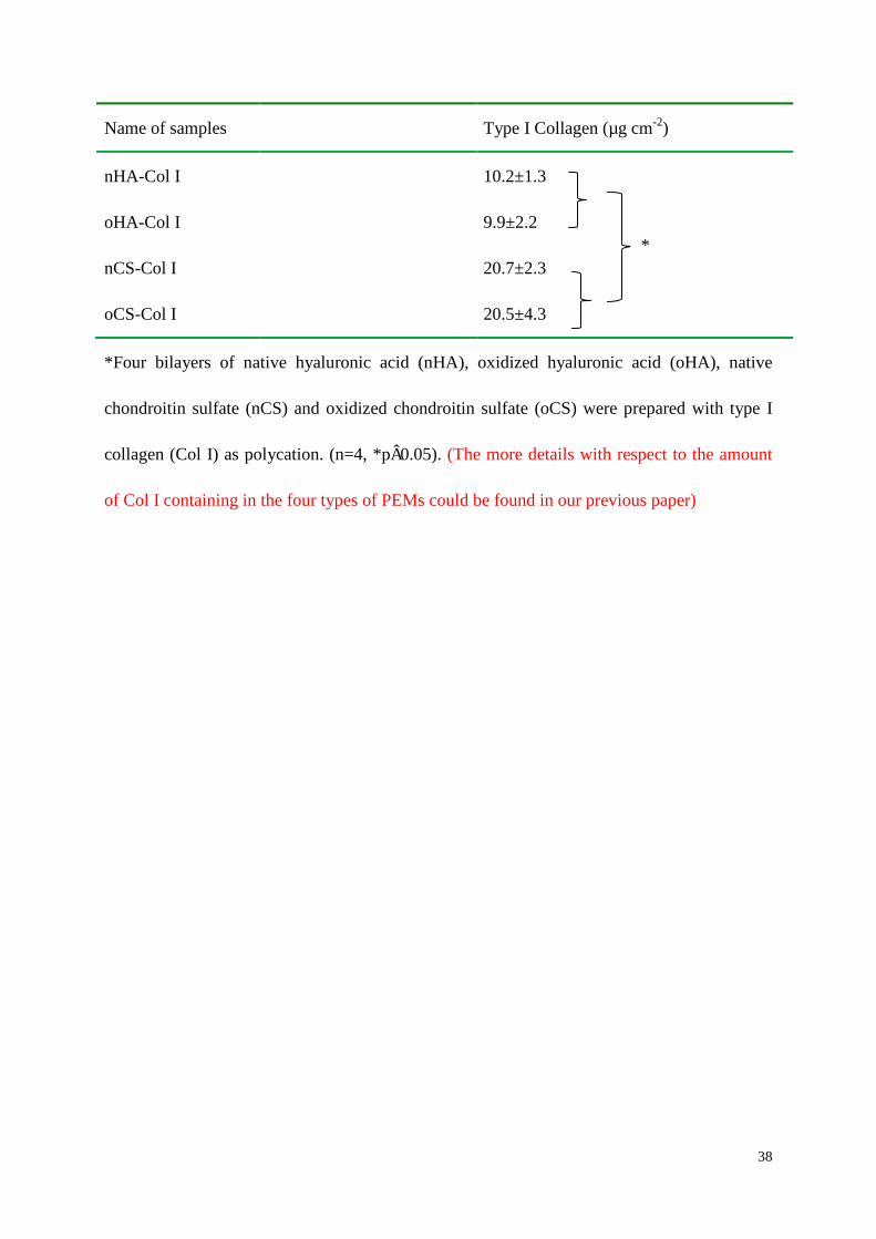
To learn more about the ability of GAG to support fibrillization of Col I in PEMs, multilayers
were investigated with AFM (see Figure 1). Col I aggregates were present in all multilayer
systems, although a prominent fibrous structure was found mostly on CS-based PEMs (both
nCS and oCS) with a tendency to form a network-like structure. Conversely, rather sparse and
short fibrils were observed in both nHA and oHA-containing PEMs, where Col I seemed to
form rather discrete aggregates than fibers. Col I fibrillogenesis is a sophisticated process
highly affected by environmental factors, such as pH, electrolyte type and collagen
concentration, but also on the presence of GAG like CS and other naturally occurring
polyanions[37, 38]. In general, at pH value <5.5 Col I forms more globular structures, while
at pH value >5.5 normally fibrillogenesis is supported[39]. The presence of GAG however,
particularly CS, seems to play an important role in regulating Col I fibril formation as it

20
happened even at pH 4. Also, the concentration of Col I affects strongly fibril formation as
found previously[40, 41]. Presumably CS binds to collagen molecules and facilitates the
organization of mature fibrils by increasing collagen concentration as it was found in the
presence of 4,6-disulfated disaccharides structures, previously[38]. In line with this, here a
pronounced fibril formation in both nCS and oCS-based PEMs was observed, when more
Col I was adsorbed, which showed also a specific network-like organization as in connective
tissue.
3.2. Short-term culture of hADSCs on multilayers
hADSCs were seeded on the samples for 1h in serum-free medium to allow a direct contact of
cells with Col I on the multilayer surfaces avoiding the interference with other proteins.
Thereafter, 10% serum was added and samples were cultured for 3 more hours. The
quantitative data of cell adhesion and spreading are shown in Figure 2. No significant
differences in cell adhesion were found, although cell number was slightly higher on
CS-based PEMs independent on oxidization (see Figure 2 A), which could be related to the
higher amount of fibrillar Col I and the lower wettability of these multilayers. By contrast,
significantly higher extent was found on CS-based (both nCS-Col I and oCS-Col I)
multilayers than on nHA-containing PEMs (p<0.05) as shown in Figure 2 B. It was also
observed that cells spread significantly more on oCS-containing PEMs compared to
oHA-based one, while no evident difference was found between nCS-Col I and oHA-Col I.
To learn more about the organization of hADSCs adhesive machinery, we followed the
formation of focal adhesions (FA), visualized by staining of vinculin and the development of
actin cytoskeleton (viewed by fluorescence-labelled phalloidin) after 4 h of incubation (see

21
Figure 2 C). To quantify the levels of vinculin expressed in hADSCs adhering on the
different PEMs, the corrected total cell fluorescence was calculated by fluorescence signal
with elimination of background signal, performed by ImageJ (shown in figure 2 D). Though a
pronounced cell spreading was found on all samples, a remarkable difference was found in the
overall appearance of FA plaques. Much less extended and rather dot-like FA plaques were
found on HA-based PEMs, while larger and elongated FA plaques where longitudinal actin
stress fibers often inserted (resulting in the yellow color of the merged images) were detected
on CS-based PEMs, which is again well in line with the presence of more fibrillar Col I.
Vinculin as part of FA complexes is directly involved in actin cytoskeleton organization,
which is critical for the development of cell spreading[42].
Since integrins are the major components of FA plaques connecting ECM proteins with the
cell cytoskeleton and signaling complexes, we studied the expression, organization and
quantification of ±2² 1 that is regarded as main collagen receptors[43, 44]. Indeed, as shown in
Figure 3 C and D, ±2² 1 were more expressed and organized in hADSCs adhering on
CS-based PEM surfaces (either nCS-Col I or oCS-Col I), forming clusters resembling small
FA plaques. Conversely, on HA-based PEMs the cells showed a weaker integrin expression
and a rather diffuse distribution with a tendency for accumulation of these integrins at the cell
edges.
The generally stronger adhesion, spreading and integrin organization of cells on
CS-containing PEMs is obviously related to the composition of these PEM systems. First,
more Col I was present in the CS-based multilayers (proven by BCA assay), which forms a
dense fibrillar network resembling the natural Col I environment. The presence of Col I fibrils

22
is known to enhance the adhesion of a variety of cells via an ±2² 1 integrin-mediated
mechanism[45], which explains also the apparently lower organization of this integrin on
HA-based PEMs that contained less Col I. The amount of GAG obviously dominates in the
HA-based systems as shown also in our previous work leading formation of smaller of Col I
[22], which are poorly recognized by integrins. On the other hand, the increased
hydrophilicity of HA-based PEMs may further hinder cell spreading due to the increased
water content of the layers coming from the ability of HA to bind large amounts of water[46].
It is also interesting to note that both spreading and organization of cell adhesion complexes
are slighty or pronounced on oCS-based PEMs compared to its native form. Here we can only
assume that intrinsic cross-linking may lead to additional changes of multilayer properties like
mechanical properties that affect adhesion and spreading of cells[47].
3.3. Remodeling of type I collagen (Col I)
Stability of PEMs made of biogenic polyelectrolytes is discussed critically because of a
possible degradation of biomolecules in the relatively “aggressive” biologic environment,
which is rich in protein degrading enzymes like matrix metalloproteases released by cells[48].
On the other hand remodeling of the adsorbed protein layers on biomaterials seems often
improves the biological performance of adhering cells[11, 28], which can be exploited as
biomimetic approach for creating conditions closer to the natural tissue [49].
3.3.1. Reorganization of type I collagen (Col I) by mechanical remodeling
Figure 4 A presents the overall morphology of hADSCs at low magnification viewed by actin
staining on the background of simultaneously visualized adsorbed Col I after 4 h of
incubation. The figure shows that the cells not only attach to the terminal layer of Col I on

23
PEMs, but also tend to remove the protein from the substratum (dark zones) and arrange it
into fibril-like pattern, particularly evident on CS-based PEMs. The reorganization is better
visible at higher magnification shown on Figure 4 B where only Col I is shown. It should be
underlined that this reorganization is not compromising stability of PEMs since the
remodeling process affected only the outermost layers (Supporting information S1),
suggesting that cells were not able to reach Col I in subzones of both native and oxidized
GAG-based PEMs. In notable contrast, much less reorganization of Col I was found in
HA-containing PEMs, especially on oHA-based PEMs.
These studies show that hADSCs are able to recognize substratum-associated Col I in PEMs
utilizing their integrin receptors, which is supported by the pronouncedly higher accumulation
of ±2² 1 integrin in focal contacts of cells plated on CS-based PEMs. It is of note that distinctly
different percentage of Col I rearrangement was found among the four types of PEM (Figure
4 C). The apparently visible fibril-like pattern particularly on CS-containing PEMs clearly
revealed the ability of hADSCs to remodel Col I involving integrins. Conversely, the less
apparent reorganization of Col I on HA-based PEMs presumably is caused by the lowered
amount of protein on this surface resulting in less binding sites for integrins leading to a
weaker cellular interaction manifested by less FA formation and diminished accumulation of
±2² 1 integrin. An interesting observation was the less pronounced reorganization of Col I on
oGAG-based PEMs, which is presumably owing to the increased stability of the protein layer
by intrinsic cross-linking[22], an additional sign for the increased overall stability of this type
of PEMs, detected also previously with other methods[22].
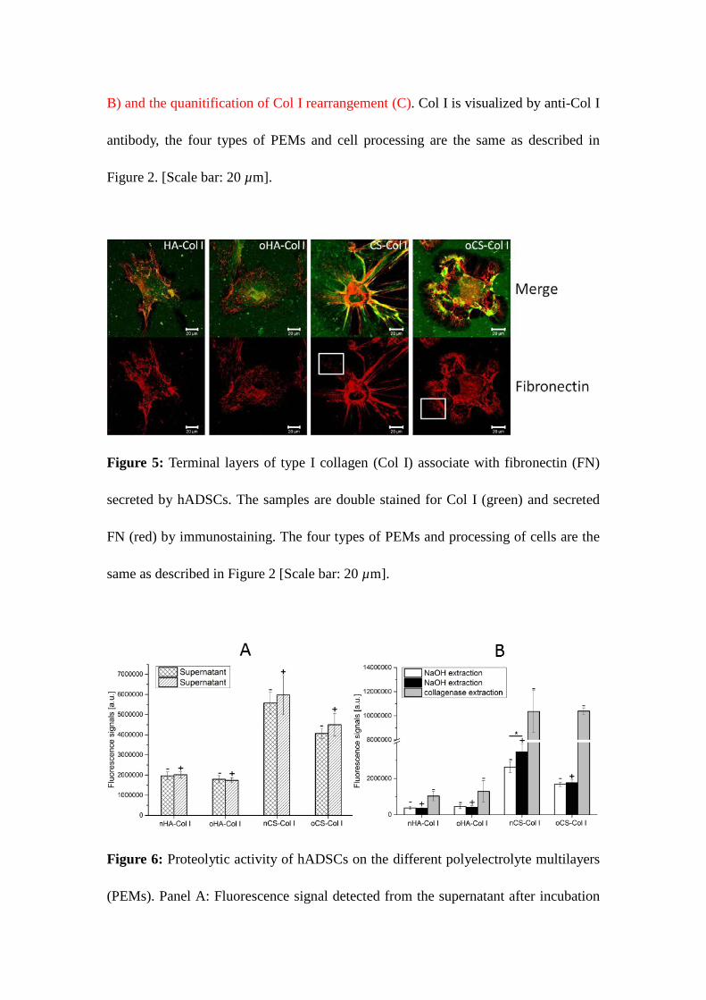
FN molecules possess two binding sites for collagens[50] and have been demonstrated to

24
drive the organization of collagen type I and III into fibrils[51, 52]. Fibril-like reorganization
in the presence of cells was found also for the non-fibrillar collagen type IV[9, 11].
Interestingly, co-staining of FN with Col I in this study showed significant FN secretion on
CS-based PEMs in comparison to HA-containing multilayers (see Figure 5). This indicates an
increased activity of hADSCs on CS-based PEMs by means of de novo synthesis of this
protein. Furthermore, the clearly expressed co-localization of FN with Col I (resulting in
orange on merges seen in the upper panel in Figure 5) indicates a close relationship between
FN fibrillogenesis and mechanical remodeling of Col I. It has to be noted however that a large
part of Col I appears still green, which points to the possibility for an additional, most
probably also integrin-dependent mechanisms contributing to the reorganization process. This
is in agreement with other studies showing that integrin signaling regulates ECM organization
and remodeling and thus contributes to the control of cell behavior in tissues[53]. Also the
enhanced ±2² 1 integrin clustering on CS-containing PEMs, where cells arranged more Col I
fibrils is in line with this (see Figure 3). However, cross-linking of proteins has been shown
to inhibit reorganization processes[54], which explains the reduced Col I reorganization on
crosslinked PEMs using oxidized GAG shown in the present study.
3.3.2. Proteolytic remodeling of FITC labelled type I collagen (FITC-Col I)
Apart from the mechanical tethering of Col I by the cells, another potential mechanism of
Col I remodeling is the pericellular proteolysis by secretion of metallomatrix proteinases
(MMPs) [8]. Here the dark zone around cells in Figure 4 and 5 represents the places from
where the protein was removed to be arranged mechanically in a fibril-like pattern, but
possibly also due to a proteolytic degradation of adsorbed Col I [9, 11]. To study the

25
contribution of proteolytic degradation and to learn also about stability of these PEMs,
FITC-Col I was used to quantify the cellular proteolytic activity by evaluating the release of
labelled FITC-Col I (or its fragments) into the supernatant.
Figure 6 shows the fluorescent signal intensity measured under different conditions, including:
(A) spontaneous release of FITC-Col I in the medium from samples with cells (+) or without
cells (-), and (B) fluorescent signal after extraction with NaOH from the same samples, again
in the presence (+) or absence (-) of cells, characterizing the de-quenching of substratum
bound FITC-Col I (see Methods section). The maximal de-quenching (e.g. complete
proteolysis) caused by the exposure of a given sample to collagenase is also shown for
comparison. Figure 6 A shows that the release of FITC-Col I from all PEM systems after 10 h
incubation did not differ significantly, either with or without cells, though a little tendency
(p>0.05) for increasing the signal in samples “+ cells” for CS-based PEMs was detected.
Significantly higher released fluorescence signal was found for CS-based PEM systems,
which correlates well with the higher Col I content measured by BCA assay. It was also
observed that spontaneous release from nCS-based PEMs was higher than from oCS-based
one, which is another hint for the increased stability due intrinsic cross-linking by imine bond
formation.
Figure 6 B shows the NaOH-extracted fluorescent signal from the samples “with cells” (+)
versus “without cells” (-) compared to the maximal signal from collagenase treated samples.
No effect of the adhering cells was observed in HA-based PEM systems suggesting an absent
or negligible proteolytic activity in comparison to the signal obtained from collagenase treated
PEMs (positive control). However, a significant increase of the signal “(+) cells” versus (–)

26
cells (p < 0.05) was found in nCS–containing PEMs suggesting a valuable proteolytic activity
of hADSCs against Col I in this system. Again absent cell-mediated proteolysis was found in
oCS–based PEMs, which seems to be again related to the enhanced stability of cross-linked
PEMs. Additionaly, we spectulate that when cells adhere onto a surface they do not like so
mcuh, they will attempt to degrade the surrounding matrix to get rid of its shackles,
conversely, when the surface is in favour of cellular interaction, it will suppress the
degradation process, while favoring reorganization ones.
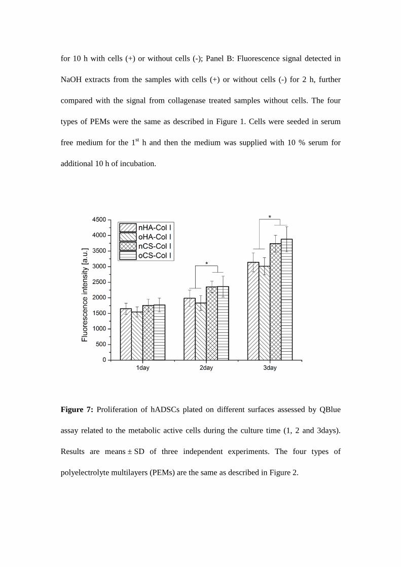
Cell proliferation
The proliferation of hADSCs over a period of 1, 2 and 3 days on the different PEMs was
studied with QBlue assay, which determines the metabolic activity of cells (Figure 7). In
correspondence to the cell adhesion results, there was no significant difference in the
metabolic activity of cells on different PEMs after one day of incubation. However, a
significantly higher metabolic activity (p<0.05) was found on CS-based compared to
HA-containing PEMs at longer incubation time, though no obvious difference was found between
the PEMs with the same type of GAG containing (as shown in Figure 7). It is well documented
that integrin-mediated cell spreading and FA formation triggers cell proliferation and support
the survival of anchorage dependent cells due to activation of MAP kinase pathway and
up-regulation of Bcl-2 family of proteins[55]. Integrin-mediated cell spreading and FA
formation was more pronounced on CS-based PEMs, obviously providing better conditions
for cell growth than PEMs based on HA (as shown in Figure 2 and Figure 3). Furthermore,
the ability of cells to develop a structured ECM at the materials interface was also supported
particularly in nCS but also on oCS-based PEMs, which seems to promote further the activity

27
of cells as shown in previous studies for other type of biomaterials systems[8, 53].
3.4. Osteogenic differentiation of hADSCs
The activity of ALP is an important prerequisite for mineral deposition by osteoblasts, since
the enzyme catalyzes the release of phosphate from phosphate donors for subsequent
precipitation of calcium ions and formation of hydroxyapatite[56]. To evaluate the function of
hADSCs on the various PEMs, ALP activity was normalized to protein content and monitored
at day 5, 11 and 15 post-differentiation (Figure 8). There was no detectable difference of ALP
activity of hADSCs cultured in BM on the different PEMs. However, ALP activity of
hADSCs increased largely after addition of OM between days 5 and 15 on all the test samples
(Figure 8), while there was only a slight increase of ALP activity within 15 days, when cells
were cultured in BM. Notably, pronounced differences were found when comparing the ALP
activity of hADSCs cultured in OM on the four types of PEMs. As shown in Figure 8, both
CS–based PEMs showed significantly higher ALP activity compared to HA-containing PEMs
within the culture time, suggesting that CS-based PEMs promote the osteogenic
differentiation of hADSCs.
The deposition of hydroxyapatite is one feature of mature osteoblasts. hADSCs cultured on
the different PEMs in BM or OM were stained at day 21 post-differentiation with Alizarin
Red S (Figure 9) to visualize calcium phosphate deposition. No significant staining was
observed when hADSCs were cultured in BM (upper panel of Figure 9) indicating the need
of osteogenic inductors for differentiation of these stem cells. However, a staining was
observed on all PEMs when hADSCs were cultured in OM (lower panel of Figure 9),
although the staining was more intense on CS-based compared to HA-based PEMs.
Specifically, oCS-based PEMs induced most extensive calcium deposition, indicating that the

28
intrinsic cross-linking of PEMs enhanced the osteogenic differentiation.
Immunofluorescent staining shown in Figure 10 A was also used to detect de novo expression
of the bone-specific ECM protein Col I (green staining) synthesized by hADSCs (blue nuclear
staining) cultured in either BM or OM after 21 days induction. The upper panel of Figure
10A shows micrographs from hADSCs cultured in BM, where no visible staining of Col I was
observed. This finding demonstrates not only that osteogenic inductors are required for
hADSCs, but also that the Col I initially deposited during PEMs formation did not contribute
to staining after this time. By contrast, intense staining with visualization of a fibrillar
structure of newly synthesized Col I became visible particularly when cells were cultured on
CS-based multilayers in OM (lower panel in Figure 10A). It was also evident here in a
qualitative manner that oCS-based PEMs promoted most Col I synthesis as a more extend
network of Col I was detected here. To further study the effect of molecular composition of
PEMs on the differentiation potential of hADSCs, the osteogenic markers Runx2 and Col I
were measured by qRT-PCR at day 14 post-differentiation. As shown in Figure 10 B,
CS-based PEMs especially the oCS-PEMs caused a significant up-regulation of Runx2
expression in comparison to HA-containing PEMs when hADSCs were cultured in OM.
Runx2 is a zinc finger transcription factor, which is essential for osteoblast differentiation,
acting on the downstream signaling and promoting the expression of important osteoblast
proteins, such as Col I. Notably, a pronounced increase of Col I expression was observed in
hADSCs cultured on oCS-based PEMs, however, no detectable differences were found among
the other three groups (see Figure 10C).
ALP activity test, histochemical staining, immunofluorescence staining, and qRT-PCR results
demonstrate that CS-containing PEM, particularly oCS-based greatly enhance hADSCs
29
osteogenic differentiation compared to cells on HA-containing PEMs in the presence of
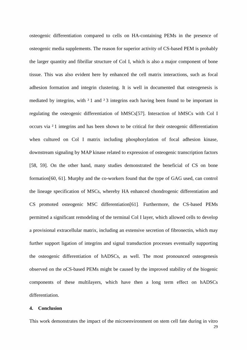
osteogenic media supplements. The reason for superior activity of CS-based PEM is probably
the larger quantity and fibrillar structure of Col I, which is also a major component of bone
tissue. This was also evident here by enhanced the cell matrix interactions, such as focal
adhesion formation and integrin clustering. It is well in documented that osteogenesis is
mediated by integrins, with ² 1 and ² 3 integrins each having been found to be important in
regulating the osteogenic differentiation of hMSCs[57]. Interaction of hMSCs with Col I
occurs via ² 1 integrins and has been shown to be critical for their osteogenic differentiation
when cultured on Col I matrix including phosphorylation of focal adhesion kinase,
downstream signaling by MAP kinase related to expression of osteogenic transcription factors
[58, 59]. On the other hand, many studies demonstrated the beneficial of CS on bone
formation[60, 61]. Murphy and the co-workers found that the type of GAG used, can control
the lineage specification of MSCs, whereby HA enhanced chondrogenic differentiation and
CS promoted osteogenic MSC differentiation[61]. Furthermore, the CS-based PEMs
permitted a significant remodeling of the terminal Col I layer, which allowed cells to develop
a provisional extracellular matrix, including an extensive secretion of fibronectin, which may
further support ligation of integrins and signal transduction processes eventually supporting
the osteogenic differentiation of hADSCs, as well. The most pronounced osteogenesis
observed on the oCS-based PEMs might be caused by the improved stability of the biogenic
components of these multilayers, which have then a long term effect on hADSCs
differentiation.
4. Conclusion
This work demonstrates the impact of the microenvironment on stem cell fate during in vitro

30
culture of hADSCs on polyelectrolyte multilayers (PEM) made of Col I and either HA or CS
by layer-by-layer technique. By varying the type of GAG, we showed that the initial cellular
behavior, and hence the fate of hADSCs regarding osteogenic differentiation was significantly
affected. The CS-based PEMs supported hADSCs osteogenesis most, which was related to an
increased cell spreading, FA formation, pronounced integrin clustering and well-defined actin
cytoskeleton organization at early stages of culture, followed by more growth and osteogenic
differentiation of hADSCs. This study also shows for the first time active remodeling of
biogenic PEMs by adhering cells, based on both mechanical reorganization and pericellular
proteolytic degradation, which obviously allowed further beneficial changes of the
microenvironment of cells. Indeed, both mechanical remodeling and proteolysis were mainly
restricted to terminal Col I layers, while leaving layers underneath unaffected. Hence, such
systems might be also used for uploading bioactive factors to achieve a further
multi-functionality of PEMs. These findings provide new insights to our understanding not
only on the importance of controlling matrix composition as a strategy to manipulate stem cell
fates, but also to the dynamic nature of PEMs in contact with cells, which may open new
avenues for their application in tissue engineering and regenerative medicine.
5. Diclosure
No conflicts of interests are declared.
6. Acknowledgments
The work was funded partly by the European Commission, (FP7-PEOPLE-2012-IAPP) under
grant agreement no. 324386 (FIBROGELNET), the EuroNanoMed project STRUCTGEL and
the Chinese Scholarship Council program funded by Chinese government. Parts of the studies

31
were supported by CIBER-BBN and the project MAT2012-38359-C03-03
HEALINSYNERGY funded by Spanish Ministry of Science and Innovation. We are thankful
to Prof. Changren Zhou and Prof. Lihua Li from Department of Materials Science and
Engineering at Jinan University for providing type I collagen.
7. Reference
1. Özbek S, Balasubramanian PG, Chiquet-Ehrismann R, Tucker RP, Adams JC. The Evolution of Extracellular Matrix. Molecular Biology of the Cell;21(24):4300-4305. 2. Watt FM, Huck WTS. Role of the extracellular matrix in regulating stem cell fate. Nat Rev Mol Cell Biol;14(8):467-473. 3. Moore KA, Lemischka IR. Stem Cells and Their Niches. Science 2006;311(5769):1880-1885. 4. Kresse H, Schönherr E. Proteoglycans of the extracellular matrix and growth control. Journal of Cellular Physiology 2001;189(3):266-274. 5. Liu Z-M, Gu Q, Xu Z-K, Groth T. Synergistic Effect of Polyelectrolyte Multilayers and Osteogenic Growth Medium on Differentiation of Human Mesenchymal Stem Cells. Macromolecular Bioscience 2010;10(9):1043-1054. 6. Phillips JE, Petrie TA, Creighton FP, García AJ. Human mesenchymal stem cell differentiation on self-assembled monolayers presenting different surface chemistries. Acta Biomaterialia 2010;6(1):12-20. 7. Hynes RO. Extracellular matrix: not just pretty fibrils. Science (New York, NY) 2009;326(5957):1216-1219. 8. Daley WP, Peters SB, Larsen M. Extracellular matrix dynamics in development and regenerative medicine. Journal of Cell Science 2008;121(3):255-264. 9. Maneva-Radicheva L, Ebert U, Dimoudis N, Altankov G. Fibroblast remodeling of adsorbed collagen type IV is altered in contact with cancer cells. Histology and histopathology 2008 Jul;23(7):833-842. 10. Tamariz E, Grinnell F. Modulation of fibroblast morphology and adhesion during collagen matrix remodeling. Mol Biol Cell 2002 2002/11//;13(11):3915-3929. 11. Coelho NM, Salmeron-Sanchez M, Altankov G. Fibroblasts remodeling of type IV collagen at a biomaterials interface. Biomaterials Science 2013;1(5):494-502. 12. Giraud Guille MM, Mosser G Fau - Helary C, Helary C Fau - Eglin D, Eglin D. Bone matrix like assemblies of collagen: from liquid crystals to gels and biomimetic materials. (0968-4328 (Print)). 13. Matsumoto N, Horibe S, Nakamura N, Senda T, Shino K, Ochi T. Effect of alignment of the transplanted graft extracellular matrix on cellular repopulation and newly synthesized collagen. Archives of Orthopaedic and Trauma Surgery 1998;117(4-5):215-221. 14. He D, Xiao X, Liu F, Liu R. Chondroitin sulfate template-mediated biomimetic synthesis of nano-flake hydroxyapatite. Applied Surface Science 2008;255(2):361-364. 15. Liu Y, Wang X, Kaufman DS, Shen W. A synthetic substrate to support early mesodermal differentiation of human embryonic stem cells. Biomaterials 2011 11//;32(32):8058-8066. 16. Chen X-D. Extracellular matrix provides an optimal niche for the maintenance and propagation of mesenchymal stem cells. Birth Defects Research Part C: Embryo Today: Reviews 2010;90(1):45-54. 17. Zuk PA, Zhu M, Mizuno H, Huang J, Futrell JW, Katz AJ, et al. Multilineage cells from human adipose tissue: implications for cell-based therapies. Tissue engineering 2001 Apr;7(2):211-228. 18. Hong JM, Kim BJ, Shim J-H, Kang KS, Kim K-J, Rhie JW, et al. Enhancement of bone regeneration through
32
facile surface functionalization of solid freeform fabrication-based three-dimensional scaffolds using mussel adhesive proteins. Acta Biomaterialia 2012 7//;8(7):2578-2586. 19. Shin H, Jo S, Mikos AG. Biomimetic materials for tissue engineering. Biomaterials 2003 11//;24(24):4353-4364. 20. Rodriguez-Velazquez E, Alatorre-Meda M Fau - Mano JF, Mano JF. Polysaccharide-Based Nanobiomaterials as Controlled Release Systems for Tissue Engineering Applications. Curr Pharm Des 2015;21(33):4837-4850. 21. Rahmany MB, Van Dyke M. Biomimetic approaches to modulate cellular adhesion in biomaterials: A review. Acta Biomaterialia 2013 3//;9(3):5431-5437. 22. Zhao M, Li L, Zhou C, Heyroth F, Fuhrmann B, Maeder K, et al. Improved Stability and Cell Response by Intrinsic Cross-Linking of Multilayers from Collagen I and Oxidized Glycosaminoglycans. Biomacromolecules 2014;15(11):4272-4280. 23. Oliveira SM, Santo VE, Gomes ME, Reis RL, Mano JF. Layer-by-layer assembled cell instructive nanocoatings containing platelet lysate. Biomaterials 2015;48:56-65. 24. Mhanna RF, Vörös J, Zenobi-Wong M. Layer-by-Layer Films Made from Extracellular Matrix Macromolecules on Silicone Substrates. Biomacromolecules 2011 2011/03/14;12(3):609-616. 25. Chaubaroux C, Vrana E, Debry C, Schaaf P, Senger B, Voegel J-C, et al. Collagen-Based Fibrillar Multilayer Films Cross-Linked by a Natural Agent. Biomacromolecules 2012 2012/07/09;13(7):2128-2135. 26. Ko WH, Suminto JT, Yeh GJ. Bonding techniques for microsensors, 1985. 27. Johansson JÅ, Halthur T, Herranen M, Söderberg L, Elofsson U, Hilborn J. Build-up of Collagen and Hyaluronic Acid Polyelectrolyte Multilayers. Biomacromolecules 2005 2005/05/01;6(3):1353-1359. 28. Altankov G, Grinnell F, Groth T. Studies on the biocompatibility of materials: Fibroblast reorganization of substratum-bound fibronectin on surfaces varying in wettability. Journal of Biomedical Materials Research 1996;30(3):385-391. 29. Ochsner M, Textor M, Vogel V, ML. S. Dimensionality controls cytoskeleton assembly and metabolism of fibroblast cells in response to rigidity and shape. PLoS ONE 2010;5(3):e9445. 30. Bathawab F, Bennett M, Cantini M, Reboud J, Dalby MJ, Salmeron-Sanchez M. Lateral Chain Length in Polyalkyl Acrylates Determines the Mobility of Fibronectin at the Cell/Material Interface. Langmuir 2016;32(3):800-809. 31. McSwain BS, Irvine RL, Hausner M, Wilderer PA. Composition and Distribution of Extracellular Polymeric Substances in Aerobic Flocs and Granular Sludge. Applied and Environmental Microbiology 2005;71(2):1051-1057. 32. Chomczynski P Fau - Sacchi N, Sacchi N. Single-step method of RNA isolation by acid guanidinium thiocyanate-phenol-chloroform extraction. (0003-2697 (Print)). 33. Livak KJ, Schmittgen TD. Analysis of Relative Gene Expression Data Using Real-Time Quantitative PCR and
the 2 鈭捨斘擟 T Method. Methods 2001;25(4):402-408.
34. Gribova V, Auzely-Velty R, Picart C. Polyelectrolyte Multilayer Assemblies on Materials Surfaces: From Cell Adhesion to Tissue Engineering. Chemistry of Materials 2012;24(5):854-869. 35. Aggarwal N, Altgärde N, Svedhem S, Zhang K, Fischer S, Groth T. Effect of Molecular Composition of Heparin and Cellulose Sulfate on Multilayer Formation and Cell Response. Langmuir 2013 2013/11/12;29(45):13853-13864. 36. Duval JFL, Küttner D, Werner C, Zimmermann R. Electrohydrodynamics of Soft Polyelectrolyte Multilayers: Point of Zero-Streaming Current. Langmuir 2011;27(17):10739-10752. 37. Wood BC. The Formation of Fibrils from Collagen Solutions 3. Effect of chondroitin sulfate and some other naturally occurring polyanions on the rate of formation. Biochem J 1960;75(3):605-612.

33
38. Kvist AJ, Johnson AE, Mörgelin M, Gustafsson E, Bengtsson E, Lindblom K, et al. Chondroitin sulfate perlecan enhances collagen fibril formation. Implications for perlecan chondrodysplasias. J Biol Chem 2006;281(44):33127-33139. 39. Jiang F, Hörber H, Howard J, Müller DJ. Assembly of collagen into microribbons: effects of pH and electrolytes. Journal of Structural Biology 2004 12//;148(3):268-278. 40. Raspanti M, Viola M, Sonaggere M, Tira ME, Tenni R. Collagen Fibril Structure Is Affected by Collagen Concentration and Decorin. Biomacromolecules 2007 2007/07/01;8(7):2087-2091. 41. Gobeaux F, Mosser G, Anglo A, Panine P, Davidson P, Giraud-Guille MM, et al. Fibrillogenesis in Dense Collagen Solutions: A Physicochemical Study. Journal of Molecular Biology 2008 3/7/;376(5):1509-1522. 42. Ezzell RM, Goldmann WH, Wang N, Parasharama N, Ingber DE. Vinculin Promotes Cell Spreading by Mechanically Coupling Integrins to the Cytoskeleton. Experimental Cell Research 1997 2/25/;231(1):14-26. 43. Hynes RO. Integrins: Bidirectional, Allosteric Signaling Machines. Cell 2002 9/20/;110(6):673-687. 44. Khoshnoodi J, Pedchenko V, Hudson BG. Mammalian collagen IV. Microscopy Research and Technique 2008;71(5):357-370. 45. Coelho NM, Gonzalez-Garcia C, Planell JA, Salmeron-Sanchez M, Altankov G. Different assembly of type IV collagen on hydrophilic and hydrophobic substrata alters endothelial cells interaction. European cells & materials 2010;19:262-272. 46. Whitson KB, Lukan AM, Marlowe RL, Lee SA, Anthony L, Rupprecht A. Binding of the water of primary hydration to the sodium and cesium salts of deoxyribonucleic acid and potassium hyaluronate. Physical Review E 1998 08/01/;58(2):2370-2377. 47. Richert L, Engler AJ, Discher DE, Picart C. Elasticity of Native and Cross-Linked Polyelectrolyte Multilayer Films. Biomacromolecules 2004;5(5):1908-1916. 48. Picart C. Polyelectrolyte multilayer films: from physico-chemical properties to the control of cellular processes. Curr Med Chem 2008;15(7):685-697. 49. Lutolf MP, Hubbell JA. Synthetic biomaterials as instructive extracellular microenvironments for morphogenesis in tissue engineering. Nat Biotech 2005 01//print;23(1):47-55. 50. Mao Y, Schwarzbauer JE. Fibronectin fibrillogenesis, a cell-mediated matrix assembly process. Matrix Biology 2005 9//;24(6):389-399. 51. Velling T, Risteli J, Wennerberg K, Mosher DF, Johansson S. Polymerization of Type I and III Collagens Is Dependent On Fibronectin and Enhanced By Integrins α11β1and α2β1. Journal of Biological Chemistry 2002;277(40):37377-37381. 52. Kadler KE, Hill A, Canty-Laird EG. Collagen fibrillogenesis: fibronectin, integrins, and minor collagens as organizers and nucleators. Current Opinion in Cell Biology 2008 10//;20(5):495-501. 53. Larsen M, Artym VV, Green JA, Yamada KM. The matrix reorganized: extracellular matrix remodeling and integrin signaling. Current Opinion in Cell Biology 2006 10//;18(5):463-471. 54. Grinnell F. Focal adhesion sites and the removal of substratum-bound fibronectin. The Journal of Cell Biology 1986;103(6):2697-2706. 55. Hynes RO. Integrins: Versatility, modulation, and signaling in cell adhesion. Cell 1992;69(1):11-25. 56. Orimo H. The Mechanism of Mineralization and the Role of Alkaline Phosphatase in Health and Disease. Journal of Nippon Medical School 2010;77(1):4-12. 57. Shekaran A, García AJ. Extracellular matrix-mimetic adhesive biomaterials for bone repair. Journal of Biomedical Materials Research Part A;96A(1):261-272. 58. Lund AW, Stegemann Jp Fau - Plopper GE, Plopper GE. Inhibition of ERK promotes collagen gel compaction and fibrillogenesis to amplify the osteogenesis of human mesenchymal stem cells in three-dimensional collagen I culture. (1557-8534 (Electronic)).

34
59. Salasznyk RM, Klees Rf Fau - Hughlock MK, Hughlock Mk Fau - Plopper GE, Plopper GE. ERK signaling pathways regulate the osteogenic differentiation of human mesenchymal stem cells on collagen I and vitronectin. (1541-9061 (Print)). 60. Rammelt S, Illert T, Bierbaum S, Scharnweber D, Zwipp H, Schneiders W. Coating of titanium implants with collagen, RGD peptide and chondroitin sulfate. Biomaterials 2006;27(32):5561-5571. 61. Murphy CM, Matsiko A, Haugh MG, Gleeson JP, O’Brien FJ. Mesenchymal stem cell fate is regulated by the composition and mechanical properties of collagen–glycosaminoglycan scaffolds. Journal of the Mechanical Behavior of Biomedical Materials 2012 7//;11(0):53-62.
8. Figure captions
Figure 1: Surface topography of type I collagen (Col I) terminated polyelectrolyte multilayer
(PEMs) (8th layer) visualized by atomic force microscopy (AFM). Four bilayers of native
hyaluronic acid (nHA), oxidized hyaluronic acid (oHA), native chondroitin sulfate (nCS) or
oxidized chondroitin sulfate (oCS) were prepared with Col I as polycation. (Scale bar: 1 µm)
(The relative high magnification images of the PEMs could be also found in our previous
published paper)
Figure 2: Cell adhesion (A, means ± SD), spreading (B, Box−whisker diagram indicating the
25th and 75th percentile and median plus mean values as small black squares), (C) confocal
laser scanning micrographs (CLSM) with staining of vinculin (green) and actin (red) of
hADSCs after 4 h incubation on different polyelectrolyte multilayers (PEMs) and
fluorescence signals of vinculin in panel (C) was quantified by ImageJ and corrected total cell
fluorescence was calculated by fluorescence signal with elimination of background signal (D).
Four bilayers of native hyaluronic acid (nHA), oxidized hyaluronic acid (oHA), native
chondroitin sulfate (nCS) or oxidized chondroitin sulfate (oCS) were prepared with type I
collagen (Col I) as terminal layer. Cells were seeded in serum free medium for the 1st hour
and then supplied with 10 % serum for the next 3 h of incubation. [Scale bar: 20 µm]

35
Figure 3: Expression of ±2² 1 integrin (green) in hADSCs adhering on different test samples
after 4 h incubation and the respective merged images with filamentous actin (red) (A);
Fluorescence signals of ±2² 1 integrin in panel (A) was quantified by ImageJ and corrected
total cell fluorescence was calculated by fluorescence signal with elimination of background
signal (B). The four types of polyelectrolyte multilayers (PEMs) and cells processing are the
same as described in Figure 2. [Scale bar: 20 µm].
Figure 4: Remodeling of type I collagen (Col I) (green) on the different polyelectrolyte
multilayers (PEMs) by adhering hADSCs (stained for actin in red) (A, B) and the
quanitification of Col I rearrangement (C). Col I is visualized by anti-Col I antibody, the four
types of PEMs and cell processing are the same as described in Figure 2. [Scale bar: 20 µm].
Figure 5: Terminal layers of type I collagen (Col I) associate with fibronectin (FN) secreted
by hADSCs. The samples are double stained for Col I (green) and secreted FN (red) by
immunostaining. The four types of PEMs and processing of cells are the same as described in
Figure 2 [Scale bar: 20 µm].
Figure 6: Proteolytic activity of hADSCs on the different polyelectrolyte multilayers (PEMs).
Panel A: Fluorescence signal detected from the supernatant after incubation for 10 h with cells
(+) or without cells (-); Panel B: Fluorescence signal detected in NaOH extracts from the
samples with cells (+) or without cells (-) for 2 h, further compared with the signal from
collagenase treated samples without cells. The four types of PEMs were the same as described
in Figure 1. Cells were seeded in serum free medium for the 1st h and then the medium was
supplied with 10 % serum for additional 10 h of incubation.
Figure 7: Proliferation of hADSCs plated on different surfaces assessed by QBlue assay

36
related to the metabolic active cells during the culture time (1, 2 and 3days). Results are
means ± SD of three independent experiments. The four types of polyelectrolyte multilayers
(PEMs) are the same as described in Figure 2.
Figure 8: Activity of alkaline phosphates (ALP) normalized to protein content of hADSCs
plated on the different polyelectrolyte multilayers after 3 weeks induction with basal medium
(BM) and osteogenic differentiation medium (OM), respectively. ALP activity was
determined after lysis of cells and use of p-nitrophenylphosphate (pNPP) as substrate,
normalized to the protein content of the lysates. The four types of PEMs were the same as
described in Figure 2.
Figure 9: Histochemical staining of calcium phosphate with Alizarin Red S at day 21
post-osteogenic differentiation. hADSCs were cultured in BM (basal medium; upper panel)
and OM (osteogenic differentiation medium; lower panel); The four types of polyelectrolyte
multilayers (PEMs) were the same as described in Figure 2.
Figure 10: Expression of osteogenic markers in hADSCs cultured on the different
polyelectrolyte multilayers (PEMs) in basal medium (BM) and osteogenic differentiation
medium (OM) respectively. (A) Immunofluorescence staining of type I collagen (Col I) in
hADSCs at day 21 post-osteogenic differentiation in presence of BM (upper panel) and OM
(lower panel); Bar charts showing the expression of Runx 2 (B) and Col IA (C) in hADSCs at
day 14 post-osteogenic differentiation measured by qRT-PCR. Relative gene expression is
presented as normalized to gene expression by hADSCs cultured on plain glass. The four
types of PEMs were the same as described in Figure 2.
9. Tables

37
Table 1: Primers used for qRT-PCR
Forward primer Reverse primer size
² -actin ACTCCTACGTGGGCGACGAGG CAGGTCCAGACGCAGGATGGC 389 bp
Col IA GCCAAGACGAAGACATCCCA CACCATCATTTCCACGAGCA 891 bp
Runx 2 CTCACTACCACACCTACCTG TCAATATGGTCGCCAAACAGATTC 320 bp
Table 2: Surface properties and thickness of type I collagen (Col I) terminated polyelectrolyte
multilayer (PEM) systems.
Multilayer systems Water contact anglea (o) Zeta potentialb (mV) Thicknessc (nm)
nHA-Col I ~34.3o±1.7 o -57.53±0.29 11.89±1.12
oHA-Col I ~26.6o±1.6 o -42.33±1.93 25.62±1.53
nCS-Col I ~45.2o±1.4 o -44.05±0.54 13.55±0.06
oCS-Col I ~43.4o±3.4 o -37.54±1.06 17.64±0.65
*Four bilayers of native hyaluronic acid (nHA), oxidized hyaluronic acid (oHA), native
chondroitin sulfate (nCS) and oxidized chondroitin sulfate (oCS) were prepared with type I
collagen (Col I) as polycation. aStatic water contact angles (WCA) for Col I terminated
polyelectrolyte multilayers (PEMs) (8th layer). (n=16, *pÂ0.05). bZeta potential of outermost
Col I layer for multilayer systems measured at pH 7.4 in 1 mM KCL. c Layer thickness was
determined by ellipsometry in PBS buffer, pH 7.4. (The more details regarding the properties
of PEM systems could be found in our previous paper)
Table 3: Type I collagen (Col I) content of polyelectrolyte multilayers (PEMs)*.
*
*
38
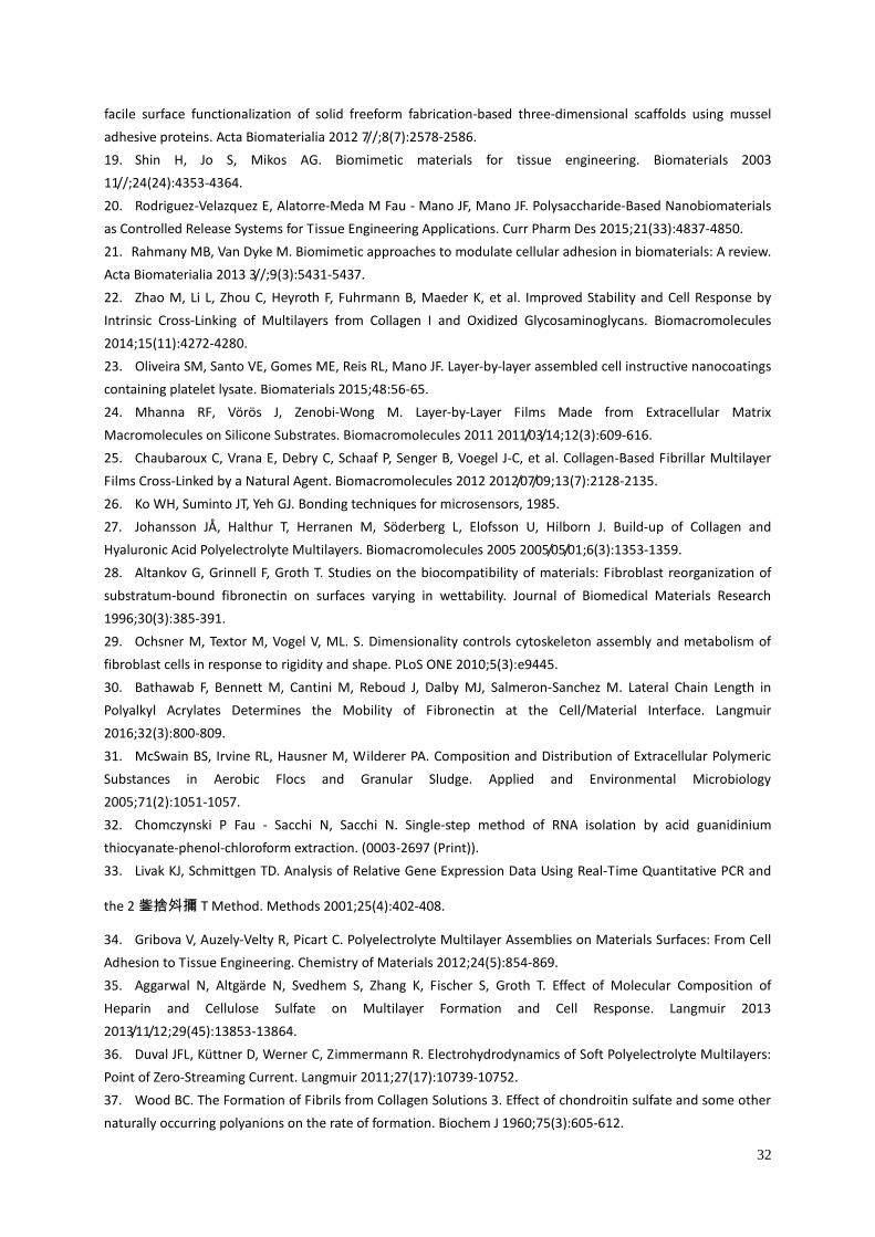
Name of samples Type I Collagen (µg cm-2)
nHA-Col I 10.2±1.3
oHA-Col I 9.9±2.2
nCS-Col I 20.7±2.3
oCS-Col I 20.5±4.3
*Four bilayers of native hyaluronic acid (nHA), oxidized hyaluronic acid (oHA), native
chondroitin sulfate (nCS) and oxidized chondroitin sulfate (oCS) were prepared with type I
collagen (Col I) as polycation. (n=4, *pÂ0.05). (The more details with respect to the amount
of Col I containing in the four types of PEMs could be found in our previous paper)
*
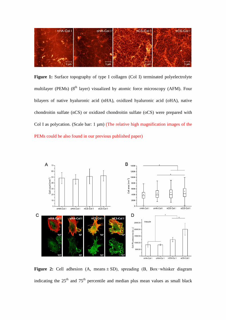
Figure 1: Surface topography of type I collagen (Col I) terminated polyelectrolyte
multilayer (PEMs) (8th layer) visualized by atomic force microscopy (AFM). Four
bilayers of native hyaluronic acid (nHA), oxidized hyaluronic acid (oHA), native
chondroitin sulfate (nCS) or oxidized chondroitin sulfate (oCS) were prepared with
Col I as polycation. (Scale bar: 1 µm) (The relative high magnification images of the
PEMs could be also found in our previous published paper)
Figure 2: Cell adhesion (A, means ± SD), spreading (B, Box−whisker diagram
indicating the 25th and 75th percentile and median plus mean values as small black

squares), (C) confocal laser scanning micrographs (CLSM) with staining of vinculin
(green) and actin (red) of hADSCs after 4 h incubation on different polyelectrolyte
multilayers (PEMs) and fluorescence signals of vinculin in panel (C) was quantified
by ImageJ and corrected total cell fluorescence was calculated by fluorescence signal
with elimination of background signal (D). Four bilayers of native hyaluronic acid
(nHA), oxidized hyaluronic acid (oHA), native chondroitin sulfate (nCS) or oxidized
chondroitin sulfate (oCS) were prepared with type I collagen (Col I) as terminal layer.
Cells were seeded in serum free medium for the 1st hour and then supplied with 10 %
serum for the next 3 h of incubation. [Scale bar: 20 µm]

Figure 3: Expression of ±2² 1 integrin (green) in hADSCs adhering on different test
samples after 4 h incubation and the respective merged images with filamentous actin
(red) (A); Fluorescence signals of ±2² 1 integrin in panel (A) was quantified by ImageJ
and corrected total cell fluorescence was calculated by fluorescence signal with
elimination of background signal (B). The four types of polyelectrolyte multilayers
(PEMs) and cells processing are the same as described in Figure 2. [Scale bar:
20 µm].
Figure 4: Remodeling of type I collagen (Col I) (green) on the different
polyelectrolyte multilayers (PEMs) by adhering hADSCs (stained for actin in red) (A,
B) and the quanitification of Col I rearrangement (C). Col I is visualized by anti-Col I
antibody, the four types of PEMs and cell processing are the same as described in
Figure 2. [Scale bar: 20 µm].
Figure 5: Terminal layers of type I collagen (Col I) associate with fibronectin (FN)
secreted by hADSCs. The samples are double stained for Col I (green) and secreted
FN (red) by immunostaining. The four types of PEMs and processing of cells are the
same as described in Figure 2 [Scale bar: 20 µm].
Figure 6: Proteolytic activity of hADSCs on the different polyelectrolyte multilayers
(PEMs). Panel A: Fluorescence signal detected from the supernatant after incubation
for 10 h with cells (+) or without cells (-); Panel B: Fluorescence signal detected in
NaOH extracts from the samples with cells (+) or without cells (-) for 2 h, further
compared with the signal from collagenase treated samples without cells. The four
types of PEMs were the same as described in Figure 1. Cells were seeded in serum
free medium for the 1st h and then the medium was supplied with 10 % serum for
additional 10 h of incubation.
Figure 7: Proliferation of hADSCs plated on different surfaces assessed by QBlue
assay related to the metabolic active cells during the culture time (1, 2 and 3days).
Results are means ± SD of three independent experiments. The four types of
polyelectrolyte multilayers (PEMs) are the same as described in Figure 2.
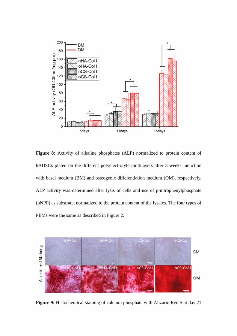
Figure 8: Activity of alkaline phosphates (ALP) normalized to protein content of
hADSCs plated on the different polyelectrolyte multilayers after 3 weeks induction
with basal medium (BM) and osteogenic differentiation medium (OM), respectively.
ALP activity was determined after lysis of cells and use of p-nitrophenylphosphate
(pNPP) as substrate, normalized to the protein content of the lysates. The four types of
PEMs were the same as described in Figure 2.
Figure 9: Histochemical staining of calcium phosphate with Alizarin Red S at day 21
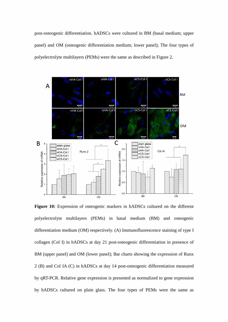
post-osteogenic differentiation. hADSCs were cultured in BM (basal medium; upper
panel) and OM (osteogenic differentiation medium; lower panel); The four types of
polyelectrolyte multilayers (PEMs) were the same as described in Figure 2.
Figure 10: Expression of osteogenic markers in hADSCs cultured on the different
polyelectrolyte multilayers (PEMs) in basal medium (BM) and osteogenic
differentiation medium (OM) respectively. (A) Immunofluorescence staining of type I
collagen (Col I) in hADSCs at day 21 post-osteogenic differentiation in presence of
BM (upper panel) and OM (lower panel); Bar charts showing the expression of Runx
2 (B) and Col IA (C) in hADSCs at day 14 post-osteogenic differentiation measured
by qRT-PCR. Relative gene expression is presented as normalized to gene expression
by hADSCs cultured on plain glass. The four types of PEMs were the same as

described in Figure 2.