Measurement of ocular anterior segment dimension and ......Measurement of ocular anterior segment...
4
Measurement of ocular anterior segment dimension and wavefront aberration simultaneously during accommodation Guohua Shi Yuanyuan Wang Yimin Yuan Lin Wei Fan Lv Yudong Zhang Downloaded From: https://www.spiedigitallibrary.org/journals/Journal-of-Biomedical-Optics on 26 Apr 2020 Terms of Use: https://www.spiedigitallibrary.org/terms-of-use
Transcript of Measurement of ocular anterior segment dimension and ......Measurement of ocular anterior segment...
Measurement of ocular anterior segmentdimension and wavefront aberrationsimultaneously during accommodation
Guohua ShiYuanyuan WangYimin YuanLin WeiFan LvYudong Zhang
Downloaded From: https://www.spiedigitallibrary.org/journals/Journal-of-Biomedical-Optics on 26 Apr 2020Terms of Use: https://www.spiedigitallibrary.org/terms-of-use
Measurement of ocularanterior segment dimensionand wavefront aberrationsimultaneously duringaccommodation
Guohua Shi,a,b* Yuanyuan Wang,c* Yimin Yuan,cLin Wei,a,b Fan Lv,c and Yudong Zhanga,baThe Chinese Academy of Sciences, The Key Laboratory on AdaptiveOptics, Chengdu 610209, ChinabThe Chinese Academy of Sciences, The Institute of Optics andElectronics, Chengdu 610209, ChinacWenzhou Medical College, School of Ophthalmology & Optometry,Wenzhou 32500, China
Abstract. In order to understand the relationship betweenaccommodation and vision quality, a custom-built ultra-long scan depth spectral domain optical coherence tomog-raphy (UL-SDOCT) and a Shack-Hartmann wavefrontsensor (HSWFS) were combined. The resolution and scandepth of UL-SDOCT are 6 μm and 15.6 mm, respectively,which allow for high-resolution imaging of the whole ante-rior segment. The HSWFS consists of a 32 × 32 microlensarray, and is able to measure first 35th-order Zernike aberra-tions with ð1∕20Þλ measurement accuracy. The integratedsystem succeeded in measuring the ocular anterior segmentdimension parameters and the ocular monochromatichigh-order aberrations simultaneously under the conditionsof nonaccommodative and accommodative stimuli. Thismay help understand the regulatory mechanism of imagequality control in the human eye. © 2012 Society of Photo-Optical
Instrumentation Engineers (SPIE). [DOI: 10.1117/1.JBO.17.12.120501]
Keywords: optical coherence tomography; wavefront sensor; accom-modation; wavefront aberration.
Paper 12629L received Sep. 22, 2012; revised manuscript receivedNov. 6, 2012; accepted for publication Nov. 8, 2012; published onlineNov. 28, 2012.
The purpose of accommodation is to compensate for retinaldefocus. According to the theory of Helmholtz1 and Fincham2
the ocular anterior segment changes significantly during accom-modation to bring the objects from different distance into focuson the retina. However, the accommodation also induces thechange in ocular wavefront aberration3 which determines thevision quality. Thus, it is necessary to explore factors, whichalter ocular aberrations during the accommodation. This mayhelp understand the regulatory mechanism of imaging qualitycontrol in the human eye.
Some imaging methods, such as magnetic resonance imaging,4
Scheimpflug photography,4 and ultrasound biomicroscopy,5
have measured the dimension change of ocular anteriorsegment. Furthermore, Shack-Hartmann wavefront sensor(SHWFS) is the successful wavefront measuring device whichis widely used in vision research,6 but previous studies separatedthe measurement of ocular anterior segment dimension andwavefront aberrations. Besides, these imaging methods havesome drawbacks, for example low resolution, slow scan speed,and contactness. So in order to understand the relationshipbetween accommodation and vision quality, an integrated sys-tem which can quantify the changes of ocular anterior segmentand wavefront synchronously is required.
Optical coherence tomography (OCT) is a rapid and non-contact imaging method. It is widely used in anterior segmentimaging.7–10 The use of near-infrared light makes it possible tomaintain the natural accommodative condition during OCTimaging, so OCT is an ideal tool to study the accommodationin vivo. In this study, we have developed an integrated systemwhich consists of a custom-built, ultra-long scan depth spectraldomain optical coherence tomography (UL-SDOCT) with aShack-Hartmann wavefront sensor (SHWFS). The systemmakes it possible to carry out dimension and wavefront aberra-tion measuring synchronously during accommodation. Thereby,the differences in the dimensional parameters and high orderaberrations (HOAs) between the accommodative and non-accommodative states can be compared to investigate therelationships between the biometric dimensions of the ocularanterior segment and the ocular high-order aberrations duringaccommodation.
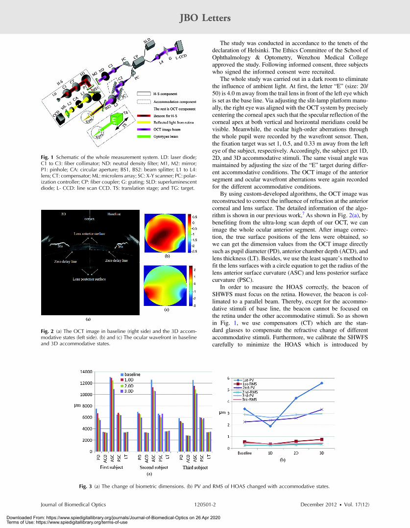
Figure 1 is the schematic of whole system combining theUL-SDOCT, SHWFS, and vision target. The UL-SDOCTcomponent is based on a classical fiber-type Michelson inter-ferometer. By using a broadband SLD (Superlum, Russia,SLD_371-HP1) which is centered at 840 nm with the bandwidthof 50 nm, the UL-SDOCT leads to 6.0 μm axial resolution in air.A custom spectrometer which consists of a volume holographicdiffraction grating (1; 800 lines∕mm; Wasatch Photonics,Logan, Uttah) and a line scans CCD (Aviiva-SM2010, 2,048pixels; Atmel, e2v Inc., Elmsford, New York) was developed.As detailed in our previous work,11 by using a custom method,the raw image with mirror artifacts was processed and recon-structed to obtain the full-range (equivalent depth was 15.6 mm)anterior segment image.
The SHWFS component has a 32 × 32 microlens array.An LD (780 nm) functions as a beacon of SHWFS. By usingP1, the paraxial illumination is formed to reduce the cornealreflected light. Besides, the CA is conjugated with the retinaso as to eliminate the stray light. It is able to measure first35th order Zernike aberrations with ð1∕20Þλ measurementaccuracy.
A fixation target component was also built. Awhite “E” fixa-tion target with a black background was displayed on the liquid-crystal display monitor, with a 20/100 letter according to theSnellen visual acuity chart that can be moved forward andbackward to guide the subject to do accommodation.
SHWFS and the sample arm of UL-SDOCT were mountedon a modified slit-lamp platform. The dichroic beam splitter(BS2) was used to combine the two measuring lights. The mea-surement of dimension and wavefront aberration was done onthe right eye while the accommodative stimuli was given on theleft eye.*These authors contributed equally to this work.
Address all correspondence to: Fan Lv or Yudong Zhang, The Chinese Academyof Sciences, The institute of Optics and Electronics, Chengdu Shuangliu 610209,China. Tel: +86-28-8510-0168; E-mail: [email protected] 0091-3286/2012/$25.00 © 2012 SPIE
Journal of Biomedical Optics 120501-1 December 2012 • Vol. 17(12)
JBO Letters
Downloaded From: https://www.spiedigitallibrary.org/journals/Journal-of-Biomedical-Optics on 26 Apr 2020Terms of Use: https://www.spiedigitallibrary.org/terms-of-use
The study was conducted in accordance to the tenets of thedeclaration of Helsinki. The Ethics Committee of the School ofOphthalmology & Optometry, Wenzhou Medical Collegeapproved the study. Following informed consent, three subjectswho signed the informed consent were recruited.
The whole study was carried out in a dark room to eliminatethe influence of ambient light. At first, the letter “E” (size: 20/50) is 4.0 m away from the trail lens in front of the left eye whichis set as the base line. Via adjusting the slit-lamp platform manu-ally, the right eye was aligned with the OCT system by preciselycentering the corneal apex such that the specular reflection of thecorneal apex at both vertical and horizontal meridians could bevisible. Meanwhile, the ocular high-order aberrations throughthe whole pupil were recorded by the wavefront sensor. Then,the fixation target was set 1, 0.5, and 0.33 m away from the lefteye of the subject, respectively. Accordingly, the subject get 1D,2D, and 3D accommodative stimuli. The same visual angle wasmaintained by adjusting the size of the “E” target during differ-ent accommodative conditions. The OCT image of the anteriorsegment and ocular wavefront aberrations were again recordedfor the different accommodative conditions.
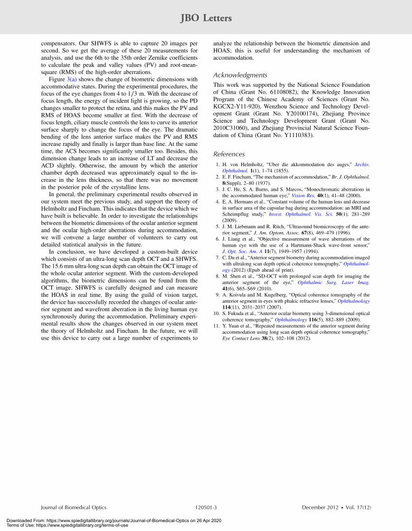
By using custom-developed algorithms, the OCT image wasreconstructed to correct the influence of refraction at the anteriorcorneal and lens surface. The detailed information of the algo-rithm is shown in our previous work,7 As shown in Fig. 2(a), bybenefiting from the ultra-long scan depth of our OCT, we canimage the whole ocular anterior segment. After image correc-tion, the true surface positions of the lens were obtained, sowe can get the dimension values from the OCT image directlysuch as pupil diameter (PD), anterior chamber depth (ACD), andlens thickness (LT). Besides, we use the least square’s method tofit the lens surfaces with a circle equation to get the radius of thelens anterior surface curvature (ASC) and lens posterior surfacecurvature (PSC).
In order to measure the HOAS correctly, the beacon ofSHWFS must focus on the retina. However, the beacon is col-limated to a parallel beam. Thereby, except for the accommo-dative stimuli of base line, the beacon cannot be focused onthe retina under the other accommodative stimuli. So as shownin Fig. 1, we use compensators (CT) which are the stan-dard glasses to compensate the refractive change of differentaccommodative stimuli. Furthermore, we calibrate the SHWFScarefully to minimize the HOAS which is introduced by
Fig. 1 Schematic of the whole measurement system. LD: laser diode;C1 to C3: fiber collimator; ND: neutral density filter; M1, M2: mirror;P1: pinhole; CA: circular aperture; BS1, BS2: beam splitter; L1 to L4:lens; CT: compensator; ML: microlens array; SC: X-Y scanner; PC: polar-ization controller; CP: fiber coupler; G: grating; SLD: superluminescentdiode; L- CCD: line scan CCD. TS: translation stage; and TG: target.
Fig. 2 (a) The OCT image in baseline (right side) and the 3D accom-modative states (left side). (b) and (c) The ocular wavefront in baselineand 3D accommodative states.
Fig. 3 (a) The change of biometric dimensions. (b) PV and RMS of HOAS changed with accommodative states.
Journal of Biomedical Optics 120501-2 December 2012 • Vol. 17(12)
JBO Letters
Downloaded From: https://www.spiedigitallibrary.org/journals/Journal-of-Biomedical-Optics on 26 Apr 2020Terms of Use: https://www.spiedigitallibrary.org/terms-of-use
compensators. Our SHWFS is able to capture 20 images persecond. So we get the average of these 20 measurements foranalysis, and use the 6th to the 35th order Zernike coefficientsto calculate the peak and valley values (PV) and root-mean-square (RMS) of the high-order aberrations.
Figure 3(a) shows the change of biometric dimensions withaccommodative states. During the experimental procedures, thefocus of the eye changes from 4 to 1∕3 m. With the decrease offocus length, the energy of incident light is growing, so the PDchanges smaller to protect the retina, and this makes the PVandRMS of HOAS become smaller at first. With the decrease offocus length, ciliary muscle controls the lens to curve its anteriorsurface sharply to change the focus of the eye. The dramaticbending of the lens anterior surface makes the PV and RMSincrease rapidly and finally is larger than base line. At the sametime, the ACS becomes significantly smaller too. Besides, thisdimension change leads to an increase of LT and decrease theACD slightly. Otherwise, the amount by which the anteriorchamber depth decreased was approximately equal to the in-crease in the lens thickness, so that there was no movementin the posterior pole of the crystalline lens.
In general, the preliminary experimental results observed inour system meet the previous study, and support the theory ofHelmholtz and Fincham. This indicates that the device which wehave built is believable. In order to investigate the relationshipsbetween the biometric dimensions of the ocular anterior segmentand the ocular high-order aberrations during accommodation,we will convene a large number of volunteers to carry outdetailed statistical analysis in the future.
In conclusion, we have developed a custom-built devicewhich consists of an ultra-long scan depth OCT and a SHWFS.The 15.6 mm ultra-long scan depth can obtain the OCT image ofthe whole ocular anterior segment. With the custom-developedalgorithms, the biometric dimensions can be found from theOCT image. SHWFS is carefully designed and can measurethe HOAS in real time. By using the guild of vision target,the device has successfully recorded the changes of ocular ante-rior segment and wavefront aberration in the living human eyesynchronously during the accommodation. Preliminary experi-mental results show the changes observed in our system meetthe theory of Helmholtz and Fincham. In the future, we willuse this device to carry out a large number of experiments to
analyze the relationship between the biometric dimension andHOAS; this is useful for understanding the mechanism ofaccommodation.
AcknowledgmentsThis work was supported by the National Science Foundationof China (Grant No. 61108082), the Knowledge InnovationProgram of the Chinese Academy of Sciences (Grant No.KGCX2-Y11-920), Wenzhou Science and Technology Devel-opment Grant (Grant No. Y20100174), Zhejiang ProvinceScience and Technology Development Grant (Grant No.2010C31060), and Zhejiang Provincial Natural Science Foun-dation of China (Grant No. Y1110383).
References1. H. von Helmholtz, “Uber die akkommodation des auges,” Archiv.
Ophthalmol. 1(1), 1–74 (1855).2. E. F. Fincham, “The mechanism of accommodation,” Br. J. Ophthalmol.
8(Suppl), 2–80 (1937).3. J. C. He, S. A. Burns, and S. Marcos, “Monochromatic aberrations in
the accommodated human eye,” Vision Res. 40(1), 41–48 (2000).4. E. A. Hermans et al., “Constant volume of the human lens and decrease
in surface area of the capsular bag during accommodation: an MRI andScheimpflug study,” Invest. Ophthalmol. Vis. Sci. 50(1), 281–289(2009).
5. J. M. Liebmann and R. Ritch, “Ultrasound biomicroscopy of the ante-rior segment,” J. Am. Optom. Assoc. 67(8), 469–479 (1996).
6. J. Liang et al., “Objective measurement of wave aberrations of thehuman eye with the use of a Hartmann-Shack wave-front sensor,”J. Opt. Soc. Am. A 11(7), 1949–1957 (1994).
7. C. Du et al., “Anterior segment biometry during accommodation imagedwith ultralong scan depth optical coherence tomography,” Ophthalmol-ogy (2012) (Epub ahead of print).
8. M. Shen et al., “SD-OCT with prolonged scan depth for imaging theanterior segment of the eye,” Ophthalmic Surg. Laser Imag.41(6), S65–S69 (2010).
9. A. Koivula and M. Kugelberg, “Optical coherence tomography of theanterior segment in eyes with phakic refractive lenses,” Ophthalmology114(11), 2031–2037 (2007).
10. S. Fukuda et al., “Anterior ocular biometry using 3-dimensional opticalcoherence tomography,” Ophthalmology 116(5), 882–889 (2009).
11. Y. Yuan et al., “Repeated measurements of the anterior segment duringaccommodation using long scan depth optical coherence tomography,”Eye Contact Lens 38(2), 102–108 (2012).
Journal of Biomedical Optics 120501-3 December 2012 • Vol. 17(12)
JBO Letters
Downloaded From: https://www.spiedigitallibrary.org/journals/Journal-of-Biomedical-Optics on 26 Apr 2020Terms of Use: https://www.spiedigitallibrary.org/terms-of-use