M.D. OBSTETRICS AND GYNAECOLOGY BRANCH II
111
ASSESSING THE ROLE OF SONOGRAPHIC CERVICAL LENGTH MEASUREMENT & VAGINAL CYTOLOGY IN PREDICTION OF THREATENED PRETERM LABOUR Dissertation Submitted to THE TAMILNADU Dr. M.G.R. MEDICAL UNIVERSITY in partial fulfilment for the award of the Degree of M.D. OBSTETRICS AND GYNAECOLOGY BRANCH II MADRAS MEDICAL COLLEGE CHENNAI APRIL – 2011
Transcript of M.D. OBSTETRICS AND GYNAECOLOGY BRANCH II

ASSESSING THE ROLE OF SONOGRAPHIC CERVICAL LENGTH MEASUREMENT & VAGINAL CYTOLOGY IN PREDICTION OF
THREATENED PRETERM LABOUR
Dissertation Submitted to
THE TAMILNADU Dr. M.G.R. MEDICAL UNIVERSITY
in partial fulfilment for the award of the Degree of
M.D. OBSTETRICS AND GYNAECOLOGY
BRANCH II
MADRAS MEDICAL COLLEGE
CHENNAI
APRIL – 2011
DECLARATION
I Dr.S.GOWRI solemnly declare that the dissertation titled
ASSESSING THE ROLE OF SONOGRAPHIC CERVICAL LENGTH
MEASUREMENT & VAGINAL CYTOLOGY IN PREDICTION OF
THREATENED PRETERM LABOUR has been prepared by me. This is
submitted to the Tamilnadu Dr. MGR Medical University, Chennai in partial
fulfillment of the rules and regulations for MD Examination in Obstetrics
and Gynaecology. This has not been previously submitted by me for the
award of any degree or diploma from any university.
Place : Chennai Date : . 12.2010
CERTIFICATE
This is to certify that the dissertation titled ASSESSING THE
ROLE OF SONOGRAPHIC CERVICAL LENGTH
MEASUREMENT AND VAGINAL CYTOLOGY IN
PREDICTION THREATENED PRETERM LABOUR is the
bonafide work done by Dr.S.GOWRI between May 2009 to
October 2010 during her M.D.O.G; Course at INSTITUTE OF
SOCIAL OBSTETRICS, GOVT. KASTURBA GANDHI
HOSPITAL, MADRAS MEDICAL COLLEGE, Chennai.
DEAN DIRECTOR
MADRAS MEDICAL COLLEGE ISO-GOVT.KGH

ACKNOWLEDGEMENT
I would like to thank Prof.Dr.J.MOHANASUNDARAM M.D,
Ph.D., DNB,Dean,Madras Medical College for having permitted me to do
this dissertation work.
It is my pleasure to express my thanks to
Prof.Dr.MOHANAMBAL MD,DGO,Director, Institute of social
obstetrics and GOVT. Kasturba Gandhi hospital, for her valuable
guidance, interest and encouragement in this study.
I take this opportunity to express my deep sense of gratitude and
humble regards to my beloved teacher and guide Prof. Dr.P.M.
GOPINATH MD,DGO for his guidance, suggestion and constant
inspiration enabled me to complete this dissertation.
My sincere thanks to Prof.Dr.Rama M.D.,(Patho)for her guidance
through out the study and Dr. Dhanraj DMRD (Dept. of radiology) ISO-
KGH for his guidance and technical support
I thank all my professors,assistant professors and paramedical staffs
of this institute.
I thank Mr.Ravanan ,Statistician who helped me for statistical
analysis. I thank all my patients for their co-operation and hence for
success of this Study.I thank my family and friends for their inspiration and
support given to me.

Last, but not the least, I whole heartedly thank the patients for their
co-operation, without whom this study would not have been materialized.

CONTENTS
S.NO CHAPTER PAGE NO.
1. INTRODUCTION 1
2. AIMS AND OBJECTIVES 19
3. REVIEW OF LITERATURE 20
4. MATERIALS AND METHODS 48
5. RESULTS AND ANALYSIS 57
6. DISCUSSION 65
7. CONCLUSION 73
8. SUMMARY 74
9. BIBLIOGRAPHY
10. PROFORMA
11. MASTER CHART
12. ABBREVIATIONS

LIST OF ABBREVIATIONS
EI Esinophillic Index
KPI Karyopyknotic Index
MI Maturation Index
TVUS Transvaginal Utrasound
CGA Cervical Gland Area
LBW Low Birth Weight
IUGR Intrauterine Growth Retardation
IVH Intraventicular Hemorrhage
RDS Respiratory Distress Syndrome
URINE C/S Urine Culture and Sensitivity

INTRODUCTION

INTRODUCTION:
“GOOD PRENATAL SUPERVISION, EARLY DIAGNOSIS, AND
TREATMENT GOES A LONG WAY IN AMELIORATING MANY
CASES SO THAT FETO-MATERNAL OUTCOME IS SATISFACTORY”
-PRITCHARD, 1978
Any endeavor to reduce perinatal mortality calls for a successful effort
to reduce problem of preterm birth, for no single obstetrical misfortune is
more wasteful as preterm birth. Half of neonatal morbidity occurs in preterm
infants.
Preterm birth occurs in about 12% of all pregnancies varying from 5-
12% . Preterm labor is defined as occurrence of regular uterine contractions
(4 or more in 20 minutes; 8 0r more in 60 minutes) and cervical changes
(effacement> 80% and dilatation > 1cm ) in females with intact membranes
and gestational age <37wks and pregnancy > 28wks.
THREATENED PRETERM LABOR:
If uterine contractions are perceived in absence of cervical change the
condition is called threatened preterm labor. This condition tends to be over
diagnosed and over treated and an objective assessment may reduce
unnecessary intervention. Early identification of at risk gravidas with timely
referral for sub specialized obstetrical care may help to identify women at

risk for preterm labour and delivery and there by reducing morbidity,
mortality and expense associated with prematurity.
NEED FOR STUDY:
In past, traditional approach to diagnosis of preterm labor in females
with no apparent cervical changes was to observe them for variable period of
time and repeat digital examination looking for effacement and dilatation.
This approach to the diagnosis of female with contractions and
minimal or no cervical changes by digital examination has changed radically
at present to determine who among them are destined to deliver prematurely,
it is necessary to perform examination of cervix by TVUS and vaginal
cytology adds value to the study.
Digital assessment of the cervix has been commonly used to diagnose
premature labour or to evaluate women perceived to be at increased risk of
preterm labour. Digital assessment of cervical length is subjective, varies
between examiners, and underestimates true anatomic length. Digital exams
before
Hysterectomy underestimated cervical length by approximately14mm,
whereas transvaginal ultrasound measured length accurately. Investigations
using transvaginal ultrasound measurement as the standard confirmed that
digital examination underestimates cervical length. This underestimation
may result from an inability to digitally assess the cervix length beyond the

vaginal fornices unless there is two cm or more of dilation and the entire
intracervical canal are examined.
COMPARISON BETWEEN TRANSVAGINAL,
TRANSABDOMINAL, AND TRANSPERINEAL ULTRASOUND
CERVICAL MEASUREMENT
Ultrasound assessment of the cervix was initially transabdominal, but
specific disadvantages led to a preference for the transvaginal examination.
Transabdominal ultrasound requires filling the bladder to assess the
cervix adequately, but this may spuriously lengthen the cervix by opposing

the anterior and posterior lower uterine segments, concealing cervical
shortening or funneling. In contrast, transvaginal ultrasound is performed
with the bladder empty.
Transabdominal resolution is hampered significantly by maternal
obesity, shadowing from fetal parts, and the need for lower frequency
transducers.
Transperineal ultrasound has been performed as less invasive than the
transvaginal examination but most studies have found that both approaches
are acceptable to women. Since image resolution is better transvaginally,
transperineal ultrasound should be reserved for and offered to women at
increased risk of preterm birth for whom vaginal assessment is unacceptably
invasive or uncomfortable.
NORMAL CERVICAL LENGTH
Cervical length is normally distributed and remains relatively constant
until the third trimester. Heath found at 23 weeks a mean length of 38 mm.
Iams found a mean length at 24 weeks of 35 mm and at 28 weeks of 34 mm.
If funneling is present, measurement should exclude the funnel and be taken
from the funnel tip to the external os.
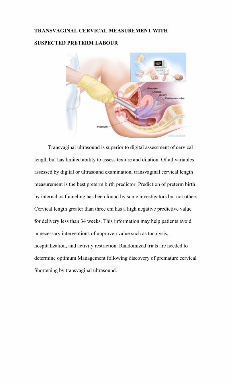
TRANSVAGINAL CERVICAL MEASUREMENT WITH
SUSPECTED PRETERM LABOUR
Transvaginal ultrasound is superior to digital assessment of cervical
length but has limited ability to assess texture and dilation. Of all variables
assessed by digital or ultrasound examination, transvaginal cervical length
measurement is the best preterm birth predictor. Prediction of preterm birth
by internal os funneling has been found by some investigators but not others.
Cervical length greater than three cm has a high negative predictive value
for delivery less than 34 weeks. This information may help patients avoid
unnecessary interventions of unproven value such as tocolysis,
hospitalization, and activity restriction. Randomized trials are needed to
determine optimum Management following discovery of premature cervical
Shortening by transvaginal ultrasound.
Cervical insufficiency defined as cervical changes in absence of
uterine contraction. It was traditionally considered as mechanical/Anatomic
defect that is either congenital/Acquired.
Current thinking favors a concept of functional insufficiency
including a spectrum of progressive degrees of insufficiency rather than
incompetence. Biochemical process involves inflammatory response with up
regulation of cytokines/prostaglandins and matrix metalloproteinase
resulting in premature cervix ripening. Process that initiate cervix change
can be either loss of mechanical / functional support /infection. These
painless cervix changes sooner or later result in premature myometrial
contraction. It is recommended that 3measurements are taken 5minutes apart
and lowest of 3 are considered.
PROCEDURE: - ( VAGINAL CYTOLOGY BY PAP SMEAR)
Contractions of sufficient frequency and intensity to effect progressive
effacement and dilatation of the cervix at 28-37 weeks gestation are
indicative of active preterm labour. If the diagnosis of preterm labor is
suspected, but not confirmed, it many be prudent to fist obtain a vaginal
smear of cytology before pelvic cervical examination. If the diagnosis of
preterm labor becomes obvious after the pelvic examination, the vaginal
smear specimen can be subsequently discarded. However, if the diagnosis

remains in doubt, the vaginal smear specimen can be sent to the lab for
analysis.
CYTOLOGICAL EVALUATION OF FETAL MATURITY BY
STUDYING THE EFFECTS OF HORMONES IN LATERAL
VAGINAL WALL SMEAR
Smears are taken from the upper two thirds of the lateral vaginal wall
by a wooden or plastic spatula. Immediately a smear is made from this on a
clean glass slide. The slide is immersed in 95% ethanol (without drying it).
Normal vaginal epithelium is lined by a stratified multilayered squamous
epithelium. The thickness and structure of the epithelium is closely related to
the conc. of the circulating ovarian hormones.
Under the influence of unopposed estrogen (no opposition by progesterone
e.g., in the proliferative phase) due to the absence of progesterone, this
vaginal epithelium is fully mature and fully developed and has 4 layers e.g.,

1) THE BASAL CELL LAYERS (THE RESERVE CELL LAYER): This
layer consists of ONE ROW of cells. These are very small cuboidal cells
with relatively large nuclei. They are firmly attached to basement membrane
and not exfoliated. THIS CELL LAYER DOES NOT EXFOLIATE. The
regeneration of vaginal epithelium occurs from the basal cell layer.
2) THE PARA BASAL CELL LAYER: This layer is composed of several
ROWS of cells. They are slightly larger with central nuclei also slightly
larger. Para basal cells exfoliate. The exfoliated Parabasal cells are
squamous, small, round or oval. They stain blue against a clear and clean
background of the vaginal smear. Cytoplasm is compact, central nuclei
relatively large. Presence of exfoliated Parabasal cells in large numbers in
vaginal smears means ovarian inactivity (no hormone stimulus e.g.,
immediately after delivery.)
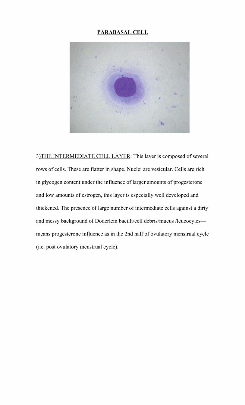
PARABASAL CELL
3)THE INTERMEDIATE CELL LAYER: This layer is composed of several
rows of cells. These are flatter in shape. Nuclei are vesicular. Cells are rich
in glycogen content under the influence of larger amounts of progesterone
and low amounts of estrogen, this layer is especially well developed and
thickened. The presence of large number of intermediate cells against a dirty
and messy background of Doderlein bacilli/cell debris/mucus /leucocytes—
means progesterone influence as in the 2nd half of ovulatory menstrual cycle
(i.e. post ovulatory menstrual cycle).

INTERMEDIATE CELL
There is a special and particular form of intermediate cell i.e. curled cell
form of intermediate sq cell called Navicular cell. Navicular cell is oval
(boat) shaped cell originally described by Papanicolaou in 1925. The cell
border of this cell is thickened and prominent. Cytoplasm stains light. Nuclei
are vesicular and eccentric. If there is subsequent insufficient progesterone
production without simultaneous estrogen reduction-the above smear will
have-
1-disappearance of navicular clusters
2-breaking up intermediate cell clusters
3-replacement of intermediate cells by superficial squamous cells
Exfoliated intermediate squamous cells are slightly smaller cells. Stain pale

blue. Cytoplasm is less transparent and rather denser. Nuclei are vesicular.
These cells have a tendency to curl at the edges. These cells are “sticky” in
nature and so they adhere to each other and clump together in masses thus
forming clusters. They exfoliate also in clusters. Navicular cells when found
in vaginal smear are found in clusters. These navicular cells are found-
1-in hormonally normal pregnancy in large numbers
2-in excessive androgen production due to progesterone effect
3-O+P pill
4-low estrogen status( with progesterone)
Navicular clusters are always found whenever there is a marked thickening
of the intermediate squamous cell layer of the vaginal epithelium.
4) SUPERFICIAL SQUAMOUS CELL LAYER: This layer is composed of
few rows of cells. These are large flat cells with pyknotic nuclei. The
percentage of squamous epithelial cells having this nuclear pyknosis is
known as karyopyknotic index (KI). The percentage of superficial squamous
cells showing cytoplasmic acidophilia is known as eosinophilic index (EI) or
cornification index (CI). Sometimes this layer contains anucleated
keratinized cells in the topmost layer. Estrogen is the only hormone that
promotes vaginal squamous proliferation to the superficial layer (means
progesterone is either low or absent simultaneously).

SUPERFICIAL CELL
Exfoliated superficial squamous cells are seen as large polyhedral-flat-lying
singly. Nuclei are pyknotic, cytoplasm thin, transparent stains pink with Pap
staining technique. These are most mature cells of the vaginal epithelium.
There presence in the smear means highest degree of vaginal proliferation-
unopposed estrogen influence (no or lack of progesterone to oppose
estrogen) simultaneously e.g., proliferative / 1st half of normal menstrual
cycle / estrogen administration / estrogen producing tumor / some androgen
due to estrogen effect and other conditions associated with excessive
estrogen production.
There is no characteristic pattern if there is lack of estrogen while

progesterone levels are maintained or if the quantities of both hormones are
simultaneously reduced.
PARABASAL CELLS INTERMEDIATE CELLS SUPERFICIAL CELLS
A shift towards this A shift towards this A shift towards this
A shift to left Shift to midzone Is shift to right
A shift towards to right If from right then a midzone shift to left
-then it is midzone shift to the right If from left then a midzone shift to the
right
In hormonally normal pregnancy. Due to large amounts of progesterone (
corpus luteum in 1st trimester and placenta later on ). And large amounts of
estrogen---- placenta and the fetus.
Karyopyknotic cell index (K.P.I): - It was estimated by the ratio of total
karyopyknotic cells(KC) to the total vesicular(intermediate) cells(VC) in a
sample.
K.P.I=KC/VC
Vaginal smears are seen as:
1st Trimester (Corpus Luteum Phase):
1) progressive enlargement of intermediate cell layers because of
progesterone influence of post ovulatory phase.
2) more dense clustering of these intermediate cells
3) marked decrease in superficial cells (present from preovulatory phase of

menstrual cycle)
4) E.I. falls below 5% and K.I. not > 10%
Mid pregnancy smear (from 3rd month onwards until 2-3 months prior to
term):
1) large numbers of clusters of intermediate cells are formed
2) large numbers of thick clusters of navicular cells are formed. Navicular
cells appear after 3rd week
3) no superficial cells are formed ( because large amounts of estrogens are
opposed by large amounts of progesterone)
4) E.I and K.I are very low --- each < 10%
5) background is dirty and messy as said earlier
At Term Smear (from 38-40 wks—beginning fall of progesterone by
placenta):
1) less no of navicular cells
2) small no of individual clusters of navicular cells
3) discrete intermediate squamous cells
4) maturity index slightly shifts to right
5) E.I rises to > 5-6% ( even 10 %)
6) K.I rises to about 15-20 % means impending delivery
Beyond Term Smear:
1) no intermediate cells

2) no navicular cells
Post Term Smear:
1) no intermediate cells
2) no navicular cells
3) no superficial cells
4) considerable number of parabolas cells appear so there is significant shift
of M.I to “left” : 100/0/0-means fetus in danger and deliver the patient
immediately.
5) Nearly all cells are single
But these are not always accurate and reliable tests so not used
routinely. MATURATION INDEX (M.I.): It is the expression of
percentage of the 3 major types of cells exfoliated from the stratified
squamous epithelium of vaginal walls eg. Parabasal/ Intermediate/
Superficial cells
The order is just like above.
Each cell type has been given a Maturation value
Parabasal =0 intermediate =0.5 superficial=5.0
In mid cycle of normal ovulatory menstrual cycle the M.I =0 / 40 / 60
In 1st half of menstrual cycle M.I=0 / 0 / 100----completely right
At midcycle ( i.e. at ovulation) it is 0 / 40 / 60 ---slight shift to right ---so a
shift of M.I to midzone
In 2nd half =0 / 70 / 30 ------shift to left in midzone
In hormonally normal pregnancy =0 / 95 / 5----shift to midzone
Soon after conception---progesterone is secreted by the corpus luteum. So
M.I shifts towards the midzone 0 / 70 / 30 it does not go in reverse order i.e.
to preovular pattern.
It continues to be there throughout pregnancy 0 / 95 / 5 due to
massive levels of O and P and other cortical steroids. It continues it’s
midzone shift until usually within a few weeks of delivery i.e. it reaches it’s
characteristic pattern of pregnancy i.e. an extreme shift to midzone of M.I to
0 / 95 / 5. This pattern is not reversed towards the preovulatory pattern as in
normal menstrual cycle.
TRANS VAGINAL ULTRA SOUND:-
POSITION: Female in recumbent position, bladder empty.
EQUIPMENT: 5MHZ/7MHZ Vaginal probe transducer.
STEPS: Probe inserted into vagina and advanced until cervix is visualized in
sagital plane with echogenic endocervix mucosa along endocervix canal.
Then probe is moved back in vagina and reapplied against cervix using
minimal/no pressure. Length of endo cervix canal, width of internal OS and
length of funnel if present are noted. Most important is measurement of
cervical length:-Measured from internal OS to notch made by external Os.

Funneling if present: - Cervix length is between upper and lower end of
closed segment of endocervix canal.
Width of Internal OS: - Measured from anterior to posterior lip of cervix.
TRANS VAGINALULTRA SOUND IN PREGNANCY
Transvaginal cervical length measurement from the internal to external
cervical os (arrows).
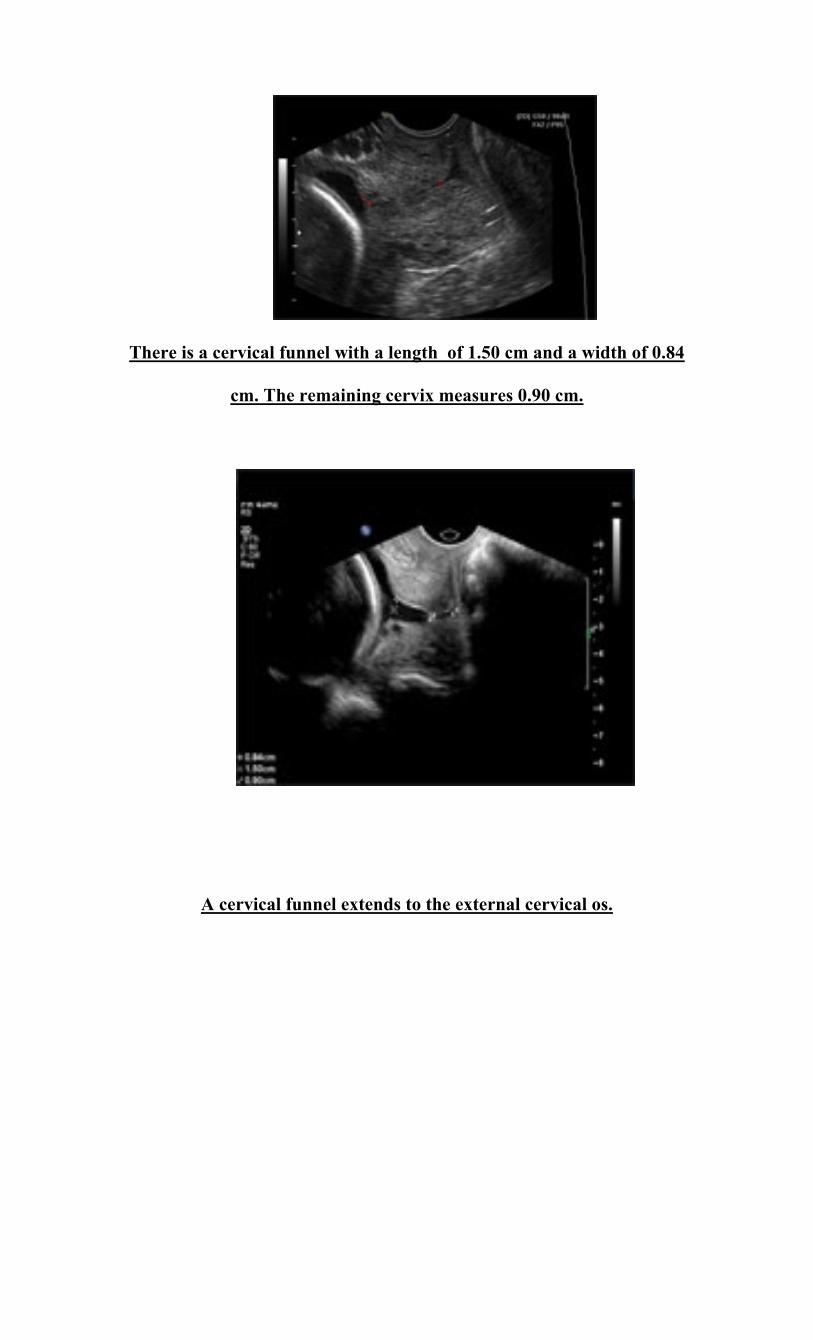
There is a cervical funnel with a length of 1.50 cm and a width of 0.84
cm. The remaining cervix measures 0.90 cm.
A cervical funnel extends to the external cervical os.

Minimal endocervical canal dilatation that has been associated with an
increased risk of preterm labor.
THE SHORT CERVIX:

Funnel length:-Will be distance between imaginary line used to measure
width of internal OS and upper end of closed segment and endocervical
canal. Examination takes not more than 5 minutes and causes minimal or no
discomfort to patient. The cervical length slowly decreases from mean of
4cm at 16wks to 3cm at 40 wks and there is no significant difference by
parity. Since risk of preterm birth increases markedly when cervix < 2.5 cm,
this measurement is widely accepted threshold to define risk of premature
birth. Possibility of preterm delivery(Positive Predictive Value) when
cervical length is less than 25mm is 17.8% , Negative Predictive Value of
cervical length >25mm is 97%. So these females are reassured and do
routine care.

DIAGNOSIS OF PRETERM LABOUR
Digital Examination
Effacement and Dilatation
80% effaced >80% effaced <80% effaced
(Dilatation > 3cm) (Dilatation >1 and <3cm) (Dilatation <1cm)
Advanced preterm labour Early Preterm labour TVUS (Cx Length)
<2.5cm >2.5cm
Threatened preterm labour False labour
High risk if <1.5cm

AIM &OBJECTIVES

AIM &OBJECTIVES:
All patients with features of threatened preterm labour are studied. In
this study we investigate potential value of cervical length by Trans Vaginal
Sonography & determination of vaginal cytology in prediction of outcome
of threatened preterm labour.
To facilitate early recognition of the condition and there by providing
prophylactic pharmacologic therapy to prolong gestation and reduce the
incidence of RDS and improve neonatal outcome.

REVIEW OF LITERATURE
REVIEW OF LITERATURE:
Iams et al (1990) used vaginal ultrasonography to measure the
length of the cervix and also documented the incidence of spontaneous
delivery before 35 weeks gestation. They examined 2915 women at
approximately 24 weeks of gestation. They examined 2915 women at
approximately 24 weeks of gestation and 2531 of these women again at
approximately 28 weeks. Spontaneous preterm delivery (at less than 35
weeks) occurred in 126 of the women (4.3 percent) examined at 24 weeks.
The length of the cervix was normally distributed at 24 and 28 weeks
[mean+/- SD], 35.2 +/- 8.3 mm and 33.7 +/- mm, respectively). The relative
risk of preterm delivery increased as the length of the cervix decreased.
When women with shorter cervixes at 24 weeks were compared with the
women with values above the 75th percentile, the relative risks of preterm
delivery among the women with shorter cervixes were as follows: 1.98 for
cervical lengths at or below the 75th percentile (40mm), 1. 35 for length at or
below the 50th percentile (35mm), 3.79 for lengths at or below the 25th
percentile (30mm), 6.19 for lengths at or below the 10th percentile (26mm),
9.49 for lengths at or below the 5th percentile (22mm), and 13.99 for lengths
at or below the 1st percentile (13mm) (p<0.001 for values at or below the
50th percentile). The risk of spontaneous preterm delivery is increased in

women who are found to have a short cervix by vaginal ultrasonography
during pregnancy. Shows the distribution of subjects among the different
percentiles and relative risk for spontaneous preterm birth.
An inverse relationship between cervical length measurement and
relative risk of preterm birth was demonstrated. Using receiver operator
curves, three potential thresholds values for clinical use were identified.
30mm, 25mm and 20mm.
VARIABLE ≤ 20 mm <25 mm < 30 mm
Sensitivity 23% 37.2% 54%
Specificity 97% 92.2% 76%
Positive
predictive value 25.7% 17.8% 93%
Negative
predictive value 96.5% 97% 97.4%
The rate of spontaneous delivery before 35 weeks was 4.3% among
female examined at 24wks. The tenth percentile cervical length
measurement at 24 weeks was found to be 25mm and this increased the risk
for preterm delivery six fold. Although a cervical length measurement of
25mm had only an 18% positive predictive value, this measurement has

subsequently been used as a benchmark of short cervical length in second
trimester in many studies.
Why is the Cervix short?
The length of the cervical canal measured by USG in the second and
early third trimester ranges from 10 to 50mm, the median length is 35mm,
the length percentile is 25mm and the 90th percentile is 45mm. The risk of
spontaneous preterm birth increases as the length of the cervix decreases
across the entire range of cervical length. A cervix length of <25mm at 22-
24 weeks is associated with a six fold increase in preterm birth before 35
weeks relative to women whose cervical length is above the 75th percentile.
Some of the range of cervical length is thought to be simply biologic.
In other cases, women may experience early effacement as a result of
inflammation due to hemorrhage, infections , or less commonly due to
biophysical effects of uterine distention or subclinical contractions.
1. Physical Factors:
A. Biological variation
1) Absolute versus relative insufficiency
2) Cervical function insufficient to carry a singleton pregnancy to 32 weeks
but not to term.
a) Cervical function insufficient to carry a singleton pregnancy to 32 weeks
but not to term.
b) Cervical function insufficient to carry a singleton pregnancy to 24 weeks,
that is the lower most end of a bell curve of cervical performance.
B. Uterine Volume
a) Multiple gestations, especially higher order
C. Cervical Injury
a) Obstetric laceration
b) Gynecological conditions: conisation, laser or LEEP
D. Contractions
2. Biochemical Factors:
a) Infection
b) Decidual Hemorrhage
c) Role of genetic polymorphisms
In a low-risk population endovaginal cervical ultrasonography helps
to rule out a preterm delivery if cervical length is long enough. It can also
detect cervical incompetence. In a high-risk population, women whose
cervix is longer than 30mm can be identified. These women have over 80%
chance to deliver on or after 36 weeks of pregnancy.
Heath VC et al (1998): stated that cervical length at 23 weeks is
<or = 15min in <2% of the populations; this group contains about 90% and
60% of the women delivering at <or = 28 and <or 32 weeks, respectively.

Measurement of cervical length provides accurate prediction of risk for early
preterm delivery.
Transvaginal ultrasound measurement of the cervix is increasingly
used for the prediction of preterm labour. In comparison to clinical vaginal
examination, it has the advantages of being highly reproducible, with a low
inter-observer variability, and of offering an evaluation of the entire cervical
canal, including the internal os. The sensitivity and specificity of
transvaginal ultrasound have been validated by several studies in women
with symptoms of preterm labour, however its clinical applications and its
limits have yet to be fully determined.
Hertzberg BS et al (1995) assessed the spontaneously changing
gravid cervix its clinical implications and prognostic features. Sonograms in
27 pregnant patient with a spontaneously changing cervix were studied
prospectively. The length and width of cervical funneling and the length of
intact cervix caudal to the funneling were measured when the cervical
dimensions were most normal and most abnormal.
Sonographic measurements were correlated with clinical and delivery
data. Twenty patients delivered preterm, although only six delivered within a
week of the ultrasound examination. Wider funneling of the internal os and a
shorter segment of intact cervix caudal to the funneling both correlated with
an increased likelihood of preterm delivery.

Most patients with a spontaneously changing cervix deliver preterm.
Measurements obtained when the cervix appears most abnormal are most
predictive of early delivery.
Is a study done by Watson WJ et al (1999): There was a positive
association between a short cervix and increased risk of preterm birth
(F=13.3, P<.0001). The variable with the highest predictive value for
preterm birth was the cervical length at 24 weeks gestation. Changes over
time did not substantially improve the predictive accuracy for spontaneous
preterm birth.
We conclude that a short cervix as determined by endovaginal
sonography has a significant association with preterm birth in a high-risk
obstetric population. Measurements taken at 24 weeks gestation are most
accurate in assessing this risk, and serial observations of the cervix over time
have less accuracy for predicting preterm birth.
Taipale P et al (1998): stated that despite much research, little
progress has been made in timely identification of the mothers at risk. He
examined the uterine cervix with ultrasonography to discover whether such a
procedure would be helpful in determining which women will deliver
prematurely.
He performed transvaginal ultrasound examinations in addition to
routine transabdominal ultrasonography at 18 to 22 weeks gestation in 3694

consecutive pregnant women with live singleton fetuses. He measured the
length of the uterine cervix and evaluated the dilatation, if any, of the
internal os. The results of cervical ultrasonography were not available to the
clinicians. Spontaneous delivery occurred before 37 completed weeks in 88
women (2.4%) and before 35 weeks in 31 (0.8%). The relative risk of
delivery risk of delivery before 35 weeks was 8 (95% confidence interval
1,267).
Transvaginal ultrasonography performed as an addition to routine
transabdominal ultrasonography at 18 to 22 weeks helps to identify many
patients at significant risk for prematurity; however, low sensitivity and low
positive predictive value limit its usefulness in screening low-risk obstetric
populations.
Fukami T et al (2003): Numerous reports have examined the
relationship between sonographically determine cervical length and
spontaneous preterm birth. Moreover, large screening studies have
consistently demonstrated that the shorter the cervical length, the higher the
rate of spontaneous preterm delivery. However, the sensitivity and positive
predictive value of the cervical length for detecting preterm birth were low.
Subsequently, developed a new sonographic cervical finding (shortened
cervical length or absence of cervical glandular area) at 16-19 weeks
gestation could predict spontaneous preterm birth. The absence of CGA as
compared to the shortened cervical length showed a higher sensitivity
(75.0% vs. 50.0%) and a significantly elevated positive predictive value
(54.5% vs. 8.3%) for preterm birth before 32 weeks gestation. It was
concluded that the absence of CGA was a novel and useful sonographic
parameter for predicting early spontaneous preterm birth.
In addition to primary predictors of preterm birth which are used to
estimate the baseline risk of preterm birth, secondary predictors (based on
examinations done during the current pregnancy) allow a more accurate
assessment of the risk of preterm birth in individual women. Screening for
early signs of spontaneous preterm labour has always been and important
topic in obstetric care. During the last two decades, the detection of fetal
fibronectin (FFN) from cervicovaginal detections and cervical shortening
diagnosed by transvaginal ultrasonography have emerged as the major
secondary factors of preterm birth.
Both markers have been extensively studied and consistently shown to
be strong short term predictors of preterm birth across a wide range of
gestational ages. Other secondary predictors that confirm the role of
intrauterine infection in the pathogenesis of preterm birth are bacterial
vaginosis (BV) and elevated levels of interleukin (IL-6, IL-8) ferritin and
granulocyte colony-stimulating factor. Apart from bacterial vaginosis,
inflammatory markers are still not routinely used.

The sensitivity of single markers in predicting preterm birth is only
moderate and serial examinations of markers, combinations of different
markers and multiple marker tests have been studied, with limited results.
Studies of interventions in order to prevent preterm birth have also yielded
mixed benefits, as a consequence of which the use of these markers to screen
low risk pregnancies is generally not recommended.
Several investigators have attempted to use cervical length in
asymptomatic women to predict preterm delivery.
Conoscenti et al (2003) prospectively followed 2469 women and
found that cervical length at 13 to 15 weeks gestation was not different in
women who delivered term and preterm.
Carvalho et al (2003) conducted a prospective study involving
529 pregnant women attending for routine antenatal care who underwent
transvaginal scan at 11 to 14 weeks and 12-24 weeks for evaluation of
cervical length. The mean cervical length was calculated at both steps of
gestation and lengths were compared between groups which delivered at
term and prematurely (<37weeks).
The mean cervical lengths were 42.4mm and 38.6mm at 11 to 14
weeks. Cervical length at 11 to 14 weeks was not significantly different
between the groups who delivered at term (42+mm) and preterm (40.6mm).
However, at 22-24 weeks evaluation, cervical length was significantly
shorter in the group which had a preterm delivery (26.7mm) and term
(39.3mm). So he concluded that there is a spontaneous shortening of the
pregnant cervix from the first to the second trimester of pregnancy.
Many studies have evaluated second trimester assessment of cervical
length as a predictor of preterm delivery. Goldenberg et al conducted
Preterm Prediction Study with the Maternal Fetal Medicine from 1993 to
1996.
They assessed about 3000 women for risk factors, biophysical
characteristics and biochemical tests that might be predictive of preterm
delivery. Using a cervical length of 25mm as the definition of short cervix
(24 to 30 wks), positive fetal fibronectin was the strongest predictor of
preterm birth followed by short cervix.
Among non-gravid women Jackson et al reported that
transvaginal and transabdominal ultrasound measurements of the cervix
agreed closely with anatomic measurements, whereas digital examination
underestimated the cervical length by an average of 13.6mm.
Honest H et al (2003): Conducted studies where they undertook
antenatal transvaginal sonographic cervical assessment among a population
of pregnant women with known gestational age of delivery. There were 46
primary articles, which included a total of 31,577 women, consisting of 33
studies in asymptomatic and 13 studied in symptomatic women. Data were
extracted for the studies characteristics and quality. Accuracy data were used
to form 2x2 contingency tables for various cervical length measurements
with birth before 32, 34 and 37 weeks gestation as the reference standards.
Our review showed that transvaginal cervical sonography identifies
women who are at higher risk of spontaneously preterm birth, although there
was a wide variation amongst studies with respect to gestational age at
testing, definition of threshold of abnormality and definition of reference
standard.
The most commonly reported sub-group was testing of asymptomatic
women at <20 weeks gestation using a threshold cervical length of 25mm
with spontaneous preterm birth before 34 weeks gestation as the reference
standard.
Both cervical length measurement and funneling, whether alone or in
combination, appear to be useful (depending on the threshold chosen to
define the abnormality) in predicting spontaneous preterm birth in
asymptomatic women. For symptomatic women there was a paucity of data,
although the degree of funneling appeared to be predictive of spontaneous
preterm birth.
From Goldenberg RC, Iams JD, Mercer BM et al: The
preterm prediction study: the value of new vs. standard risk factors in
predicting early of all spontaneous preterm births (Am J Public Health 88:
233, 1998). In symptomatic women with suspected preterm labour a cervical
length of <20mm is not necessarily predictive of preterm birth, but a length
of > 30mm can reliable exclude preterm birth.
Hasegawa et al conducted a study in 729 pregnant women with
no risk factors between 15 and 34 weeks gestation in Japan. Cervical
parameters, including cervical length, internal os dilatation and funneling
depth, more measured by transvaginal ultrasound. The predictive value of
these measurements for preterm delivery was investigated in a prospective
fashion.
Among various parameters, cervical length (mm) showed the best
correlation with pregnancy outcome. The group with a shortened cervix
(mean cervix length – ISD as cut off value) showed a significantly high
preterm delivery rate exclusively in primigravidae (odds ratio: 4.86, 95%).
Internal os dilation, in contrast, was a useful predictor in multiparous women
(odds ratio 6, 98%). It was concluded that TVS cervical assessment,
especially the measurement of cervical length, was effective for the
prediction of preterm delivery in the primigravidae.
The accurate predictor, prophylaxis and management of preterm
labour are a challenge for every obstetrician. This study conducted in a
multi-disciplinary tertiary institute correlates cervical length by TVS with
the occurrence of preterm labour.

Several published studies have demonstrated inverse relationship
between cervix length and frequency of preterm delivery. The group with a
shortened cervix showed a significantly higher preterm delivery rate
exclusive in the primigravid population. In contrast, internal os dilation was
a more useful predictor in multiparous women. The author concluded that
the length of the cervix was possible an indirect indicator of cervical
competence.
Although the predictive value of ultrasonography was low in this low
risk population it is postulated that the predictive value will raise as the risk
of prematurity in the study population increases.
Cervical effacement in pregnancy has been demonstrated by USG to
begin at approximately 32 weeks for term births and as early as 16-24 weeks
for preterm birth. The process of change of the internal os often is well
established before recognition of external os changes. Cervical effacement
may occur slowly and often precedes clinically evident preterm labour.
TRANSVAGINAL CERVICAL MEASUREMENT IN
ASYMPTOMATIC PREGNANT WOMEN
Cervical length is inversely related to preterm birth risk in
asymptomatic women. The largest study of this relationship noted relative
risks, compared to women above the 75th percentile, of approximately four
if length was less than 30 mm (25th percentile), six if less than 26 mm (10th
percentile), nine if less than 22 mm (5th percentile), and 14 if less than 13
mm (1st percentile). The positive predictive value was poor (35%). Heath
and colleagues19 studied women who were not at increased risk of preterm
birth and, using transvaginal ultrasound at 23 weeks, found that 1.7 percent
had a cervix length less than or equal to 15mm. These women accounted for
90 percent of deliveries at less than 28 weeks and 60 percent of deliveries at
less than or equal to 32 weeks. This suggests that the positive predictive
value of a short cervix (≤15 mm) is much greater for extreme prematurity
(≤28 weeks). The authors have created a formula to predict the risk of
spontaneous delivery at less than or equal to 32 weeks based on cervical
length at 23 weeks.
Although transvaginal ultrasound screening of cervical length can
predict increased risk of preterm birth,23 there is no evidence that this
information can be used to improve outcomes. Consultation and the
proposed location of birth should be considered. Other management options,
such as cerclage, activity restriction, tocolytics, and prophylactic steroids
await appropriate evaluation by randomized trials. The significant
association between cervical length and preterm birth risk may not apply to
women who have undergone cervical surgery resulting in permanent
shortening.

First detailed histologic description of changes in vaginal epithelium
in pregnant female was made by Faverger in 1913.
Papanicolaou (1925) postulated that navicular cells might be
used as diagnostic criterian for early pregnancy.
Pundel has emphasized the importance of obtaining vaginal
scraping from lateral wall of vagina in its upper 1/3 rd and cites the loss of
cell clustering and a sharp increase in KPI as among the most distinctive and
significant changes related to impending onset of labor;
When smears are procured in this manner according to Pundel smears
of late pregnancy contain <10% of superficial cells until shortly before onset
of labor when there is rise of 15% or more.
Lemberg-Siegfried and Samm and Pundel suggest useful
practical applicaton to be made from smears of late pregnancy, showing
1) Recognition of Impending onset of labour
2) Recognition of Post maturity of fetus
3) Progress fro success when medical induction of labour is
contemplated Barner and Zuspan in applying the criteria of
Lemberg-Siegfried and Stamm; were able to distinguish
labour smears from prenatal smears with only 60% accuracy.

The typical smear in Normal Pregnancy shows a predominantly bluish
green staining reaction and its cell population is almost exclusively derived
from intermediate layer with large clusters of navicular cells. The cells have
a pronounced tendency to crowd together in large clumps and curl their
edges.
The pregnancy pattern described above denotes adequate progesterone
effects. According to Lichtfus(1959) and Pundel(1959) the smear
appearances change in the last 2 weeks before delivery.
The large clusters of navicular cells break up, into smaller clusters and
later only isolated navicular cells are seen. Until they finally disappear
altogether. At the same time there is generally less crowding pattern shows
more discrete cells. Later still, the intermediate cells are replaced by
superficial squamous so that eventually the smear pattern resembles that of
the proliferative phase of the cycle. When this stage is reached, the onset of
labour is expected within the next 48 to 72 hours.
Pundel described in 1959, the existence of the following three
different smear types in the vaginal smear of a pregnancy near term and
pointed out their practical significance.
PREGNANCY SMEAR BEFORE TERM:
This smear is the type which is found in the last trimester of
pregnancy, namely exclusively intermediate cells, most of which are of the

navicular type and many exfoliated in placards. The acidophilic index is less
than 2.3% the KPI under 10% multiple Doderlein bacteria are found in the
background. This type of smear represents a hormonologically normal
pregnancy with an adequately functioning placenta.
PREGNANCY SMEAR AT TERM:
A few days before normal labor starts spontaneously, the previously
described smear will start to change. The cluster will be smaller. The
karyopyknotic index will increase to 15-20%. The Doderlein bacilli will
decrease in number and background will convey a “cleaner” impression.
This type of smear indicates that the function of the placenta, particularly
with respect to progesterone production, has decreased.
THE REGRESSIVE SMEAR:
In the regressive smear type not only have the placards disappeared
and the intermediate cells been scattered discretely, but the number of
acidophilic cells and pyknotic nuclei has increased considerably. The
characteristic of this type of smear, however is the presence of basal and
parabasal cells. In extreme cases red parabasal cells will be present-actually,
the same cells that will be called ’lactation cells’ when found in a
postpartum smear. This smear type may proceed to show the typical aspect
of a postpartum smear and this seems to express the fact that as far as the
vaginal epithelium is concerned, the pregnancy is hormonologically

terminated. The significance of this smear type is that it serves as a warning
that placental function is completely exhausted.
Pundel(1959) found in 1000 patients who were clinically at term
that 414 cases had the smear type of pregnancy before term, 574 that of
pregnancy at term, and 12 the regressive type. Out of the 414 cases with the
before term smear type only 18(4.3%) delivered spontaneously within 5 days
after the smear was taken.
Of the 574 cases with the at term smear type, 528 (92%) delivered within 5
days. He also found that the spontaneously delivery of the 18 cases which
were cytologically ‘before term’ could be attributed to nonhormonal factors
such as hydramnios, toxemia and multiple pregnancy.
In the 46 with the at term smear type who did not start labour within
5 days 42 started labour between sixth and twelfth day after the first smear
was taken. In two cases the smear showed a change towards the regressive
type and labour had to be induced artificially.
From Pundel’s material it appears that in over 90% of cases it can
be predicted whether or not the patient will start spontaneous labour within 5
days. It should be understood that this is not entirely confirmed to the 40
weeks gestation. This change in smear type only reflects a decrease in
placental hormonal activity and this may occur before the fortieth week.

In 12 cases with the regressive smear type, labour was artificially
induced, but only eight babies could be saved. All twelve babies showed
classical signs of post-maturity. Death of the four infants was attributed to
post-maturity. This regressive smear type seems to be associated with
serious fetal distress.
Pundel found in material of over 12,000 deliveries that true
postmaturity existed in only 37 cases and in all cases the smear was of the
regressive type. The smear reflects placental insufficiency and this is often
accompanied by fetal distress or even death. But there are cases of fetal
distress, for instance from erythroblastosis, malformations and infection
which are not accompanied by placental dysfunction. In such cases the fetal
distress may not show in the vaginal smear. After the baby dies from any
cause however, the placenta will degenerated and evidence of this
degeneration will be found in the smear.
` Pundel(1959) also obtained smears from 517 patients who had
carried the pregnancy beyond the calculated day of delivery and who had
been judged by skilled obstetricians to be ‘ripe’ for induction of labour 400
cases showed at term smear pattern 82 the pattern before term and 35 the
regressive type of smear. In the82 patients who showed before term pattern
the induction of labour was unsuccessful in any of the three attempts with
pitocin. Of the 400 cases with the at term smear type 300 inductions were

successful at the first attempt and 98 on the second attempt, again without
artificial rupture of membrane.
Prior to Pundel’s publications in 1959, Barnes and
Zuspan(1956) had published their results with this method. They reported
an accuracy of more than 70% in determining the date of confinement, but
doubted its use as a routine method. Zidovsky(1961) warned that
traumatic lesions and infections may be responsible for the appearance of
parabasal cells in the smears of women with prolonged pregnancy and this
may make the interpretation less valid. Nikilicek (1963) however agreed
with Pundel that these conditions can usually be recognized and seldom
lead to errors of judgement. Pundel’s cytologic conclusions depend on the
assumption that prior to the onset of labour there is indeed a decrease of
progesterone activity.
STUDY IN J.J. HOSPITALS BOMBAY
INTRODUCTION
The increasing levels of the hormones after conception bring about
changes in the vaginal epithelium. They can be detected by examination of
the vaginal smear which reveals intermediate cell-clusters and navicular
cells. This cytological pattern changes suddenly at the end of pregnancy due

to drop in the. hormonal levels. The latter also brings about spontaneous
labour within 5 days. However, there is no unanimity about the relationship
between the changed smear pattern and the onset of labour.
In India, determination of biological pregnancy terns is of
considerable practical importance, as many of the pregnant women attending
general hospitals do not know the date of their last menstrual period. Also, in
patients with irregular menses, the date of the last menstrual period cannot
be relied upon to determine the expected date of delivery. In such case, to
predict the date of labour, vaginal cytology is a simple and inexpensive
parameter as compared to estimation of urinary total oestrogen, ultrasound
studies and amniotic fluid analysis. In order to increase the predictability of
vaginal cytology, in addition to subjective, classification of the smears into
'pre-term' and 'at-term' patterns, eosinophilic and karyopyknotic indices can
be calculated
Lykke et al found that spontaneous preterm delivery, especially if
the complications were severe. In a registry-based cohort study of 536,419
Danish women, delivery between 32 and 36 weeks of gestation increased the
risk of preterm delivery in the second pregnancy from 2.7% to 14.7% (odds
ratio [OR] 6.12; 95% confidence interval [CI], 5.84-6.42) and increased the
risk of preterm birth from 1.1% to 1.8% (OR 1.60; 95% CI, 1.41-1.81). A
first delivery before 28 weeks increased the risk of a second preterm

delivery to 26.0% (OR 13.1; 95% CI, 10.8-15.9) and increased the risk of
preterm birth to 3.2% (OR 2.96; 95% CI, 1.80-4.88).
MATERIALS AND METHODS
Single smears were collected from 75 pregnant women in the age
group of 18 to 40 years, attending antenatal clinic of J. J. Group of
Hospitals, Bombay. The smears were obtained one week before the
due date of delivery. They were collected from the upper one third of the
lateral vaginal wall and were immediately fixed in ether-alcohol. The smears
were stained with Papanicolaou method and were categorized into three
patterns: 1.'Pre-term' type, when the smear showed large clumps of
cyanophilic intermediate cells and navicular cells uniformly distributed on
the slide; 2. 'At-term' pattern which revealed single intermediate cells or
occasional small clumps of intermediate cells and a very few navicular cells;
3. 'Post-term' or 'regressive' pattern, in which the cellular characteristics
were similar to those noted in the 'at term' category, but they were more
clear and distinctive. Parabasal cells were also seen in these smears.
In addition to this categorization of the smears, eosinophilic and
karyopyknotic indices were calculated. For this investigation, a total of one
hundred cells were counted in four different microscopic fields under high
power lens (magnification 40 x 10).
The time interval between the day of obtaining the smear and the

date of delivery was noted, and its relationship with the smear pattern was
recorded. The data was analyzed by discriminant analysis.
RESULTS
Out of 75 smears examined, 37 were of 'pre-term' type, 34 showed 'at-
term' pattern and 4 smears were of 'post-term' type.
The time interval between the date of obtaining the smear and the
actual date of delivery is shown in.
For the statistical analysis, eosinophilic and karyopyknotic indices
were calculated in each case. Discriminant analysis was carried out with this
two indices. The separation function, T was obtained as:
T = X + 0.8763Y, where
X = v Eosinophilic index
and
Y = vKaryopyknotic index
This produced a highly effective separation of 'pre-term' and 'at-term'
smears. The discriminant function for 'pre-term' type: was 4.58 (Tp) and for
'at-term' pattern was 5.24 (Ta). The mean of the two discriminants was 4.91,
which was the cut off value for 'pre-term and 'at-term' patterns. In our data,
none of the 'at-term' patterns had discriminant: less than 4.91, whereas only
one 'pre-term' pattern showed a value greater than 4.91 (i.e. 4.92).
DISCUSSION
The usefulness of vaginal cytology at the end of pregnancy for the
prediction of labour has been studied by many workers. Some found it to be
useful for the prediction of onset of labour whereas many others came to the
conclusion that there was no relationship between the smear pattern and the
onset of labour. Therefore, Ortner carried out discriminant analysis of
eosinophilic and karyopyknotic indices and thus supplemented subjective
criteria of cytological pattern determination with the objective criteria. The
same method was followed by us. Our values of eosinophilic and
karyopyknotic indices were lower than those obtained by Ortner and T
value was also low. We could separate 'pre-term' and 'at-term' patterns quite
well without any overlapping. Our prediction of onset of labour from the
smear pattern was correct in 73% of the patients. Thus, we find that vaginal
cytology at the end of pregnancy is a useful parameter to predict the onset of
labour in most cases
Lichtfus recorded “at-term” changes in 97.3% and “prior-to-term”
changes in 2.7% of patients within 5 days of spontaneous delivery in a series
of 369 patients. In 88 patients examined by Sammour spontaneous
delivery occurred within 5 days of the smear showing “at- term” changes in
73, or 83%, of patients.

In a study of 130 patients who had smears taken within 8 days, the
spontaneous onset of labor, Osmond-Clarke et al, found that 8%
showed a normal pregnancy smear of the clumped variety and 92% showed
some cytologic changes-partly discrete smears in 34% and discrete smears in
58% of patients.
At variance with these reports are the findings of Birtch and
Abrams and Abrams. Of 208 patients, Birtch obtained “before-
term” smears in 53%, “approaching-term” smears in 42%, and “at-term”
smears in only 5% of patients within 5 days of the onset of labour.
Abrams and Abrams were unable to demonstrate the abrupt change
from the third trimester to the “at-term” pattern within 5 days of the onset of
labour in any of the smears from 193 patients.
Vaginal cytology changes during labor or immediately after delivery
have been reported by Birtch, Ruiz and Soule. Birtch, in a study of
208 patients, recorded “before-term” smear in 50%, “approaching-term”
smears in 29.3%, and “at-term” smears in only 20% of patients.
Ruiz studied smears from 200 patients and observed that during
delivery vaginal cytology had the typical characteristics of pregnancy in
60% of patients, changes due to infection in 27%, and in only 13% of these
patients described as precursors of imminent delivery.
Soule concluded that vaginal cytology obtained during spontaneous
labor is of less correlative value than the clinical history.
Stricter criteria for a more reliable classification of smears are
desirable because the partly discrete smears considered by Osmond-
Clarke et al, as representing “prior-to-term” changes, might be
interpreted as representing “approaching-term” or even “at-term” changes.
Nevertheless, there is no doubt that alterations in the vaginal cytology
frequently occur towards the end of pregnancy. In 78% of patients reported
here changes were found when the patients were in labor. If the patient were
about to come into labor within 8 days, similar changes were found less
often (62% of this series). It is likely that the changes would be detected
more often before the onset of labor if smear could be taken every day or
two rather than at weekly intervals, but this poses administrative problems
that are likely to reduce the value of the method in general obstetric practice.
Neonatal Morbidity and Mortality by Gestational Age:
A study at University of California showing that between 24 and 33
weeks’ gestation, benefits of tocolytic therapy are generally accepted to
outweigh the risk of maternal and/or fetal complications and these agents
should be initiated provided no contraindications exist. The following table

depicts survival, major short-term morbidity, and intact long-term survival
by gestational age. The risk of neonatal mortality and morbidity is low after
34 completed weeks of gestation.
Gestational Age, wk
Survival Respiratory Distress Syndrome
Intraventricular Hemorrhage
Sepsis Necrotizing Enterocolitis
Intact
24 40% 70% 25% 25% 8% 5%
25 70% 90% 30% 29% 17% 50%
26 75% 93% 30% 30% 11% 60%
27 80% 84% 16% 36% 10% 70%
28 90% 65% 4% 25% 25% 80%
29 92% 53% 3% 25% 14% 85%
30 93% 55% 2% 11% 15% 90%
31 94% 37% 2% 14% 8% 93%
32 95% 28% 1% 3% 6% 95%
33 96% 34% 0% 5% 2% 96%
34 97% 14% 0% 4% 3% 97%

MATERIALS AND METHODS
MATERIALS AND METHODS:
This prospective clinical trial of 100 cases comprised of patients with
feature suggestive of threatened preterm labour and multigravida with
previous history of preterm labour who attended the Obstetrics and
Gynaecology Department of Institute of Social Obstetrics and Kasturbha
Gandhi Hospital for routine antenatal checkup.
The study was undertaken for a period of one year between May
2009-October 2010
Inclusion Criteria:
1) All Singleton live pregnancies with features of threatened preterm
labour included regular uterine contractions, low abdominal cramping,
low back pain, pelvic pressure & increased vaginal discharge.
2) Gestational age between 28weeks- 36 weeks
3) With Intact membranes
4) Cervical dilatation <3cms
Exclusion Criteria:
1) H/O 1st Trimester bleeding
2) Presence of uterine malformations and leiomyoma
3) H/O PIH, GDM, Essential HT
4) Multiple Pregnancies
5) Hydramnios

6) Low lying placenta
7) Pt in active labour, ruptured membranes.
8) Cervical Dilatation >3cms
9) Those who underwent permanent cervical shortening/surgery
Examination:
1) Maternal vital signs
2) Uterine activity, Tenderness, Fundal height, Presentation
3) Speculum examination to exclude rupture of membrane and assess
cervical change
a) High vaginal swab performed for culture and sensitivity
b) Vaginal cytology if cervix <3cm dilated, no liquor/blood is visible
Investigation:
1) Midstream urine
2) High vaginal swab
3) Vaginal cytology
4) TVUS
VAGINAL CYTOLOGY:
For staining exfoliated cells in cytological specimens.
Summary and Explanation:
The Papanicolaou Staining procedure is used for examining exfoliated
cells in secretions, exudates, transudates or scrapings of various internal
organs and tissue. Cells are fixed to a slide and stained first with
Hematoxylin, which stains the nuclei followed by OG-6 and EA-65 as a
counter stain.
Reagents:
EA-65 Multiple Polychrome Stain
1000mL Catalogue # IMEA65
Light Green S.F. Yellowish, Fast Green FCF, Bismark Brown Eosin Y,
Phosphotungstic Acid,Acid-Glacial Acetic Acid. Filter before use.
OG-6 Orange G Stain
1000mL Catalogue # IMOG6
Orange G, Phosphotungstic Acid. Filter before use.
Harris Hematoxylin
1000mL Catalogue # IMHARRISHX
Hematoxylin, Potassium Alum, Glacial Acetic Acid, Sodium Iodate. Filter
before use.
EA-50 and OG-6 were developed as general stains for vaginal
smears. EA-65, a modification of EA-50, has been especially developed for
use with OG-6 in the study of smears from urinary, paracentetic,

thoracentetic material, from sputum, from gastric and external ulcerated
sources as well as those smears which, are heavily mixed with mucus. In
such smears EA-65 helps retain the translucency essential to proper
cytological study. EA-65 is interchangeable with EA-50 in the staining
procedure. Staining times and technique remain the same.
Storage and Stability:
Store at 15°-30°C. Reagents are stable until expiration dates shown on
the label.
Warnings and Precautions:
Papanicolaou Staining reagents are flammable and toxic. Keep away
from sources of ignition. In case of contact with eyes, rinse immediately
with water and seek medical advice. May be fatal or cause blindness if
swallowed. Dispose of waste in accordance with applicable laws.
Materials Required:
95% Ethanol, Hydrochloric acid, Cover slips, Microscope,
Ammonium Hydroxide, and Microscope slides, Xylene Methanol.

Specimen Collection:
In the collection and preparation of smears for cytological examination, the
major objectives are:
1. Specimens should have a sufficient number of cells from the upper third
of lateral vaginal wall. Smears should contain well preserved cells uniformly
distributed so that each cell can be individually examined.
3. The staining procedure should clearly define the details of all structures.
Cytological preparations are obtained from patient by approved methods and
techniques. Scraping, obtained from the upper third of lateral vaginal wall is
spread directly on a clean microscope slide. The smear is immediately fixed
with a cytological spray fixative or in an alcohol-ether dip. Fixation or
preservation is one of the most important steps in the procedure.

Drying of the cells prior to fixation will usually result in artifacts such
as nuclear distortion and vacuolization. After fixation there are no special
handling requirements for cytological smears. However, smears should
remain in the fixative for about one hour. A second clean glass slide may be
placed on each fixed slide for protection.
Procedure:
Filter the Harris Haematoxylin immediately before use.
1. Dip slide(s) gently 5-10 times in 95% ethanol.
2. Dip slide(s) gently 5-10 times in 70% ethanol.
3. Dip slide(s) gently 5-10 times in distilled water.
4. Stain 5 minutes in Harris Hematoxylin.
5. Place smears in distilled water. Rinse in successive changes of distilled
water
until the water remains colorless.

6. Dip slide(s) gently 5-10 times in 70% ethanol.
7. Dip slide(s) in a 1% solution of HCl in 70% ethanol until the smear shows
a
Salmon color.
8. Rinse slide(s) well in 2 changes of 70% ethanol.
9. Dip slide(s) gently in a 3% solution of ammonium hydroxide in 70%
ethanol
until the smear takes on a blue color.
10. Rinse the slide(s) in two changes of 70% ethanol.
11. Dip slide(s) 5-10 times in 95% ethanol.
12. Stain slide(s) in OG-6 for 2 minutes.
13. Rinse slide(s) in two changes of 95% ethanol.
14. Stain slide(s) in EA-50 or EA-65 for 3-6 minutes.
15. Rinse slide(s) well in two changes of 100% methanol.
16. Rinse slide(s) in one part absolute methanol one part xylene.
17. Clean smear in xylene mounting.
Procedure:
1. After the smear has been completely cleaned in xylene it is mounted with
a
Microscope slide cover glass preferably 22x40mm, #1 thinness.
2. A permanent clean mounting medium should be used.

3. The excess xylene should be drained, in order to avoid the appearance of
air
spaces when xylene evaporates.
4. Place the required amount of mounting medium along an edge of one of
the
longer borders of the cover slip.
5. Place the slide at right angles to the edge of the cover slip so that the side
containing the cells is facing the mounting medium.
6. Slowly lower the slide and permit the mounting medium to spread
between the
slide and cover slip.
Results:
Nuclei are stained blue while cytoplasm displays varying shades of
pink, orange, yellow and green.
Limitations:
1. Proper specimen collection and fixation of cells is essential for
interpretation.
TRANS VAGINAL ULTRASOUND:
Before proceeding to transvaginal ultrasound, the woman was asked
to empty the bladder. With patient in lithotomy position, 5 MHz vaginal
probe was introduced into the vagina and the length & width of the cervix

was measured with the probe placed in the anterior fornix of the vagina. The
appropriate sagital view of cervix was obtained by simultaneous imaging of
external and internal os. External os was identified by its triangular echo
density and internal os by its V-shape appearance. The cervical canal was
seen as a translucent line connecting these two points. The distance between
the external and internal os was taken as cervical length. The width was
measured at the level of internal os. All these measurements were repeated
thrice and the averaged readings were taken for statistical analysis. These
measurements were repeated every four weeks till delivery.
To reduce the interobserver variability and improve reproducibility of
cervical measurements using transvaginal ultrasound, the following criteria
were adopted;
* The internal os is visualized as a flat dimple or an isosceles triangle.
* The whole length of the cervix is visualized.
* The whole length of the cervix is visualized.
* The external os appears symmetric
Method of Statistical Analysis:
The following methods of statistical analysis have been used in this
study.The data were entered into a Microsoft Excel Worksheet and analysed
using SPSS(ver 7.5) statistical package.

The results were presented in number and percentage in tables and
figures.
Cervical length and Karyopyknotic Index are plotted in Chi Square Table
and the results were analysed.

RESULTS AND ANALYSIS
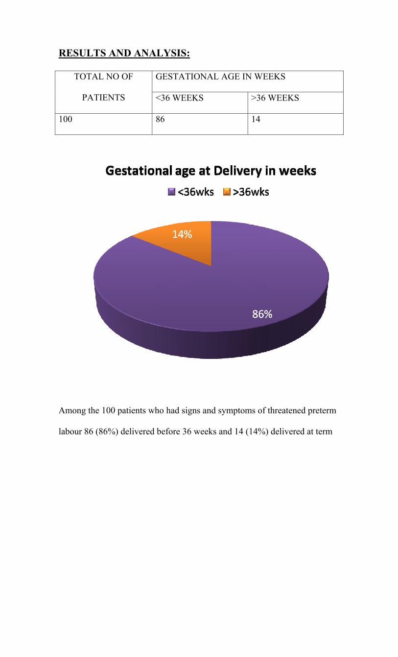
RESULTS AND ANALYSIS:
TOTAL NO OF
PATIENTS
GESTATIONAL AGE IN WEEKS
<36 WEEKS >36 WEEKS
100 86 14
Among the 100 patients who had signs and symptoms of threatened preterm
labour 86 (86%) delivered before 36 weeks and 14 (14%) delivered at term

KPI - Gestational age at Delivery in weeks
Gestational age at Delivery in weeks Total
Below 36 Above 36
KPI
Below 10
Count 37 12 49
% within KPI 75.5% 24.5% 100.0%
% within Gestational age at Delivery in weeks 43.0% 85.7% 49.0%
Above 10
Count 49 2 51
% within KPI 96.1% 3.9% 100.0%
% within Gestational age at Delivery in weeks 57.0% 14.3% 51.0%
Total
Count 86 14 100
% within KPI
86.0% 14.0% 100.0%
% within Gestational age at Delivery in weeks 100.0% 100.0% 100.0%

KPI - Gestational age at Delivery in weeks
Among the vaginal cytology smears collected from 100 patients; 51 smears showed KPI >10 showing progesterone deficiency and impending delivery, of them 49(96.1%) delivered before 36weeks and 2(3.9%) delivered >36weeks. Remaining 49 smears showed KPI<10;of which 37(75.5%) delivered before 36 weeks and 12 (24.5%) delivered after 36 weeks. This shows significant P value (0.003).

Cervix Length in cms - Gestational age at Delivery in weeks
Gestational age at Delivery in weeks Total
Below 36 Above 36
Cervix Length in cms
Below 2.5
Count 83 1 84
% within Cervix Length in cms 98.8% 1.2% 100.0%
% within Gestational age at Delivery in weeks 96.5% 7.1% 84.0%
Above 2.5
Count 3 13 16
% within Cervix Length in cms 18.8% 81.3% 100.0%
% within Gestational age at Delivery in weeks 3.5% 92.9% 16.0%
Total
Count 86 14 100
% within Cervix Length in cms 86.0% 14.0% 100.0%
% within Gestational age at Delivery in weeks
100.0% 100.0% 100.0%
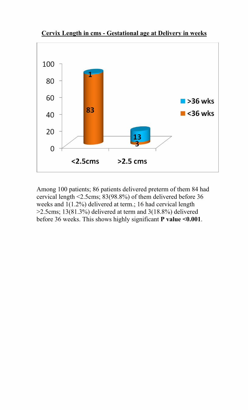
Cervix Length in cms - Gestational age at Delivery in weeks
Among 100 patients; 86 patients delivered preterm of them 84 had cervical length <2.5cms; 83(98.8%) of them delivered before 36 weeks and 1(1.2%) delivered at term.; 16 had cervical length >2.5cms; 13(81.3%) delivered at term and 3(18.8%) delivered before 36 weeks. This shows highly significant P value <0.001.

Complication * Gestational age at Delivery in weeks
Gestational age at Delivery in
weeks Total
Below 36 Above 36
Complication
No complication Count 1 9 10 % within Complication 10% 90% 100.0% % within Gestational age at
Delivery in weeks 1.2% 64.29% 11.0%
LBW Count 31 1 32 % within Complication 96.9% 3.1% 100.0% % within Gestational age at
Delivery in weeks 36.0% 7.14% 31.0%
RDS Count 22 1 23 % within Complication 95.7% 4.3% 100.0% % within Gestational age at
Delivery in weeks 25.6% 7.14% 23.0%
LBW and RDS Count 26 1 27 % within Complication 96.3% 3.7% 100.0% % within Gestational age at
Delivery in weeks 30.2% 7.14% 26.0%
IUGR Count 5 2 7 % within Complication 71.4% 28.6% 100.0% % within Gestational age at
Delivery in weeks 5.8% 14.29% 8.0%
IVH Count 1 0 1 % within Complication 100.0% .0% 100.0% % within Gestational age at
Delivery in weeks 1.2% .0% 1.0%
Total
Count 86 14 100
% within Complication 86.0% 14.0% 100.0%% within Gestational age at Delivery in weeks 100.0% 100.0% 100.0%

Complications - Gestational age at Delivery in weeks
Among 100 patients delivered ; 86 babies delivered <36 weeks and 14 babies delivered at term; of them 90(90%) babies had different postnatal complications and 10(10%) babies had no complications.
Among 100 patients delivered; 10 babies had no complication; of them 1(10%) delivered <36 wks and 9(90%) delivered at term

Among 100 patients delivered; 32 babies were Low Birth Weight; of them 31(96.9%) delivered <36 weeks and 1(3.1%) delivered at term
Among 100 patients delivered; 23 babies had Respiratory Distress Syndrome; of them 22(95.7%) delivered <36 weeks and 1(4.3%) delivered at term

Among 100 patients delivered; 27 babies had Low Birth Weight and Respiratory Distress Syndrome; of them 26(96.3%) delivered <36 weeks and 1(3.7%) delivered at term
Among 100 patients delivered; 7 babies had Intra Uterine Growth Retardation; of them 5(71.4%) delivered <36 weeks and 2(28.6%) delivered at term

Among 100 patients delivered; 1 baby had Intraventricular hemorrhage which delivered at < 36 weeks. Previous History of Preterm - Gestational age at Delivery in weeks
Gestational Age at Delivery in weeks
Total
Below 36 Above 36
Positive Count % within Previous H/O Preterm
40 5 45

Previous History of Preterm * Gestational age at Delivery in weeks
Among the 100 patients 45 of them had Previous H/O Preterm and 40 of them had Recurrent Preterm Labour and only 5 delivered at term. This shows P value is highly significant i.e., <0.001.
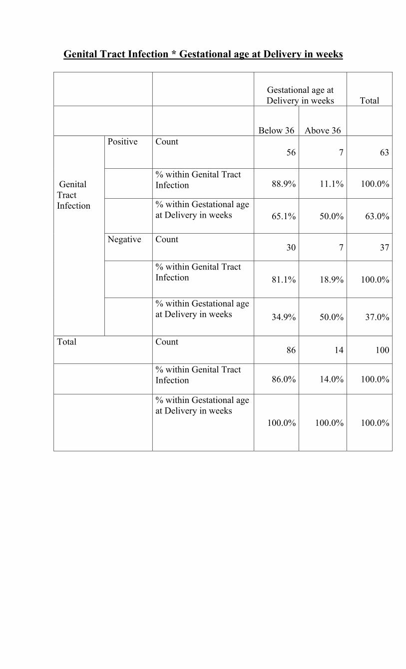
Genital Tract Infection * Gestational age at Delivery in weeks
Gestational age at Delivery in weeks Total
Below 36 Above 36 Genital Tract Infection
Positive Count 56 7 63
% within Genital Tract Infection 88.9% 11.1% 100.0%
% within Gestational age at Delivery in weeks 65.1% 50.0% 63.0%
Negative Count 30 7 37
% within Genital Tract Infection 81.1% 18.9% 100.0%
% within Gestational age at Delivery in weeks 34.9% 50.0% 37.0%
Total Count 86 14 100
% within Genital Tract Infection 86.0% 14.0% 100.0%
% within Gestational age at Delivery in weeks
100.0% 100.0% 100.0%

Genital Tract Infection - Gestational age at Delivery in weeks
Among patients 100 patients, 63 patients had Genital Tract Infection; of
them 56(88.9%) delivered before 36 weeks and 7(11.1%) delivered at term;
37 patients did not had Genital Tract Infection; of them 30 (81.1%) delivered
before 36 weeks and 7(18.9%) delivered at term. So P value is insignificant.

Urine C/S - Gestational age at Delivery in weeks
Gestational age at Delivery in weeks Total
Below 36 Above 36 Urine C/S
Positive Count 50 11 61
% within Urine C/S 82.0% 18.0% 100.0%
% within Gestational age at Delivery in weeks 58.1% 78.6% 61.0%
Negative Count 36 3 39
% within Urine C/S 92.3% 7.7% 100.0%
% within Gestational age at Delivery in weeks 41.9% 21.4% 39.0%
Total Count 86 14 100
% within Urine C/S 86.0% 14.0% 100.0%
% within Gestational age at Delivery in weeks 100.0% 100.0% 100.0%

Urine C/S - Gestational age at Delivery in weeks
Among 100 patients examined; 61 patients has Positive Urine C/S;
of them 50(82%) delivered before 36 weeks and 11(18%)
delivered at term; 39 patients has Negative Urine C/S; of them
36(92.3%) delivered before 36 weeks and 3(7.7%) delivered at
term. So P value is insignificant.

PARITY - Gestational age at Delivery in weeks
Gestational age at Delivery in weeks Total
Below 36 Above 36
PARITY
PRIMI
Count 23 7 30
% within PARITY 76.7% 23.3% 100.0%
% within Gestational age at Delivery in weeks 26.7% 50.0% 1.0%
MULTI
Count 63 7 70
% within PARITY 90.0% .10% 100.0%
% within Gestational age at Delivery in weeks 73.3% 50.0% 3.0%
Total Count 86 14 100
% within PARITY 86.0% 14.0% 100.0%
% within Gestational age at Delivery in weeks
100.0% 100.0% 100.0%

PARITY - Gestational age at Delivery in weeks
Among 100 patients examined; 30 patients were PRIMI of them
23(76.7%) delivered before 36 weeks and 7(23.3%) delivered at
term; 70 patients were MULTI of them 63(90%) delivered before
36 weeks and 7(10%) delivered at term. So P value is insignificant.

DISCUSSION

DISCUSSION
The studies of cervical length and karyopyknotic index for assessing the risk for preterm labour is very limited. By combining those two parameters
increases the specificity. Vaginal cytology is a simple and inexpensive
parameter as compared to estimation of urinary total oestrogen, ultrasound
studies and amniotic fluid analysis. In order to increase the predictability of
vaginal cytology, in addition to subjective, classification of the smears into
'pre-term' and 'at-term' patterns, eosinophilic and karyopyknotic indices can
be calculated.
Iams et al (1990)
Taipale P et al (1998):
Carvalho et al (2003)
Heath VC et al
(1998) Present study
Total pts 2531 3694 529 27 100
Initial cervical length
assement done on
24wks 24wks 14wks 30wks 34wks
Preterm labour & its week
4.3% (<35wks)
2.4% (<34wks)
3.2% (<34wks)
74% (<36wks)
83% (<36wks)
Cutoff value of cervical length
<3.5cm 2.5cm 4.2 - 3.8cm <2.5cm <2.5cm

In most studies the addition of cervical funneling does not improve the
predictive accuracy of cervical length in preterm labor prediction . This
may, in part, be due to the wide variations noted in funnel measurement.
Rust et al 14 have found that as a categorical variable (present or absent), a
funnel is a significant risk factor for preterm labor. However, the latter study
had a small sample size and was retrospective in nature. Additional
prospective studies will be required to determine if the presence of a funnel
adds to the predictive accuracy of cervical length in predicting preterm
delivery.
Iams et al (1990)
Taipale P et al (1998):
Carvalho et al (2003)
Heath VC et al
(1998) Present study
Total pts 2531 3694 529 27 100
Initial cervical length
assement done on
24wks 24wks 14wks 30wks 34wks
Preterm labour & its week
4.3% (<35wks)
2.4% (<34wks)
3.2% (<34wks)
74% (<36wks)
83% (<36wks)
Cutoff value of cervical length
<3.5cm 2.5cm 4.2 - 3.8cm <2.5cm <2.5cm

Pundel et al Present study
KP index <10 414 49
Outcome of preterm
KPI <10
4.3% 70%
KPI >10 574 51
Occurrence of
preterm
92 96.1%
The Karyopyknotic index of vaginal cytology for assessing the time
of delivery is very low cost method and it can be done in all centers. But the
studies are very less. So the supportive evidence for this test is very less. By
adding this to cervical lengthening increases the specificity for assessing the
gestational age of pregnancy.
Very limited studies are available to explain the correlation between
vaginal cytology and preterm birth .Pundel et all study clearly tells , if the
KP index more than 10 they may go for preterm labour(92%) which is very
much comparable to present study where the occurrence of preterm in KP
index more than 10 is (96%).
Pandit et al studied the eosinophillic and karyopyknotic index to
assess the preterm delivery. And the results are well correlating with our
study to assess the preterm pregnancies

A similar study was done J.J.Group of Hospitals, Mumbai by
collecting single smears from 75 pregnant women with features of preterm
labour. In addition to this categorization of the smears, eosinophilic and
karyopyknotic indices were calculated. For this investigation, a total of one
hundred cells were counted in four different microscopic fields under high
power lens (magnification 40 x 10).
The time interval between the day of obtaining the smear and the date of
delivery was noted, and its relationship with the smear pattern was recorded.
The data was analysed by discriminant analysis.
Ortner carried out discriminant analysis of eosinophilic and
karyopyknotic indices and thus supplemented subjective criteria of
cytological pattern determination with the objective criteria. The same
method was followed by them. They found the values of eosinophilic and
karyopyknotic indices were lower than those obtained by Ortner and T value
was also low and could separate 'pre-term' and 'at-term' patterns quite well
without any overlapping. Their prediction of onset of labour from the smear
pattern was correct in 73% of the patients. Thus, they found that vaginal
cytology at the end of pregnancy is a useful parameter to predict the onset of
labour in most cases, supporting our study.
By doing both these investigations give clue to the risk of preterm
pregnancy. By adding these investigation together increases the specificity
of assessing the risk of preterm pregnancy. Also we have shown previous
H/O preterm birth increases the recurrence risk which was supported by
Yost and co-workers as a patient's number of preterm births increases and
the gestational age at preterm delivery decreases, the likelihood of another
preterm delivery rises by 3 to 4-fold. They found that these historical
variables in 22 patients did not affect the predictive accuracy of a cervical
length < 25 mm between 16 and 19 weeks' gestation. However, because of
the small sample size, the power of the study to detect a clinically significant
difference was low. Until additional studies are reported, prior preterm
delivery should be considered a categorical variable that increases the risk of
preterm labor
Complications related to the preterm are very high. All are managed
well by expecting the complications and be prepare for them.
Iams et all study explains the need of test to asses the gestational age
by cervical length. Complication like IUD and LBW are very common. By
doing this two techniques increases the sensitivity and specificity of the pt
that whom are going for labour.

GENITAL TRACT INFECTION:
The young gynecology patient diagnosed with gonorrhea, chlamydia,
or trichomoniasis has an approximate 25% risk of reinfection during the
subsequent 12 months, but a clear association between these organisms and
preterm delivery has not been established. BV is a vaginal syndrome
associated with an alteration of the normal vaginal flora rather than an
infection specific to any one organism and a lack of vaginal inflammation is
evident when compared with vaginitis. The diagnosis of BV should be
suspected with a positive Gram stain result or the presence of 3 of 4
traditional diagnostic signs (homogenous gray-white discharge, >20% clue
cells on saline wet smear, positive whiff test, and a vaginal pH >4.50)
Patients should be treated per the US Centers for Disease Control and
Prevention guidelines, with test-of-cure sampling and subsequent treatment
if necessary.
PRETERM LABOR/BIRTH HISTORY:
A history of prior preterm deliveries places the patient in the high-risk
category. Of the predictors of preterm birth, past obstetric history may be
one of the strongest predictors of recurrent preterm birth. Given a baseline
risk of 10-12%, the risk of recurrent preterm birth after 1, 2, and 3
consecutive preterm births may be increased to approximately 15%, 30%,

and 45%, respectively. Pre-conceptual counseling should help encourage
patients to make informed decisions concerning future pregnancy in light of
prematurity risk in the presence of previous preterm delivery. Often the best
time to counsel the patient is at her 4- to 6-week postpartum check after a
preterm delivery.
In our study, complications like LBW, RDS, IUGR and IUD are
common in preterm babies. Usually those preterm babies will go for high
mortality and morbidity. In our study it’s very less. Because, we were
expected those preterm delivery and neonatal complications. Because of
these we could able to reduce the morbidity and mortality of neonates of
preterm.
Those patients who are admitted in hospital for threatened labour with
KPI more than 10 and cervical length <2.5cm are very high risk patients for
preterm labour so, those patients must be admitted in hospital and they
should be started on tocolytic and other measures. The neonatal
complications must be expected and the precautions have to be taken to
prevent the mortality and morbidity of neonates.
Our study did not show any relation between parity and cervical
dimensions. Gramellini et al (2002) reported similar finding in their cross
sectional study involving 321 subjects. They showed that cervical length was
comparable in nulliparous and parous women throughout pregnancy and

shows a progressive, linear reduction between the 10th and 40th weeks in
both the groups. But some studies have reported that cervices of parous
women are thicker and lengthier. They have attributed it to the fact that
elastin, collagen and water content of parous cervices is different than
nulliparous cervices because of changes that the cervix undergoes after
delivery.

CONCLUSION

CONCLUSION:
The incidence of preterm birth remains around 12%. Preterm labor is a
heterogeneous process. The presence of multiple interactive continuous
variables explains the relatively low predictive value of any one variable. A
transvaginal assessment of cervical length is one of the best tests for
predicting preterm birth. A cervical length < 25 mm (10th percentile)
between 28 to 36 weeks' gestation is generally agreed upon as increasing the
risk of preterm labor. The shorter the cervical length, the greater the
likelihood of preterm labor. The positive predictive value of a 25 mm cervix
for preterm labor is dependent upon the patient's pretest probability (i.e.
high-risk or low-risk for preterm labor).
Prior spontaneous preterm birth, fetal fibronectin, cervical length, and
vaginal cytology are associated with early preterm birth. The association of
the first 4 with preterm delivery is more than additive. These associations
indicate that there is more than one pathway resulting in preterm delivery. In
the future, a multifactorial assessment of patients at risk for preterm delivery
should improve the positive predictive value of our testing schema. Specific
therapeutic regimens depending upon the etiology or etiologies of preterm
labor will be required and should improve our current limited successfully
inhibition of preterm labor.

BIBILOGRAPHY

BIBILOGRAPHY:
References:
1. Papanicolaou GN: Atlas of Exfoliative Cytology, Harvard University
Press,
Cambridge, 1954. United States, Naval Medical School, National
Naval Medical Center, Bethesda, Maryland, Manual of gynecological
exfoliative cytology. US Government PrintingOffice, Washington,
D.C., 1965.
2. A Manual for Cytotechnology, C. Keebler, J. Reagen, Editors,
American Societyof Clinical Pathologists Press, Chicago (IL), 1983.
3. Ventura SJ, Martin JA, Curtain SC , Mathews TJ. Births: Final Data
for 1997. Nat Vital Stat Report 1999;47(18):1-96.
4. Cicero S, Skentou C, Souka A et al. Cervical length at 22-24 weeks of
gestation: comparison of transvaginal and transperineal-translabial
ultrasonography. Ultrasound Obstet Gynecol 2001;17:335-340.
5. Iams JD, Goldenberg RL, Meis PJ, Mercer BM, Moawad A et al. The
length of the cervix and the risk of spontaneous premature delivery.
National Institute of Child Health and Human Development Maternal
Fetal Medicine Unit Network. N Engl J Med 1996;334:567-572.
6. Edwards RK, Ferguson RJ, Shuster JJ, Theriaque D, Gentry S et al.
Assessment of cervical antibody concentrations fails to enhance the
value of cervical length as a predictor of preterm delivery. Am J
Obstet Gynecol 2005;192:887-891.
7. Phelps JY, Higby K, Smyth MW, Ward JA, Arredondo F et al.
Acuracy and intraobserver variability of simulated cervical dilatation
measurements. Am J Obstet Gynecol 1995;173:942-945.
8. Gomez R, Galasso M, Romero R, Mazor M, Sorukin Y et al.
Ultrasonographic examination of the uterine cervix is better than
cervical digital examination as a predictor of the likelihood of
premature delivery in patients with preterm labor and intact
membranes. Am J Obstet Gynecol 1994;171:956-965.
9. Rozenberg P, Rudant J, Chevret S, Boulogne AI. Repeat measurement
of cervical length after successful tocolysis. Obstet Gynecol
2004;104:995-999.
10. Burger M, Weber-Rössler TW, Willmann M. Measurement of the
pregnant cervix by transvaginal sonography: an interobserver study
and new standards to improve the interobserver variability.
Ultrasound Obstet Gynecol 1997;9:188-193.
11. Saito M, Kozuma S, Kikuchi A, Sakai M, Fujii T et al. Sonographic
assessment of the cervix before, during and after a uterine contraction
is effective in predicting the course of labor. Ultrasound Obstet
Gynecol 2003;22:604-608.
12. Berghella V, Bega G, Tolosa JE, Berghela M. Ultrasound assessment
of the cervix. Clin Obstet Gynecol 2003;46:947-962.
13. Kurtzman JT, Jenkins SM, Brewster WR. Dynamic cervical changes
during real-time ultrasound: prospective characterization and
comparison in patients with and without symptoms of preterm labor.
Ultrasound Obstet Gynecol 2004;23:574-578.
14. Owen J, Yost N, Berghella V, Thom E, Swain GA et al. Mid-trimester
endovaginal sonography in women at high risk for spontaneous
preterm birth. JAMA 2001;286:1340-1348.
15. Owen J, Yost N, Berghella V, MacPherson C, Swain M et al. Can
shortened mid-trimester cervical length predict very early spontaneous
preterm birth? Am J Obstet Gynecol 2004;191:298-303.
16. Rust OA, Atlas RO, Kimmel S, Roberts WE, Hess LW. Does the
presence of a funnel increase the risk of adverse perinatal outcome in
a patient with a short cervix? Am J Obstet Gynecol 2005;192:1060-
1066.
17. Anderson HF, Nugent CE, Wanty SO, Hayashi RH. Prediction of risk
for preterm delivery by ultrasonographic measurement of cervical
length. Am J Obstet Gyecol 1990;163:859-867.
18. Berghella V, Tallucci M, Desai A. Does transvaginal sonographic
measurement of cervical length before 14 weeks predict preterm

delivery in high-risk pregnancies? Ultrasound Obstet Gynecol
2003;21:140-144.
19. Heath VCF, Southall TR, Souka AP, Elisseou A, Nicolaides KH.
Cervical length at 23 weeks of gestation: prediction of spontaneous
preterm delivery. Ultrasound Obstet Gynecol 1998;12:312-317.
20. Honest H, Bachman LM, Gupta JK, Kleijnen J, Khan KS . Accuracy
of cervicovaginal fetal fibronectin test in predicting risk of
spontaneous preterm birth: systematic review. BMJ 2002;325:301-
320.
21. Peaceman AM, Andrews WW, Thorp JM, Cliven SP, Lukes A et al.
Fetal fibronectin as a predictor of preterm birth in patients with
symptoms: a multicenter trial. Am J Obstet Gynecol 1997;177:13-18.
22. Goldenberg RL, Iams JD, Das A, Mercer BM, Meis PJ et al. The
Preterm Prediction Study: sequential cervical length and fetal
fibronectin testing for the prediction of spontaneous preterm birth. Am
J Obstet Gynecol 2000;182:636-643.
23. Tsoi E, Fuchs IB, Rane S, Geents L, Nicolaides KH. Sonographic
measurement of cervical length in threatened preterm labor in
singleton pregnancies with intact membranes. Ultrasound Obstet
Gynecol 2005;25:353-356.

24. Yost NP, Owen J, Berghella V, MacPherson C, Swain M et al.
Number and gestational age of prior preterm births does not modify
the predictive value of a short cervix. Am J Obstet Gynecol
2004;191:241-246.
25. Airoldi J, Berghella V, Sehdeu H, Ludmin J. Transvaginal
ultrasonography of the cervix to predict preterm birth in women with
uterine anomalies. Obstet Gynecol 2005;106:553-556.
26. 24. Odibo AO, Talucci M, Berghella V. Prediction of preterm
premature rupture of the membranes by transvaginal ultrasound
features and risk factors in a high-risk population. Ultrasound Obstet
Gynecol 2002;20:245-251.
27. 25. Berghella V, Tolosa JE, Kuhlman KA, Weine S, Bolagnese RJ et
al. Cervical ultrasonography compared to manual examination as a
predictor of preterm delivery. Am J Obstet Gynecol 1997;177:723-
730.
28. Guzman ER, Mellon C, Vintzileos AM, Ananth CV, Walters C et al.
Longitudinal assessment of endocervical canal length between 15 and
24 weeks' gestation in women at risk for pregnancy loss or preterm
birth. Obstet Gynecol 1998;92:31-37.

29. Yost N for the NICHD MFMU Nework: Mid-trimester vaginal
sonography: Ancillary markers for spontaneous preterm birth. Am J
Obstet Gynecol 2001;185:(suppl)144.
30. Gomez R, Romero R, Nien JK, Chaiworapongsa T, Medina L et al. A
short cervix in women with preterm labor and intact membranes: A
risk factor for microbial invasion of the amniotic cavity. Am J Obstet
Gynecol 2005;192:678-689.
31. Watts DH, Krohn MA, Hillier SL, Eschenbach DA. The association of
occult amniotic fluid infection with gestational age and neonatal
outcome among women in preterm labor. Obstet Gynecol
1992;79:351-357.
32. Vayssiére C, Favre R, Audibert F, Chauvet MP, Gaucherand P et al.
Cervical assessment at 22 and 27 weeks for the prediction of
spontaneous birth before 34 weeks in twin pregnancies: is
transvaginal sonography more accurate than digital examination?
Ultrasound Obstet Gynecol 2005;26:707-712.
33. Gibson JL, Macara LM, Owen P, Young D, Macauley J et al.
Prediction of preterm delivery in twin pregnancy: a prospective,
observational study of cervical length and fetal fibronectin testing.
Ultrasound Obstet Gynecol 2004;23:561-566.

34. Imseis HM, Albert A, Iams JD. Identifying twin gestations at low risk
for preterm birth with a transvaginal ultrasonographic cervical
measurement at 24 to 26 weeks' gestation. Am J Obstet Gynecol
1997;177:1149-1155.
35. Bega G, Lev-Toaff A, Khulman K et al. Three-dimensional
multiplanar transvaginal ultrasound of the cervix in pregnancy.
Ultrasound Obstet Gynecol 2000;16:351-358.
36. Towner D, Boe N, Lou K, Gilbert WM. Cervical length measurements
in pregnancy are longer when measured with three-dimensional
transvaginal ultrasound. J Maternal Fetal Neonat Med 2004;16:167-
170.
37. Severi FM, Bocchi C, Florio P, Picciolini E, D'Aniello G et al.
Comparison of two-dimensional and three-dimensional ultrasound in
the assessment of the cervix to predict preterm delivery. Ultrasound
Med Biol 2003;29:1261-1265.
38. Berghella V, Odibo AO, To MS, Rust OA, Althuisius SM. Cerclage
for short cervix on ultrasonography: meta-analysis of trials using
individual patient-level data. Obstet Gynecol 2005;106:181-189.
39. Althuisius SM, Dekker GA , van Geijn HP, Hummel P. The effect of
therapeutic McDonald cerclage on cervical length as assessment by

transvaginal ultrasonography. Am J Obstet Gynecol 1999;180:366-
370.
40. Guzman ER, Houlihan C, Vintzileos A, Ivan J, Berito C et al. The
significance of transvaginal ultrasonographic evaluation of the cervix
in women treated with emergency cerclage. Am J Obstet Gynecol
1996;175:471-476.
41. Hassan SS, Romero R, berry SM, Dang K, Blackwell SC et al .
Patients with an ultrasonographic cervical length £ 15 mm have nearly
a 50% risk of early spontaneous preterm delivery. Am J Obstet
Gynecol 2000;182:1458-1467.
42. To MS, Skentou C, Liao AW, Cacho A, Nicolaides KH. Cervical
length and funneling at 23 weeks of gestation in the prediction of
spontaneous preterm delivery. Ultrasound Obstet Gynecol
2001;18:200-203.
43. Goldenberg RL, Iams JD, Mercer BM, Meis PJ, Moawad AH et al.
The preterm prediction study: the value of new vs. standard risk
factors in predicting early and all spontaneous preterm births. Am J
Public Health 1998;88:233-238.

ANNEXURES

ANNEXURES
PROFORMA
1.Patient particular:
Name: Hospital No:
Age: G P L A
D.O.A: I.P. No 2.Complaints: H/O any imminent symptoms 3.Menstrual History: Previous cycles
LMP
EDD
4.Marital History:
Married life
5.Obstetric History:
H/O any significant obstetric complications in the previous pregnancies
6.Past History of any medical or surgical illness

7.Family History:
Any History of Preterm in the mother, siblings or cousins
8.General Physical Examination:
Ht, Wt, Pallor, Edema, Breasts, Thyroid, PR, BP
9.Systemic Infections:
CVS
RS
P/A- Uterine size, liquor, FHR
10.Investigations:
Hb, Urine routine, (Plt count, LFT, RFT, Fundoscopy-if patient had high
BP)
11. Cervical Length Measurement:
12.Vaginal Cytology Test Result:
13.GA at delivery
14.NN Outcome