Mccaughey, Laura C., Grinter, Rhys, Josts, Inokentijs ...eprints.gla.ac.uk/91918/1/91918.pdf ·...
16
Mccaughey, Laura C., Grinter, Rhys, Josts, Inokentijs, Roszak, Aleksander W., Waløen, Kai, Cogdell, Richard J., Milner, Joel, Evans, Tom, Kelly, Sharon, Tucker, Nicholas P., Byron, Olwyn, Smith, Brian, and Walker, Daniel (2014) Lectin-like bacteriocins from Pseudomonas spp. utilise D- rhamnose containing lipopolysaccharide as a cellular receptor. PLoS Pathogens, 10 (2). e1003898. ISSN 1553-7366 Copyright © 2014 The Authors http://eprints.gla.ac.uk/91918 Deposited on: 28 February 2014 Enlighten – Research publications by members of the University of Glasgow http://eprints.gla.ac.uk
Transcript of Mccaughey, Laura C., Grinter, Rhys, Josts, Inokentijs ...eprints.gla.ac.uk/91918/1/91918.pdf ·...

Mccaughey, Laura C., Grinter, Rhys, Josts, Inokentijs, Roszak, Aleksander W., Waløen, Kai, Cogdell, Richard J., Milner, Joel, Evans, Tom, Kelly, Sharon, Tucker, Nicholas P., Byron, Olwyn, Smith, Brian, and Walker, Daniel (2014) Lectin-like bacteriocins from Pseudomonas spp. utilise D-rhamnose containing lipopolysaccharide as a cellular receptor. PLoS Pathogens, 10 (2). e1003898. ISSN 1553-7366 Copyright © 2014 The Authors http://eprints.gla.ac.uk/91918 Deposited on: 28 February 2014
Enlighten – Research publications by members of the University of Glasgow
http://eprints.gla.ac.uk

Lectin-Like Bacteriocins from Pseudomonas spp. UtiliseD-Rhamnose Containing Lipopolysaccharide as a CellularReceptorLaura C. McCaughey1., Rhys Grinter1., Inokentijs Josts1, Aleksander W. Roszak2,3, Kai I. Waløen1,4,
Richard J. Cogdell , Joel Milner , Tom Evans , Sharon Kelly , Nicholas P. Tucker , Olwyn Byron ,
Brian Smith ,
3 3 1 3 5 4
3 Daniel Walker1*
1 Institute of Infection, Immunity and Inflammation, College of Medical, Veterinary and Life Sciences, University of Glasgow, Glasgow, United Kingdom, 2 WestCHEM,
School of Chemistry, College of Science and Engineering, University of Glasgow, Glasgow, United Kingdom, 3 Institute of Molecular Cell and Systems Biology, College of
Medical, Veterinary, and Life Sciences, University of Glasgow, Glasgow, United Kingdom, 4 School of Life Sciences, College of Medical, Veterinary and Life Sciences,
University of Glasgow, Glasgow, United Kingdom, 5 Strathclyde Institute for Pharmaceutical and Biomedical Sciences, University of Strathclyde, Glasgow, United Kingdom
Abstract
Lectin-like bacteriocins consist of tandem monocot mannose-binding domains and display a genus-specific killing activity.Here we show that pyocin L1, a novel member of this family from Pseudomonas aeruginosa, targets susceptible strains ofthis species through recognition of the common polysaccharide antigen (CPA) of P. aeruginosa lipopolysaccharide that ispredominantly a homopolymer of D-rhamnose. Structural and biophysical analyses show that recognition of CPA occursthrough the C-terminal carbohydrate-binding domain of pyocin L1 and that this interaction is a prerequisite for bactericidalactivity. Further to this, we show that the previously described lectin-like bacteriocin putidacin L1 shows a similarcarbohydrate-binding specificity, indicating that oligosaccharides containing D-rhamnose and not D-mannose, as waspreviously thought, are the physiologically relevant ligands for this group of bacteriocins. The widespread inclusion of D-rhamnose in the lipopolysaccharide of members of the genus Pseudomonas explains the unusual genus-specific activity ofthe lectin-like bacteriocins.
Citation: McCaughey LC, Grinter R, Josts I, Roszak AW, Waløen KI, et al. (2014) Lectin-Like Bacteriocins from Pseudomonas spp. Utilise D-Rhamnose ContainingLipopolysaccharide as a Cellular Receptor. PLoS Pathog 10(2): e1003898. doi:10.1371/journal.ppat.1003898
Editor: Guy Tran Van Nhieu, College de France, France
Received July 18, 2013; Accepted December 10, 2013; Published February 6, 2014
Copyright: � 2014 McCaughey et al. This is an open-access article distributed under the terms of the Creative Commons Attribution License, which permitsunrestricted use, distribution, and reproduction in any medium, provided the original author and source are credited.
Funding: LCM and IJ are supported by 4-year studentships from the Wellcome Trust. RG is supported by a Kelvin Smith Scholarship from the University ofGlasgow. We would like to acknowledge the Diamond Light Source for access to I04, I04-1 and I24 (proposal number MX6683). The funders had no role in studydesign, data collection and analysis, decision to publish, or preparation of the manuscript.
Competing Interests: The authors have declared that no competing interests exist.
* E-mail: [email protected]
. These authors contributed equally to this work.
Introduction
The ability to target a subgroup of pathogenic bacteria in a
complex bacterial community has potential applications in
medicine and agriculture where the maintenance of a ‘normal’
microbiome is beneficial. For example, the use of broad spectrum
antibiotics to treat bacterial infections is known to cause a range of
complications associated with collateral damage to the micro-
biome, including antibiotic associated diarrhea and Clostridium
difficile infection [1,2]. In addition, there is growing evidence to
suggest that microbial dysbiosis may play a role in a range of
chronic diseases such as inflammatory bowel disease, diabetes,
obesity and rheumatoid arthritis [3,4,5,6]. Indeed, for Crohn’s
disease, where the link with dysbiosis is well established, the
administration of multiple courses of antibiotics is associated with
an increased risk factor for the development of this chronic form of
inflammatory bowel disease [7,8,9].
In contrast to the broad spectrum antibiotics that are widely
used in medicine and agriculture, protein antibiotics known as
bacteriocins often target a specific bacterial species or a group of
closely related bacterial species [10,11,12,13]. Well characterised
bacteriocins include the S-type pyocins from P. aeruginosa and the
closely related colicins of E. coli [12,13]. The colicin-like
bacteriocins form a diverse family of multidomain protein
antibiotics which share similar mechanisms of uptake and kill
cells through either a pore-forming activity, a specific nuclease
activity against DNA, tRNA or rRNA or through inhibition of cell
wall synthesis [14,15,16,17]. In the case of S-type pyocins it is
thought that their activity is limited to strains of P. aeruginosa,
whereas colicins show activity against E. coli and some strains of
closely related bacteria such as Salmonella spp. [18]. In the case of
colicins and S-type pyocins, killing specificity is primarily
determined by the presence of a specific outer membrane receptor
on the cell surface. For example, the well characterised E group
colicins utilise the TonB-dependent BtuB receptor, which has a
normal physiological role in vitamin B12 uptake [19]. Colicin-like
bacteriocins have also been shown to have a potent antibiofilm
activity, indicating their potential as useful therapeutics for the
treatment of chronic biofilm mediated infections [20,21]. In the
case of the opportunistic human pathogen P. aeruginosa there is an
urgent requirement for the development of novel therapeutic
options since its ability to form drug-resistant biofilms in
PLOS Pathogens | www.plospathogens.org 1 February 2014 | Volume 10 | Issue 2 | e1003898

combination with the presence of an outer membrane that is
highly impermeable to many classes of antibiotics can make this
pathogen essentially untreatable in some groups of patients. This is
exemplified in cystic fibrosis patients where chronic lung infection
with P. aeruginosa is the leading cause of mortality [22].
An interesting addition to this group of protein antibiotics is the
recently discovered lectin-like bacteriocins that contain two
carbohydrate-binding domains of the monocot mannose-binding
lectin (MMBL) family [23,24,25,26,27]. Lectin-like bacteriocins
from P. putida (putidacin L1 or LlpABW) P. syringae (LlpAPss642) and
P. fluorescens (LlpA1Pf-5) have been characterised and have the
unprecedented ability to kill strains of a broad range of bacterial
species within the genus Pseudomonas, but are not active outside this
genus [24,26,27]. Similarly the lectin-like bacteriocin LlpAXcm761
from Xanthomonas citri pv. malvacearum LMG 761 has the ability to
kill various species within the genus Xanthomonas [24]. The
molecular basis of this unusual genus specific activity has not
been explained.
Lectins are a structurally and evolutionarily diverse class of
proteins produced widely by prokaryotes and eukaryotes and are
defined by their ability to recognise and bind carbohydrates. This
binding is generally highly specific and mediates a range of diverse
functions, including cell-cell interaction, immune recognition and
cytotoxicity [28,29] MMBLs represent a structurally conserved
lectin subclass, of which the mannose-binding Galanthus nivalis
agglutinin (GNA) was the first to be characterised [30]. The
MMBL-fold consists of a three sided b-prism; each face of which
contains a sugar binding motif with the conserved sequence
QxDxNxVxY [31]. While originally identified in monocots like G.
nivalis or Allium sativum, it is now recognised that proteins of this
class are distributed widely throughout prokaryotes and eukary-
otes, where they have evolved to mediate diverse functions
[30,32,33,34]. Structural and biochemical analysis of MMBLs has
shown that they are generally translated as a single polypeptide
chain containing tandem b-prism domains that are then proteo-
lytically processed into monomers. These domains often form
homo- or hetero-dimers by strand exchange and p-stacking [35].
The lectin-like bacteriocins are not proteolytically processed and
thus consist of a single peptide chain, containing tandem b-prism
domains. Sequence alignments of members of this class from
Pseudomonas spp. show complete conservation of two sugar binding
motifs on the C-terminal domain and partial conservation of two
sites on the N-terminal domain [23]. Recent work by Ghequire et
al [23] on the characterisation of putidacin L1 shows these motifs
to be important for cytotoxicity. Mutagenesis of the first C-
terminal motif has the most dramatic effect on activity, while
mutagenesis of the second C-terminal and first N-terminal sugar
binding motifs leads to a synergistic reduction in activity. This
study also showed low-affinity binding between putidacin L1 and
methyl-a-D-mannose or a range of mannose containing oligosac-
charides. However, Kds for these protein-carbohydrate complexes
were reported in the range from 46 mM for methyl-a-D-manno-
side to 2 mM for a mannose containing pentasaccharide [23]. An
extensive search for high affinity carbohydrate binding through
the use of glycan arrays failed to detect high affinity carbohydrate
binding for this lectin-like bacteriocin [23].
Despite progress in our understanding of the structure and host
range of MMBL-like bacteriocins, the mechanism by which these
bacteriocins target susceptible strains and exert their antimicrobial
effects is unknown. Here we report on the discovery of a novel
member of this family, pyocin L1 from P. aeruginosa, and show that
it utilises lipopolysaccharide (LPS) as a surface receptor, specifi-
cally targeting the common polysaccharide antigen (CPA) that is a
conserved homopolymer of D-rhamnose. Structural and biophys-
ical analysis shows that the C-terminal carbohydrate binding
motifs are responsible for D-rhamnose recognition and that these
sites are specific for this sugar over D-mannose. Further to this, we
show that the previously described putidacin L1 also selectively
binds LPS from susceptible, but not from resistant, P. syringae
isolates and shows selectivity for D-rhamnose over D-mannose.
This work shows that the physiologically relevant ligand for the
QxDxNxVxY carbohydrate binding site of the lectin-like bacte-
riocins is indeed D-rhamnose and not D-mannose as previously
thought. As such, the genus-specific activity of lectin-like
bacteriocins from Pseudomonas spp. can be attributed to the
widespread inclusion of the rare D-rhamnose in the LPS of
members of the genus Pseudomonas.
Results
Identification and characterisation of pyocin L1As part of a wider project, aimed at identifying bacteriocins that
could be used as novel therapeutics in the treatment of P. aeruginosa
infections, we searched the genomes of 10 recently sequenced
clinical and environmental isolates of P. aeruginosa for genes with
homology to known bacteriocins. One putative bacteriocin gene
identified in strain C1433, an isolate from a patient with cystic
fibrosis, encodes a protein with 31% identity to the lectin-like
bacteriocin LlpA1Pf-5, from P. fluorescens. This protein, designated
pyocin L1, contained 256-amino acids with a predicted molecular
mass of 28413 Da. Alignment of the pyocin L1 protein sequence
with other lectin-like bacteriocins, LlpA1Pf-5, LlpAPss642, putidacin
L1 (LlpABW), LlpAAu1504 from Burkholderia cenocepacia and
LlpAXcm761 from Xanthomonas citri pv. malvacearum shows that
pyocin L1 contains tandem MMBL domains with three conserved
QxDxNxVxY MMBL sugar-binding motifs (Figure S1). Two of
these motifs are located in the C-terminal domain of the protein
and one in the N-terminal domain. Comparison with the
sequences of other lectin-like bacteriocins shows that the C-
terminal QxDxNxVxY motifs are highly conserved, with only
LlpAXcm761 lacking one C-terminal motif. In contrast the
Author Summary
Due to rapidly increasing rates of antibiotic resistanceobserved among Gram-negative pathogens, such asPseudomonas aeruginosa, there is an urgent requirementfor novel approaches to the treatment of bacterial infec-tions. Lectin-like bacteriocins are highly potent proteinantibiotics that display an unusual ability to kill a selectgroup of bacteria within a specific genus. In this work, weshow how the lectin-like protein antibiotic, pyocin L1, cankill Pseudomonas aeruginosa with extraordinary potencythrough specific binding to the common polysaccharideantigen (CPA) of P. aeruginosa lipopolysaccharide. The CPAis predominantly a homopolymer of the sugar D-rhamnosethat although generally rare in nature is found frequentlyas a component of the lipopolysaccharide of members ofthe genus Pseudomonas. The targeting of D-rhamnosecontaining polysaccharides by pyocin L1 and a relatedlectin-like protein antibiotic, putidacin L1, explains theunusual genus- specific killing activity of the lectin-likebacteriocins. As we learn more about the link betweenchanges to the microbiome and a range of chronicdiseases there is a growing realisation that the ability totarget specific bacterial pathogens while maintaining thenormal gut flora is a desirable property for next generationantibiotics.
Pyocin L1 Uses LPS as a Cell Surface Receptor
PLOS Pathogens | www.plospathogens.org 2 February 2014 | Volume 10 | Issue 2 | e1003898

N-terminal sugar-binding motifs are less well conserved with only
LlpAAu1504 possessing two fully conserved QxDxNxVxY motifs
(Figure S1).
In order to determine the killing spectrum of pyocin L1 we
cloned the pyocin L1 open reading frame into the pET21a vector
and expressed and purified the protein by nickel affinity, anion
exchange and size exclusion chromatography. Purified pyocin L1
was tested for its ability to inhibit the growth of 32 environmental
and clinical isolates of P. aeruginosa using an overlay spot plate
method on LB agar [36]. Under these conditions, pyocin L1
showed killing activity against nine of the P. aeruginosa strains
tested. Strain E2, an environmental isolate from a tomato plant for
which the genome sequence is available, and strain P8, a clinical
isolate from a cystic fibrosis patient, showed the greatest sensitivity
to pyocin L1 with killing observed down to concentrations of
27 nM and 7 nM, respectively. Pyocin L1 also showed activity
against 5 of the 11 P. syringae strains tested, although the effect was
much weaker, with cell killing observed at high mM concentra-
tions.
Pyocin L1 targets the common polysaccharide antigen(CPA) of P. aeruginosa LPS
In order to gain insight into the bacteriocidal activity of pyocin
L1, we subjected P. aeruginosa E2 to high concentrations of
recombinant protein and recovered mutants with greatly increased
tolerance to pyocin L1 (Figure 1A). The genomes of two of these
mutants were sequenced and comparative analysis with the
genome of wild-type E2 revealed a dinuclear deletion, C710 and
T711, of the 1146-bp wbpZ gene. This deletion was common to
both mutants. wbpZ encodes a glycosyltransferase of 381 amino
acids that plays a key role in lipopolysaccharide synthesis,
specifically in the synthesis of the common polysaccharide antigen
(CPA) also known as A-band LPS [37], (Figure S2). Most strains of
P. aeruginosa produce two distinct LPS-types that differ in their O-
antigen, but share the same core oligosaccharide. The CPA is
predominantly a homopolymer of D-rhamnose and the O-specific
antigen contains a heteropolymeric repeating unit that varies
widely among strains [38]. Consistent with mutation of wbpZ, we
found that production of CPA, as determined by immunoblotting
with a CPA-specific monoclonal antibody [39], in both M4(E2)
and M11(E2) was reduced to undetectable levels (Figure 1B).
Visualisation of LPS from these strains was performed via silver
staining and comparable quantities of LPS were shown to be
present. These observations suggest that pyocin L1 may utilise
CPA as a cellular receptor. To test this idea further, we obtained
two transposon insertion mutants of P. aeruginosa PAO1, which is
sensitive to pyocin L1, with insertions in the genes responsible for
the transport of CPA to the periplasm [40]. These two genes, wzt
and wzm, encode the ATP-binding component and membrane
component of a CPA dedicated ABC transporter [38]. Pyocin L1,
which shows good activity against PAO1 showed no activity
against strains with insertions in wzm and wzt (Figure 1C) and
immunoblotting with a CPA-specific antibody confirmed the
absence of the CPA in these pyocin L1 resistant strains (Figure 1D).
Thus, the presence of CPA on the cell surface is required for
pyocin L1 killing.
In order to determine if the requirement for CPA is due to a
direct interaction with pyocin L1 we purified LPS from wild-type
PAO1 and from the pyocin L1 resistant, wzm and wzt mutants
(which produce no CPA but do produce the O-specific antigen)
and analysed the pyocin-CPA interaction by isothermal titration
calorimetry (ITC). Titration of pyocin L1 into isolated LPS-
derived polysaccharides (a mixture of CPA and the O-specific
antigen containing polysaccharides) from PAO1 gave rise to strong
saturable exothermic heats of binding (Figure 2A), whereas no
binding was detected on titration of pyocin L1 into an equivalent
concentration of LPS carbohydrates from PAO1 wzt, which
produces the O-specific antigen but not the CPA (Figure 2B).
These data show that pyocin L1 binds directly to the CPA and that
this interaction is required for killing. The CPA is therefore likely
to be the cellular receptor for pyocin L1.
Pyocin L1 binds the monosaccharide D-rhamnoseThe evolutionary relationships between MMBL-like bacterio-
cins and the originally identified mannose-binding members of this
protein family, led to the assumption that carbohydrate binding of
polysaccharides by the lectin-like bacteriocins is primarily medi-
ated through binding of D-mannose at one or more of their
conserved QxDxNxVxY carbohydrate binding motifs. Indeed, the
recent structures [23] of putidacin L1 bound to mannose-
containing monosaccharides adds weight to this idea, although
measured affinities between polysaccharides and putidacin L1 are
weak (mM) and so may not be physiologically relevant. However,
the strong interaction between pyocin L1 and CPA, is incompat-
ible with this and suggests that D-rhamnose and not D-mannose is
the likely physiological substrate for the QxDxNxVxY carbohy-
drate binding motifs.
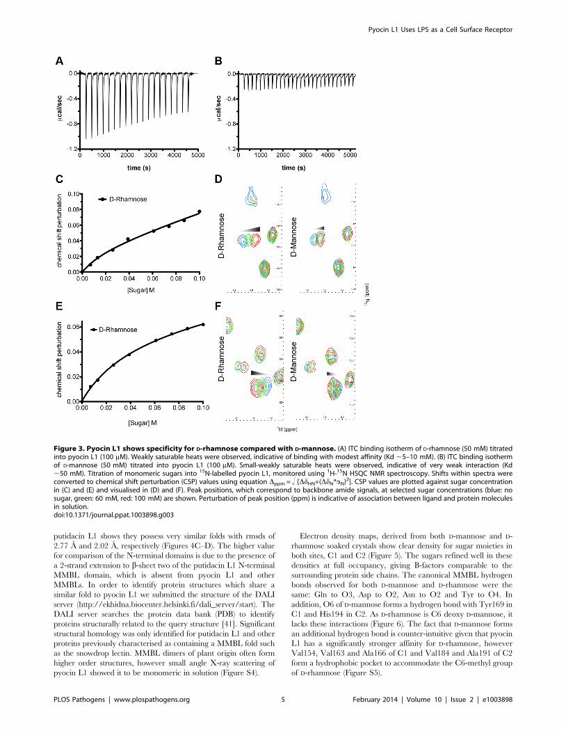
To determine the affinity of pyocin L1 for D-rhamnose and D-
mannose, isothermal titration calorimetry (ITC) was performed.
Titration of pyocin L1 into D-rhamnose gave rise to weakly
saturable heats of binding that are significantly larger than the
heats observed on titration of pyocin L1 into an identical
concentration of D-mannose (Figure 3). From this experiment an
apparent Kd of 5–10 mM was estimated for the interaction of
pyocin L1 with D-rhamnose with apparently weaker binding for
Figure 1. CPA production correlates with pyocin L1 killing. (A)Inhibition of growth of P. aeruginosa E2 and tolerant mutants M4 andM11 by pyocin L1, as shown by a soft agar overlay spot-test. 5 ml ofpurified pyocin L1 (1.5 mg ml21) was spotted onto a growing lawn ofcells. Clear zones indicate cell death. (B) Expression of CPA by P.aeruginosa E2 and tolerant mutants, visualised by immunoblotting withthe CPA specific antibody N1F10. (C) Inhibition of growth of P.aeruginosa PAO1 and PAO1 wzm and wzt mutants by pyocin L1 (detailsas for A). (D) Expression of CPA by PAO1 and wzm and wzt strains(details as for B).doi:10.1371/journal.ppat.1003898.g001
Pyocin L1 Uses LPS as a Cell Surface Receptor
PLOS Pathogens | www.plospathogens.org 3 February 2014 | Volume 10 | Issue 2 | e1003898

D-mannose, Kd.50 mM. The interaction between pyocin L1 and
these monosaccharides was also probed using NMR with 15N
labelled pyocin L1, monitoring changes to its 15N-heteronuclear
single quantum correlation (15N-HSQC) spectra on addition of D-
rhamnose or D-mannose. In the absence of added monosaccharide15N-HSQC spectra of pyocin L1, which should contain one
crosspeak for each non-proline amide NH as well as peaks for the
NH groups in various side chains, were well resolved and
dispersed, indicative of a folded protein. Chemical shift perturba-
tion monitored by 15N-HSQC allows the mapping of changes to a
protein that occur on ligand binding. Addition of either D-
rhamnose or D-mannose up to a concentration of 100 mM did not
give rise to large or global changes in chemical shifts (Figure S3).
On addition of D-rhamnose significant chemical shift changes were
observed for a discrete subset of peaks including some in the amide
side chain region of the spectra, while changes of a smaller
magnitude were observed on the addition of equal concentrations
of D-mannose (Figure S3). Fitting the chemical shift changes that
occur on addition of D-rhamnose, for peaks showing strong shifts,
to a single site binding model indicates a Kd for the pyocin L1- D-
rhamnose complex in the range of 5–20 mM (Figures 3C–F).
These data correlated well with the ITC sugar binding data, with
low mM binding of pyocin L1 to D-rhamnose and much weaker
binding to D-mannose.
D-rhamnose and the CPA bind to the C-terminalQxDxNxVxY motifs of pyocin L1
In an attempt to determine the location of the pyocin L1 D-
rhamnose binding site(s) and the structural basis of the D-rhamnose
specificity of pyocin L1 we determined the X-ray structures of
pyocin L1 with bound D-mannose, D-rhamnose and in the unbound
form (Table 1). Pyocin L1, as predicted by sequence homology to
MMBL proteins, consists of two tandem b-prism domains
characteristic of MMBLs, connected by antiparallel strands
propagating from the end of each MMBL domain and lending a
strand to the reciprocal b-prism. The strands contain a tryptophan
residue which forms p-stacking interactions with two other
tryptophans in the b-prism to stabilise the structure (Figure 4A).
This interaction is conserved throughout MMBLs, with most
members of the class utilising it to form either homo- or hetero-
dimers of single MMBL subunits. However, in pyocin L1, as with
the recently described structure of putidacin L1, both domains are
from a single polypeptide chain [23]. Other structural elements are
also common between the two bacteriocins, namely a C-terminal
extension of 30 amino acids and a two-turn a-helix insertion into
loop 6 of the N-terminal MMBL domain (Figure 4B). The overall
root mean square deviation (rmsd) of backbone atoms for pyocin L1
and putidacin L1 is 7.5 A, which is relatively high due to a
difference in the relative orientation of the two MMBL domains. In
contrast, the relative orientation of the tandem MMBL domains of
pyocin L1 matches those of the dimeric plant lectins very closely,
with alignment of pyocin L1 with the snowdrop lectin homodimer
(pdb ID: 1MSA) giving an rmsd of 4.81 A. Comparison of the
respective N- and C- terminal domains from pyocin L1 and
Figure 2. Pyocin L1 binds strongly to CPA from P. aeruginosaPAO1. (A) ITC binding isotherm of pyocin L1 (150 mM) titrated intoisolated LPS-derived polysaccharide (1 mg ml21) from wild-type P.aeruginosa PAO1. Strong, saturable heats were observed indicative of astrong interaction. Curve fitted with a single binding site model. (B) ITCisotherm of pyocin L1 (150 mM) titrated into isolated LPS-derivedpolysaccharide (1 mg ml21) from PAO1 wzt. No saturable bindingisotherm was observed.doi:10.1371/journal.ppat.1003898.g002
Pyocin L1 Uses LPS as a Cell Surface Receptor
PLOS Pathogens | www.plospathogens.org 4 February 2014 | Volume 10 | Issue 2 | e1003898
putidacin L1 shows they possess very similar folds with rmsds of
2.77 A and 2.02 A, respectively (Figures 4C–D). The higher value
for comparison of the N-terminal domains is due to the presence of
a 2-strand extension to b-sheet two of the putidacin L1 N-terminal
MMBL domain, which is absent from pyocin L1 and other
MMBLs. In order to identify protein structures which share a
similar fold to pyocin L1 we submitted the structure of the DALI
server (http://ekhidna.biocenter.helsinki.fi/dali_server/start). The
DALI server searches the protein data bank (PDB) to identify
proteins structurally related to the query structure [41]. Significant
structural homology was only identified for putidacin L1 and other
proteins previously characterised as containing a MMBL fold such
as the snowdrop lectin. MMBL dimers of plant origin often form
higher order structures, however small angle X-ray scattering of
pyocin L1 showed it to be monomeric in solution (Figure S4).
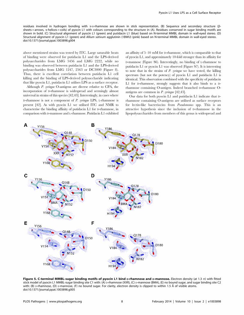
Electron density maps, derived from both D-mannose and D-
rhamnose soaked crystals show clear density for sugar moieties in
both sites, C1 and C2 (Figure 5). The sugars refined well in these
densities at full occupancy, giving B-factors comparable to the
surrounding protein side chains. The canonical MMBL hydrogen
bonds observed for both D-mannose and D-rhamnose were the
same: Gln to O3, Asp to O2, Asn to O2 and Tyr to O4. In
addition, O6 of D-mannose forms a hydrogen bond with Tyr169 in
C1 and His194 in C2. As D-rhamnose is C6 deoxy D-mannose, it
lacks these interactions (Figure 6). The fact that D-mannose forms
an additional hydrogen bond is counter-intuitive given that pyocin
L1 has a significantly stronger affinity for D-rhamnose, however
Val154, Val163 and Ala166 of C1 and Val184 and Ala191 of C2
form a hydrophobic pocket to accommodate the C6-methyl group
of D-rhamnose (Figure S5).
Figure 3. Pyocin L1 shows specificity for D-rhamnose compared with D-mannose. (A) ITC binding isotherm of D-rhamnose (50 mM) titratedinto pyocin L1 (100 mM). Weakly saturable heats were observed, indicative of binding with modest affinity (Kd ,5–10 mM). (B) ITC binding isothermof D-mannose (50 mM) titrated into pyocin L1 (100 mM). Small-weakly saturable heats were observed, indicative of very weak interaction (Kd,50 mM). Titration of monomeric sugars into 15N-labelled pyocin L1, monitored using 1H-15N HSQC NMR spectroscopy. Shifts within spectra wereconverted to chemical shift perturbation (CSP) values using equation Dppm = ! [DdHN+(DdN*aN)2]. CSP values are plotted against sugar concentrationin (C) and (E) and visualised in (D) and (F). Peak positions, which correspond to backbone amide signals, at selected sugar concentrations (blue: nosugar, green: 60 mM, red: 100 mM) are shown. Perturbation of peak position (ppm) is indicative of association between ligand and protein moleculesin solution.doi:10.1371/journal.ppat.1003898.g003
Pyocin L1 Uses LPS as a Cell Surface Receptor
PLOS Pathogens | www.plospathogens.org 5 February 2014 | Volume 10 | Issue 2 | e1003898

Weak density was observed for both sugars at site N1, however
given the high concentrations used in the soak and the overall low
binding affinity of pyocin L1 for monomeric sugars, it is unlikely
that N1 represents a primary binding site for D-rhamnose (Figure
S5). The conserved residues in site N2 form interactions with the
C-terminal extension of the protein and as such are inaccessible.
Weak density was also observed adjacent to the binding site C1 of
mol B in both the soaks and in mol A of the D-rhamnose form.
This density may correspond to a peripheral binding site utilised in
binding to the carbohydrate chain of LPS, as is observed in the
structure of putidacin L1 bound to oligosaccharides [23].
To test the idea that the observed binding of D-rhamnose to sites
C1 and C2 is reflective of CPA binding and that this binding is
critical to pyocin L1 cytotoxicity, we created pyocin L1 variants in
which the conserved aspartic acids of the QxDxNxVxY motifs of
the C1 and C2 sugar binding sites were mutated to alanine and
compared their cytotoxicity and ability to bind the CPA by ITC
with the wild-type protein. Titrations with wild-type pyocin L1 and
the D150A (C1) and D180A (C2) variants were performed by
titrating protein at a concentration of 100 mM into a solution of
LPS-derived polysaccharide (1 mg ml21) from strain PAO1
(Figure 7). Under these conditions we were able to generate binding
isotherms that enabled us to accurately determine an apparent Kd
of 0.15 (60.07) mM for the wild-type pyocin L1-CPA complex. For
both the D150A (C1) and D180A (C2) variants, affinity for CPA was
reduced. For the pyocin L1 D150A-CPA complex a Kd of 1.52
(60.51) mM was determined, a 10-fold increase in Kd relative to the
wild-type pyocin L1-CPA complex. However, CPA binding to the
D180A variant was severely weakened and although heats of
binding were still observed the Kd for this complex, which could not
be accurately determined, is likely .500 mM. We also produced a
double mutant in which both D150A and D180A mutations were
present. For this double mutant, no binding to CPA was observed
by ITC. These data show that both the C1 and C2 sugar binding
motifs are required for full CPA binding, but that the C2 binding
site is the major CPA binding determinant. The killing activity of
these sugar binding motif variants showed a good correlation with
their ability to bind the CPA. Both the D150A and D180A variants
showed reduced cytotoxicity against PAO1 relative to pyocin L1,
with the D150A showing a greater reduction in activity and for the
D150A/D180A variant very low levels of cytotoxicity were
observed (Figure 7).
Putidacin L1 binds to P. syringae LPS and D-rhamnosePyocin L1 targets sensitive strains of P. aeruginosa through
binding to LPS and utilises this as a cell surface receptor. To
determine if LPS binding is common to the homologous and
previously characterised lectin-like bacteriocin putidacin L1, we
purified this protein and determined if the susceptibility of a
number of strains of P. syringae correlated with the ability of
putidacin L1 to bind to LPS-derived carbohydrates from these
strains.
From the five strains of P. syringae tested, LMG 5456 and LMG
2222 were found to be highly susceptible to putidacin L1 with
killing down to concentrations of 0.3 and 7.6 nM respectively.
DC3000 and NCPPB 2563 showed complete resistance and LMG
1247 was highly tolerant (killing down to 0.6 mM). Binding of
putidacin L1 to the isolated LPS-derived polysaccharides of the
Table 1. Crystallographic data collection and refinement statistics.
Sugar Free Form D-Rhamnose Soak D-Mannose Soak
Data collectiona
Space group C2221 C2221 C2221
Cell dimensions, a, b, c (A) 53.41, 158.40, 147.67 52.99, 160.65, 150.57 53.42, 162.1, 152.5
Resolution (A) 36.42 - 2.09 (2.14 - 2.09) 54.99 - 2.37 (2.43 - 2.37) 55.53 -2.55 (2.67 - 2.55)
Solvent content (%) 56 55 56
No. of unique observations 37131 (2751) 26242 (1922) 22096 (2901)
Multiplicity 4.8 (4.9) 4.4 (4.5) 5.5 (5.7)
Completeness (%) 99.0 (99.8) 99.1 (99.5) 99.9 (100.0)
Rmerge (%) 7.2 (59.2) 5.9 (83.0) 7.1 (85.6)
Rpim (%)b 4.1 (33.0) 3.4 (44.9) 3.3 (39.2)
Mean I/sigma (I) 14.3 (2.1) 19.0 (2.1) 13.3 (2.3)
Refinement statistics
Rwork/Rfree (%) 17.8/22.2 20.9/25.7 19.4/24.8
No. of non-hydrogen atoms 4505 4178 4138
RMSD of bond lengths (A) 0.02 0.015 0.013
RMSD of bond angles (u) 1.96 1.63 1.70
No. of waters 344 95 27
Mean/Wilson plot B-value (A2) 40.2/33.8 54.2/43.6 65.9/59.1
Ramachandran plot (%)c
Favoured/Allowed/Outliers 97.2/2.2/0.6 97.4/2.2/0.4 96.6/3.0/0.4
PDB identifier 4LE7 4LED 4LEA
aValues in parentheses refer to the highest resolution shell.bRpim =Shkl[1/(N21)]1/2Si|Ii(hkl)2,I(hkl).|/ShklSiIi(hkl).cPercentages of residues in favored/allowed regions calculated by the program RAMPAGE [68].doi:10.1371/journal.ppat.1003898.t001
Pyocin L1 Uses LPS as a Cell Surface Receptor
PLOS Pathogens | www.plospathogens.org 6 February 2014 | Volume 10 | Issue 2 | e1003898

Figure 4. Crystal structure of pyocin L1 reveals tandem MMBL domains and sugar-binding motifs. (A) Ribbon diagram of structure ofpyocin L1 in complex with a-D-rhamnose, amino acids 2-256. N-terminal domain (green), C-terminal domain (pink), C-terminal extension (red), a-D-rhamnose (spheres) and sugar binding sites containing the conserved or partially conserved QxDxNxVxY motif are highlighted (blue) and aredesignated N1, N2 and C1, C2 according to order of appearance in the primary sequence of the N- and C-terminal domains, respectively. Pyocin L1
Pyocin L1 Uses LPS as a Cell Surface Receptor
PLOS Pathogens | www.plospathogens.org 7 February 2014 | Volume 10 | Issue 2 | e1003898
above mentioned strains was tested by ITC. Large saturable heats
of binding were observed for putidacin L1 and the LPS-derived
polysaccharides from LMG 5456 and LMG 2222, while no
binding was observed between putidacin L1 and the LPS-derived
polysaccharides from LMG 1247, 2563 or DC3000 (Figure 8).
Thus, there is excellent correlation between putidacin L1 cell
killing and the binding of LPS-derived polysaccharide indicating
that like pyocin L1, putidacin L1 utilises LPS as a surface receptor.
Although P. syringae O-antigens are diverse relative to CPA, the
incorporation of D-rhamnose is widespread and seemingly almost
universal in strains of this species [42,43]. Interestingly, in cases where
D-rhamnose is not a component of P. syringae LPS, L-rhamnose is
present [42]. As with pyocin L1 we utilised ITC and NMR to
characterise the binding affinity of putidacin L1 for D-rhamnose, in
comparison with D-mannose and L-rhamnose. Putidacin L1 exhibited
an affinity of 5–10 mM for D-rhamnose, which is comparable to that
of pyocin L1, and approximately 10-fold stronger than its affinity for
D-mannose (Figure S6). Interestingly, no binding of L-rhamnose to
putidacin L1 or pyocin L1 was observed (Figure S7). It is interesting
to note that in the strains of P. syringae we have tested, the killing
spectrum (but not the potency) of pyocin L1 and putidacin L1 is
identical. This observation combined with the specificity of putidacin
L1 for D-rhamnose, strongly suggests that it also binds to a D-
rhamnose containing O-antigen. Indeed branched D-rhamnose O-
antigens are common in P. syringae [42,43].
Our data for both pyocin L1 and putidacin L1 indicate that D-
rhamnose containing O-antigens are utilised as surface receptors
for lectin-like bacteriocins from Pseudomonas spp. This is an
attractive hypothesis since the inclusion of D-rhamnose in the
lipopolysaccharides from members of this genus is widespread and
residues involved in hydrogen bonding with a-D-rhamnose are shown in stick representation. (B) Sequence and secondary structure (b-sheets = arrows, a-helices = coils) of pyocin L1 with colours corresponding to the structure in (A). Residues conserved in sugar binding motifs areshown in bold. (C) Structural alignment of pyocin L1 (green) and putidacin L1 (blue) based on N-terminal MMBL domain in wall-eyed stereo. (D)Structural alignment of pyocin L1 (green) and Allium sativum agglutinin (1BWU) (pink) based on N-terminal MMBL domain in wall-eyed stereo.doi:10.1371/journal.ppat.1003898.g004
Figure 5. C-terminal MMBL-sugar binding motifs of pyocin L1 bind D-rhamnose and D-mannose. Electron density (at 1.3 s) with fittedstick model of pyocin L1 MMBL-sugar binding site C1 with: (A) D-rhamnose (XXR), (C) D-mannose (BMA), (E) no bound sugar, and sugar binding site C2with: (B) D-rhamnose, (D) D-mannose, (F) no bound sugar. For clarity, electron density is clipped to within 1.5 A of visible atoms.doi:10.1371/journal.ppat.1003898.g005
Pyocin L1 Uses LPS as a Cell Surface Receptor
PLOS Pathogens | www.plospathogens.org 8 February 2014 | Volume 10 | Issue 2 | e1003898

could form an important component of the genus specific activity
of this group of bacteriocins.
Discussion
In this work we have shown that pyocin L1 targets susceptible
cells through binding to the CPA component of LPS and that
primary recognition of CPA occurs through binding of D-
rhamnose at the conserved QxDxNxVxY sugar binding motifs
of the C-terminal lectin domain. The ability of both pyocin L1 and
putidacin L1 to recognise D-rhamnose containing carbohydrates is
an important component of their ability to target sensitive strains
of Pseudomonas spp. The use of the O-antigen as a primary receptor
differentiates the lectin-like bacteriocins from other multidomain
bacteriocins such as colicins and S-type pyocins (colicin-like
bacteriocins) which utilise outer membrane proteins as their
primary cell surface receptors [44]. The colicin-like bacteriocins
also possess a flexible, or natively disordered N-terminal region
that is thought to pass through the lumen of a coreceptor and
interact with the periplasmic Tol or Ton complexes that mediate
translocation of the bacteriocin across the outer membrane
[11,44]. The lack of such a flexible N-terminal region in the
lectin-like bacteriocins suggests that either they do not need to
cross the outer membrane in order to mediate their cytotoxicity or
they do so by a mechanism that is fundamentally different to the
diverse family of colicin-like bacteriocins. Given the extensive
structural homology between the lectin-like bacteriocins and plant
lectins it seems likely that these bacteriocins share a common
ancestor with plant lectins and from an evolutionary perspective
are unrelated to the colicin-like bacteriocins.
In addition to O-antigen recognition, additional factors, as yet
to be determined, are clearly also important in strain and species
specificity among the lectin-like bacteriocins. Indeed, recent work
from Ghequire et al. has shown through domain swapping
experiments that for putidacin L1 (LlpABW) and the homologous
lectin-like bacteriocin LlpA1Pf-5 from Pseudomonas fluorescens, species
specificity is governed by the identity of the N-terminal lectin
domain [23]. Thus, in view of these data and our own data it
seems likely that the C-terminal lectin domain of this class of
bacteriocins plays a general role in the recognition of D-rhamnose
containing O-antigens, with the N-terminal domain interacting
with species-specific factors and thus determining the precise
species and strain specificity of these bacteriocins. Although there
are few clues as to how the lectin-like bacteriocins ultimately kill
susceptible cells, we have established a clear role for the C-
terminal MMBL domain of these proteins. The roles of the N-
terminal MMBL domain and the C-terminal extension remain to
be discovered [23]. However, from the previous work of Ghequire
et al, it is clear that all three of these regions are required for killing
of susceptible cells.
Interestingly, although rhamnose is frequently a component of
plant and bacterial glycoconjugates, such as the rhamnolipids of P.
aeruginosa [45] and pectic polysaccharides of plant cell walls [46], it
is generally the L-form of this sugar that is found in nature.
Although otherwise rare, D-rhamnose is found frequently as a
component of the LPS of plant pathogens and plant associated
bacteria such as P. syringae [42,43], P. putida [47], Xanthomonas
campestris [48] and Burkholderia spp. [49], but is a relatively rare
component of the O-antigens of animal pathogens such as E. coli,
Salmonella and Klebsiella. It is interesting to speculate that since D-
rhamnose is a common component of the LPS of bacterial plant
pathogens, that some of the many lectins produced by plants may
have evolved to target D-rhamnose as part of plant defence to
bacterial pathogens.
The specificity of lectin-like bacteriocins suggests that these
protein antibiotics may be useful in combating plant pathogenic
bacteria, either through the use of bacteriocin expressing
biocontrol strains or by the production of transgenic plants
engineered to express these proteins. The specific targeting
mechanism described here, binding of D-rhamnose containing
polymers, indicates that the lectin-like bacteriocins would not
interact with either plant or animal cells, since these lack D-
rhamnose containing glycoconjugates. In addition, these narrow
spectrum antibiotics would leave the majority of the soil
microbiome and the gut microbiome of plant-eating animals
intact and so would be likely to have minimal environmental
impact and minimal impact on animal health. This latter property
and the potency of these protein antibiotics could also make the
use of lectin-like bacteriocins in the treatment of chronic
multidrug-resistant P. aeruginosa infections in humans an attractive
proposition.
Materials and Methods
Bacterial strains, plasmids and growth conditionsStrains and plasmids utilised in this study are presented in
Supplementary Table S1. Strains of P. aeruginosa were grown in LB
at 37uC, P. syringae were grown in King’s B Media (KB) (20 g
Figure 6. Hydrogen-bonding interactions between pyocin L1 MMBL sugar-binding motif C1 with D-rhamnose and D-mannose.Hydrogen bonds between protein side chains with (A) D-rhamnose and (B) D-mannose are shown; all distances are in A.doi:10.1371/journal.ppat.1003898.g006
Pyocin L1 Uses LPS as a Cell Surface Receptor
PLOS Pathogens | www.plospathogens.org 9 February 2014 | Volume 10 | Issue 2 | e1003898

Figure 7. Binding of the CPA at the C-terminal sugar binding motifs, C1 and C2, is critical to pyocin L1 cytotoxicity. ITC bindingisotherms of (A) wild-type (B) D180A (C) D150A and (D) D150A/D180A pyocin L1 all at (100 mM) titrated into isolated LPS-derived polysaccharide(1 mg ml21) from wild-type P. aeruginosa PAO1. Fit to a single binding site model is shown. (E) Spot tests to determine cytotoxic activity of wild-type
Pyocin L1 Uses LPS as a Cell Surface Receptor
PLOS Pathogens | www.plospathogens.org 10 February 2014 | Volume 10 | Issue 2 | e1003898

peptone, 10 g glycerol, 1.5 g MgSO4, 1.5 g K2HPO4 per liter
adjusted to pH 7.5) at 28uC.
Cloning and purification of lectin-like bacteriocinsPyocin L1 was amplified from the genomic DNA of the
producing strain P. aeruginosa C1433 [50] by PCR using primers
designed to introduce an NdeI site at the start of the pyoL1 gene
(ACA GAT CAT ATG AAG TCT CCA AAC AAA AGG AGG)
and an XhoI site at the end of the gene (ACA GAT CTC GAG
GAC CAC GGC GCG CCG TCG TGG ATA GTC GTG GGG
CCA A). The PCR product was ligated into the corresponding
sites of the E. coli expression vector pET21a to give pETPyoL1
which encodes pyocin L1 with a C-terminal His6 tag separated
from the C-terminus of pyocin L1 by a 6 amino acid linker
(RRRAVV). Pyocin L1 was overexpressed from E. coli
BL21(DE3)pLysS carrying the plasmid pETPyoL1. Five litres of
LB broth was inoculated (1:100) from an overnight culture and
cells were grown at 37uC in a shaking incubator to an
OD600 = 0.6. Protein production was induced by the addition of
0.3 mM isopropyl b-D-1-thiogalactopyranoside (IPTG), the cells
were grown at 22uC for a further 20 hand harvested by
centrifugation. Cells were resuspended in 20 mM Tris-HCl,
500 mM NaCl, 5 mM imidazole (pH 7.5) and lysed using an
MSE Soniprep 150 (Wolf Laboratories) and the cell debris was
separated by centrifugation. The cell-free lysate was applied to a 5-
ml His Trap HP column (GE Healthcare) equilibrated in 20 mM
Tris-HCl, 500 mM NaCl, 5 mM imidazole (pH 7.5) and pyocin
L1 was eluted over a 5–500 mM imidazole gradient. Pyocin L1
containing fractions were identified by SDS PAGE, pooled and
dialyzed overnight into 50 mM Tris-HCl, 200 mM NaCl, pH 7.5
and remaining contaminants were removed by gel filtration
chromatography on a Superdex S75 26/600 column (GE
Healthcare) equilibrated in the same buffer. The protein was
concentrated using a centrifugal concentrator (Vivaspin 20) with a
molecular weight cut off of 5 kDa and stored at 280uC until
required. The putidacin L1 open reading frame was synthesised
(DNA 2.0) and cloned into pET21a via 59 NdeI and 39 XhoI
restriction sites. The stop codon was removed in order to utilise the
pET21a C-terminal His6 tag. Purification of putidacin L1 was
performed as for pyocin L1. Constructs to express the pyocin L1
mutants D31A, D97A, D150A and D180A were created using the
QuikChange Site Directed Mutagenesis Kit (Stratagene) utilising
pETPyoL1 as a template. The primers used were CAA ATT GGT
CAT GCA AGC GGC TGG CAA CTT GGT CCT TTA CG
and CGT AAA GGA CCA AGT TGC CAG CCG CTT GCA
TGA CCA ATT TG for D31A, GCG TAC CTG AAT CTT
CAA GAT GCT GGG GAC TTC GGT ATA TTT TC and
GAA AAT ATA CCG AAG TCC CCA GCA TCT TGA AGA
TTC AGG TAC GC for D97A, CGC CTA GCG TTT CAG
GGA GCT GGC AAC CTA GTG ATC TAT C and GAT AGA
TCA CTA GGT TGC CAG CTC CCT GAA ACG CTA GGC
G for D150A and GAT AGA GCA GTA GTG CAA GAG GCT
GGA AAT TTT GTT ATC TAC AAA G and CTT TGT AGA
TAA CAA AAT TTC CAG CCT CTT GCA CTA CTG CTC
and pyocin L1 variants against of P. aeruginosa PAO1. Purified protein (starting concentration 400 mg ml21 with 2-fold sequential dilutions) wasspotted onto a growing lawn of P. aeruginosa PAO1. Clear zones indicate pyocin L1 cytotoxicity.doi:10.1371/journal.ppat.1003898.g007
Figure 8. Putidacin L1 binds strongly to LPS-derived polysaccharides from susceptible but not tolerant or resistant P. syringaeisolates. ITC isotherm of LPS-derived polysaccharides (3 mg ml21) from strains highly sensitive to putdacin L1: (A) P. syringae LMG 2222, (B) P.syringae LMG 5456 titrated into putidacin L1 (60 mM). Large, saturable heats are indicative of binding. LPS-derived polysaccharides (3 mg ml21) fromstrains non-sensitive to putidacin L1: (C) P. syringae NCPPB 2563, (D) P. syringae DC3000, or highly tolerant (E) P. syringae LMG 1247 to putidacin L1,show no heats of binding when titrated into putidacin L1 (60 mM).doi:10.1371/journal.ppat.1003898.g008
Pyocin L1 Uses LPS as a Cell Surface Receptor
PLOS Pathogens | www.plospathogens.org 11 February 2014 | Volume 10 | Issue 2 | e1003898

TAT C for D180A. Mutant proteins were purified as described
above for wild-type pyocin L1.
Pyocin sensitivity assays: Overlay spot plate methodSoft agar overlay spot plates were performed using the method
of [35]. 150 ml of test strain culture at OD600 = 0.6 was added to
6 ml of 0.8% soft agar and poured over an LB or KB agar plate.
5 ml of bacteriocin at varying concentrations was spotted onto the
plates and incubated for 20 h at 37 or 28uC.
Isolation of pyocin L1 tolerant mutants1.5 ml of a culture of P. aeruginosa E2 (OD600 = 0.6) was
centrifuged and resuspended in 100 ml of LB, to which 100 ml
(8 mg ml21) of purified pyocin L1 was added. The culture was
grown for 1 h, plated onto a LB agar plate and incubated for 20 h
at 37uC. Isolated colonies were identified as P. aeruginosa using 16S
PCR as described previously [51].
Whole genome sequencingThe genomes of P. aeruginosa E2 and derived pyocin L1 tolerant
mutants were sequenced at the Glasgow Polyomics Facility,
generating paired-end reads on an Illumina MiSeq Personal
Sequencer. Reads were mapped to the previously sequenced
parent genomes of P. aeruginosa E2 using the CLC genomics
workbench, MAUVE and RAST to create an ordered annotated
genome. The CLC genomics workbench was used for genome
comparisons and the identification of SNPs/INDELs.
LPS purification and isolation of LPS-derivedpolysaccharide
LPS was purified from 1 litre cultures of P. aeruginosa and P.
syringae strains as described previously, with modifications includ-
ing the omission of the final trifluoroacetic acid hydrolysis and
chromatography steps [52]. Cells were grown for 20 h at 37uCand 28uC for P. aeruginosa and P. syringae respectively, pelleted by
centrifugation at 6000 g for 20 min, and resuspended in 50 mM
Tris, pH 7.5 containing lysozyme (2 mg ml21) and DNase I
(0.5 mg ml21). Cells were lysed by sonication and the cell lysate
was incubated at 20uC for 30 min before EDTA was added to a
final concentration of 2 mM. An equal volume of aqueous phenol
was added and the solution was heated at 70uC for 20 min, with
vigorous mixing. The solution was then cooled on ice for 30 min,
centrifuged at 7000 g for 20 min and the aqueous phase extracted.
Proteinase K was added to a final concentration of 0.05 mg ml21
and dialysed for 12 h against 265 L H2O. LPS was pelleted by
ultracentrifugation at 100,000 g for 1 h, resuspended in H2O and
heated to 60uC for 30 min to remove residual proteinase K
activity. LPS-derived carbohydrates were isolated by heating LPS
in 2% acetic acid for 1.5 h at 96uC. Lipid A was removed by
centrifugation at 13,500 g for 3 min followed by extraction with
an equal volume of chloroform. The aqueous phase was then
lyophilised.
SDS-PAGE, silver staining and immunoblottingPurified LPS from wild-type and mutant samples were resolved
by electrophoresis on 12% SDS-polyacrylamide gels. The LPS
banding patterns were visualised by the Invitrogen ultrafast silver
staining method. For immunoblotting LPS was transferred onto
nitrocellulose membranes and western immunoblotting was
performed as previously described using the CPA-specific mono-
clonal antibody N1F10 and alkaline phosphatase-conjugated goat
anti-mouse Fab2 as the secondary antibody [39]. The blots were
developed using SIGMAFAS BCIP/NBT tablets.
Isothermal titration calorimetryITC experiments were performed on a VP-ITC microcalorim-
eter (MicroCal LLC). For monosaccharide binding, titrations were
carried out at 299 K with regular 15 ml injections of ligands into
60–100 mM pyocin L1 or putidacin L1 at 300 s intervals. 50 mM
D-rhamnose, D-mannose or L-rhamnose were used as titrants and
reactions were performed in 0.2 M sodium phosphate buffer,
pH 7.5. D-rhamnose (.97%) was obtained from Carbosynth
Limited (UK) and D-mannose and L-rhamnose (.99%) from
Sigma-Aldrich (UK). For O-antigen-pyocin L1 binding reactions,
pyocin L1 or pyocin L1 variants were used as titrant at 100 or
150 mM with cleaved O-antigen sugars dissolved at 1 mg ml21 in
the chamber. For curve fitting we estimated the molar concen-
tration of LPS-derived CPA containing carbohydrate chains at
20 mM based on an estimated average molecular weight of 10 kDa
for CPA containing polysaccharides and estimating the percentage
of total LPS represented by CPA containing carbohydrates as 20%
of the total by weight [53]. This value may not be accurate and as
such the stoichiometry implied by the fit is likely to be unreliable.
However, the use of this estimated value has no impact on the
reported parameters of DH, DS and Kd. For O-antigen-putidacin
L1 binding reactions, O-antigen was used as the titrant at
3 mg ml21 with 60 mM putidacin L1 in the chamber. Reactions
were performed in 20 mM HEPES buffer pH 7.5. All samples
were degassed extensively prior to the experiments. Calorimetric
data were calculated by integrating the area under each peak and
fitted with a single-site binding model with Microcal LLC Origin
software. The heats of dilution for each titration were obtained
and subtracted from the raw data.
NMR titration experimentsNMR chemical shift perturbation analysis of sugar binding by
pyocin L1 and putidacin L1 was carried out at 305 K and 300 K
respectively. Fast-HSQC spectra [54] were recorded using 15N
labelled proteins (0.1–0.2 mM) and unlabelled ligands, D-rham-
nose and D-mannose (100 mM), on a Bruker AVANCE 600 MHz
spectrometer. Protein samples were prepared with and without the
sugars present and volumes were exchanged at fixed ratios,
making sure the protein concentration remained unchanged. The
spectra were processed with Topspin and analysed with CCPNmr
analysis [55].
Crystallisation and data collection for pyocin L1Purified pyocin L1 at a concentration of 15 mg ml21 was
screened for crystallisation conditions using the Morpheus and
PGA crystallisation screens (Molecular Dimensions) [56]. Screens
were prepared using a Cartesian Honeybee 8+1 dispensing robot,
into 96-well, MRC-format, sitting drop plates (reservoir volume of
80 ml; drop size of 0.5 ml of protein and 0.5 ml of reservoir
solution). Clusters of needle shaped crystals grew in a number of
conditions in each screen over 3 to 7 days. Two of these
conditions, condition 1 (20% v/v ethylene glycol, 10% w/v PEG
8000, 0.03 M CaCl2, 0.03 M MgCl2, 0.1 M Tris/Bicine, pH 8.5)
and condition 2 (20% PEG 550 MME, 20% PEG 20 K, 0.03 M
CaCl2, 0.03 M MgCl2 0.1 M MOPS/HEPES, pH 7.5) from the
Morpheus screen were selected for optimisation by vapour
diffusion in 24 well plates (reservoir volume 500 ml, drop size
1 ml protein and 1 ml reservoir solution). Clusters of needles from
these trays grew after 3–7 days and were mechanically separated.
The un-soaked crystals were from condition 1, while soaked
crystals were from condition 2. Un-soaked crystals were looped
and directly cryo-cooled to 110 K in liquid nitrogen; D-mannose
and D-rhamnose soaked crystals were soaked for 2–12 min in
Pyocin L1 Uses LPS as a Cell Surface Receptor
PLOS Pathogens | www.plospathogens.org 12 February 2014 | Volume 10 | Issue 2 | e1003898

artificial mother liquor containing 4 M D-mannose or 2 M D-
rhamnose, before cryo-cooling to 110 K. X-ray diffraction data
were collected at the Diamond Light Source, Oxfordshire, UK at
beam lines I04, I04-1 and I24. Automatic data processing was
performed with Xia2 within the EDNA package [57].
Structure solution and refinement for pyocin L1A dataset from an un-soaked pyocin L1 crystal was submitted to
the Balbes pipeline along with the amino acid sequence for pyocin
L1 [58]. Balbes produced a partial molecular replacement solution
based on the structure of Galanthus nivalis agglutinin (PDB ID:
1MSA). Initial phases from Balbes were improved via density
modification and an initial model was built using Phase and Build
from the Phenix package [59]. The model was then built and
refined using REFMAC5 and Coot 0.7 [60,61]. Validation of all
models was performed using the Molprobity web server and
Procheck from CCP4-I [62,63]. Two structures of sugar soaked
pyocin L1 were solved by molecular replacement using Phaser
[64], with the sugar-free pyocin L1 as the search model.
Additional electron density corresponding to bound sugars, was
observed in both 2Fo-2Fc and Fo-Fc maps [65]. Sugars were fitted
and structures refined using Coot 0.7 and REFMAC5. b-D-
mannose (PDB ID: BMA) corresponded best to the density of
bound D-mannose. The density in the D-rhamnose complex best
corresponded to a-D-rhamnose, for which no PDB ligand exists; a
model for a-D-rhamnose was prepared by removing the oxygen
from carbon 6 of a-D-mannose and submitting these PDB
coordinates to the Prodrg server, which generated the model
and modeling restraints [65]. The resultant a-D-rhamnose was
designated with the PDB ID: XXR.
Small angle X-ray scatteringSAXS was carried out on the X33 beamline at the Deutsches
Elektronen Synchrotron (DESY, Hamburg, Germany). Data were
collected on samples of Pyocin L1 in the range of 0.5–5 mg ml21.
Buffer was read before and after each sample and an average of
the buffer scattering was subtracted from the sample scattering.
The data obtained for each sample were analysed using PRIMUS
[66], merging scattering data at low angles with high angle data.
The distance distribution function, p(r), was obtained by indirect
Fourier transform of the scattering intensity using GNOM [67]. A
Guinier plot (ln I(s) vs s2) was used to calculate the molecular
weight at I(0) and radius of gyration, Rg, of PyoL1. Ab initio models
of the protein in solution were built using DAMMIF [68],
averaged with DAMAVER [69] and overlaid with the available
crystal structure using SUPCOMB [70].
Supporting Information
Figure S1 Sequence alignment of pyocin L1 and previ-ously reported MMBL-like bacteriocins. Dark blue shading
designates sequence identity, light blue designates chemically
conserved residues. The three conserved MMBL sugar-binding
motifs (N1, C1 and C2) and the partially conserved motif (N2) are
boxed in red.
(JPG)
Figure S2 Genetics of CPA biosynthesis in P. aerugi-nosa. (A) CPA operon, annotated with location of P. aeruginosa E2
tolerant mutant (M4 and M11) deletion and PAO1 transposon
insertion mutants. (B) Summary of CPA biosynthetic pathway,
showing function performed by genes, shown to induce pyocin L1
tolerance or resistance.
(TIF)
Figure S3 1H-15N HSQC spectra of 15N-labelled pyocinL1 in presence (red) and absence (black) of 100 mM (A)
D-rhamnose and (B) D-mannose, showing distinctive
chemical shifts upon addition of associating sugars.Chemical shift changes specific to a small number of cross-peaks
illustrates association of the sugars with a small subset of amino
acids, which likely correspond to the residues within the binding
sites. Analogous changes are observed for D-rhamnose and D-
mannose titrations indicative that the same sites are binding both
ligands. Greater shift magnitude is observed for D-rhamnose,
indicative of a greater affinity towards this monosaccharide. Boxed
regions include cross-peaks used for chemical shift perturbation
analysis as shown in Figure 3.
(TIF)
Figure S4 Small angle X-ray scattering of pyocin L1. (A)
Ab initio model of pyocin L1 computed with DAMMIF overlaid
with the crystal structure. (B) Guinier plot of scattering data
indicates that the protein is monomeric in solution (I(0) gives a
molecular mass of 29.53 kDa) by extrapolation of scattering
intensity to zero scattering angle. Radius of gyration is 2.72 nm,
indicative of a folded, globular monomeric particle in solution.
(TIF)
Figure S5 Coordination of D-rhamnose in C1, C2 and N2binding sites of pyocin L1. (A) Stereo view of D-rhamnose
coordination by binding site C1 (A), C2 (C) and N1 (E), from D-
rhamnose soak data. Core binding motif residues (blue) and
additional residues contributing to the pocket (white) are shown.
Omit map density for D-rhamnose in binding site C1 (B), C2 (D),
N1 (F) calculated by refinement of data from D-rhamnose soaked
crystal with model built from unsoaked crystal. Density for all sites
contoured to 0.15e/A3.
(TIF)
Figure S6 Putidacin L1 shows specificity for D-rham-nose, compared with D-mannose. (A) ITC isotherm of D-
rhamnose (50 mM) titrated into putidacin L1 (0.1 mM). Weakly
saturable heats are indicative of binding with modest affinity (Kd
,5–10 mM). (B) ITC isotherm of D-mannose (50 mM) titrated
into putidacin L1 (0.1 mM). Binding is undetectable under
reaction conditions.
(TIF)
Figure S7 Putidacin L1 and pyocin L1 do not bind L-rhamnose. ITC isotherms of L-rhamnose (50 mM) titrated into
putidacin L1 (A) and pyocin L1 (B) both at (0.1 mM). Binding is
undetectable under these conditions.
(TIF)
Table S1 Strains and plasmids used in this work.(PDF)
Text S1 References for supplementary information.(DOCX)
Acknowledgments
We thank Joseph Lam and Erin Anderson (University of Guelph) for kindly
supplying an anti-CPA monoclonal antibody.
Author Contributions
Conceived and designed the experiments: DW LCM RG IJ. Performed the
experiments: LCM RG IJ. Analyzed the data: DW LCM RG IJ NPT SK BS
KIW AWR OB. Contributed reagents/materials/analysis tools: DW NPT
SK BS. Wrote the paper: DW LCM RG IJ AWR KIW RJC JM TE SK
NPT OB BS. Provided strains and genome sequences used in the study: NPT.
Pyocin L1 Uses LPS as a Cell Surface Receptor
PLOS Pathogens | www.plospathogens.org 13 February 2014 | Volume 10 | Issue 2 | e1003898

References
1. Gorkiewicz G (2009) Nosocomial and antibiotic-associated diarrhoea caused by
organisms other than Clostridium difficile. International Journal of Antimicro-bial Agents 33: S37–S41.
2. Carroll KC, Bartlett JG (2011) Biology of Clostridium difficile: Implications for
Epidemiology and Diagnosis. Annual Review of Microbiology 65: 501–521.
3. Manichanh C, Borruel N, Casellas F, Guarner F (2012) The gut microbiota inIBD. Nature Reviews Gastroenterology & Hepatology 9: 599–608.
4. Qin J, Li Y, Cai Z, Li S, Zhu J, et al. (2012) A metagenome-wide association
study of gut microbiota in type 2 diabetes. Nature 490: 55–60.
5. Henao-Mejia J, Elinav E, Jin C, Hao L, Mehal WZ, et al. (2012) Inflammasome-mediated dysbiosis regulates progression of NAFLD and obesity. Nature 482:
179–U167.
6. Scher JU, Abramson SB (2011) The microbiome and rheumatoid arthritis.Nature Reviews Rheumatology 7: 569–578.
7. Hviid A, Svanstrom H, Frisch M (2011) Antibiotic use and inflammatory bowel
diseases in childhood. Gut 60: 49–54.
8. Shaw SY, Blanchard JF, Bernstein CN (2011) Association Between the Use ofAntibiotics and New Diagnoses of Crohn’s Disease and Ulcerative Colitis.
American Journal of Gastroenterology 106: 2133–2142.
9. Spehlmann ME, Begun AZ, Saroglou E, Hinrichs F, Tiemann U, et al. (2012)Risk factors in German twins with inflammatory bowel disease: Results of a
questionnaire-based survey. Journal of Crohns & Colitis 6: 29–42.
10. Grinter R, Milner J, Walker D (2012) Ferredoxin containing bacteriocins suggesta novel mechanism of iron uptake in Pectobacterium spp. PLoS ONE 7: e33033.
11. Grinter R, Roszak AW, Cogdell RJ, Milner JJ, Walker D (2012) The Crystal
Structure of the Lipid II-degrading Bacteriocin Syringacin M SuggestsUnexpected Evolutionary Relationships between Colicin M-like Bacteriocins.
Journal of Biological Chemistry 287: 38876–38888.
12. Cascales E, Buchanan SK, Duche D, Kleanthous C, Lloubes R, et al. (2007)Colicin biology. Microbiology and Molecular Biology Reviews 71: 158–229.
13. Michel-Briand Y, Baysse C (2002) The pyocins of Pseudomonas aeruginosa.
Biochimie 84: 499–510.
14. Walker D, Moshbahi K, Vankemmelbeke M, James R, Kleanthous C (2007)The role of electrostatics in colicin nuclease domain translocation into bacterial
cells. Journal of Biological Chemistry 282: 31389–31397.
15. Ogawa T, Tomita K, Ueda T, Watanabe K, Uozumi T, et al. (1999) A cytotoxicribonuclease targeting specific transfer RNA anticodons. Science 283: 2097–
2100.
16. Ng CL, Lang K, Meenan NAG, Sharma A, Kelley AC, et al. (2010) Structuralbasis for 16S ribosomal RNA cleavage by the cytotoxic domain of colicin E3.
Nature Structural & Molecular Biology 17: 1241–+.
17. Zeth K, Roemer C, Patzer SI, Braun V (2008) Crystal structure of colicin M, anovel phosphatase specifically imported by Escherichia coli. Journal of Biological
Chemistry 283: 25324–25331.
18. Graham AC, Stocker BAD (1977) GENETICS OF SENSITIVITY OFSALMONELLA SPECIES TO COLICIN-M AND BACTERIOPHAGES
T5 T1, AND ES18. Journal of Bacteriology 130: 1214–1223.
19. Kurisu G, Zakharov SD, Zhalnina MV, Bano S, Eroukova VY, et al. (2003) Thestructure of BtuB with bound colicin E3 R-domain implies a translocon. Nature
Structural Biology 10: 948–954.
20. Smith K, Martin L, Rinaldi A, Rajendran R, Ramage G, et al. (2012) Activity ofPyocin S2 against Pseudomonas aeruginosa Biofilms. Antimicrobial Agents and
Chemotherapy 56: 1599–1601.
21. Brown CL, Smith K, McCaughey L, Walker D (2012) Colicin-like bacteriocinsas novel therapeutic agents for the treatment of chronic biofilm-mediated
infection. Biochemical Society Transactions 40: 1549–1552.
22. Lyczak JB, Cannon CL, Pier GB (2002) Lung Infections Associated with CysticFibrosis. Clinical Microbiology Reviews 15: 194–222.
23. Ghequire MGK, Garcia-Pino A, Lebbe EKM, Spaepen S, Loris R, et al. (2013)
Structural Determinants for Activity and Specificity of the Bacterial Toxin LlpA.PLoS pathogens 9: e1003199–e1003199.
24. Ghequire MGK, Li W, Proost P, Loris R, De Mot R (2012) Plant lectin-like
antibacterial proteins from phytopathogens Pseudomonas syringae and Xantho-
monas citri. Environmental Microbiology Reports 4: 373–380.25. Ghequire MGK, Loris R, De Mot R (2012) MMBL proteins: from lectin to
bacteriocin. Biochemical Society Transactions 40: 1553–U1433.
26. Parret AHA, Schoofs G, Proost P, De Mot R (2003) Plant lectin-like bacteriocinfrom a rhizosphere-colonizing Pseudomonas isolate. Journal of Bacteriology 185:
897–908.
27. Parret AHA, Temmerman K, De Mot R (2005) Novel lectin-like bacteriocins ofbiocontrol strain Pseudomonas fluorescens Pf-5. Applied and Environmental
Microbiology 71: 5197–5207.
28. Sharon N (2001) Lectins. eLS: John Wiley & Sons, Ltd.
29. Sharon N, Lis H (2004) History of lectins: from hemagglutinins to biologicalrecognition molecules. Glycobiology 14: 53R–62R.
30. Van Damme EJM, Nakamura-Tsuruta S, Smith DF, Ongenaert M, Winter HC,
et al. (2007) Phylogenetic and specificity studies of two-domain GNA-relatedlectins: generation of multispecificity through domain duplication and divergent
evolution. Biochemical Journal 404: 51–61.
31. Chandra NR, Ramachandraiah G, Bachhawat K, Dam TK, Surolia A, et al.(1999) Crystal structure of a dimeric mannose-specific agglutinin from garlic:
Quaternary association and carbohydrate specificity. Journal of Molecular
Biology 285: 1157–1168.
32. Vasta GR, Nita-Lazar M, Giomarelli B, Ahmed H, Du S, et al. (2011) Structural
and functional diversity of the lectin repertoire in teleost fish: Relevance to
innate and adaptive immunity. Developmental and Comparative Immunology35: 1388–1399.
33. Kurimoto E, Suzuki M, Amemiya E, Yamaguchi Y, Nirasawa S, et al. (2007)Curculin Exhibits Sweet-tasting and Taste-modifying Activities through Its
Distinct Molecular Surfaces. Journal of Biological Chemistry 282: 33252–33256.
34. Shimokawa M, Fukudome A, Yamashita R, Minami Y, Yagi F, et al. (2012)Characterization and cloning of GNA-like lectin from the mushroom Marasmius
oreades. Glycoconjugate Journal 29: 457–465.
35. Hester G, Wright CS (1996) The Mannose-specific bulb lectin from Galanthusnivalis (snowdrop) binds mono- and dimannosides at distinct sites. Structure
analysis of refined complexes at 2.3 angstrom and 3.0 angstrom resolution.Journal of Molecular Biology 262: 516–531.
36. Fyfe JAM, Harris G, Govan JRW (1984) Revised Pyocin Typing Method For
Pseudomonas-Aeruginosa. Journal of Clinical Microbiology 20: 47–50.
37. Rocchetta HL, Burrows LL, Pacan JC, Lam JS (1998) Three rhamnosyl-
transferases responsible for assembly of the A-band D-rhamnan polysaccharidein Pseudomonas aeruginosa: a fourth transferase, WbpL, is required for the
initiation of both A-band and B-band lipopolysaccharide synthesis. Molecular
Microbiology 30: 1131–1131.
38. Lam JS, Taylor VL, Islam ST, Hao Y, Kocincova D (2011) Genetic and
Functional Diversity of Pseudomonas aeruginosa Lipopolysaccharide. Frontiersin microbiology 2: 118–118.
39. Hao Y, King JD, Huszczynski S, Kocincova D, Lam JS (2013) Five New Genes
Are Important for Common Polysaccharide Antigen Biosynthesis in Pseudo-monas aeruginosa. Mbio 4.
40. Jacobs MA, Alwood A, Thaipisuttikul I, Spencer D, Haugen E, et al. (2003)Comprehensive transposon mutant library of Pseudomonas aeruginosa.
Proceedings of the National Academy of Sciences of the United States of
America 100: 14339–14344.
41. Holm L, Rosenstrom P (2010) Dali server: conservation mapping in 3D. Nucleic
Acids Research 38: W545–W549.
42. Ovod V, Rudolph K, Knirel Y, Krohn K (1996) Immunochemicalcharacterization of O polysaccharides composing the alpha-D-rhamnose
backbone of lipopolysaccharide of Pseudomonas syringae and classification ofbacteria into serogroups O1 and O2 with monoclonal antibodies. Journal of
Bacteriology 178: 6459–6465.
43. Ovod VV, Knirel YA, Samson R, Krohn KJ (1999) Immunochemicalcharacterization and taxonomic evaluation of the O polysaccharides of the
lipopolysaccharides of Pseudomonas syringae serogroup O1 strains. Journal ofBacteriology 181: 6937–6947.
44. Kleanthous C (2010) Swimming against the tide: progress and challenges in our
understanding of colicin translocation. Nature Reviews Microbiology 8: 843–848.
45. Abdel-Mawgoud AM, Lepine F, Deziel E (2010) Rhamnolipids: diversity ofstructures, microbial origins and roles. Applied Microbiology and Biotechnology
86: 1323–1336.
46. Caffall KH, Mohnen D (2009) The structure, function, and biosynthesis of plantcell wall pectic polysaccharides. Carbohydrate Research 344: 1879–1900.
47. Knirel YA, Shashkov AS, Senchenkova S, Ajiki Y, Fukuoka S (2002) Structure of
the O-polysaccharide of Pseudomonas putida FERM p-18867. CarbohydrateResearch 337: 1589–1591.
48. Molinaro A, Silipo A, Lanzetta R, Newman MA, Dow JM, et al. (2003)Structural elucidation of the O-chain of the lipopolysaccharide from Xantho-
monas campestris strain 8004. Carbohydrate Research 338: 277–281.
49. Vinion-Dubiel AD, Goldberg JB (2003) Lipopolysaccharide of Burkholderiacepacia complex. Journal of Endotoxin Research 9: 201–213.
50. Stewart L, Ford A, Sangal V, Jeukens J, Boyle B, et al. (2013) Draft genomes of
twelve host adapted and environmental isolates of Pseudomonas aeruginosa andtheir position in the core genome phylogeny. Pathogens and Disease [epub
ahead of print]
51. Claesson MJ, Wang Q, O’Sullivan O, Greene-Diniz R, Cole JR, et al. (2010)
Comparison of two next-generation sequencing technologies for resolving highly
complex microbiota composition using tandem variable 16S rRNA gene regions.Nucleic Acids Research 38.
52. Ramm M, Lobe M, Hamburger M (2003) A simple method for preparation ofD-rhamnose. Carbohydrate Research 338: 109–112.
53. Rivera M, Bryan LE, Hancock REW, McGroarty EJ (1988) Heterogeneity Of
Lipopolysaccharides From Pseudomonas-Aeruginosa - Analysis Of Lipopoly-saccharide Chain-Length. Journal of Bacteriology 170: 512–521.
54. Mori S, Abeygunawardana C, Johnson MO, vanZijl P (1996) Improvedsensitivity of HSQC spectra of exchanging protons at short interscan delays
using a new fast HSQC (FHSQC) detection scheme that avoids water saturation
(vol 108, pg 94, 1995). Journal of Magnetic Resonance Series B 110: 321–321.
55. Vranken WF, Boucher W, Stevens TJ, Fogh RH, Pajon A, et al. (2005) The
CCPN data model for NMR spectroscopy: Development of a software pipeline.Proteins-Structure Function and Bioinformatics 59: 687–696.
Pyocin L1 Uses LPS as a Cell Surface Receptor
PLOS Pathogens | www.plospathogens.org 14 February 2014 | Volume 10 | Issue 2 | e1003898
56. Gorrec F (2009) The MORPHEUS protein crystallization screen. Journal of
Applied Crystallography 42: 1035–1042.
57. Incardona M-F, Bourenkov GP, Levik K, Pieritz RA, Popov AN, et al. (2009)
EDNA: a framework for plugin-based applications applied to X-ray experiment
online data analysis. Journal of Synchrotron Radiation 16: 872–879.
58. Long F, Vagin AA, Young P, Murshudov GN (2008) BALBES: a molecular-
replacement pipeline. Acta Crystallographica Section D-Biological Crystallog-
raphy 64: 125–132.
59. Adams PD, Afonine PV, Bunkoczi G, Chen VB, Davis IW, et al. (2010)
PHENIX: a comprehensive Python-based system for macromolecular structure
solution. Acta Crystallographica Section D-Biological Crystallography 66: 213–
221.
60. Emsley P, Lohkamp B, Scott WG, Cowtan K (2010) Features and development
of Coot. Acta Crystallographica Section D-Biological Crystallography 66: 486–
501.
61. Murshudov GN, Skubak P, Lebedev AA, Pannu NS, Steiner RA, et al. (2011)
REFMAC5 for the refinement of macromolecular crystal structures. Acta
Crystallographica Section D-Biological Crystallography 67: 355–367.
62. Chen VB, Arendall WB, III, Headd JJ, Keedy DA, Immormino RM, et al.
(2010) MolProbity: all-atom structure validation for macromolecular crystallog-
raphy. Acta Crystallographica Section D-Biological Crystallography 66: 12–21.
63. Laskowski RA, Macarthur MW, Moss DS, Thornton JM (1993) PROCHECK -
A program to check the stereochemical quality of protein structures. Journal ofApplied Crystallography 26: 283–291.
64. McCoy AJ, Grosse-Kunstleve RW, Adams PD, Winn MD, Storoni LC, et al.
(2007) Phaser crystallographic software. Journal of Applied Crystallography 40:658–674.
65. Schuttelkopf AW, van Aalten DMF (2004) PRODRG: a tool for high-throughput crystallography of protein-ligand complexes. Acta Crystallographica
Section D-Biological Crystallography 60: 1355–1363.
66. Konarev PV, Volkov VV, Sokolova AV, Koch MHJ, Svergun DI (2003)PRIMUS: a Windows PC-based system for small-angle scattering data analysis.
Journal of Applied Crystallography 36: 1277–1282.67. Svergun DI (1992) Determination Of The Regularization Parameter In Indirect-
Transform Methods Using Perceptual Criteria. Journal of Applied Crystallog-raphy 25: 495–503.
68. Franke D, Svergun DI (2009) DAMMIF, a program for rapid ab-initio shape
determination in small-angle scattering. Journal of Applied Crystallography 42:342–346.
69. Volkov VV, Svergun DI (2003) Uniqueness of ab initio shape determination insmall-angle scattering. Journal of Applied Crystallography 36: 860–864.
70. Kozin MB, Svergun DI (2001) Automated matching of high- and low-resolution
structural models. Journal of Applied Crystallography 34: 33–41.
Pyocin L1 Uses LPS as a Cell Surface Receptor
PLOS Pathogens | www.plospathogens.org 15 February 2014 | Volume 10 | Issue 2 | e1003898