MCB
12
MOLECULAR AND CELLULAR BIOLOGY, May 2007, p. 3417–3428 Vol. 27, No. 9 0270-7306/07/$08.000 doi:10.1128/MCB.02249-06 Copyright © 2007, American Society for Microbiology. All Rights Reserved. Molecular Insights into the Klotho-Dependent, Endocrine Mode of Action of Fibroblast Growth Factor 19 Subfamily Members Regina Goetz, 1 Andrew Beenken, 1 Omar A. Ibrahimi, 1 Juliya Kalinina, 1 Shaun K. Olsen, 1 Anna V. Eliseenkova, 1 ChongFeng Xu, 1,7 Thomas A. Neubert, 1 Fuming Zhang, 2,7 Robert J. Linhardt, 2 Xijie Yu, 3 Kenneth E. White, 3 Takeshi Inagaki, 4 Steven A. Kliewer, 4 Masaya Yamamoto, 5 Hiroshi Kurosu, 5 Yasushi Ogawa, 5 Makoto Kuro-o, 5 Beate Lanske, 6 Mohammed S. Razzaque, 6 and Moosa Mohammadi 1 * Department of Pharmacology 1 and Skirball Institute of Biomolecular Medicine, 7 New York University School of Medicine, New York, New York 10016; Department of Chemistry, Biology, and Chemical and Biological Engineering, Rensselaer Polytechnic Institute, Troy, New York 12180 2 ; Department of Medical and Molecular Genetics, Indiana University School of Medicine, Indianapolis, Indiana 46202 3 ; Department of Molecular Biology 4 and Department of Pathology, 5 The University of Texas Southwestern Medical Center at Dallas, 6000 Harry Hines Blvd., Dallas, Texas 75390; and Department of Oral and Developmental Biology, Harvard School of Dental Medicine, Boston, Massachusetts 02115 6 Received 30 November 2006/Accepted 15 February 2007 Unique among fibroblast growth factors (FGFs), FGF19, -21, and -23 act in an endocrine fashion to regulate energy, bile acid, glucose, lipid, phosphate, and vitamin D homeostasis. These FGFs require the presence of Klotho/Klotho in their target tissues. Here, we present the crystal structures of FGF19 alone and FGF23 in complex with sucrose octasulfate, a disaccharide chemically related to heparin. The conformation of the heparin-binding region between strands 10 and 12 in FGF19 and FGF23 diverges completely from the common conformation adopted by paracrine-acting FGFs. A cleft between this region and the 1-2 loop, the other heparin-binding region, precludes direct interaction between heparin/heparan sulfate and backbone atoms of FGF19/23. This reduces the heparin-binding affinity of these ligands and confers endocrine function. Klotho/Klotho have evolved as a compensatory mechanism for the poor ability of heparin/heparan sulfate to promote binding of FGF19, -21, and -23 to their cognate receptors. The mammalian fibroblast growth factor (FGF) family com- prises 18 polypeptides (FGF1 to FGF10 and FGF16 to FGF23) which participate in a myriad of biological processes during embryogenesis, including but not limited to gastrulation, body plan formation, somitogenesis, and morphogenesis of essen- tially every tissue/organ such as limb, lung, brain and kidney (3, 30). FGFs execute their biological actions by binding to, dimerizing, and activating FGF receptor (FGFR) tyrosine kinases, which are encoded by four distinct genes (Fgfr1 to Fgfr4). Prototypical FGFRs consist of an extracellular do- main composed of three immunoglobulin-like domains, a single-pass transmembrane domain, and an intracellular do- main responsible for the tyrosine kinase activity (16). The number of principal FGFRs is increased from four to seven due to a major tissue-specific alternative splicing event in the second half of the immunoglobulin-like domain 3 of FGFR1 to FGFR3, which creates epithelial lineage-specific b and mesenchymal lineage-specific c isoforms (16, 21). Generally, the receptor-binding specificity of FGFs is di- vided along this major alternative splicing of receptors whereby FGFRb-interacting FGFs are produced by mesen- chymal cells and FGFRc-interacting FGFs are produced by epithelial cells (21). These reciprocal expression patterns of FGFs and FGFRs result in the establishment of a paracrine epithelial-mesenchymal signaling which is essential for proper organogenesis and patterning during development as well as tissue homeostasis in the adult organism. Based on phylogeny and sequence identity, FGFs are grouped into seven subfamilies (21). The FGF core homology domain (approximately 120 amino acids long) is flanked by N- and C-terminal sequences that are highly variable in both length and primary sequence, particularly among different FGF subfamilies. The core region of FGF19 shares the highest sequence identity with FGF21 (38%) and FGF23 (36%), and therefore, these ligands are considered to form a subfamily. However, the degree of identity within the FGF19 subfamily is only 2 to 3% greater than that between FGF19 subfamily members and members of other FGF subfamilies, making this subfamily the most divergent one. FGF19 subfamily members regulate diverse physiological processes uncommon to classical FGFs, namely, energy (32) and bile acid homeostasis (FGF19) (5, 8, 13), glucose and lipid metabolism (FGF21) (10), and phosphate and vitamin D homeostasis (FGF23) (27). More- over, unlike classical FGFs, FGF19 subfamily members achieve their unconventional activities in an endocrine fashion. To date, only a single structure from the endocrine-acting FGF19 subfamily has been reported (4), whereas there are crystal structures available for eight classical, paracrine-acting FGFs (2, 20, 22, 37, 38, 40). The structures from the paracrine class of FGFs (FGF1, -2, -4, -7, -8, -9, -10, and -21) show that the core homology region folds into a globular domain com- posed of 12 antiparallel -strands (1 to 12) known as the * Corresponding author. Mailing address: Department of Pharmacol- ogy, New York University School of Medicine, New York, NY 10016. Phone: (212) 263-2907. Fax: (212) 263-7133. E-mail: mohammad@saturn .med.nyu.edu. Published ahead of print on 5 March 2007. 3417
description
KLOTHO, FGF23,
Transcript of MCB

MOLECULAR AND CELLULAR BIOLOGY, May 2007, p. 3417–3428 Vol. 27, No. 90270-7306/07/$08.00�0 doi:10.1128/MCB.02249-06Copyright © 2007, American Society for Microbiology. All Rights Reserved.
Molecular Insights into the Klotho-Dependent, Endocrine Mode ofAction of Fibroblast Growth Factor 19 Subfamily Members�
Regina Goetz,1 Andrew Beenken,1 Omar A. Ibrahimi,1 Juliya Kalinina,1 Shaun K. Olsen,1Anna V. Eliseenkova,1 ChongFeng Xu,1,7 Thomas A. Neubert,1 Fuming Zhang,2,7 Robert J. Linhardt,2
Xijie Yu,3 Kenneth E. White,3 Takeshi Inagaki,4 Steven A. Kliewer,4 Masaya Yamamoto,5Hiroshi Kurosu,5 Yasushi Ogawa,5 Makoto Kuro-o,5 Beate Lanske,6
Mohammed S. Razzaque,6 and Moosa Mohammadi1*Department of Pharmacology1 and Skirball Institute of Biomolecular Medicine,7 New York University School of Medicine, NewYork, New York 10016; Department of Chemistry, Biology, and Chemical and Biological Engineering, Rensselaer Polytechnic
Institute, Troy, New York 121802; Department of Medical and Molecular Genetics, Indiana University School of Medicine,Indianapolis, Indiana 462023; Department of Molecular Biology4 and Department of Pathology,5 The University of Texas
Southwestern Medical Center at Dallas, 6000 Harry Hines Blvd., Dallas, Texas 75390; and Department of Oral andDevelopmental Biology, Harvard School of Dental Medicine, Boston, Massachusetts 021156
Received 30 November 2006/Accepted 15 February 2007
Unique among fibroblast growth factors (FGFs), FGF19, -21, and -23 act in an endocrine fashion to regulateenergy, bile acid, glucose, lipid, phosphate, and vitamin D homeostasis. These FGFs require the presence ofKlotho/�Klotho in their target tissues. Here, we present the crystal structures of FGF19 alone and FGF23 incomplex with sucrose octasulfate, a disaccharide chemically related to heparin. The conformation of theheparin-binding region between � strands 10 and 12 in FGF19 and FGF23 diverges completely from thecommon conformation adopted by paracrine-acting FGFs. A cleft between this region and the �1-�2 loop,the other heparin-binding region, precludes direct interaction between heparin/heparan sulfate and backboneatoms of FGF19/23. This reduces the heparin-binding affinity of these ligands and confers endocrine function.Klotho/�Klotho have evolved as a compensatory mechanism for the poor ability of heparin/heparan sulfate topromote binding of FGF19, -21, and -23 to their cognate receptors.
The mammalian fibroblast growth factor (FGF) family com-prises 18 polypeptides (FGF1 to FGF10 and FGF16 to FGF23)which participate in a myriad of biological processes duringembryogenesis, including but not limited to gastrulation, bodyplan formation, somitogenesis, and morphogenesis of essen-tially every tissue/organ such as limb, lung, brain and kidney(3, 30). FGFs execute their biological actions by binding to,dimerizing, and activating FGF receptor (FGFR) tyrosinekinases, which are encoded by four distinct genes (Fgfr1 toFgfr4). Prototypical FGFRs consist of an extracellular do-main composed of three immunoglobulin-like domains, asingle-pass transmembrane domain, and an intracellular do-main responsible for the tyrosine kinase activity (16). Thenumber of principal FGFRs is increased from four to sevendue to a major tissue-specific alternative splicing event inthe second half of the immunoglobulin-like domain 3 ofFGFR1 to FGFR3, which creates epithelial lineage-specificb and mesenchymal lineage-specific c isoforms (16, 21).Generally, the receptor-binding specificity of FGFs is di-vided along this major alternative splicing of receptorswhereby FGFRb-interacting FGFs are produced by mesen-chymal cells and FGFRc-interacting FGFs are produced byepithelial cells (21). These reciprocal expression patterns of
FGFs and FGFRs result in the establishment of a paracrineepithelial-mesenchymal signaling which is essential for properorganogenesis and patterning during development as well astissue homeostasis in the adult organism.
Based on phylogeny and sequence identity, FGFs aregrouped into seven subfamilies (21). The FGF core homologydomain (approximately 120 amino acids long) is flanked by N-and C-terminal sequences that are highly variable in bothlength and primary sequence, particularly among differentFGF subfamilies. The core region of FGF19 shares the highestsequence identity with FGF21 (38%) and FGF23 (36%), andtherefore, these ligands are considered to form a subfamily.However, the degree of identity within the FGF19 subfamily isonly 2 to 3% greater than that between FGF19 subfamilymembers and members of other FGF subfamilies, making thissubfamily the most divergent one. FGF19 subfamily membersregulate diverse physiological processes uncommon to classicalFGFs, namely, energy (32) and bile acid homeostasis (FGF19)(5, 8, 13), glucose and lipid metabolism (FGF21) (10), andphosphate and vitamin D homeostasis (FGF23) (27). More-over, unlike classical FGFs, FGF19 subfamily membersachieve their unconventional activities in an endocrine fashion.
To date, only a single structure from the endocrine-actingFGF19 subfamily has been reported (4), whereas there arecrystal structures available for eight classical, paracrine-actingFGFs (2, 20, 22, 37, 38, 40). The structures from the paracrineclass of FGFs (FGF1, -2, -4, -7, -8, -9, -10, and -21) show thatthe core homology region folds into a globular domain com-posed of 12 antiparallel �-strands (�1 to �12) known as the
* Corresponding author. Mailing address: Department of Pharmacol-ogy, New York University School of Medicine, New York, NY 10016.Phone: (212) 263-2907. Fax: (212) 263-7133. E-mail: [email protected].
� Published ahead of print on 5 March 2007.
3417

�-trefoil motif (18). In the reported FGF19 structure (4), theregion between �10 and �12 is missing, and therefore, FGF19has only the corresponding 11 � strands, namely, �1 through�10 and �12.
Stable FGF-FGFR binding and dimerization are regulatedby heparan sulfate (HS) (15), which is a polymer of variablysulfated, repeating GlcN(S)6O(S)-IdoA/GlcA(2S) disaccha-ride units. In all cases studied so far, HS can be replaced byheparin, which has the same disaccharide building block as HSbut is more densely and uniformly sulfated along the polysac-charide chain. The crystal structure of a symmetric 2:2:2 FGF2-FGFR1c-heparin dimer has provided the molecular basis forthe mechanism by which HS promotes FGF-FGFR bindingand dimerization (25). Within the 2:2 FGF-FGFR dimer, theindividual heparin-binding sites (HBS) of two FGFs andFGFRs are merged together and act in unison to bind specificHS sequences, leading us to propose that HS selection in FGFsignaling is achieved in the context of a 2:2 FGF-FGFR dimerrather than by FGF or FGFR alone, or even by a 1:1 FGF-FGFR monomer. The heparin-binding residues of FGFs,which are generally basic, reside in the �1-�2 loop and theregion encompassing the �10 strand, the �10-�11 loop, the �11strand, and the �11-�12 loop. These solvent-exposed basicresidues are in proximity to each other on the FGF �-trefoilfold and form a contiguous, positively charged surface on oneside of the �-trefoil. Superimposition of the crystal structuresof paracrine-acting FGFs reveals that their �10-�12 regionsadopt a very similar conformation even though they differ inprimary amino acid sequence.
In addition to HS, FGF19 subfamily members requireKlotho/�Klotho proteins in their target tissues to exert theirendocrine functions (12, 31, 33). �Klotho-deficient mice shareremarkable phenotypic similarities not only with Fgfr4 knock-out mice but also with Fgf15 knockout mice, including anincreased synthesis and excretion of bile acids concomitantwith activation of CYP7A1 gene expression (9). The overlap-ping phenotypes strongly suggest that �Klotho may function-ally interact in vivo with the FGF19-FGFR4 signaling axis toregulate bile acid homeostasis. Similarly, Fgf23-null mice de-velop phenotypes associated with premature aging (24) whichresemble those seen in mice deficient in Klotho (11), indicatinga cross talk between FGF23 and Klotho in vivo. Immunopre-cipitation studies have shown that Klotho forms a ternary com-plex with FGF23 and its cognate FGFRs (12, 33).
To begin to understand the molecular basis for the Klotho-dependent, endocrine mode of action of FGF19 subfamilymembers, we determined the crystal structures of FGF19 aloneand of FGF23 in complex with sucrose octasulfate (SOS), adisaccharide chemically related to heparin. We show that theheparin-binding regions of FGF19 and FGF23 adopt uniqueconformations that translate into poor binding affinity for HS/heparin and hence the endocrine mode of action of theseligands. The poor heparin-binding affinity of FGF19 subfamilymembers restricts signaling of these ligands to tissues express-ing Klotho/�Klotho proteins. Klotho/�Klotho partially makeup for the poor ability of HS/heparin to promote FGF19/21/23-FGFR binding and dimerization by interacting concomi-tantly with ligand and receptor and enhancing their bindingaffinity.
MATERIALS AND METHODS
Purification and crystallization of FGF19 and FGF23 proteins. HumanFGF19 and FGF23 proteins were expressed in Escherichia coli, refolded in vitro,and purified by previously published protocols (6, 23). Crystals of FGF19 and theFGF23 core domain were grown by hanging-drop vapor diffusion. The FGF19protein was concentrated to �12 mg ml�1 in 25 mM HEPES-NaOH (pH 7.5),417 mM NaCl, and the FGF23 protein was concentrated to 3.71 mg ml�1 in 25mM HEPES-NaOH (pH 7.5), 150 mM NaCl, 50 mM (NH4)2SO4, 205 mMimidazole, 25 mM SOS. Concentrated protein was mixed 1:1 with reservoirsolution and equilibrated against 750 �l of reservoir solution at 20°C. FGF19crystallized under three sets of crystallization conditions: (i) 100 mM trisodiumcitrate (pH 5.75) and 14% (vol/vol) polyethylene glycol 1000; (ii) 85 mM sodiumcacodylate (pH 6.5), 170 mM (NH4)2SO4, 25.5% (vol/vol) polyethylene glycol8000, and 15% (vol/vol) glycerol; and (iii) 100 mM Tris-HCl (pH 8.5), 200 mMsodium acetate, and 15% (vol/vol) polyethylene glycol 4000. FGF23 crystals weregrown over a reservoir of 100 mM Tris-HCl (pH 8.5), 1.0 M (NH4)2SO4, and 10mM [Co(NH3)6]Cl3. Cryoprotection was achieved by soaking crystals in thereservoir solution supplemented with 20 to 25% (vol/vol) glycerol before flashfreezing under a liquid nitrogen stream. Diffraction data were collected andprocessed for each of the three FGF19 crystals. All FGF19 crystals were of spacegroup P3 and contained two FGF19 molecules in the asymmetric unit. The datacollected for FGF19 crystals grown over a reservoir of 100 mM trisodium citrate(pH 5.75) and 14% (vol/vol) polyethylene glycol 1000 were used for structuredetermination. The unit cell dimensions of these crystals are as follows: a � 67.36Å, b � 67.36 Å, and c � 54.64 Å. FGF23 crystals were of space group P212121,with unit cell dimensions as follows: a � 38.81 Å, b � 47.09 Å, and c � 84.93 Å.These crystals contained one FGF23 molecule in the asymmetric unit.
X-ray diffraction data collection and structure determination. Diffraction datawere collected at the National Synchrotron Light Source beam line X4A, anddata sets were indexed, integrated, and scaled using DENZO and SCALEPACK.The FGF19 structure was determined by molecular replacement using the pro-gram AMoRe and the published FGF19 structure (Protein Database identifica-tion [PDB ID], 1PWA) (4) as the search model. The FGF23 structure was alsosolved by molecular replacement, with our FGF19 structure minus the �10-�12region as the search model. Models were built into 2Fo-Fc and Fo-Fc electrondensity maps using program O and refined with the CNS suite. The final modelfor the FGF19 structure contains residues Asp40 to Glu175 of two FGF19molecules; residues 23 to 39 at the N terminus and 176 to 216 at the C terminusare disordered in each of the two molecules. The final FGF23 model containsresidues Ser29 to Asn170; the N-terminal residues 25 to 28 and the C-terminalresidues 171 to 179 are disordered. Analysis of FGF23 crystals by MALDI-TOF(matrix-assisted laser desorption ionization–time of flight) mass spectrometry(TofSpec 2E; Micromass/Waters) revealed that the N-terminal hexahistidine taghad been cleaved from the protein in the course of crystallization.
SPR analysis of FGF19/21/23-heparin binding. Binding of FGF19, -21, and -23to heparin was analyzed by surface plasmon resonance (SPR) spectroscopy by apreviously reported protocol (7). A heparin sensor chip was prepared by immo-bilizing biotinylated heparin on a research-grade streptavidin chip (Biacore AB,Uppsala, Sweden). Increasing concentrations of FGF19, FGF21, or FGF23 inHBS-EP buffer (10 mM HEPES-NaOH [pH 7.4], 150 mM NaCl, 3 mM EDTA,0.005% [vol/vol] polysorbate 20) were injected over the neoproteoglycan chip ata flow rate of 50 �l min�1. At the end of each FGF injection (180 s), HBS-EPbuffer (50 �l min�1) was passed over the chip to monitor dissociation for 180 s.The chip surface was then regenerated by injecting 50 �l of 2.0 M NaCl in 10 mMsodium acetate (pH 4.5). The data were processed with BiaEvaluation software(Biacore AB). For each FGF injection, responses from the control flow cell (dueto nonspecific binding to streptavidin) were subtracted from the responses re-corded for the heparin flow cell. The sensorgrams were then used to determinekinetic parameters by globally fitting the entire association and dissociationphases to a 1:1 interaction as previously described (7). Finally, the sensorgramswere manually examined for accuracy of the model fit. �2 was less than 10% ofRmax for each fit. For comparison, heparin binding was also determined forFGF1, FGF2, FGF4, FGF7, and FGF10. Additionally, HBS mutants of FGF19and FGF23 were analyzed.
Analysis of CYP7A1 gene expression in mice in response to FGF19. In primarycultures of human hepatocytes, FGF19 was shown to downregulate expression ofthe gene encoding cholesterol 7�-hydroxylase (CYP7A1), an enzyme whichcatalyzes the first and rate-limiting step in bile acid synthesis (5). To assessbiological activity of our recombinant FGF19 protein in vivo, we analyzed itseffect on CYP7A1 gene expression. FGF19 protein (1.3 to 333.3 �g kg bodyweight�1) or vehicle (isotonic saline) was injected into the jugular veins ofwild-type mice. At 6 h after injection, the mice were killed, and liver tissue was
3418 GOETZ ET AL. MOL. CELL. BIOL.
excised and flash-frozen in liquid nitrogen. Total RNA was isolated from livertissue, and CYP7A1 mRNA levels were determined by quantitative real-timereverse transcription (RT)-PCR as previously described (8). Cyclophilin wasused as internal standard. All animal care and experiments were approved by theAnimal Care and Research Advisory Committee at the University of TexasSouthwestern Medical Center and complied with the Guide for the Care and Useof Laboratory Animals (19).
Determination of serum phosphate levels in mice in response to FGF23.Recombinant FGF23 proteins or vehicle (25 mM HEPES-NaOH [pH 7.5], 1.0 MNaCl) was injected intraperitoneally into Fgf23 knockout mice (29). Each animalreceived two injections at 8-h intervals and 5 �g of protein per injection. Beforethe first injection and 8 h after the second injection, blood was drawn from thetail vein and spun at 3,000 g for 10 min to obtain serum. Blood samples werealso taken from wild-type mice not receiving any protein injection. Serum phos-phate levels were determined colorimetrically using the Phosphorus Liqui-UVreagent (Stanbio Laboratory). All animal care and experiments were approvedby the Harvard University Animal Care and Research Committee and compliedwith the Guide for the Care and Use of Laboratory Animals (19).
Analysis of EGR1 mRNA expression in response to FGF23. To assess biolog-ical activity of our FGF23 proteins at the cellular level, we studied the ability ofthese proteins to activate early growth response 1 (EGR1) gene expression, FGFRsubstrate 2� (FRS2�), and 44/42 MAP kinase, all of which can serve as a tool tomeasure FGFR activation. For these studies, we used human embryonic kidney293 (HEK293) cells, which endogenously express at least three of the fourFGF23 cognate receptors, namely, FGFR1c, FGFR2c, and FGFR3c (12).HEK293 cells transiently transfected with the full-length transmembrane isoformof Klotho were starved with serum-free DMEM/F12 medium plus 0.2% bovineserum albumin for 24 h and then stimulated with FGF23 proteins (1 ng ml�1) for30 min. After stimulation, total RNA was extracted from the cells, and EGR1mRNA levels were determined by quantitative real-time RT-PCR using �-actinas the internal standard. The primers and probes used for EGR1 were 5-GGACACGGGCGAGCAG-3, 5-CGTTGTTCAGAGAGATGTCAGGA-3, and5-CCTACGAGCACCTGACCGCAGAGTCT-3; the primers and probes for�-actin were 5-GGCACCCAGCACAATGAAG-3, 5-GCCGATCCACACGGAGTACT-3, and 5-TCAAGATCATTGCTCCTCCTGAGCGC-3. Each RNAsample was analyzed in triplicate on an ABI-PRISM 7700 sequence detectionsystem (Applied Biosystems), and relative mRNA levels were calculated usingthe comparative cycle threshold method.
Analysis of phosphorylation of FRS2� and 44/42 MAP kinase in response toFGF19 and FGF23. Subconfluent cells of a HEK293 cell line stably expressingthe full-length transmembrane isoform of Klotho (12) were serum starved for16 h and then stimulated with recombinant FGF23 proteins (3 to 3,000 pM) for10 min. Similarly, subconfluent cells of the H4IIE hepatoma cell line, whichendogenously expresses �Klotho, were treated with recombinant FGF19 protein.After stimulation, the cells were snap-frozen in liquid nitrogen and lysed (12),and total cellular proteins were resolved on sodium dodecyl sulfate (SDS)-polyacrylamide gels and transferred to nitrocellulose membranes. The proteinblots were probed with antibodies to phosphorylated FRS2� and phosphorylated44/42 MAP kinase. Antibodies to Klotho and nonphosphorylated 44/42 MAPkinase were used to control for even expression of Klotho and 44/42 MAP kinaseproteins among the cell samples. Except for the anti-Klotho antibody, which wasdeveloped in the Tokyo Research Laboratories, all antibodies were from CellSignaling Technology.
Analysis of FGF23 binding to Klotho. Subconfluent cells of a HEK293 cell linestably expressing the full-length transmembrane isoform of Klotho (12) werelysed in 25 mM HEPES buffer (pH 7.5) containing 150 mM NaCl, 1 mM EDTA,20 mM CHAPS (3-[(3-cholamidopropyl)-dimethylammonio]-1-propanesulfo-nate), and protease inhibitors. Recombinant FGF23 proteins (10 �g per p150dish of lysed cells) and anti-FLAG M2 agarose (Sigma-Aldrich) were added tothe cell lysate, and the samples were incubated for 2 h at 4°C. FGF21 protein wasused as a negative control. Agarose beads were collected and washed four timeswith 25 mM HEPES buffer (pH 7.5) containing 150 mM NaCl, 1 mM EDTA,and 12 mM CHAPS. Bead-bound proteins were resolved on 12% SDS–polyacryl-amide gels, and the gels were stained with Coomassie brilliant blue.
Statistical analysis. Unless stated otherwise, data are presented as means �
standard errors of the means and were analyzed by the Tukey-Kramer test.Differences were considered statistically significant when P was less than 0.01.
Protein structure accession numbers. The atomic coordinates and structurefactors have been deposited into the RSCB Protein Data Bank at http://www.rscb.org/pdb/ with accession numbers PDB 1D, 2P23 (FGF19), and 2P39(FGF23).
RESULTS AND DISCUSSION
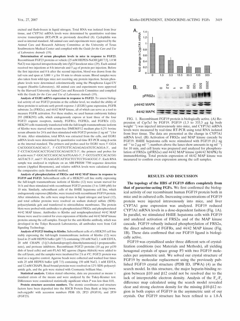
The topology of the HBS of FGF19 differs completely fromthat of paracrine-acting FGFs. We first confirmed the biolog-ical activity of our recombinant human FGF19 protein both inmice and in cultured cells. Increasing concentrations of FGF19protein were injected intravenously into mice, and liverCYP7A1 gene expression was analyzed. FGF19 reducedCYP7A1 mRNA levels in a dose-dependent fashion (Fig. 1A).In parallel, we stimulated H4IIE hepatoma cells with FGF19and analyzed activation of FRS2� and of the MAP kinasecascade. FGF19 robustly induced phosphorylation of FRS2�,the direct substrate of FGFRs, and 44/42 MAP kinase (Fig.1B). These data confirmed that our FGF19 ligand is biologi-cally active.
FGF19 was crystallized under three different sets of crystal-lization conditions (see Materials and Methods), all yieldinghexagonal crystals of space group P3 with two FGF19 mole-cules per asymmetric unit. We solved our crystal structure ofFGF19 by molecular replacement using the previously pub-lished FGF19 crystal structure (PDB ID, 1PWA) (4) as thesearch model. In this structure, the major heparin-binding re-gion between �10 and �12 could not be resolved due to thelack of interpretable electron density. Analysis of the Fo-Fc
difference map calculated using the search model revealedclear and strong electron density for the missing �10-�12 re-gion in both copies of FGF19 in the asymmetric unit of ourcrystals. Our FGF19 structure has been refined to a 1.8-Å
FIG. 1. Recombinant FGF19 protein is biologically active. (A) Re-pression of Cyp7a1 by FGF19. FGF19 (1.3 to 333.3 �g kg bodyweight�1) was injected intravenously into mice, and CYP7A1 mRNAlevels were measured by real-time RT-PCR using total RNA isolatedfrom liver tissue. The data are presented as the change in CYP7A1mRNA level. (B) Activation of FRS2� and MAP kinase cascade byFGF19. H4IIE hepatoma cells were stimulated with FGF19 (0.2 ngml�1 to 2 �g ml�1; numbers above the lanes show amounts in ng ml�1)for 10 min, and cell lysate was prepared and analyzed for phosphory-lation of FRS2� (pFRS2�) and 44/42 MAP kinase (p44/42 MAPK) byimmunoblotting. Total protein expression of 44/42 MAP kinase wasmeasured to confirm even expression among the cell samples.
VOL. 27, 2007 Klotho-DEPENDENT, ENDOCRINE-ACTING FGFs 3419

resolution with working and free R values of 24.2% and 25.9%,respectively (Tables 1 and 2), and the final model consists oftwo copies of FGF19 residues 40 to 176 (Fig. 2A).
The conformation of the segment between �10 and �12 inour FGF19 structure is completely different from the commonconformation adopted by paracrine-acting FGFs (Fig. 2B andC). The C� path of FGF19 starts to diverge from the commonpath adopted by these FGFs at Leu145 and converges againwith these FGFs at Leu162, three residues before the �12strand (Fig. 2B and 3B). Residues Lys149 to Lys155 in thisregion of FGF19 adopt a helical conformation, whereas para-crine-acting FGFs have the canonical �11 strand. This helix inFGF19, which we have termed �11 to reflect the lack of �11,bulges out of the rest of the protein fold, which has the corre-sponding 11 � strands, namely, �1 through �10 and �12 (Fig.2A and 3A). The presence of the �11 helix in this segmentgives FGF19 an atypical trefoil appearance.
Superimposition of the 11 structurally homologous � strandsbetween FGF19 and other FGF structures shows that the con-formation of the �1-�2 loop of FGF19, the other major hep-arin-binding region, is also substantially different from that ofother FGFs (Fig. 2B). This loop, which is the longest among allFGFs, also stretches out of the �-trefoil like core region ofFGF19 at the same side of the trefoil where the �11 helix islocated (Fig. 2A and 3A). Due to the protrusion of both hep-arin-binding regions, the HBS of FGF19 is lifted atop of theremaining �-trefoil like domain and thus appears structurallyseparated from the rest of FGF19 (Fig. 2A). This is in starkcontrast to all other FGFs, where the heparin-binding regionsare structurally integrated into the �-trefoil core domain asthey directly participate in the folding of the �-trefoil. TheHBS of FGF19 distinguishes itself further from that of para-crine-acting FGFs by the presence of a cleft between the �1-�2loop and �10-�12 region (Fig. 2A). The formation of this cleftis due to the fact that in FGF19, there are no intramolecularinteractions between these two heparin-binding elements(Fig. 2B).
The divergent conformation of the region between �10 and�12 observed in our crystal structure is not biased by crystalpacking forces for several reasons. Firstly, this region is missingin 1PWA, the search model used for solving our FGF19 struc-ture. Secondly, we obtained the same structure from crystalsgrown in different crystallization buffers (see Materials andMethods). Thirdly, the conformation of this region is virtuallyindistinguishable between the two independent copies ofFGF19 in the asymmetric unit of our crystals; since the twoFGF19 molecules experience different lattice contacts in thecrystal, the observed conformation/ordering of this region can-not be biased by crystal packing forces. Fourthly, even algo-rithms such as AGADIR, used to assess the helical propensity
of short peptides, predict an � helix at precisely the samelocation within the segment between �10 and �12 as observedin our FGF19 crystal structure. Lastly, reanalysis of 1PWA inlight of our FGF19 structure shows that even in 1PWA, the C�positions of Ser147 and Phe159 at either end of the disorderedregion have already begun to diverge from the C� backbone ofparacrine-acting FGFs and point towards the � helix seen inour FGF19 crystal structure.
The topology of the HBS of FGF23 also differs completelyfrom that of paracrine-acting FGFs. FGF23 circulates in thebloodstream in two distinct forms: a full-length mature form(Tyr25-Ile252; FGF23wt) and a shorter form (Tyr25-Arg179;FGF23core) lacking the unique 73-amino-acid C-terminal tail(1, 36). The shorter form arises from proteolytic cleavage atthe 176RXXR179 site, which follows the predicted FGF corehomology region of FGF23 (28, 35). Mutations at either of thetwo Arg residues result in accumulation of circulating full-length FGF23, which signals in the kidney to cause phosphatewasting in patients with autosomal-dominant hypophos-phatemic rickets (ADHR) (34). Since ADHR is inherited in anautosomal dominant fashion, it has been postulated that theC-terminal tail of FGF23 is required for regulation of phos-phate homeostasis by this FGF (28, 35).
We expressed and purified FGF23wt, FGF23core, andFGF23ADHR, an FGF23 protein harboring ADHR mutations,and assessed their biological activity in mice and in culturedcells. Both FGF23wt and FGF23ADHR reduced serum phos-phate to near-normal levels in Fgf23-null mice, whereasFGF23core had no statistically significant effect (Fig. 4A). InHEK293 cells overexpressing Klotho, both FGF23wt andFGF23ADHR robustly induced EGR1 gene expression,whereas FGF23core had almost no activity (Fig. 4B). Similarly,both FGF23wt and FGF23ADHR robustly activated FRS2� and44/42 MAP kinase, whereas FGF23core failed to induce phos-phorylation of these downstream mediators of FGF signaling(Fig. 4C). These data provide the first evidence that FGF23requires its C terminus to signal. To gain insights into themolecular basis for this requirement, we compared the abilityof FGF23wt and FGF23core to bind Klotho. Coimmunoprecipi-tation studies showed that FGF23core failed to bind Klotho,indicating the involvement of the C-terminal tail of FGF23 inthe interaction of FGF23wt with Klotho (Fig. 4D). These datasuggest that Klotho proteins interact simultaneously with theC-terminal tail of FGF23 and an unknown region of FGFR toenhance ligand-receptor affinity.
TABLE 1. Data collection statistics from crystallographic analysis
Protein Resolution(A)
Reflections(total/unique)
Completeness(%)a
Rsym(%)a,b
Signal(�I/ I�)
FGF19 50–1.8 117,476/25,585 99.9 (99.9) 5.0 (34.3) 25.6FGF23core 50–1.5 305,261/25,550 99.5 (95.6) 5.9 (29.7) 26.1
a Values in parentheses are for the highest-resolution shell: 2.07 to 1.8 A(FGF19) and 1.55 to 1.5 A (FGF23core).
b Calculated as 100 �hkl�iPIi(hkl) � �I(hkl)�P/�hkl�iIi(hkl).
TABLE 2. Refinement statistics from crystallographic analysisa
Protein Resolution(A) Reflection Rcryst/Rfree
(%)b
Root-mean-squaredeviations
Bond(A)
Angle(°)
Bfactor(A2)c
FGF19 25–1.8 25,259 25.1/28.5 0.006 1.3 1.1FGF23core 25–1.5 25,152 20.3/21.4 0.006 1.4 1.5
a Atomic model: 2,092 protein atoms (FGF19), 1,113 protein atoms(FGF23core), 55 SOS atoms, and 98 (FGF19) and 93 (FGF23) water molecules.
b Calculated as 100 �hkl �Fo(hkl)P � PFC (hkl)�/�hklPFo(hkl)P, where Fo(�0) and Fc are the observed and calculated structure factors, respectively. 10%of the reflections were used for calculation of Rfree.
c For bonded protein atoms.
3420 GOETZ ET AL. MOL. CELL. BIOL.

FIG. 2. The HBS topology of FGF19 is completely different from that of classical, paracrine-acting FGFs. (A) Molecular surface and ribbonrepresentation of the FGF19 crystal structure. The molecular surface is shown as transparent. The � strands of FGF19 are labeled according tothe conventional strand nomenclature for FGF1 and FGF2. Note that FGF19 lacks the �11 strand present in classical, paracrine-acting FGFs. TheHBS, consisting of the loop between �1 and �2 and the segment between �10 and �12, is in blue. A secondary structure element unique to theHBS of FGF19 is the �11 helix located in the �10-�12 segment. Note that �11 and the �1-�2 loop protrude from the �-trefoil like core domainof FGF19 (orange) and that there is a cleft between these two heparin-binding regions. Cysteines 58 (in �2) and 70 (in �3) form a disulfide bridgewhich stabilizes the altered conformation of the heparin-binding region between �10 and �12 (explained in more detail below). Sulfur atoms arein green. NT and CT, N and C termini of FGF19. (B) Superimposition of the C� trace of the FGF19 �-trefoil like core onto the C� trace of theFGF2 �-trefoil from the FGF2-FGFR1c-heparin structure (PDB ID, 1FQ9). A close-up view of the heparin-binding regions is shown on the right,and to aid the reader, a view of the whole structure is shown on the left. FGF19 and FGF2 are in orange and cyan blue, respectively. Blackarrowheads mark leucines 145 and 162, at which the C� trace of FGF19 diverges from that of FGF2 and converges again, respectively. Note thatcysteines 58 and 70 of FGF19 form a disulfide bridge which packs against these two leucine residues, thereby stabilizing the altered conformationof the �10-�12 segment. Also note that there are no intramolecular interactions between the �1-�2 loop and the �10-�12 segment in FGF19 (indicatedby a dashed orange line with arrowheads; see also the cleft illustrated in panel A), whereas in FGF2, these regions interact with one another (indicatedby a cyan blue line with arrowheads). Black circles denote glycine and threonine residues of the GXXXXGXX(T/S) motif present in FGF2 and otherclassical FGFs (see also panel C). (C) Superimposition of the C� traces of the �-trefoil core domain of classical, paracrine-acting FGFs onto oneanother. The view is from the top looking down into the �-trefoil core. A close-up view of the heparin-binding region encompassing �10 and �12is shown on the right, and to orient the reader, a view of the whole structure is shown on the left. The FGF ligands are colored as follows: FGF1(PDB ID, 1EVT), green; FGF2 (PDB ID, 1FQ9), cyan blue; FGF4 (PDB ID, 1IJT), red; FGF7 (PDB ID, 1QQL), blue; FGF9 (PDB ID, 1IHK),yellow; FGF10 (PDB ID, 1NUN), purple; and FGF8b (PDB ID, 2FDB), brown. Note that the C� traces of these seven classical FGFs take nearlyidentical paths in the �10-�12 region. In panel B, FGF2 was chosen from this set of FGFs to illustrate the divergence of FGF19 at this region.Glycine and threonine/serine residues of the GXXXXGXX(T/S) motif conserved in these classical FGFs are marked by black arrows.
VOL. 27, 2007 Klotho-DEPENDENT, ENDOCRINE-ACTING FGFs 3421
3422 GOETZ ET AL. MOL. CELL. BIOL.

Efforts to crystallize FGF23wt or FGF23ADHR did not yieldcrystals, presumably due to the inherent flexibility of the 73-residue C terminus of this ligand. Therefore, we decided tocrystallize FGF23core, which yielded crystals only in the pres-ence of SOS. These crystals belong to the orthorhombic spacegroup P212121 and have one FGF23 molecule per asymmetricunit. We solved the crystal structure of FGF23core by molecularreplacement using our FGF19 crystal structure as the searchmodel. The �1-�2 loop and the segment between �10 and �12were omitted from the search model, because of very poor
sequence homology. The Fo-Fc difference map showed clearand strong electron density not only for both omitted regionsbut also for a SOS molecule, allowing us to unambiguouslybuild these heparin-binding regions as well as a SOS moleculebound to these regions. The FGF23-SOS complex structurehas been refined to a 1.5-Å resolution with working and free Rvalues of 24.3% and 25.5%, respectively (Table 1), and thefinal model consists of one copy of FGF23 residues 32 to 169and one SOS molecule (Fig. 5A).
Like FGF19, FGF23 adopts an atypical �-trefoil fold be-
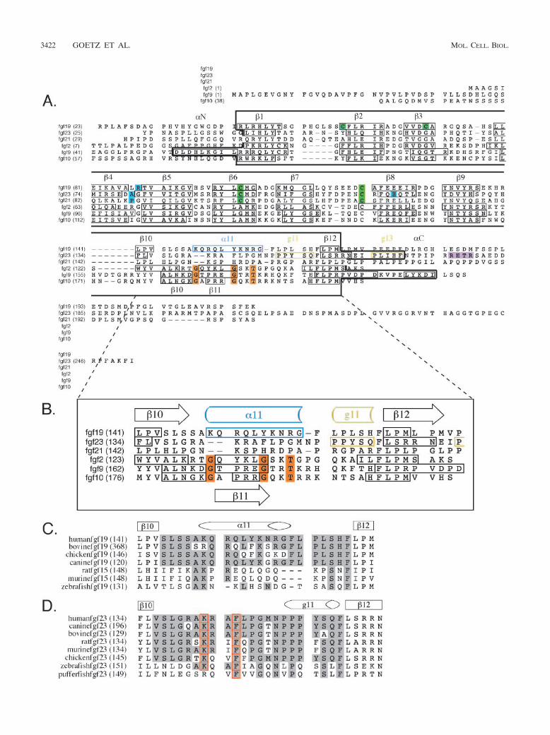
FIG. 3. Structure-based sequence analysis of FGF19 and FGF23. (A) Structure-based sequence alignment of the FGF19 subfamily membersand selected paracrine-acting FGFs. Predicted signal sequences have been omitted. Residue numbers are in parentheses on the left of thealignment. Secondary structure elements are given on top of the sequence alignment. The locations and lengths of the secondary structure elementsare indicated by boxes in the sequences. A dash in the sequence represents a gap introduced to optimize the alignment. A black box drawn aroundthe sequences marks the boundaries of the �-trefoil core domain for each FGF. Glycine and threonine residues of the GXXXXGXX(T/S) motifare in orange. Note that these residues are conserved among classical, paracrine-acting FGFs but absent in the sequences of the FGF19 subfamily.Residues which, based on published FGF-FGFR structures, likely account for low receptor-binding affinity of FGF19, -21, and -23 are in cyan blue.Cysteine residues forming disulfide bridges are in green. The proteolytic cleavage site motif RXXR in FGF23 is in purple. (B) Close-up view ofthe sequence alignment of the heparin-binding region encompassing �10 and �12. Sequence labeling is the same as in panel A. (C) Sequencealignment of the heparin-binding �10-�12 region of FGF19 orthologs. Sequence identity to human FGF19 within the �10-�12 region is highlightedin gray. Note the low degree of sequence identity between human FGF19 and rodent and fish orthologs. (D) Sequence alignment of theheparin-binding �10-�12 region of FGF23 orthologs. Sequence labeling is the same as in panel C. Residues critical for the conformation of thisregion, such as lysine 142 and phenylalanine 145, are indicated by red boxes.
FIG. 4. Recombinant FGF23 protein is biologically active. (A) The unique C-terminal tail of FGF23 is required for phosphaturic activity ofFGF23. FGF23wt, FGF23ADHR, and FGF23core were injected intraperitoneally into Fgf23-null mice. Serum phosphate levels were determinedbefore and after protein injection. Note that Fgf23-null mice show hyperphosphatemia compared to wild-type mice. (B) The C-terminal tail ofFGF23 is required for activation of EGR1 gene expression by FGF23. HEK293 cells transiently transfected with Klotho were stimulated withFGF23wt, FGF23ADHR, and FGF23core, 1 ng ml�1 each, for 30 min. Total RNA was extracted from the cells, and EGR1 mRNA levels weremeasured by real-time RT-PCR. Data are plotted as change in EGR1 mRNA expression. (C) The C-terminal tail of FGF23 is required foractivation of FRS2� and MAP kinase cascade by FGF23. HEK293 cells stably expressing Klotho were stimulated with FGF23wt, FGF23ADHR, andFGF23core, 3 nM to 3 �M each, for 10 min. Cell lysate was prepared and analyzed for phosphorylation of FRS2� (pFRS2�) and 44/42 MAP kinase(p44/42 MAPK) by immunoblotting. Total protein levels of 44/42 MAP kinase and Klotho were measured to control for equal sample loading.(D) The C-terminal tail of FGF23 is required for binding to Klotho. Lysate of HEK293 cells stably expressing Klotho was incubated withFGF23ADHR, FGF23core, FGF21, or protein sample buffer (control). Klotho was immunoprecipitated from cell lysate (IP) and analyzed for boundFGF proteins.
VOL. 27, 2007 Klotho-DEPENDENT, ENDOCRINE-ACTING FGFs 3423

FIG. 5. The HBS topology of FGF23 differs from that of classical, paracrine-acting FGFs and from that of FGF19. (A) Molecular surface andribbon representation of the crystal structure of the FGF23 core domain in complex with a SOS molecule, shown as sticks. The molecular surfaceis shown as transparent. A view of the whole structure is shown on the left; a detailed view of the SOS interactions with FGF23 is shown on theright. The � strands of FGF23 are labeled according to the conventional strand nomenclature for FGF1 and FGF2. Note that, like FGF19, FGF23lacks the �11 strand present in classical, paracrine-acting FGFs. The HBS, consisting of the loop between �1 and �2 and the segment between �10and �12, is in blue. Note that these regions do not protrude from the �-trefoil like core like those in FGF19 and that the cleft separating theseregions from one another is not as prominent as the cleft seen in the FGF19 structure (compare Fig. 2A). A secondary structure element uniqueto the HBS of FGF23 is the g11 helix, located in the segment between �10 and �12. Also note the C-terminal g13 helix, which is tethered to thecore. NT and CT, N and C termini of the FGF23 core domain. The SOS molecule makes hydrogen bonds with arginine 48 and asparagine 49 ofthe �1-�2 loop and with arginines 140 and 143 of the �10-�12 segment. Note that the sulfated fructose ring of SOS also interacts with backboneatoms of these regions. (B) Superimposition of the C� trace of the FGF23 �-trefoil like core onto the C� trace of the FGF2 �-trefoil from theFGF2-FGFR1c-heparin structure (PDB ID, 1FQ9). The viewpoint is from top of the �-trefoil like core. A close-up view of the heparin-bindingregions is shown on the right, and to assist the reader, a view of the whole structure is shown on the left. FGF23 and FGF2 are in orange and cyanblue, respectively. Black arrows mark leucine 138 and proline 153, at which the C� trace of FGF23 diverges from that of FGF2 and converges again,respectively. Glycine and threonine residues of the GXXXXGXX(T/S) motif present in FGF2 and other classical FGFs are labeled with blackcircles (see also Fig. 2C). The cyan blue arrowhead marks a one-residue insertion in the �9-�10 loop of FGF2 which is sterically incompatible withthe conformation of the �10-�12 segment in FGF23. (C) Superimposition of the C� trace of the FGF23 core domain onto the C� trace of theFGF19 core. A close-up view of the heparin-binding regions is shown on the right, and to aid the reader, a view of the whole structure is shownon the left. FGF23 and FGF19 are in orange and green, respectively. Black arrows mark glycine 139 and serine 155, at which the C� trace of FGF23diverges from that of FGF19 and converges again, respectively. Also note the different �1-�2 loop conformations of FGF23 and FGF19 concurrentwith differences in the length of the �1-�2 loop (see Fig. 3A). As in FGF19, there are no intramolecular interactions between the �1-�2 loop andthe �10-�12 segment (see also the clefts illustrated in panel A and in Fig. 2A).
3424 GOETZ ET AL. MOL. CELL. BIOL.
cause it also lacks �11. Moreover, the conformation of thesegment between �10 and �12 in FGF23 also differs from thecanonical conformation of paracrine-acting FGFs (Fig. 5B).The C� trace of FGF23 diverges from that of paracrine FGFsat exactly the same position as FGF19 does but converges atPro153, one residue earlier than FGF19 does. Notably, theconformation of the segment between �10 and �12 in theFGF23-SOS complex also differs completely from that ofFGF19 (Fig. 5C). The C� trace of FGF23 diverges from that ofFGF19 at Gly139 (homologous to Ser146 in FGF19) and con-verges again at Ser155, which corresponds to Ser163 in FGF19(Fig. 3B and 5C). The only secondary structure element as-signed to this region by PROCHECK (17) is a g helix (g11) atthe C-terminal end of this segment. Additional structural dif-ferences between FGF19 and FGF23 are seen at the �1-�2loop, the other heparin-binding region (Fig. 5C). This is antic-ipated because FGF23’s �1-�2 loop is two residues shorterthan that of FGF19 (Fig. 3A). There is also a cleft between the�1-�2 loop and the segment encompassing �10 and �12 (Fig.5A); this cleft, however, is not as prominent as the one ob-served in the FGF19 structure.
The �10-�12 region of FGF21 has no sequence identity toFGF19 and only 11% identity to FGF23, and in fact, it isshorter than those of FGF19 and FGF23 (Fig. 3B). This sug-gests that FGF21 should have yet another HBS topology. Thehigh degree of sequence divergence at this region accounts forthe overall low sequence identity among these ligands, as evi-denced by the fact that sequence identity is increased to 40 to45% when these regions are omitted from multiple sequencealignment.
The altered HBS topologies of FGF19 subfamily membersare consistent with the lack of the GXXXXGXX(T/S) motif inthis subfamily of FGFs. The unconventional conformation ofthe stretch between �10 and �12 strands observed in ourFGF19 and FGF23 structures, and by extension in FGF21, isconsistent with the major primary sequence divergence foundat this region between FGF19 subfamily members and classi-cal, paracrine-acting FGFs. This region is shorter in FGF19,FGF23, and FGF21 than in paracrine-acting FGFs by one, two,and three residues, respectively, and, most notably, lacks theGXXXXGXX(T/S) motif present in other FGFs (Fig. 3B)(14). The �10-�12 region of reported vertebrate FGF19 andFGF23 orthologs also lacks the GXXXXGXX(T/S) motif(Fig. 3C and D). The FGF19 and FGF23 crystal structuresreveal that this sequence motif plays a crucial role in pre-serving the common conformation of the stretch between�10 and �12 strands among paracrine-acting FGFs. The firstglycine from the motif makes hydrogen bonds with a highlyconserved glycine in �3, and the second glycine engages in ahydrogen bond with an FGF-invariant glycine in �7. Theseconserved hydrogen bonds promote formation of �11 andpin down the �10-�12 region to the �-trefoil core. Thethreonine/serine residue of the GXXXXGXX(T/S) motif,found in 12 of 15 paracrine-acting FGFs, also contributes tothe common conformation of the �10-�12 region in theseFGFs by forming hydrogen bonds with the second glycinefrom the motif. However, as is evident from the crystalstructure of FGF4, which has a valine instead of the T or Sof the motif, those hydrogen bonds are not absolutely re-quired for the observed canonical conformation.
The altered conformation of the �10-�12 region in FGF19and -23 is consistent with other unique structural features inthe vicinity of this region. For example, FGF19 has a cysteine(Cys70) in place of the highly conserved glycine residue in �3(Fig. 3A). Due to spatial constraints, the presence of a cysteinein this location in FGF19 is incompatible with the canonicalconformation of the heparin-binding region seen in paracrine-acting FGFs. It is noteworthy that Cys70 forms a bridge withCys58 in �2, which facilitates the altered conformation of theregion encompassing �10 and �12 in FGF19. Specifically,Leu145 and Leu162, which are positioned at the divergenceand convergence points of this region relative to other FGFs,pack against the Cys58-Cys70 disulfide bridge, thereby shield-ing it against the solvent on one side (Fig. 2B). Hence, visual-ization of the �10-�12 region in our FGF19 crystal structureshows that this disulfide bridge plays a broader role thanmerely stabilizing the tertiary structure of FGF19, as initiallysuggested by Harmer and coworkers (4).
Superimposition of the structurally homologous 11 � strandsof FGF23 onto paracrine FGFs shows that the unique confor-mation of the �10-�12 segment in FGF23 is influenced bystructural differences at the �9-�10 loop between this ligandand classical FGFs. FGF19 subfamily members have the short-est �9-�10 loop (Fig. 3A), and structural analysis shows thatthe longer �9-�10 loop of other FGFs would sterically clashwith the conformation of the �10-�12 region observed in theFGF23 structure. Hence, our structural data indicate a previ-ously unrecognized role for the �9-�10 loop in configuring theHBS and may provide an explanation for previously publisheddata showing that mutations in the �9-�10 loop or loop ex-change impair FGF biological activity (26).
The altered HBS topology of FGF19/23 is responsible forpoor heparin-binding affinity of these FGFs and for their en-docrine mode of action. In order to investigate how the alteredHBS topology of FGF19/23 would impact the mode of hepa-rin-induced FGFR dimerization by these ligands, we superim-posed FGF19/23 onto FGF2-FGFR1c-heparin (PDB ID,1FQ9) to create 2:2:2 FGF19-FGFR1c-heparin and 2:2:2FGF23-FGFR1c-heparin dimer models. In each model, theheparin-binding regions sterically clash with sugar backboneand sulfate moieties of the heparin oligosaccharide (Fig. 6). Toavoid these steric conflicts, it would be necessary to translateheparin further up from the FGF19/23 core region. The trans-lation of heparin away from FGF19/23 core domains and thepresence of a cleft in the HBS of these ligands should translateinto poor heparin-binding affinity. This is because the back-bone atoms of FGF19/23 would not be able to form hydrogenbonds with N-sulfate and 2-O-sulfate groups of rings 4[GlcN(S)6O(S)] and 5 [IdoA(2S)] of heparin; in structures ofclassical FGFs (e.g., FGF2), backbone atoms of the ligandprovide the tightest hydrogen bonds with these two sulfategroups of heparin. The orientation of SOS binding relative toFGF23 is perpendicular to that of heparin binding observed inthe FGF23-FGFR-heparin model as well as to that of SOSbinding seen in the FGF1-SOS structure (39). This observationfurther supports our prediction that linear heparin/HS cannotengage the backbone atoms of FGF23. Moreover, FGF23 hasa triple proline sequence in this region, which is incapable ofmaking hydrogen bonds with heparin/HS (Fig. 3B).
To test our structural prediction, we used SPR spectroscopy
VOL. 27, 2007 Klotho-DEPENDENT, ENDOCRINE-ACTING FGFs 3425

to compare heparin binding of FGF19, -21, and -23 with that ofparacrine-acting FGFs, including FGF1, FGF2, FGF4, FGF7,and FGF10. In support of our structural prediction, the SPRdata show that FGF19, -21, and -23 bind poorly to heparin(Fig. 7A). Importantly, the poor binding affinities of FGF19,-21, and -23 for heparin are in keeping with the endocrinebehavior of FGF19 subfamily members. The poor heparin-binding affinity should allow FGF19 subfamily members toescape the pericellular HS of the tissues where they are pro-duced. Owing to their high affinity for heparin/HS, however,paracrine-acting FGFs are entrapped in the pericellular envi-ronment of tissues at or near the site of their synthesis andrelease, and these FGFs can diffuse over a short range only,locally within tissues.
The FGF19-FGFR-heparin model suggested that Lys149,Gln152, Lys155, Asn156, and Arg157 of the �10-�12 segment
and His53 of the �1-�2 loop bind heparin in FGF19 becausethese residues are in the vicinity of the heparin oligosaccha-ride. To test this hypothesis, we mutated Lys149 and Arg157alone and in combination with alanine. SPR analysis showsthat these mutations impair the ability of FGF19 to bind hep-arin (Fig. 7B). In the FGF23-SOS structure, SOS binds with itssulfated fructose ring facing into the cleft (Fig. 5A). Arg48,Asn49, Arg140, and Arg143 of FGF23 coordinate four differ-
FIG. 6. The HBS topologies of FGF19 and FGF23 impact themode of heparin binding. (A) Detailed view of heparin binding toFGF19 in a 2:2:2 FGF19-FGFR1c-heparin model created by superim-posing FGF19 onto FGF2 in the FGF2-FGFR1c-heparin dimer (PDBID, 1FQ9). The heparin-binding regions of FGF19 are shown in ribbonand transparent surface representation; the heparin molecule is shownin stick representation. Carbon atoms are in gray, nitrogen atoms areblue, oxygen atoms are red, and sulfur atoms are yellow. Note thatthere are major steric clashes between the heparin-binding regions ofFGF19 and sugar backbone and sulfate groups of the heparin oligo-saccharide. A translation of the heparin molecule away from the HBSwould be necessary to avoid these clashes, and as a result, the heparinmolecule would not be able to interact with backbone atoms of thecleft. (B) Detailed view of heparin binding to FGF23 in a 2:2:2 FGF23-FGFR1c-heparin model created by superimposing the FGF23 coredomain onto FGF2 in the FGF2-FGFR1c-heparin dimer (PDB ID,1FQ9). Representation of the heparin-binding regions of FGF23 andthe heparin oligosaccharide, and atom coloring are the same as inpanel A. Note that the heparin-binding regions of FGF23 also steri-cally clash with sugar backbone and sulfate groups of the heparinmolecule, although not to the extent as seen in the FGF19-heparinmodel (compare panels A and B).
FIG. 7. FGF19 subfamily members exhibit unusually low bindingaffinity for heparin, and structure-based mutagenesis identifies resi-dues engaged in heparin binding in FGF19 and FGF23. (A) Repre-sentative SPR sensorgram of heparin binding of FGF19 (orange),FGF21 (brown), FGF23 (black), FGF10 (purple), FGF7 (blue), FGF4(red), FGF2 (cyan blue) and FGF1 (green), 100 nM each. (B) Represen-tative SPR sensorgram of heparin-binding of wild-type FGF19 (red)and FGF19 HBS mutants, FGF19K149A (blue) and FGF19K149A,R157A
(green), 800 nM each. (C) SPR sensorgram illustrating heparin-binding of HBS mutants of FGF23, FGF23R48,N49 (green), andFGF23R140,R143 (blue), 800 nM each. The mutations were introducedinto the ADHR mutant of FGF23 (red). FGF-heparin binding wasstudied at 25°C. The biosensor chip response is plotted as a functionof time.
3426 GOETZ ET AL. MOL. CELL. BIOL.

ent sulfates of SOS, suggesting that these are potential hepa-rin-binding residues in this ligand (Fig. 5A). To test whetherthese SOS-coordinating residues of FGF23 are indeed en-gaged in heparin binding, we introduced two double mutationsinto FGF23 (R48A-N49A and R140A-R143A). SPR analysisshows that these FGF23 mutants failed to bind heparin (Fig.7C), providing experimental evidence for the unique HBS to-pology seen in the crystal structure of FGF23.
Concluding remarks. The requirement for HS is universalfor signaling by all FGFs. While conferring endocrine ability toFGF19 subfamily members, the poor heparin-binding affinityof these ligands will reduce the ability of HS/heparin to pro-mote binding of these ligands to their cognate FGFR andhence should negatively impact signaling by these FGFs. Mod-eling studies reveal that in each of the three FGF19 subfamilymembers, a predicted key residue for FGFR binding is re-placed, so that these FGFs have inherently low affinity for theircognate receptors (Fig. 3A). The low receptor-binding affinitytogether with the low HS/heparin-binding affinity of theseFGFs is the molecular basis for the dependence of these li-gands on Klotho proteins to signal in their target tissues. Bysimultaneously interacting with FGF19, -21, and -23 and theircognate FGFRs, Klotho/�Klotho bring about binding affinitythat is just sufficient to produce a metabolic but not mitogenicresponse. The restricted expression of Klotho proteins alsocontributes to the endocrine behavior of FGF19, -21, and -23by limiting the signaling of these ligands to specific tissues. Inaddition to providing molecular insights into the endocrinemode of action of the FGF19 subfamily, our structural datamay also provide blueprints for rational drug design for met-abolic syndromes, such as diabetes, obesity, and hypercholes-terolemia, as well as disordered phosphate and vitamin Dhomeostasis.
ACKNOWLEDGMENTS
This work was supported by grants from the National Institutes ofHealth (DE13686 to M.M.; DK063934 to K.W.; AG19712 andAG25326 to M.K.; HL62244, HL52622, and GM38060-17 to R.L.;DK067158 to S.K.; S10 RR017990 to T.N.), the Irma T. Hirschl Fund(to M.M.), the Robert A. Welch Foundation (to S.K.), the Eisai Re-search Fund (to M.K.), and the Ellison Medical Foundation (to M.K.)and by institutional support from Harvard School of Dental Medicine(to B.L. and M.S.R.). We are grateful to H. C. Deitz and D. E. Arkingfor providing Klotho cDNA (to K.W.).
We are grateful to R. Abramowitz and J. Schwanof for assistance atbeam line X4A at the National Synchrotron Light Source, a DOEfacility. Beam line X4A is supported by the New York StructuralBiology Center.
REFERENCES
1. Araya, K., S. Fukumoto, R. Backenroth, Y. Takeuchi, K. Nakayama, N. Ito,N. Yoshii, Y. Yamazaki, T. Yamashita, J. Silver, T. Igarashi, and T. Fujita.2005. A novel mutation in fibroblast growth factor 23 gene as a cause oftumoral calcinosis. J. Clin. Endocrinol Metab. 90:5523–5527.
2. Bellosta, P., A. Iwahori, A. N. Plotnikov, A. V. Eliseenkova, C. Basilico, andM. Mohammadi. 2001. Identification of receptor and heparin binding sites infibroblast growth factor 4 by structure-based mutagenesis. Mol. Cell. Biol.21:5946–5957.
3. Bottcher, R. T., and C. Niehrs. 2005. Fibroblast growth factor signalingduring early vertebrate development. Endocr. Rev. 26:63–77.
4. Harmer, N. J., L. Pellegrini, D. Chirgadze, J. Fernandez-Recio, and T. L.Blundell. 2004. The crystal structure of fibroblast growth factor (FGF) 19reveals novel features of the FGF family and offers a structural basis for itsunusual receptor affinity. Biochemistry 43:629–640.
5. Holt, J. A., G. Luo, A. N. Billin, J. Bisi, Y. Y. McNeill, K. F. Kozarsky, M.Donahee, Y. Wang da, T. A. Mansfield, S. A. Kliewer, B. Goodwin, and S. A.
Jones. 2003. Definition of a novel growth factor-dependent signal cascade forthe suppression of bile acid biosynthesis. Genes Dev. 17:1581–1591.
6. Ibrahimi, O. A., F. Zhang, A. V. Eliseenkova, N. Itoh, R. J. Linhardt, and M.Mohammadi. 2004. Biochemical analysis of pathogenic ligand-dependentFGFR2 mutations suggests distinct pathophysiological mechanisms forcraniofacial and limb abnormalities. Hum. Mol. Genet. 13:2313–2324.
7. Ibrahimi, O. A., F. Zhang, S. C. Hrstka, M. Mohammadi, and R. J. Linhardt.2004. Kinetic model for FGF, FGFR, and proteoglycan signal transductioncomplex assembly. Biochemistry 43:4724–4730.
8. Inagaki, T., M. Choi, A. Moschetta, L. Peng, C. L. Cummins, J. G.McDonald, G. Luo, S. A. Jones, B. Goodwin, J. A. Richardson, R. D. Gerard,J. J. Repa, D. J. Mangelsdorf, and S. A. Kliewer. 2005. Fibroblast growthfactor 15 functions as an enterohepatic signal to regulate bile acid homeosta-sis. Cell Metab. 2:217–225.
9. Ito, S., T. Fujimori, A. Furuya, J. Satoh, Y. Nabeshima, and Y. Nabeshima.2005. Impaired negative feedback suppression of bile acid synthesis in micelacking �Klotho. J. Clin. Investig. 115:2202–2208.
10. Kharitonenkov, A., T. L. Shiyanova, A. Koester, A. M. Ford, R. Micanovic,E. J. Galbreath, G. E. Sandusky, L. J. Hammond, J. S. Moyers, R. A. Owens,J. Gromada, J. T. Brozinick, E. D. Hawkins, V. J. Wroblewski, D. S. Li, F.Mehrbod, S. R. Jaskunas, and A. B. Shanafelt. 2005. FGF-21 as a novelmetabolic regulator. J. Clin. Investig. 115:1627–1635.
11. Kuro-o, M., Y. Matsumura, H. Aizawa, H. Kawaguchi, T. Suga, T. Utsugi, Y.Ohyama, M. Kurabayashi, T. Kaname, E. Kume, H. Iwasaki, A. Iida, T.Shiraki-Iida, S. Nishikawa, R. Nagai, and Y. I. Nabeshima. 1997. Mutationof the mouse klotho gene leads to a syndrome resembling ageing. Nature390:45–51.
12. Kurosu, H., Y. Ogawa, M. Miyoshi, M. Yamamoto, A. Nandi, K. P. Rosen-blatt, M. G. Baum, S. Schiavi, M. C. Hu, O. W. Moe, and M. Kuro-o. 2006.Regulation of fibroblast growth factor-23 signaling by klotho. J. Biol. Chem.281:6120–6123.
13. Lundasen, T., C. Galman, B. Angelin, and M. Rudling. 2006. Circulatingintestinal fibroblast growth factor 19 has a pronounced diurnal variation andmodulates hepatic bile acid synthesis in man. J. Intern. Med. 260:530–536.
14. Luo, Y., W. Lu, K. A. Mohamedali, J. H. Jang, R. B. Jones, J. L. Gabriel, M.Kan, and W. L. McKeehan. 1998. The glycine box: a determinant of speci-ficity for fibroblast growth factor. Biochemistry 37:16506–16515.
15. Mohammadi, M., S. K. Olsen, and R. Goetz. 2005. A protein canyon in theFGF-FGF receptor dimer selects from an a la carte menu of heparan sulfatemotifs. Curr. Opin. Struct. Biol. 15:506–516.
16. Mohammadi, M., S. K. Olsen, and O. A. Ibrahimi. 2005. Structural basis forfibroblast growth factor receptor activation. Cytokine Growth Factor Rev.16:107–137.
17. Morris, A. L., M. W. MacArthur, E. G. Hutchinson, and J. M. Thornton.1992. Stereochemical quality of protein structure coordinates. Proteins 12:345–364.
18. Murzin, A. G., A. M. Lesk, and C. Chothia. 1992. beta-Trefoil fold. Patternsof structure and sequence in the Kunitz inhibitors interleukins-1 beta and 1alpha and fibroblast growth factors. J. Mol. Biol. 223:531–543.
19. National Research Council. 1996. Guide for the care and use of laboratoryanimals. National Academy Press, Washington, DC.
20. Olsen, S. K., J. Y. Li, C. Bromleigh, A. V. Eliseenkova, O. A. Ibrahimi, Z.Lao, F. Zhang, R. J. Linhardt, A. L. Joyner, and M. Mohammadi. 2006.Structural basis by which alternative splicing modulates the organizer activityof FGF8 in the brain. Genes Dev. 20:185–198.
21. Ornitz, D. M., and N. Itoh. 2001. Fibroblast growth factors. Genome Biol.2:reviews3005.1-reviews3005.12.
22. Plotnikov, A. N., A. V. Eliseenkova, O. A. Ibrahimi, Z. Shriver, R. Sasisekha-ran, M. A. Lemmon, and M. Mohammadi. 2001. Crystal structure of fibro-blast growth factor 9 reveals regions implicated in dimerization and autoin-hibition. J. Biol. Chem. 276:4322–4329.
23. Plotnikov, A. N., S. R. Hubbard, J. Schlessinger, and M. Mohammadi. 2000.Crystal structures of two FGF-FGFR complexes reveal the determinants ofligand-receptor specificity. Cell 101:413–424.
24. Razzaque, M. S., D. Sitara, T. Taguchi, R. St-Arnaud, and B. Lanske. 2006.Premature aging-like phenotype in fibroblast growth factor 23 null mice is avitamin D-mediated process. FASEB J. 20:720–722.
25. Schlessinger, J., A. N. Plotnikov, O. A. Ibrahimi, A. V. Eliseenkova, B. K.Yeh, A. Yayon, R. J. Linhardt, and M. Mohammadi. 2000. Crystal structureof a ternary FGF-FGFR-heparin complex reveals a dual role for heparin inFGFR binding and dimerization. Mol. Cell 6:743–750.
26. Sher, I., A. Weizman, S. Lubinsky-Mink, T. Lang, N. Adir, D. Schomburg,and D. Ron. 1999. Mutations uncouple human fibroblast growth factor(FGF)-7 biological activity and receptor binding and support broad speci-ficity in the secondary receptor binding site of FGFs. J. Biol. Chem. 274:35016–35022.
27. Shimada, T., M. Kakitani, Y. Yamazaki, H. Hasegawa, Y. Takeuchi, T.Fujita, S. Fukumoto, K. Tomizuka, and T. Yamashita. 2004. Targeted abla-tion of Fgf23 demonstrates an essential physiological role of FGF23 inphosphate and vitamin D metabolism. J. Clin. Investig. 113:561–568.
28. Shimada, T., T. Muto, I. Urakawa, T. Yoneya, Y. Yamazaki, K. Okawa, Y.Takeuchi, T. Fujita, S. Fukumoto, and T. Yamashita. 2002. Mutant FGF-23
VOL. 27, 2007 Klotho-DEPENDENT, ENDOCRINE-ACTING FGFs 3427

responsible for autosomal dominant hypophosphatemic rickets is resistant toproteolytic cleavage and causes hypophosphatemia in vivo. Endocrinology143:3179–3182.
29. Sitara, D., M. S. Razzaque, M. Hesse, S. Yoganathan, T. Taguchi, R. G.Erben, H. Juppner, and B. Lanske. 2004. Homozygous ablation of fibroblastgrowth factor-23 results in hyperphosphatemia and impaired skeletogenesis,and reverses hypophosphatemia in Phex-deficient mice. Matrix Biol. 23:421–432.
30. Thisse, B., and C. Thisse. 2005. Functions and regulations of fibroblastgrowth factor signaling during embryonic development. Dev. Biol. 287:390–402.
31. Tohyama, O., A. Imura, A. Iwano, J. N. Freund, B. Henrissat, T. Fujimori,and Y. Nabeshima. 2004. Klotho is a novel beta-glucuronidase capable ofhydrolyzing steroid beta-glucuronides. J. Biol. Chem. 279:9777–9784.
32. Tomlinson, E., L. Fu, L. John, B. Hultgren, X. Huang, M. Renz, J. P.Stephan, S. P. Tsai, L. Powell-Braxton, D. French, and T. A. Stewart. 2002.Transgenic mice expressing human fibroblast growth factor-19 display in-creased metabolic rate and decreased adiposity. Endocrinology 143:1741–1747.
33. Urakawa, I., Y. Yamazaki, T. Shimada, K. Iijima, H. Hasegawa, K. Okawa,T. Fujita, S. Fukumoto, and T. Yamashita. 2006. Klotho converts canonicalFGF receptor into a specific receptor for FGF23. Nature.
34. White, K. E., W. E. Evans, J. L. H. O’Riordan, M. C. Speer, M. J. Econs, B.Lorenz-Depiereux, M. Grabowski, T. Meitinger, and T. M. Strom. 2000.
Autosomal dominant hypophosphataemic rickets is associated with muta-tions in FGF23. Nat. Genet. 26:345–348.
35. White, K. E., G. Carn, B. Lorenz-Depiereux, A. Benet-Pages, T. M. Strom,and M. J. Econs. 2001. Autosomal-dominant hypophosphatemic rickets(ADHR) mutations stabilize FGF-23. Kidney Int. 60:2079–2086.
36. Yamazaki, Y., R. Okazaki, M. Shibata, Y. Hasegawa, K. Satoh, T. Tajima, Y.Takeuchi, T. Fujita, K. Nakahara, T. Yamashita, and S. Fukumoto. 2002.Increased circulatory level of biologically active full-length FGF-23 in pa-tients with hypophosphatemic rickets/osteomalacia. J. Clin. Endocrinol.Metab. 87:4957–4960.
37. Ye, S., Y. Luo, W. Lu, R. B. Jones, R. J. Linhardt, I. Capila, T. Toida, M.Kan, H. Pelletier, and W. L. McKeehan. 2001. Structural basis for interactionof FGF-1, FGF-2, and FGF-7 with different heparan sulfate motifs. Bio-chemistry 40:14429–14439.
38. Yeh, B. K., M. Igarashi, A. V. Eliseenkova, A. N. Plotnikov, I. Sher, D. Ron,S. A. Aaronson, and M. Mohammadi. 2003. Structural basis by which alter-native splicing confers specificity in fibroblast growth factor receptors. Proc.Natl. Acad. Sci. USA 100:2266–2271.
39. Zhu, X., B. T. Hsu, and D. C. Rees. 1993. Structural studies of the binding ofthe anti-ulcer drug sucrose octasulfate to acidic fibroblast growth factor.Structure 1:27–34.
40. Zhu, X., H. Komiya, A. Chirino, S. Faham, G. M. Fox, T. Arakawa, B. T. Hsu,and D. C. Rees. 1991. Three-dimensional structures of acidic and basicfibroblast growth factors. Science 251:90–93.
3428 GOETZ ET AL. MOL. CELL. BIOL.