
Maxillry Central incsor ppt
23
Maxillary central Incisor Prof. Dr. Mohamad Helal Professor& Head of Oral Biology Dept . Mansoura Faculty of Dentistry
Transcript of Maxillry Central incsor ppt
Maxillary central Incisor
Prof. Dr. Mohamad HelalProfessor& Head of Oral Biology Dept.
Mansoura Faculty of Dentistry
Maxillary Incisor Principal Identifying Features
The maxillary central incisor is the tooth in each maxillary quadrant of the permanent dentition which is on each side of the midline.
Normally, the max central incisors are the most prominent teeth in the oral cavity and so they are most noticeable in the dental arch, hence they contribute a focal point for the eye of the observer. In general, the labial outline of the crown conform to the general outline of the face.
The max central incisor (MCI) is the widest tooth mesio-distally (MD), the labial surface is less convex than lateral incisor and canine that give the tooth a rectangular appearance. It is the first tooth from the midline. It has a slightly straight incisal edge.
Maxillary Central Incisor
Maxillary first incisor[FDI: right=11, left=21]
Principal Identifying Features
One tapering root which, when looked at in cross-section, is roughly triangular with rounded angles.
There is only one root canal but two pulp horns (cornua), one mesial and one distal.
The crown, when viewed vestibular, has a smooth surface and is usually slightly convex.
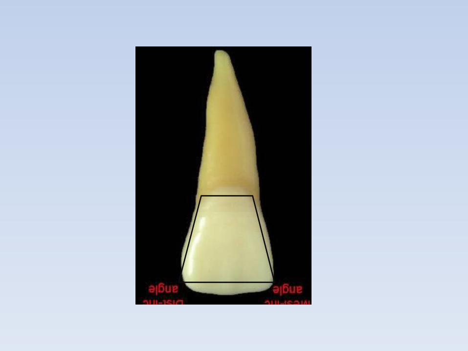
The mesio-incisal angle sharp and the disto-incisal angle rounded.
The lingual surface is concave with mesial and distal marginal ridges which meet at the neck (cervix) of the tooth forming a convex cingulum.
When viewed mesially or distally, the crown appears wedge-shaped and leans lingually.
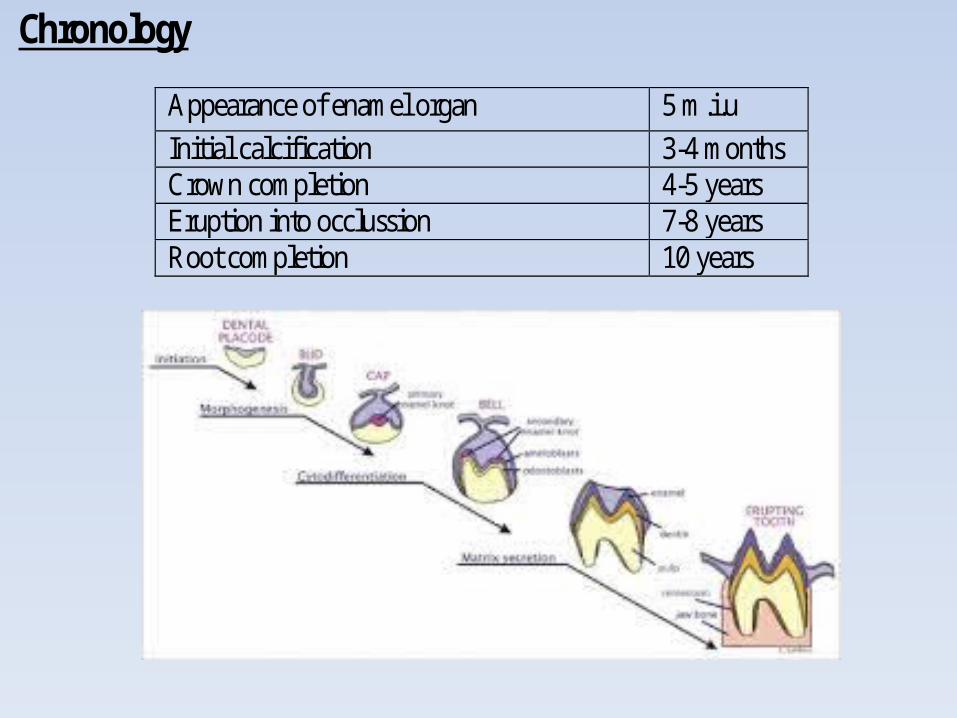
Chronology
Appearance of enamel organ 5 m.i.u Initial calcification 3-4 months Crown completion 4-5 years Eruption into occlussion 7-8 years Root completion 10 years
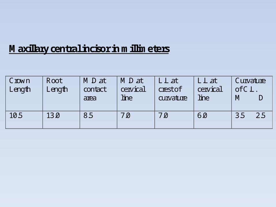
Maxillary central incisor in millimeters
Crown Length
Root Length
M.D.at contact area
M.D.at cervical line
L.L.at crest of curvature
L.L.at cervical line
Curvature of C.L. M D
10.5
13.0 8.5 7.0
7.0
6.0
3.5 2.5

Distal
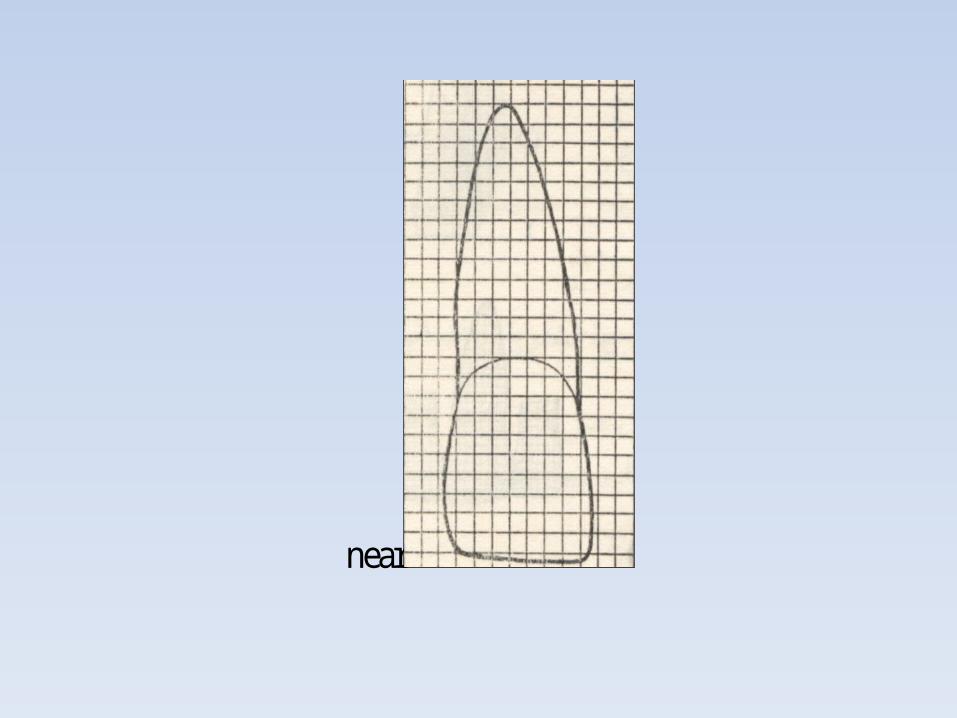
near

Lingual & Palatal aspect
cingulumm

Mesial Distal
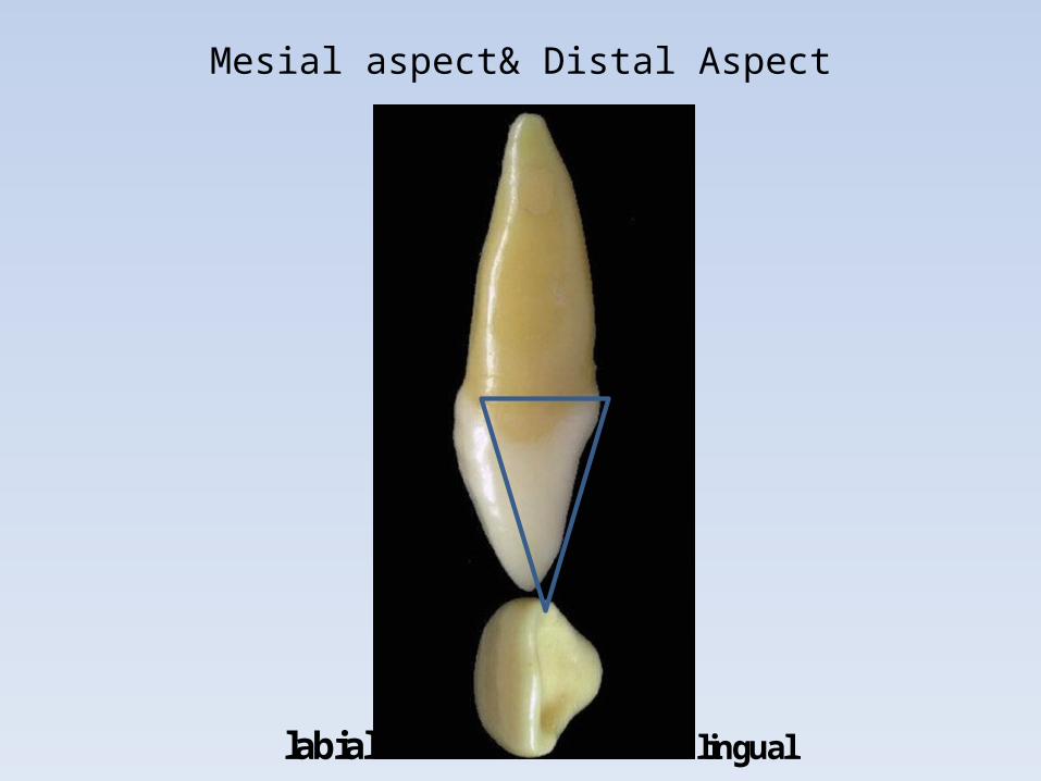
Mesial aspect& Distal Aspect
labial lingual

Root apex & incisal edge
Long axis
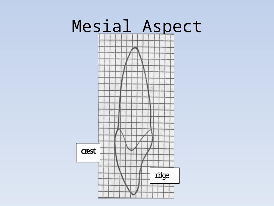
Mesial Aspect
crest
ridge
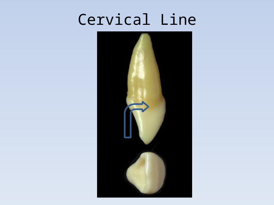
Cervical Line

Measurement of Cevical Line
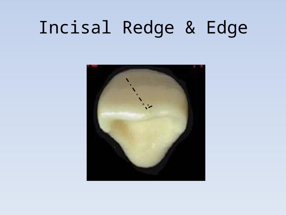
Incisal Redge & Edge
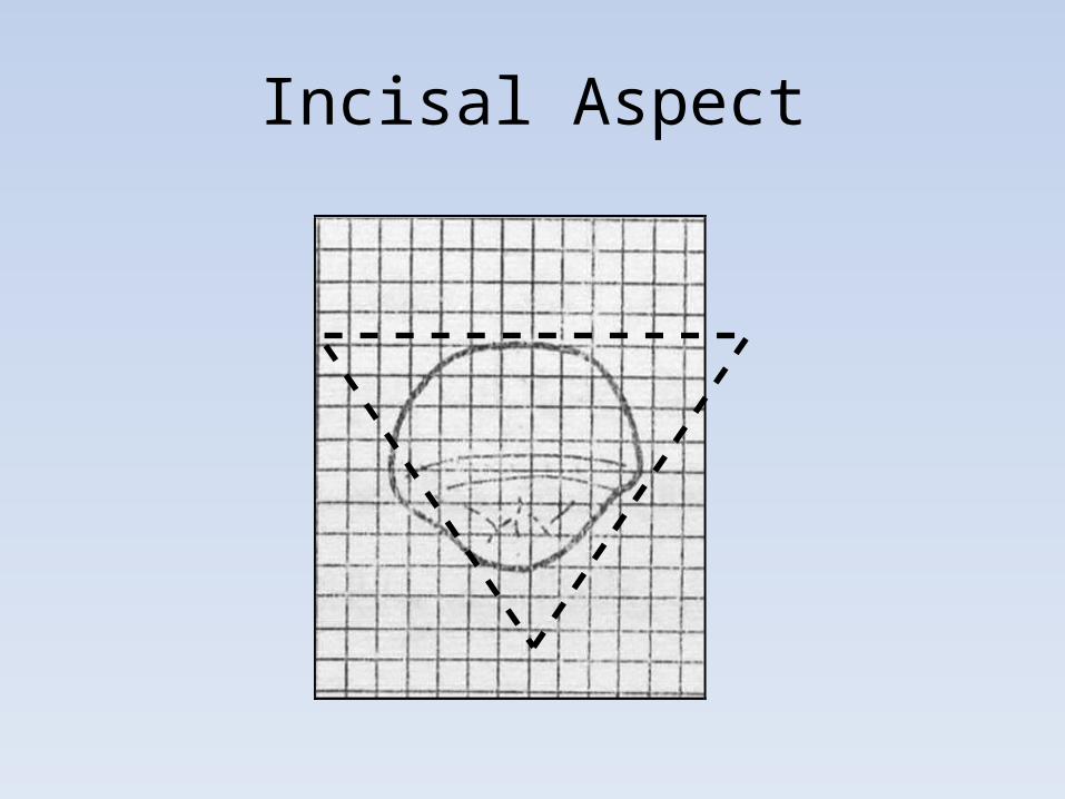
Incisal Aspect
Variations and anomalies
a) Of all the crown surfaces, the lingual exhibits the greatest variation. As previously mentioned, a pit may occasionally be present, and the depth of the fossa has a considerable range.
b) When viewed from the labial or lingual aspects, a wide variation occurs in the amount of convergence of the mesial and distal surfaces toward the cervical. When there is little convergence, the outline of the surface resembles a rectangle, but when great convergence is present, it is more nearly triangular.
c) Root length may vary considerably, but deflections of the root are relatively rare. When the root is exceptionally short, in conjunction with an abnormal contour of the crown, this anomalous condition is referred to as dwarfed root, and the lack of root support may endanger the tooth's longevity in the mouth.
a) Hutchinson's incisors: Congenital syphilis sometimes manifests itself in the central incisor by producing a screwdriver shaped crown, when it is viewed from the labial aspect.
b) Talon cusp: A large accessory cusp on the lingual surface of maxillary incisors characterizes this anomaly. Involved teeth often bear a resemblance to a Phillips screwdriver.
c) The alveolar bone between the roots of the two central incisors is occasionally the site of supernumerary teeth or extra teeth, known as mesiodens. Cysts may also be found in this area.
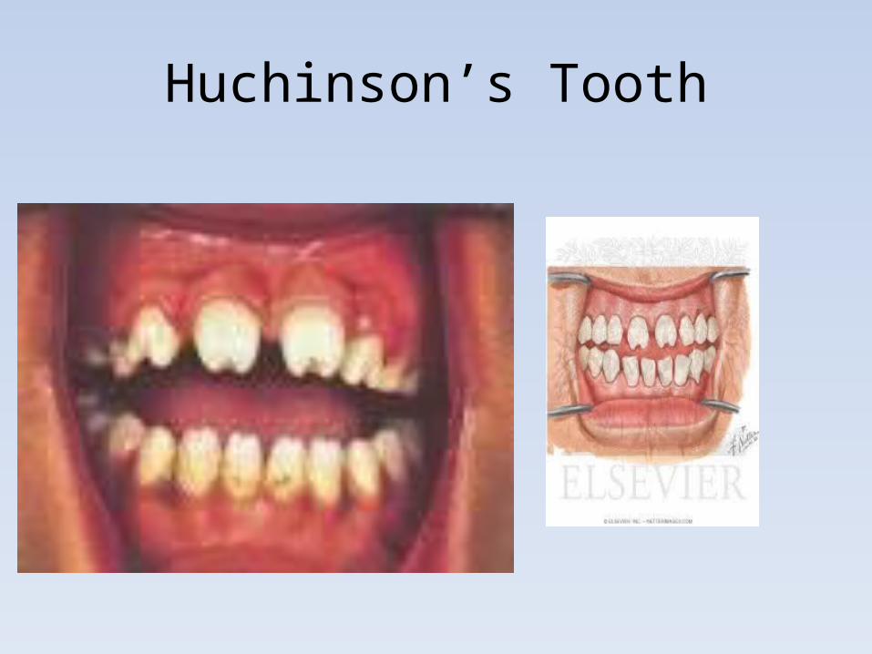
Huchinson’s Tooth
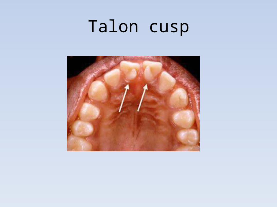
Talon cusp
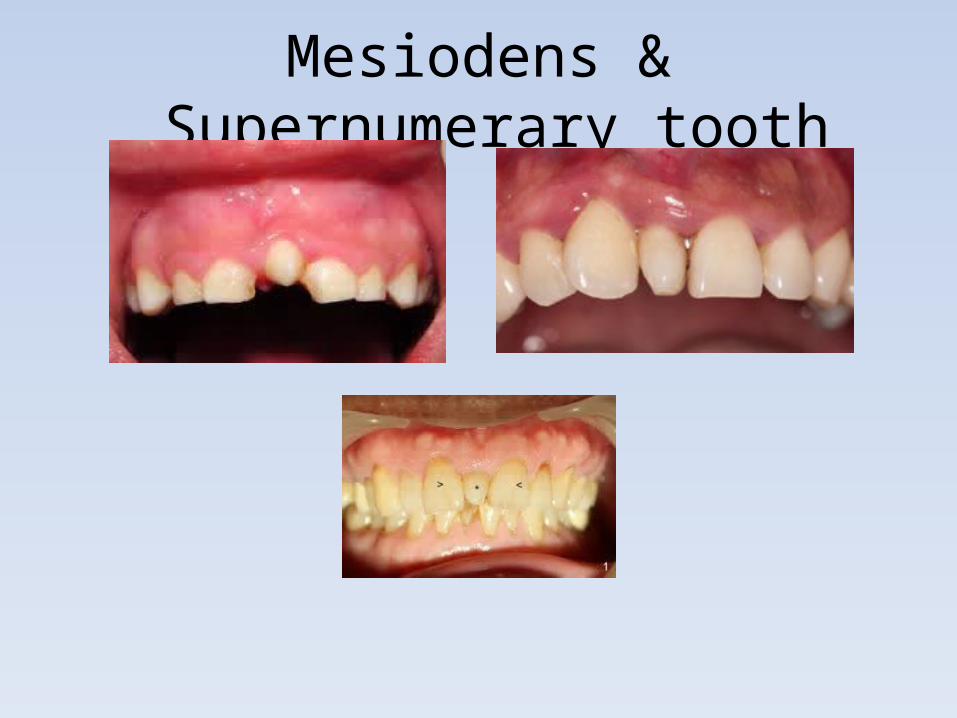
Mesiodens & Supernumerary tooth
![[PPT]PowerPoint Presentation 12.ppt · Web viewChapter 12 Incarceration of Women Central California Women’s Facility world’s largest female prison; Chowchilla, Ca. Death row in](https://static.fdocuments.in/doc/165x107/5ab5773e7f8b9a86428cbc58/pptpowerpoint-12pptweb-viewchapter-12-incarceration-of-women-central-california.jpg)


![08082015.ppt - European Central Bank · 2016. 3. 18. · Microsoft PowerPoint - 08082015.ppt [Compatibility Mode] Author: User Created Date: 3/18/2016 5:34:05 AM ...](https://static.fdocuments.in/doc/165x107/60c69e4e722d2b416a16781e/-european-central-bank-2016-3-18-microsoft-powerpoint-compatibility-mode.jpg)
![[PPT]PowerPoint Presentation - University of Central Floridaczou/CNT3004-12/ch07.ppt · Web viewAdvantages and Disadvantages Wireless Communication Advantages User Mobility Easy to](https://static.fdocuments.in/doc/165x107/5ae99d757f8b9ad73f8c0d47/pptpowerpoint-presentation-university-of-central-czoucnt3004-12ch07pptweb.jpg)


![ACCOUDOIR CENTRAL BMW E60.ppt [Mode de …lebougnat.free.fr/ACCOUDOIR_CENTRAL_BMW_E60.pdf · Title: Microsoft PowerPoint - ACCOUDOIR_CENTRAL_BMW_E60.ppt [Mode de compatibilité]](https://static.fdocuments.in/doc/165x107/5b0064bf7f8b9ad85d8c8d42/accoudoir-central-bmw-e60ppt-mode-de-microsoft-powerpoint-accoudoircentralbmwe60ppt.jpg)
