Maxillofacial Trauma: Challenges In ED Diagnosis And Management
20
February 2008 Volume 10, Number 2 Authors Charles Stewart, MD, FACEP, FAAEM Director of Research, Department of Emergency Medicine, University of Oklahoma, Oklahoma Institute of Disaster and Emergency Medicine, Tulsa, OK Peer Reviewers James F. Fiechtl, MD Assistant Professor, Department of Emergency Medicine, Vanderbilt University Medical Center Stephen J. Wolf, MD Director, Residency in Emergency Medicine, Denver Health Medical Center, Denver, Colorado CME Objectives Upon completion of this article, you should be able to: 1. List the steps in assessing a maxillofacial injury. 2. Describe immediate emergency interventions for specific maxillofacial injuries. 3. Evaluate the severity of an emergency involving maxillofacial trauma. 4. Identify appropriate imaging for dental, oral, and maxillofacial trauma. Date of original release: February 1, 2008 Date of most recent review: January 10, 2008 Termination date: February 1, 2011 Time to complete activity: 4 hours Medium: Print & online Method of participation: Print or online answer form and evaluation Prior to beginning this activity, see “Physician CME Information” on the back page. Maxillofacial Trauma: Challenges In ED Diagnosis And Management 2:20 a.m. Saturday morning: You just finished with a nine-year-old, new- onset diabetic in ketoacidosis and a 37-year-old with an inferior myocardial infarction. There aren’t any patients in the waiting room and the house supervisor is visiting. She uses the ‘Q’ word and asks if all is quiet now … “Bad choice,” you think. Just then, the survival ambulance service calls with an intoxicated patient who has obviously been assaulted. This 19-year-old male was at a bar when he ‘laid moves’ on a female patron. Her husband objected to the situation and applied ‘remedial correction’ several times with a beer bottle to the patient’s face. He apparently stopped only when the beer bottle broke. The patient is responsive to verbal stimuli, with abundant curses and imprecations. He is in spinal immobilization and strapped to a back board both for his protection and the protection of the EMS crew. The paramedics note that the patient’s respiratory rate is 20, his heart rate is 108, and his bedside glucose test measures 92. They have been unable to obtain intravenous access. They note significant facial edema on the left side, a nasal deformity, jaw deformity, and abundant epistaxis. A mong the myriad injuries seen in the emergency department, facial trauma is one of the most common. Trauma to the maxillofacial area mandates special attention. Due to their close proximity and frequent involvement, the vital structures in the head and neck region must be evaluated whenever the head and face are injured. Additionally, the psychological impact of disfigurement associated with facial and maxillary trauma can be devastating. 1-3 Given the broad variety of facial injuries and potential concomitant Editor-in-Chief Andy Jagoda, MD, FACEP Professor and Vice-Chair of Academic Affairs, Department of Emergency Medicine; Mount Sinai School of Medicine; Medical Director, Mount Sinai Hospital, New York, NY. Associate Editor John M. Howell, MD, FACEP Clinical Professor of Emergency Medicine, George Washington University, Washington, DC; Director of Academic Affairs, Best Practices, Inc, Inova Fairfax Hospital, Falls Church, VA. Editorial Board William J. Brady, MD Associate Professor and Vice Chair, Department of Emergency Medicine, University of Virginia, Charlottesville, VA. Peter DeBlieux, MD Professor of Clinical Medicine, LSU Health Science Center, New Orleans, LA. Wyatt W. Decker, MD Chair and Associate Professor of Emergency Medicine, Mayo Clinic College of Medicine, Rochester, MN. Francis M. Fesmire, MD, FACEP Director, Heart-Stroke Center, Erlanger Medical Center; Assistant Professor, UT College of Medicine, Chattanooga, TN. Michael J. Gerardi, MD, FAAP, FACEP Director, Pediatric Emergency Medicine, Children’s Medical Center, Atlantic Health System; Department of Emergency Medicine, Morristown Memorial Hospital, NJ. Michael A. Gibbs, MD, FACEP Chief, Department of Emergency Medicine, Maine Medical Center, Portland, ME. Steven A. Godwin, MD, FACEP Assistant Professor and Emergency Medicine Residency Director, University of Florida HSC/Jacksonville, FL. Gregory L. Henry, MD, FACEP CEO, Medical Practice Risk Assessment, Inc; Clinical Pro- fessor of Emergency Medicine, University of Michigan, Ann Arbor. Keith A. Marill, MD Instructor, Department of Emer- gency Medicine, Massachusetts General Hospital, Harvard Medical School, Boston, MA. Charles V. Pollack, Jr, MA, MD, FACEP Professor and Chair, Department of Emergency Medicine, Pennsyl- vania Hospital, University of Pennsylvania Health System, Philadelphia, PA. Michael S. Radeos, MD, MPH Research Director, Department of Emergency Medicine, New York Hospital Queens, Flushing, NY; Assistant Professor of Emergency Medicine, Weill Medical College of Cornell University, New York, NY. Robert L. Rogers, MD, FAAEM Assistant Professor and Resi- dency Director, Combined EM/IM Program, University of Maryland, Baltimore, MD. Alfred Sacchetti, MD, FACEP Assistant Clinical Professor, Department of Emergency Medicine, Thomas Jefferson University, Philadelphia, PA. Corey M. Slovis, MD, FACP, FACEP Professor and Chair, Department of Emergency Medicine, Vanderbilt University Medical Center, Nashville, TN. Jenny Walker, MD, MPH, MSW Assistant Professor; Division Chief, Family Medicine, Department of Community and Preventive Medicine, Mount Sinai Medical Center, New York, NY. Ron M. Walls, MD Professor and Chair, Department of Emergency Medicine, Brigham & Women’s Hospital, Boston, MA. Research Editors Nicholas Genes, MD, PhD Mount Sinai Emergency Medicine Residency. Beth Wicklund, MD Regions Hospital Emergency Medicine Residency, EMRA Representative. International Editors Valerio Gai, MD Senior Editor, Professor and Chair, Dept of EM, University of Turin, Italy. Peter Cameron, MD Chair, Emergency Medicine, Monash University; Alfred Hospital, Melbourne, Australia. Amin Antoine Kazzi, MD, FAAEM Associate Professor and Vice Chair, Department of Emergency Medicine, University of California, Irvine; American University, Beirut, Lebanon. Hugo Peralta, MD Chair of Emergency Services, Hospital Italiano, Buenos Aires, Argentina. Maarten Simons, MD, PhD Emergency Medicine Residency Director, OLVG Hospital, Amsterdam, The Netherlands. Accreditation: This activity has been planned and implemented in accordance with the Essentials and Standards of the Accreditation Council for Continuing Medical Education (ACCME) through the joint sponsorship of Mount Sinai School of Medicine and Emergency Medicine Practice. The Mount Sinai School of Medicine is accredited by the ACCME to provide continuing medical education for physicians. Faculty Disclosure: Dr. Stewart, Dr. Fiechtl, and Dr. Wolf report no significant financial interest or other relationship with the manufacturer(s) of any commercial product(s) discussed in this educational presentation. Commercial Support: Emergency Medicine Practice does not accept any commercial support.
Transcript of Maxillofacial Trauma: Challenges In ED Diagnosis And Management
February 2008Volume 10, Number 2
Authors
Charles Stewart, MD, FACEP, FAAEMDirector of Research, Department of EmergencyMedicine, University of Oklahoma, Oklahoma Institute ofDisaster and Emergency Medicine, Tulsa, OK
Peer Reviewers
James F. Fiechtl, MD Assistant Professor, Department of Emergency Medicine,Vanderbilt University Medical Center
Stephen J. Wolf, MDDirector, Residency in Emergency Medicine, DenverHealth Medical Center, Denver, Colorado
CME Objectives
Upon completion of this article, you should be able to:
1. List the steps in assessing a maxillofacial injury.
2. Describe immediate emergency interventions forspecific maxillofacial injuries.
3. Evaluate the severity of an emergency involvingmaxillofacial trauma.
4. Identify appropriate imaging for dental, oral, andmaxillofacial trauma.
Date of original release: February 1, 2008
Date of most recent review: January 10, 2008
Termination date: February 1, 2011
Time to complete activity: 4 hours
Medium: Print & online
Method of participation: Print or online answer form and evaluation
Prior to beginning this activity, see “Physician CMEInformation” on the back page.
Maxillofacial Trauma:Challenges In ED Diagnosis AndManagement2:20 a.m. Saturday morning: You just finished with a nine-year-old, new-onset diabetic in ketoacidosis and a 37-year-old with an inferior myocardialinfarction. There aren’t any patients in the waiting room and the housesupervisor is visiting. She uses the ‘Q’ word and asks if all is quiet now …“Bad choice,” you think.
Just then, the survival ambulance service calls with an intoxicatedpatient who has obviously been assaulted. This 19-year-old male was at abar when he ‘laid moves’ on a female patron. Her husband objected to thesituation and applied ‘remedial correction’ several times with a beer bottleto the patient’s face. He apparently stopped only when the beer bottlebroke. The patient is responsive to verbal stimuli, with abundant cursesand imprecations. He is in spinal immobilization and strapped to a backboard both for his protection and the protection of the EMS crew. Theparamedics note that the patient’s respiratory rate is 20, his heart rate is108, and his bedside glucose test measures 92. They have been unable toobtain intravenous access. They note significant facial edema on the leftside, a nasal deformity, jaw deformity, and abundant epistaxis.
Among the myriad injuries seen in the emergency department,facial trauma is one of the most common. Trauma to the
maxillofacial area mandates special attention. Due to their closeproximity and frequent involvement, the vital structures in the headand neck region must be evaluated whenever the head and face areinjured. Additionally, the psychological impact of disfigurementassociated with facial and maxillary trauma can be devastating.1-3
Given the broad variety of facial injuries and potential concomitant
Editor-in-Chief
Andy Jagoda, MD, FACEPProfessor and Vice-Chair ofAcademic Affairs, Department ofEmergency Medicine; Mount SinaiSchool of Medicine; MedicalDirector, Mount Sinai Hospital,New York, NY.
Associate Editor
John M. Howell, MD, FACEPClinical Professor of EmergencyMedicine, George WashingtonUniversity, Washington, DC;Director of Academic Affairs, BestPractices, Inc, Inova FairfaxHospital, Falls Church, VA.
Editorial Board
William J. Brady, MDAssociate Professor and ViceChair, Department of EmergencyMedicine, University of Virginia,Charlottesville, VA.
Peter DeBlieux, MDProfessor of Clinical Medicine,LSU Health Science Center, NewOrleans, LA.
Wyatt W. Decker, MDChair and Associate Professor ofEmergency Medicine, Mayo ClinicCollege of Medicine, Rochester,MN.
Francis M. Fesmire, MD, FACEPDirector, Heart-Stroke Center,Erlanger Medical Center;Assistant Professor, UT College ofMedicine, Chattanooga, TN.
Michael J. Gerardi, MD, FAAP,FACEPDirector, Pediatric EmergencyMedicine, Children’s MedicalCenter, Atlantic Health System;Department of EmergencyMedicine, Morristown MemorialHospital, NJ.
Michael A. Gibbs, MD, FACEPChief, Department of EmergencyMedicine, Maine Medical Center,Portland, ME.
Steven A. Godwin, MD, FACEPAssistant Professor andEmergency Medicine ResidencyDirector, University of FloridaHSC/Jacksonville, FL.
Gregory L. Henry, MD, FACEPCEO, Medical Practice RiskAssessment, Inc; Clinical Pro-fessor of Emergency Medicine,University of Michigan, Ann Arbor.
Keith A. Marill, MDInstructor, Department of Emer-gency Medicine, MassachusettsGeneral Hospital, HarvardMedical School, Boston, MA.
Charles V. Pollack, Jr, MA, MD,FACEPProfessor and Chair, Departmentof Emergency Medicine, Pennsyl-vania Hospital, University ofPennsylvania Health System,Philadelphia, PA.
Michael S. Radeos, MD, MPHResearch Director, Department ofEmergency Medicine, New YorkHospital Queens, Flushing, NY;Assistant Professor of EmergencyMedicine, Weill Medical Collegeof Cornell University, New York,NY.
Robert L. Rogers, MD, FAAEMAssistant Professor and Resi-dency Director, Combined EM/IM
Program, University of Maryland,Baltimore, MD.
Alfred Sacchetti, MD, FACEPAssistant Clinical Professor,Department of EmergencyMedicine, Thomas JeffersonUniversity, Philadelphia, PA.
Corey M. Slovis, MD, FACP,FACEPProfessor and Chair, Departmentof Emergency Medicine,Vanderbilt University MedicalCenter, Nashville, TN.
Jenny Walker, MD, MPH, MSWAssistant Professor; DivisionChief, Family Medicine,Department of Community andPreventive Medicine, Mount SinaiMedical Center, New York, NY.
Ron M. Walls, MDProfessor and Chair, Departmentof Emergency Medicine, Brigham& Women’s Hospital, Boston, MA.
Research Editors
Nicholas Genes, MD, PhDMount Sinai Emergency MedicineResidency.
Beth Wicklund, MDRegions Hospital EmergencyMedicine Residency, EMRARepresentative.
International Editors
Valerio Gai, MDSenior Editor, Professor andChair, Dept of EM, University ofTurin, Italy.
Peter Cameron, MDChair, Emergency Medicine,Monash University; AlfredHospital, Melbourne, Australia.
Amin Antoine Kazzi, MD, FAAEMAssociate Professor and ViceChair, Department of EmergencyMedicine, University of California,Irvine; American University, Beirut,Lebanon.
Hugo Peralta, MDChair of Emergency Services,Hospital Italiano, Buenos Aires,Argentina.
Maarten Simons, MD, PhDEmergency Medicine ResidencyDirector, OLVG Hospital,Amsterdam, The Netherlands.
Accreditation: This activity has been planned and implemented in accordance with the Essentials and Standards of the Accreditation Council for Continuing Medical Education(ACCME) through the joint sponsorship of Mount Sinai School of Medicine and Emergency Medicine Practice. The Mount Sinai School of Medicine is accredited by the ACCME
to provide continuing medical education for physicians. Faculty Disclosure: Dr. Stewart, Dr. Fiechtl, and Dr. Wolf report no significant financial interest or other relationshipwith the manufacturer(s) of any commercial product(s) discussed in this educational presentation. Commercial Support: Emergency Medicine Practice does not accept any
commercial support.
Emergency Medicine Practice® 2 February 2008 • EBMedicine.net
complications, management can be challenging evenfor the most experienced clinicians.
Goals in the treatment of facial injuries include areturn of normal ocular, masticatory, and nasalfunction, restoration of speech, rapid bone healing,and an acceptable facial and dental esthetic result.With ever-increasing sophistication in imaging,emergency providers can rapidly diagnose small facialfractures. However, subtle complex facial fractureswith CSF leaks, temporal bone fractures, and cranialnerve injuries can remain undiagnosed. These missedor delayed diagnoses can lead to significant morbidityor death.
The goal of this article is to assist the emergencyphysician in the initial management of patients whohave sustained a facial injury. Since the emergencyphysician is rarely involved in operative decisions,there is no effort to discuss the surgical treatment ofthese fractures and soft tissue injuries beyond theinitial stabilization. This article does not discuss pureocular or lid lesions.
Critical Appraisal Of The Literature
An electronic literature search of MEDLINE andPubMed was performed to obtain the references forthis publication. The search words maxillofacial,trauma, mandibular, maxillary, facial, and imagingwere used. The reference section of each article wasreviewed for additional references pertinent to thesubject matter. There are few prospective studies ofmaxillofacial fractures and their complications. Thereare many anecdotal case reports and retrospectiveseries examinations. As such, the majority of thestudies reviewed were retrospective in nature andwere observational rather than designed.
There are no national practice guidelines availablefor patients presenting with maxillofacial trauma, andthere are no Cochrane studies that deal directly withmaxillofacial trauma. Throughout the literature, theindications for surgery and the timing of surgery varytremendously. This lack of clear data makes themanagement of many facial fractures controversial.
Best Evidence Topics (Best Bets, available atwww.bestbets.org) were found for the followingconsiderations:
• Radiographs for facial trauma• The use of antibiotics for orbital floor fractures• Radiological diagnosis of mandibular fractures• Early manipulation of nasal fractures• The utility of the tongue blade test for the
diagnosis of mandibular fractures
Epidemiology And Etiology
More than three million facial injuries occur in theUnited States each year.4 Sports, accidental falls,motor vehicle accidents, assaults, and work-relatedaccidents account for the majority of maxillofacialinjuries.4,5 Depending on the trauma center, assaults
vie with motor vehicle accidents for the number onespot.6-9 Among sport-related injuries, boxing isparticularly associated with a high incidence facialinjuries.10
In a series of over 200 consecutive facial fracturesseen in an urban trauma center, assaults accounted fornearly 50%.4 The adult male to female ratio is 3:1, butthis is reduced to 3:2 in pediatric patients.
The incidence of concomitant major injuries isreported to be as high as 50% in high-impact facialfractures, compared to 21% for lower impactfractures.6 Fractures are more commonly associatedwith motor vehicle collisions, rather than other blunttrauma. With respect to this topic, it is worthwhile topoint out that airbag deployment considerablydecreases the incidence and severity of orbital frac-tures for front-seat occupants in frontal automobilecrashes.5,11
Fractures of the facial skeleton are relativelyuncommon in children and adolescents.12,13 This lowincidence may reflect the underdeveloped facialskeleton and paranasal sinuses as well as the un-erupted dentition which provide additional strengthto the mandible and maxilla. The well-known flexibil-ity of the pediatric skeleton will also reduce thefrequency of fractures.
Pathophysiology
Nasal FracturesThe nose is the most frequently injured facial struc-ture, accounting for approximately 40% of bonyinjuries in facial trauma.14 Assaults and sportsinjuries cause most nasal fractures in adults, followedby falls and motor vehicle collisions. Play and sportsaccount for most nasal fractures in children. Nasalfractures may occur in association with other facialinjuries or by themselves.
The nose is supported by cartilage anteriorly andinferiorly and by bone posteriorly and superiorly. Thepaired nasal bones, the maxilla, and the nasal processof the frontal bone form a framework that supportsthe cartilaginous parts of the nose. The paired nasalbones are wedge-shaped and joined at the midline.The lower half of the nasal bone is thin and broad,whereas the upper portion is thicker and is firmlysupported by an articulation with the frontal bone andthe frontal process of the maxilla. The thinner portionof the nose is more liable to fracture while the thickerportion near the frontal bone is more difficult toinjure.15 Nonetheless, the force required to fracturethe nasal bones is less than for any other facial bone.15
Likewise, due to the natural taper of the nose, thesupporting nasal septum becomes increasingly thinand tends to fracture more towards the tip of the nose.A fracture of the septum unfavorably affects thealignment of the nose during the healing process.
Many different methods have been proposed forclassifying nasal and septal fractures. Factors to
EBMedicine.net • February 2008 3 Emergency Medicine Practice®
consider in patients with an injury to the nose include:1. Cause of the trauma
a. A direct frontal blow can depress thedorsum of the nose, causing the bones totelescope posteriorly.
b. A strong force from any direction cancomminute the nasal bones and cause an“open-book” fracture of the nose.15
c. A lateral blow can cause displacement tothe opposite side of the face and leave adepression on the side of the impact.
d. Traction and torsion injuries can causedisruption of the cartilage.16
2. History of prior facial injuries3. Any prior nasal deformity4. Any prior nasal obstruction
The patient’s conception of the original shape ofhis/her nose is often inaccurate, and edema maymask bone deviation. Comparison with a picturetaken prior to trauma is often helpful. Such picturesare often found in ID documents such as a passport ordriver’s license.
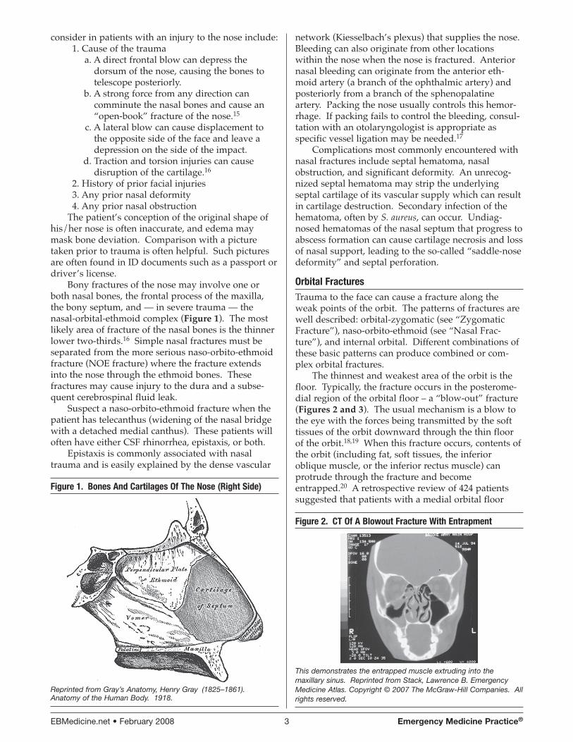
Bony fractures of the nose may involve one orboth nasal bones, the frontal process of the maxilla,the bony septum, and — in severe trauma — thenasal-orbital-ethmoid complex (Figure 1). The mostlikely area of fracture of the nasal bones is the thinnerlower two-thirds.16 Simple nasal fractures must beseparated from the more serious naso-orbito-ethmoidfracture (NOE fracture) where the fracture extendsinto the nose through the ethmoid bones. Thesefractures may cause injury to the dura and a subse-quent cerebrospinal fluid leak.
Suspect a naso-orbito-ethmoid fracture when thepatient has telecanthus (widening of the nasal bridgewith a detached medial canthus). These patients willoften have either CSF rhinorrhea, epistaxis, or both.
Epistaxis is commonly associated with nasaltrauma and is easily explained by the dense vascular
network (Kiesselbach’s plexus) that supplies the nose.Bleeding can also originate from other locationswithin the nose when the nose is fractured. Anteriornasal bleeding can originate from the anterior eth-moid artery (a branch of the ophthalmic artery) andposteriorly from a branch of the sphenopalatineartery. Packing the nose usually controls this hemor-rhage. If packing fails to control the bleeding, consul-tation with an otolaryngologist is appropriate asspecific vessel ligation may be needed.17
Complications most commonly encountered withnasal fractures include septal hematoma, nasalobstruction, and significant deformity. An unrecog-nized septal hematoma may strip the underlyingseptal cartilage of its vascular supply which can resultin cartilage destruction. Secondary infection of thehematoma, often by S. aureus, can occur. Undiag-nosed hematomas of the nasal septum that progress toabscess formation can cause cartilage necrosis and lossof nasal support, leading to the so-called “saddle-nosedeformity” and septal perforation.
Orbital FracturesTrauma to the face can cause a fracture along theweak points of the orbit. The patterns of fractures arewell described: orbital-zygomatic (see “ZygomaticFracture”), naso-orbito-ethmoid (see “Nasal Frac-ture”), and internal orbital. Different combinations ofthese basic patterns can produce combined or com-plex orbital fractures.
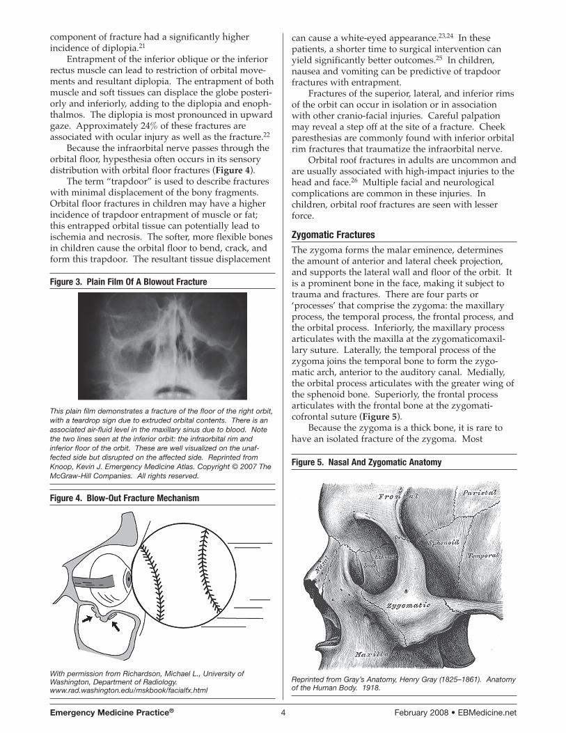
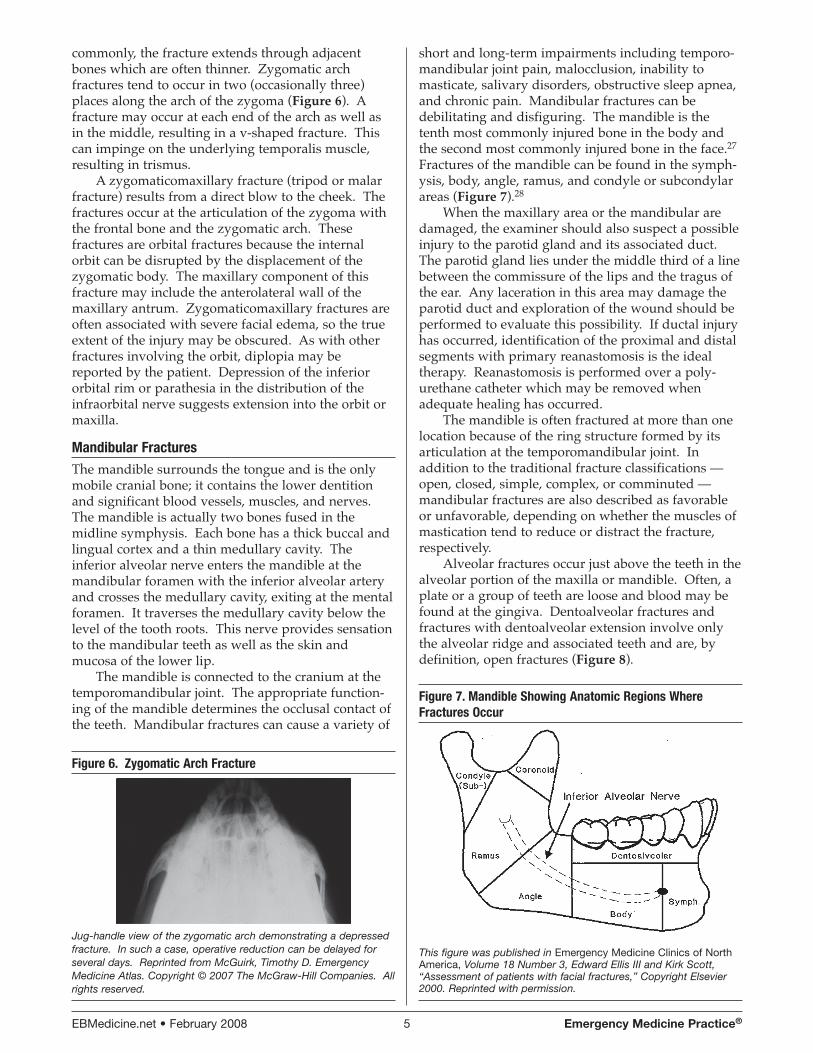
The thinnest and weakest area of the orbit is thefloor. Typically, the fracture occurs in the posterome-dial region of the orbital floor – a “blow-out” fracture(Figures 2 and 3). The usual mechanism is a blow tothe eye with the forces being transmitted by the softtissues of the orbit downward through the thin floorof the orbit.18,19 When this fracture occurs, contents ofthe orbit (including fat, soft tissues, the inferioroblique muscle, or the inferior rectus muscle) canprotrude through the fracture and becomeentrapped.20 A retrospective review of 424 patientssuggested that patients with a medial orbital floor
Figure 1. Bones And Cartilages Of The Nose (Right Side)
Reprinted from Gray’s Anatomy, Henry Gray (1825–1861).Anatomy of the Human Body. 1918.
Figure 2. CT Of A Blowout Fracture With Entrapment
This demonstrates the entrapped muscle extruding into themaxillary sinus. Reprinted from Stack, Lawrence B. EmergencyMedicine Atlas. Copyright © 2007 The McGraw-Hill Companies. Allrights reserved.
Emergency Medicine Practice® 4 February 2008 • EBMedicine.net
component of fracture had a significantly higherincidence of diplopia.21
Entrapment of the inferior oblique or the inferiorrectus muscle can lead to restriction of orbital move-ments and resultant diplopia. The entrapment of bothmuscle and soft tissues can displace the globe posteri-orly and inferiorly, adding to the diplopia and enoph-thalmos. The diplopia is most pronounced in upwardgaze. Approximately 24% of these fractures areassociated with ocular injury as well as the fracture.22
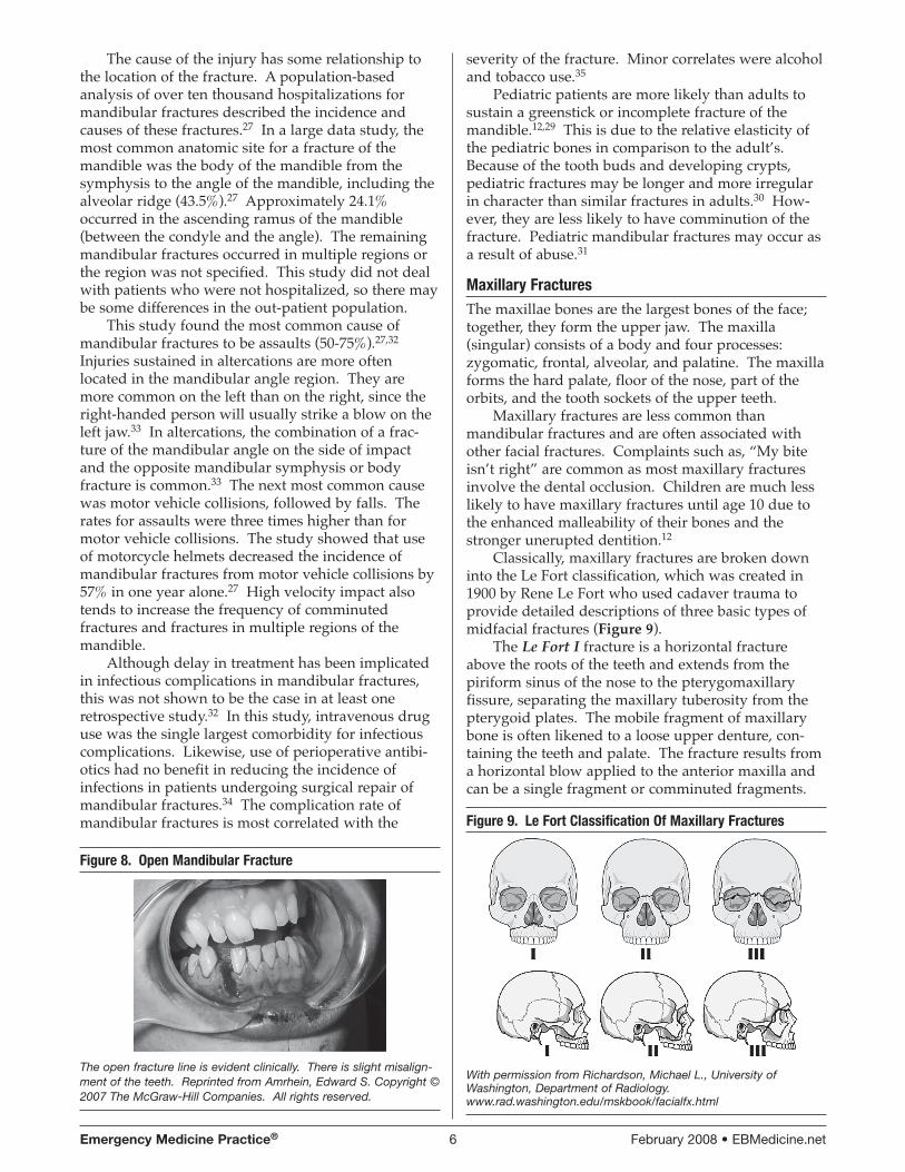
Because the infraorbital nerve passes through theorbital floor, hypesthesia often occurs in its sensorydistribution with orbital floor fractures (Figure 4).
The term “trapdoor” is used to describe fractureswith minimal displacement of the bony fragments.Orbital floor fractures in children may have a higherincidence of trapdoor entrapment of muscle or fat;this entrapped orbital tissue can potentially lead toischemia and necrosis. The softer, more flexible bonesin children cause the orbital floor to bend, crack, andform this trapdoor. The resultant tissue displacement
can cause a white-eyed appearance.23,24 In thesepatients, a shorter time to surgical intervention canyield significantly better outcomes.25 In children,nausea and vomiting can be predictive of trapdoorfractures with entrapment.
Fractures of the superior, lateral, and inferior rimsof the orbit can occur in isolation or in associationwith other cranio-facial injuries. Careful palpationmay reveal a step off at the site of a fracture. Cheekparesthesias are commonly found with inferior orbitalrim fractures that traumatize the infraorbital nerve.
Orbital roof fractures in adults are uncommon andare usually associated with high-impact injuries to thehead and face.26 Multiple facial and neurologicalcomplications are common in these injuries. Inchildren, orbital roof fractures are seen with lesserforce.
Zygomatic FracturesThe zygoma forms the malar eminence, determinesthe amount of anterior and lateral cheek projection,and supports the lateral wall and floor of the orbit. Itis a prominent bone in the face, making it subject totrauma and fractures. There are four parts or‘processes’ that comprise the zygoma: the maxillaryprocess, the temporal process, the frontal process, andthe orbital process. Inferiorly, the maxillary processarticulates with the maxilla at the zygomaticomaxil-lary suture. Laterally, the temporal process of thezygoma joins the temporal bone to form the zygo-matic arch, anterior to the auditory canal. Medially,the orbital process articulates with the greater wing ofthe sphenoid bone. Superiorly, the frontal processarticulates with the frontal bone at the zygomati-cofrontal suture (Figure 5).
Because the zygoma is a thick bone, it is rare tohave an isolated fracture of the zygoma. Most
Figure 4. Blow-Out Fracture Mechanism
With permission from Richardson, Michael L., University ofWashington, Department of Radiology.www.rad.washington.edu/mskbook/facialfx.html
Figure 5. Nasal And Zygomatic Anatomy
Reprinted from Gray’s Anatomy, Henry Gray (1825–1861). Anatomyof the Human Body. 1918.
Figure 3. Plain Film Of A Blowout Fracture
This plain film demonstrates a fracture of the floor of the right orbit,with a teardrop sign due to extruded orbital contents. There is anassociated air-fluid level in the maxillary sinus due to blood. Notethe two lines seen at the inferior orbit: the infraorbital rim andinferior floor of the orbit. These are well visualized on the unaf-fected side but disrupted on the affected side. Reprinted fromKnoop, Kevin J. Emergency Medicine Atlas. Copyright © 2007 TheMcGraw-Hill Companies. All rights reserved.
EBMedicine.net • February 2008 5 Emergency Medicine Practice®
commonly, the fracture extends through adjacentbones which are often thinner. Zygomatic archfractures tend to occur in two (occasionally three)places along the arch of the zygoma (Figure 6). Afracture may occur at each end of the arch as well asin the middle, resulting in a v-shaped fracture. Thiscan impinge on the underlying temporalis muscle,resulting in trismus.
A zygomaticomaxillary fracture (tripod or malarfracture) results from a direct blow to the cheek. Thefractures occur at the articulation of the zygoma withthe frontal bone and the zygomatic arch. Thesefractures are orbital fractures because the internalorbit can be disrupted by the displacement of thezygomatic body. The maxillary component of thisfracture may include the anterolateral wall of themaxillary antrum. Zygomaticomaxillary fractures areoften associated with severe facial edema, so the trueextent of the injury may be obscured. As with otherfractures involving the orbit, diplopia may bereported by the patient. Depression of the inferiororbital rim or parathesia in the distribution of theinfraorbital nerve suggests extension into the orbit ormaxilla.
Mandibular FracturesThe mandible surrounds the tongue and is the onlymobile cranial bone; it contains the lower dentitionand significant blood vessels, muscles, and nerves.The mandible is actually two bones fused in themidline symphysis. Each bone has a thick buccal andlingual cortex and a thin medullary cavity. Theinferior alveolar nerve enters the mandible at themandibular foramen with the inferior alveolar arteryand crosses the medullary cavity, exiting at the mentalforamen. It traverses the medullary cavity below thelevel of the tooth roots. This nerve provides sensationto the mandibular teeth as well as the skin andmucosa of the lower lip.
The mandible is connected to the cranium at thetemporomandibular joint. The appropriate function-ing of the mandible determines the occlusal contact ofthe teeth. Mandibular fractures can cause a variety of
short and long-term impairments including temporo-mandibular joint pain, malocclusion, inability tomasticate, salivary disorders, obstructive sleep apnea,and chronic pain. Mandibular fractures can bedebilitating and disfiguring. The mandible is thetenth most commonly injured bone in the body andthe second most commonly injured bone in the face.27
Fractures of the mandible can be found in the symph-ysis, body, angle, ramus, and condyle or subcondylarareas (Figure 7).28
When the maxillary area or the mandibular aredamaged, the examiner should also suspect a possibleinjury to the parotid gland and its associated duct.The parotid gland lies under the middle third of a linebetween the commissure of the lips and the tragus ofthe ear. Any laceration in this area may damage theparotid duct and exploration of the wound should beperformed to evaluate this possibility. If ductal injuryhas occurred, identification of the proximal and distalsegments with primary reanastomosis is the idealtherapy. Reanastomosis is performed over a poly-urethane catheter which may be removed whenadequate healing has occurred.
The mandible is often fractured at more than onelocation because of the ring structure formed by itsarticulation at the temporomandibular joint. Inaddition to the traditional fracture classifications —open, closed, simple, complex, or comminuted —mandibular fractures are also described as favorableor unfavorable, depending on whether the muscles ofmastication tend to reduce or distract the fracture,respectively.
Alveolar fractures occur just above the teeth in thealveolar portion of the maxilla or mandible. Often, aplate or a group of teeth are loose and blood may befound at the gingiva. Dentoalveolar fractures andfractures with dentoalveolar extension involve onlythe alveolar ridge and associated teeth and are, bydefinition, open fractures (Figure 8).
Figure 7. Mandible Showing Anatomic Regions WhereFractures Occur
This figure was published in Emergency Medicine Clinics of NorthAmerica, Volume 18 Number 3, Edward Ellis III and Kirk Scott,“Assessment of patients with facial fractures,” Copyright Elsevier2000. Reprinted with permission.
Figure 6. Zygomatic Arch Fracture
Jug-handle view of the zygomatic arch demonstrating a depressedfracture. In such a case, operative reduction can be delayed forseveral days. Reprinted from McGuirk, Timothy D. EmergencyMedicine Atlas. Copyright © 2007 The McGraw-Hill Companies. Allrights reserved.
Emergency Medicine Practice® 6 February 2008 • EBMedicine.net
The cause of the injury has some relationship tothe location of the fracture. A population-basedanalysis of over ten thousand hospitalizations formandibular fractures described the incidence andcauses of these fractures.27 In a large data study, themost common anatomic site for a fracture of themandible was the body of the mandible from thesymphysis to the angle of the mandible, including thealveolar ridge (43.5%).27 Approximately 24.1%occurred in the ascending ramus of the mandible(between the condyle and the angle). The remainingmandibular fractures occurred in multiple regions orthe region was not specified. This study did not dealwith patients who were not hospitalized, so there maybe some differences in the out-patient population.
This study found the most common cause ofmandibular fractures to be assaults (50-75%).27,32
Injuries sustained in altercations are more oftenlocated in the mandibular angle region. They aremore common on the left than on the right, since theright-handed person will usually strike a blow on theleft jaw.33 In altercations, the combination of a frac-ture of the mandibular angle on the side of impactand the opposite mandibular symphysis or bodyfracture is common.33 The next most common causewas motor vehicle collisions, followed by falls. Therates for assaults were three times higher than formotor vehicle collisions. The study showed that useof motorcycle helmets decreased the incidence ofmandibular fractures from motor vehicle collisions by57% in one year alone.27 High velocity impact alsotends to increase the frequency of comminutedfractures and fractures in multiple regions of themandible.
Although delay in treatment has been implicatedin infectious complications in mandibular fractures,this was not shown to be the case in at least oneretrospective study.32 In this study, intravenous druguse was the single largest comorbidity for infectiouscomplications. Likewise, use of perioperative antibi-otics had no benefit in reducing the incidence ofinfections in patients undergoing surgical repair ofmandibular fractures.34 The complication rate ofmandibular fractures is most correlated with the
severity of the fracture. Minor correlates were alcoholand tobacco use.35
Pediatric patients are more likely than adults tosustain a greenstick or incomplete fracture of themandible.12,29 This is due to the relative elasticity ofthe pediatric bones in comparison to the adult’s.Because of the tooth buds and developing crypts,pediatric fractures may be longer and more irregularin character than similar fractures in adults.30 How-ever, they are less likely to have comminution of thefracture. Pediatric mandibular fractures may occur asa result of abuse.31
Maxillary FracturesThe maxillae bones are the largest bones of the face;together, they form the upper jaw. The maxilla(singular) consists of a body and four processes:zygomatic, frontal, alveolar, and palatine. The maxillaforms the hard palate, floor of the nose, part of theorbits, and the tooth sockets of the upper teeth.
Maxillary fractures are less common thanmandibular fractures and are often associated withother facial fractures. Complaints such as, “My biteisn’t right” are common as most maxillary fracturesinvolve the dental occlusion. Children are much lesslikely to have maxillary fractures until age 10 due tothe enhanced malleability of their bones and thestronger unerupted dentition.12
Classically, maxillary fractures are broken downinto the Le Fort classification, which was created in1900 by Rene Le Fort who used cadaver trauma toprovide detailed descriptions of three basic types ofmidfacial fractures (Figure 9).
The Le Fort I fracture is a horizontal fractureabove the roots of the teeth and extends from thepiriform sinus of the nose to the pterygomaxillaryfissure, separating the maxillary tuberosity from thepterygoid plates. The mobile fragment of maxillarybone is often likened to a loose upper denture, con-taining the teeth and palate. The fracture results froma horizontal blow applied to the anterior maxilla andcan be a single fragment or comminuted fragments.
Figure 9. Le Fort Classification Of Maxillary Fractures
With permission from Richardson, Michael L., University ofWashington, Department of Radiology.www.rad.washington.edu/mskbook/facialfx.html
Figure 8. Open Mandibular Fracture
The open fracture line is evident clinically. There is slight misalign-ment of the teeth. Reprinted from Amrhein, Edward S. Copyright ©2007 The McGraw-Hill Companies. All rights reserved.
EBMedicine.net • February 2008 7 Emergency Medicine Practice®
The Le Fort II fracture courses upward throughthe infraorbital rim, through the medial orbit and thenasal bones. Since the fragment forms a triangularshape, this is often called a pyramidal fracture.
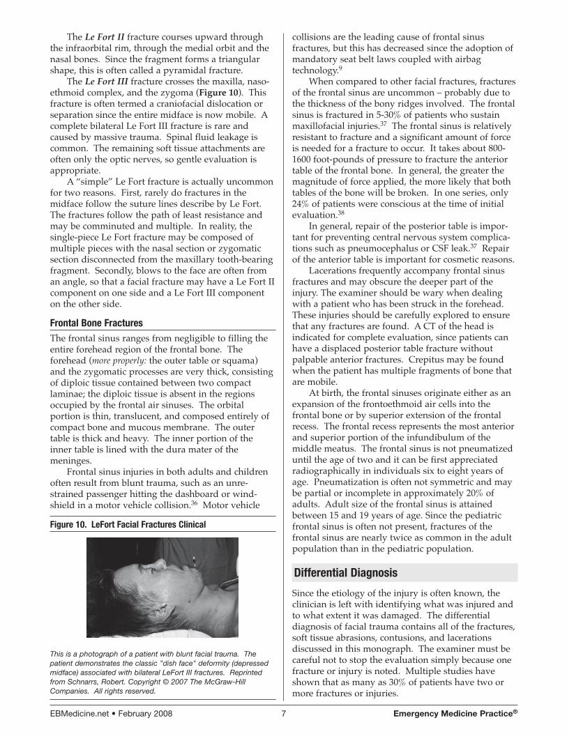
The Le Fort III fracture crosses the maxilla, naso-ethmoid complex, and the zygoma (Figure 10). Thisfracture is often termed a craniofacial dislocation orseparation since the entire midface is now mobile. Acomplete bilateral Le Fort III fracture is rare andcaused by massive trauma. Spinal fluid leakage iscommon. The remaining soft tissue attachments areoften only the optic nerves, so gentle evaluation isappropriate.
A “simple” Le Fort fracture is actually uncommonfor two reasons. First, rarely do fractures in themidface follow the suture lines describe by Le Fort.The fractures follow the path of least resistance andmay be comminuted and multiple. In reality, thesingle-piece Le Fort fracture may be composed ofmultiple pieces with the nasal section or zygomaticsection disconnected from the maxillary tooth-bearingfragment. Secondly, blows to the face are often froman angle, so that a facial fracture may have a Le Fort IIcomponent on one side and a Le Fort III componenton the other side.
Frontal Bone FracturesThe frontal sinus ranges from negligible to filling theentire forehead region of the frontal bone. Theforehead (more properly: the outer table or squama)and the zygomatic processes are very thick, consistingof diploic tissue contained between two compactlaminae; the diploic tissue is absent in the regionsoccupied by the frontal air sinuses. The orbitalportion is thin, translucent, and composed entirely ofcompact bone and mucous membrane. The outertable is thick and heavy. The inner portion of theinner table is lined with the dura mater of themeninges.
Frontal sinus injuries in both adults and childrenoften result from blunt trauma, such as an unre-strained passenger hitting the dashboard or wind-shield in a motor vehicle collision.36 Motor vehicle
collisions are the leading cause of frontal sinusfractures, but this has decreased since the adoption ofmandatory seat belt laws coupled with airbagtechnology.9
When compared to other facial fractures, fracturesof the frontal sinus are uncommon – probably due tothe thickness of the bony ridges involved. The frontalsinus is fractured in 5-30% of patients who sustainmaxillofacial injuries.37 The frontal sinus is relativelyresistant to fracture and a significant amount of forceis needed for a fracture to occur. It takes about 800-1600 foot-pounds of pressure to fracture the anteriortable of the frontal bone. In general, the greater themagnitude of force applied, the more likely that bothtables of the bone will be broken. In one series, only24% of patients were conscious at the time of initialevaluation.38
In general, repair of the posterior table is impor-tant for preventing central nervous system complica-tions such as pneumocephalus or CSF leak.37 Repairof the anterior table is important for cosmetic reasons.
Lacerations frequently accompany frontal sinusfractures and may obscure the deeper part of theinjury. The examiner should be wary when dealingwith a patient who has been struck in the forehead.These injuries should be carefully explored to ensurethat any fractures are found. A CT of the head isindicated for complete evaluation, since patients canhave a displaced posterior table fracture withoutpalpable anterior fractures. Crepitus may be foundwhen the patient has multiple fragments of bone thatare mobile.
At birth, the frontal sinuses originate either as anexpansion of the frontoethmoid air cells into thefrontal bone or by superior extension of the frontalrecess. The frontal recess represents the most anteriorand superior portion of the infundibulum of themiddle meatus. The frontal sinus is not pneumatizeduntil the age of two and it can be first appreciatedradiographically in individuals six to eight years ofage. Pneumatization is often not symmetric and maybe partial or incomplete in approximately 20% ofadults. Adult size of the frontal sinus is attainedbetween 15 and 19 years of age. Since the pediatricfrontal sinus is often not present, fractures of thefrontal sinus are nearly twice as common in the adultpopulation than in the pediatric population.
Differential Diagnosis
Since the etiology of the injury is often known, theclinician is left with identifying what was injured andto what extent it was damaged. The differentialdiagnosis of facial trauma contains all of the fractures,soft tissue abrasions, contusions, and lacerationsdiscussed in this monograph. The examiner must becareful not to stop the evaluation simply because onefracture or injury is noted. Multiple studies haveshown that as many as 30% of patients have two ormore fractures or injuries.
Figure 10. LeFort Facial Fractures Clinical
This is a photograph of a patient with blunt facial trauma. Thepatient demonstrates the classic "dish face" deformity (depressedmidface) associated with bilateral LeFort III fractures. Reprintedfrom Schnarrs, Robert. Copyright © 2007 The McGraw-HillCompanies. All rights reserved.
Emergency Medicine Practice® 8 February 2008 • EBMedicine.net
Prehospital Care
There is very little research on the prehospital care ofthe patient with maxillofacial trauma. When emer-gency services personnel are evaluating the patient,they need to ensure the ABC’s of trauma care (asdiscussed in the next section). This is particularlyimportant in the management of facial trauma wherecervical spine damage can simultaneously occur withairway damage or impingement by swelling orsignificant bleeding.
ED Management
Upon arrival to the emergency department, thephysician should re-evaluate the patient with thesame four emergency steps recommended for prehos-pital providers (listed below).
Severe facial trauma often results in partial airwayocclusion. The airway risk may increase due to bleed-ing, swelling in soft tissues, and hematoma formation.Patients with severe facial fractures may havedecreased level of consciousness due to intracranialinjury. While lifting the jaw and inserting an oral ornasal airway may temporarily help the situation, neckfractures are also common in these patients. Onlywhen the patient’s airway is stable, breathing is intact,bleeding has been controlled, and the cervical spinehas been immobilized should the physician proceed inthe secondary evaluation.1. The physician MUST ensure an airway if this has
not been done by prehospital providers.Airway compromise is common in persons
with severe maxillofacial injures. Assess thepatient for airway obstruction. Look for evidenceof injury to the larynx and trachea, includingcrepitus of the surrounding soft tissues. Clinically,the patient may have noisy breathing, snoring,gurgling, or croaking. Remove foreign bodieswith finger sweep, strong suction, or Magillforceps under direct visualization. After foreignbodies are removed, endotracheal intubation andassisted ventilation may be appropriate. In somepatients, cricothyroidotomy or emergent tra-cheotomy may be necessary. (Emergent tra-cheotomy may be needed with an associatedfractured larynx. Hoarseness, subcutaneousemphysema, and a palpable fracture are sugges-tive of laryngeal fracture.) Airway compromisemay require an immediate operation to reduce thefractured facial bones that are impinging in theairway.
2. The physician MUST ensure that the patient isbreathing and continues to breathe.
Blood or edema resulting from the injury cancause upper airway obstruction. The tongue mayobstruct the airway in a patient with a mandibularfracture. A fractured free-floating maxilla can fallback, obstructing the airway. Displaced tooth
fragments can become foreign bodies in theairway.
3. The physician MUST assess the circulation andcontrol bleeding.
Blood is supplied to the midface region fromthe branches of the sphenopalatine artery andgreater palatine artery as well as branches of theinternal carotid artery (such as the anterior andposterior ethmoid branch of the ophthalmicartery). Assuming that coagulopathy is absent,severe bleeding resulting from maxillofacialtrauma is rare and the source can often be prop-erly controlled in the ED.
Control of facial vascular injuries proceedsfrom simple direct wound pressure to vesselligation for significant bleeding. Vessel ligationshould only be performed under direct visualiza-tion of the bleeding vessel. Blind clamping candamage critical structures such as the facial nerveor the parotid duct. A Foley catheter placed into awound and inflated may control bleeding frompenetrating wounds.
Oropharyngeal bleeding must be controlled toensure that the airway remains patent. Thisbleeding is often from the nose and associatedstructures that drains through the posteriornasopharynx into the oral cavity.40 Posterior nasalpacking should be considered early in the controlof oropharyngeal bleeding in any patient who hassignificant facial trauma. As mentioned, a nasalballoon or Foley catheter may be inserted into thecranial vault in a patient with severe facial traumaand basilar skull or cribriform plate fracture. Ifsevere disruption of the hard palate or maxillawithin the oral cavity occurs, control of the airwayand packing of the oral cavity may also berequired.
Continued severe bleeding may requireimmediate surgery to ligate associated majorvessels or to reduce broken facial bones andcontrol hemorrhage. Transcatheter arterialembolization may be an alternative to surgicalcontrol of hemorrhage.41 This requires an arteri-ogram to visualize the site of bleeding beforeintroduction of the embolus.40
Large open wounds should be debrided andclosed in a layered fashion. This will bothdecrease blood loss and subsequent infectiouscomplications. Wounds that may be used later foraccess to repair fractures may be closed in atemporary fashion.
4. The physician MUST assess for neurologicaldisability and ensure that the patient’s cervicalspine is protected from further damage.
During airway control, maintain cervicalspinal immobilization in bluntly injured patients.Unstable cervical spine injury is quite rare inneurologically intact penetrating facial wounds,but up to 10% of patients with significant bluntfacial injuries will also have cervical spine injury.39
EBMedicine.net • February 2008 9 Emergency Medicine Practice®
Conversely, 15-20% of cervical spine injuries areassociated with facial fractures.
History
Ask the responsive patient the following questions:1. Which areas on you face hurt? Although this is a
basic question, the presence of pain in a specificlocation can direct the examiner towards the siteof a fracture. This question may not be as reliablein the intoxicated patient or the patient withmultiple injuries.
2. Are there any areas of numbness on your face? Anysensory deficit may show where a facial fracturehas occurred, and the fragments either impingeupon or have damaged the bony canals / grooves/ foramina where the branches of the trigeminalnerve run.
a. Is your lip or chin numb? The inferior alveolarnerve runs through the center of the mandiblefrom the middle of the ramus to the mentalforamen, where it exists to provide sensationto the lower lip and chin. If the patient feelsnumb on the lower lip or chin, it is likely thatthere is a fracture on the side of thenumbness.
b. Is your cheek, upper lip, side of the nose, or uppergingiva numb? The infraorbital and superioralveolar nerves provide sensation to themaxillary teeth and gingiva, the upper lip, theside of the nose, and the lower eyelid. If thepatient has numbness here, it is likely that afracture exists in the maxilla or orbital floor.
c. Is your lower eyelid, cheek, or upper lip numb?The infraorbital nerve courses along the floorof the orbit. It is often disrupted by a zygo-maticomaxillary complex fracture.
3. Does your bite feel ‘normal?’ Mandibular and/ormaxillary fractures are commonly associated withthe feeling that the bite is not ‘normal.’ Thelocation of contact of the teeth can often help theclinician find the site of the fracture.
4. Does it hurt when you open your mouth? Where doesit hurt? Pain when the patient attempts functionalmovements of the mandible can indicate thepresence of fractures of the mandible or maxilla.Contusions of the mandible or thetemporomandibular joint can also produce similarpain. For example, pre-auricular tenderness withmandibular movement can indicate a condylarprocess fracture. Pain in the area of the cheekwhen the patient attempts to open their mouthcan indicate a zygomaticomaxillary complexfracture. Pain at the angle of the mandible canindicate a fracture in that area. The massetermuscle attaches to the body of the zygoma andinserts onto the mandibular ramus. When frac-tures of the zygomaticomaxillary complex occur,they cause contraction of the masseter muscle andsubsequent trismus. Rarely, the zygoma can be
displaced so that it impinges on the coronoidprocess of the mandible, limiting the opening ofthe jaw.
5. Are you having trouble seeing? If possible, pre-injury visual status should be reviewed with thepatient. The state of the vision immediately afterthe trauma should also be determined. Loss oflight perception that returns suggests either avascular occlusion or an optic nerve contusion.Immediate and persistent loss of light perceptionimplies severe damage to the retina or optic nerve.Initial good vision that deteriorates may suggest acompressive ocular neuropathy and constitutes anemergency. If the patient reports flashing lights or‘floaters,’ a retinal tear or detachment or a vitreoushemorrhage should be suspected. Other ophthal-mologic conditions to include in the differentialare corneal abrasions, traumatic iritis, globerupture, and lens dislocation. See our September2007 issue, An Evidence-Based Approach To AbnormalVision, at www.ebmedicine.net/redirect/?topic=emp.
6. Do you see double? The presence of diplopia canindicate a periorbital fracture with or withoutimpingement of the extraocular muscles. It is arelatively non-specific symptom and can becaused by periorbital edema alone. If the patientnotes diplopia, CT of the orbits/facial bones isindicated. Ocular symptoms of orbital facialfractures include orbital pain, enophthalmos, andvertical diplopia.
7. Does your neck hurt? This is not an unreasonablequestion given the association of facial traumaand cervical spine injuries.
If possible, the events that surround the injuryshould be obtained from the patient, the police, orEMS providers. Remember that the patient’s accountmay differ from other potentially more reliablesources. This history can provide clues to the type ofinjuries that the patient has sustained. For example,blunt trauma to the face is more likely to result infractures, while a sharp penetrating injury can injurenerves and major vessels. Interpersonal altercationstend to result in a higher incidence of nasal, blow-out,and mandibular fractures, while motor vehicleaccidents result in more serious trauma. Heightenedconcern for serious injury should be present whenevaluating the patient who has had a high velocityblow to the face, such as a baseball, baseball bat, orgolf club impact.
Past medical history should always be assessedwhen possible. Look for seizure disorders, alcoholabuse, prior head and neck trauma or surgery, tem-poromandibular joint problems, and nutritional ormetabolic derangements. Use of ‘blood thinning’agents such as aspirin, warfarin, clopidogrel (Plavix®),or enoxaparin (Lovenox®) should always be ascer-tained as these agents may increase bleeding both inthe wounds and within the skull.
Emergency Medicine Practice® 10 February 2008 • EBMedicine.net
Physical Examination
A careful physical examination is paramount for thediagnosis of craniofacial injury, since additional andpotentially life-threatening injuries are not uncom-mon. During this initial survey, life-threateninginjuries and systemic medical problems should beaddressed.
The airway is the first critical injury that may beassociated with facial trauma. The patient who hassustained a maxillofacial fracture may have airwaycompromise due to loss of tongue support secondaryto facial fractures or obstruction of the airway due toblood or debris. The unconscious patient withsignificant facial trauma needs rapid sequence intuba-tion or a surgical airway as indicated by the patientcondition and ease of intubation. This intubationshould be performed with appropriate cervical spinalprecautions.
The patient who has sustained a significant facialfracture should be assumed to have an associatedcervical spine injury. Studies have shown thatapproximately 10% of patients with facial fractureshave injuries of the cervical spine. Suspicion ofconcomitant spinal injury can range from low in theisolated nasal fracture from a blow to very high in thecomplex facial fracture sustained in a high-speedmotor vehicle accident.
Once the patient’s airway, breathing, and hemo-dynamic status have been stabilized and the patienthas been evaluated for cervical spinal trauma, per-form a systematic secondary survey. Determine andappropriately manage the tetanus status of allpatients. Gently remove dried blood and foreignbodies from wound sites in order to evaluate thedepth and extent of the injury.
As always in the trauma patient, seek associatedinjuries. The patient with facial injuries often has analtered sensorium and may not relate such details assustaining a fight bite laceration to the hand. Acomprehensive physical examination to ensure that nosignificant other lesions are missed is not just ‘nice tohave’… it’s imperative.
Examine the patient for dental trauma andmalocclusion.42 Examine the oral cavity for lacera-tions, penetrating injuries, and continuing bleeding.The tongue is frequently lacerated in facial traumaand can produce both airway compromise withswelling and significant bleeding. Explore soft tissueinjuries within the oral cavity for tooth fragments andother foreign bodies. Note areas of ecchymosis andfacial swelling.
Evaluate the dentition and account for all emptytooth sockets. Teeth can be displaced into soft tissues,pushed into the socket, avulsed with subsequentaspiration, swallowed, or left at the scene. Anymissing teeth require a chest x-ray to ensure that atooth has not been aspirated. Evaluate the dentitionfor mobility which would indicate an underlying
alveolar fracture. The presence of a step-off or irregu-larity of the dentition may indicate an underlyingfracture. The presence of blood at the gingiva shouldalso prompt a search for underlying fracture. Fre-quently, hematomas on the lingular side of the jawmay extend into the floor of the mouth, causingdiscoloration and swelling of the floor of the mouth.Check for stability of the teeth in both the upper andlower jaw.
The clinical examination of the face begins with adetailed examination of the area for localized tender-ness, numbness, bleeding, deformity, ecchymosis,periorbital edema, otorrhea, rhinorrhea, and facialasymmetry.43 Facial asymmetry is often easiest toexamine by looking down from the head of the bed.The superior and inferior orbital rims, zygomaticarches, nose, maxilla, mandible, and both alveolarridges should be palpated and evaluated.
Zygoma fractures commonly present with perior-bital ecchymosis, lateral subconjunctival hemorrhage,infraorbital hypoesthesia, bony step-off of the orbitalrim, and depression of the malar eminence.Displacement of the bone medially may impinge onthe coronoid process of the mandible, resulting intrismus. Most zygomaticomaxillary fractures occurthrough the frontozygomatic suture, and a step-offmay be found at the junction of the superior one-thirdand inferior two-thirds of the lateral orbital rim.
A depression of the malar eminence with tender-ness suggests either a zygoma, zygomatic arch, ortripod fracture. A zygomatic arch fracture can beclinically difficult to find as the only sign may be adepression of the arch or a decreased range of mouthopening. The patient may have marked edema due tothe associated soft tissue trauma, so the depressionmay be obscured. The patient may note pain in thecheek, pain in the cheek on movement of the jaw, ortrismus. Trismus may be marked when the zygomaticfracture impinges on the temporalis muscle. A flatmalar arch may best be assessed by palpation frombehind the patient’s head or viewing the supinepatient from above the patient’s head. Comparesymmetry with the opposite side.
Suspect a tripod fracture after a blow to the cheekresulting in marked periorbital edema andecchymosis. There may be flattening of the malareminence, but resultant soft tissue trauma can obscurethe flattening.
If the zygoma is displaced inferiorly, it may causedepression of the lateral canthus. When the fractureextends through the orbit, the infraorbital nerve maybe damaged or bruised which results in hypesthesiaof the distribution of the infraorbital nerve.
If the examiner palpates the zygomaticomaxillaryarch from within the mouth, a step-off may be found.Another step-off point is at the zygomaticofrontalsuture or on the zygoma.
Closely examine the eyes, even if the eye isswollen shut. Check for injury, abnormality of ocularmovements, and visual acuity when possible. Eyelid
EBMedicine.net • February 2008 11 Emergency Medicine Practice®
ecchymosis, subcutaneous emphysema, ptosis,epistaxis, lacrimal system injuries, and pupillarydilation may each be associated with facial fractures.Pay special attention to assessing extraocular motilityfor extraocular muscle entrapment and to ensure thatthere is no orbital compartment syndrome present.25
Whenever there is enough lid swelling or perior-bital edema to restrict voluntary eyelid opening, usegreat caution. Consider the mechanism of injury andsearch for a penetrating wound. The presence ofhyphema, vitreous hemorrhage, or inability to visual-ize the fundi should prompt an urgent ophthalmo-logic consultation. If there is a through and throughlid laceration, call the ophthalmologist to repair thetarsal plate which gives structure to the lids.
Midface stability should also be evaluated. Thiscan be accomplished by grasping the teeth and hardpalate and gently pushing back and forth and then upand down. Look for movement or instability of themidface. If mobility is detected, determine the level ofthe fractures by palpating with the other hand on theface over the bridge of the nose, the infraorbital rims,and along the zygoma.
A Le Fort I fracture includes facial edema andmobility of the hard palate. Grasp the incisors andhard palate and gently push in and out. In a Le Fort Ifracture, these structures will move.
A Le Fort II fracture has marked facial edema,bilateral subconjunctival hemorrhage, and mobility ofthe maxilla. Telecanthus is usually appreciated. The
patient may have either epistaxis or CSF rhinorrhea.If the nasal bridge moves along with the maxilla, a LeFort II fracture should be suspected
A Le Fort III fracture has facial flattening andelongation. The maxilla may be displaced posteriorly,leaving the mouth open. Grasping the anterior teethand moving them will result in movement of theentire front face (craniofacial dislocation). CSFrhinorrhea and epistaxis are likely both present. If thenose, infraorbital rims, and the zygoma move togetherwith the maxilla, a Le Fort III fracture is probable.
The physical examination should involve bothinternal and external evaluation of the nose, regardlessof the mechanism of injury. Palpate the nose for ten-derness and crepitus. Clinical evidence of a nasal frac-ture includes swelling, tenderness, deformity, epistaxis,crepitus, nasal airway obstruction, and periorbitalecchymosis. In examination of the nose, note thedegree of bony deformity (laterally or depressed) aswell as the presence of cartilaginous deformity andassociated soft tissue injury (such as mucosal lacera-tion, soft tissue swelling, epistaxis, septal or orbitalhematoma, and subcutaneous emphysema).
Inspect the nasal septum for septal hematoma. Abulging, bluish, tender septal swelling or mass indi-cates a septal hematoma and requires evacuation ofthe hematoma.
Examine the nose for CSF fluid. The presence ofCSF rhinorrhea indicates disruption of the base of theskull, most commonly at the cribriform plate of the
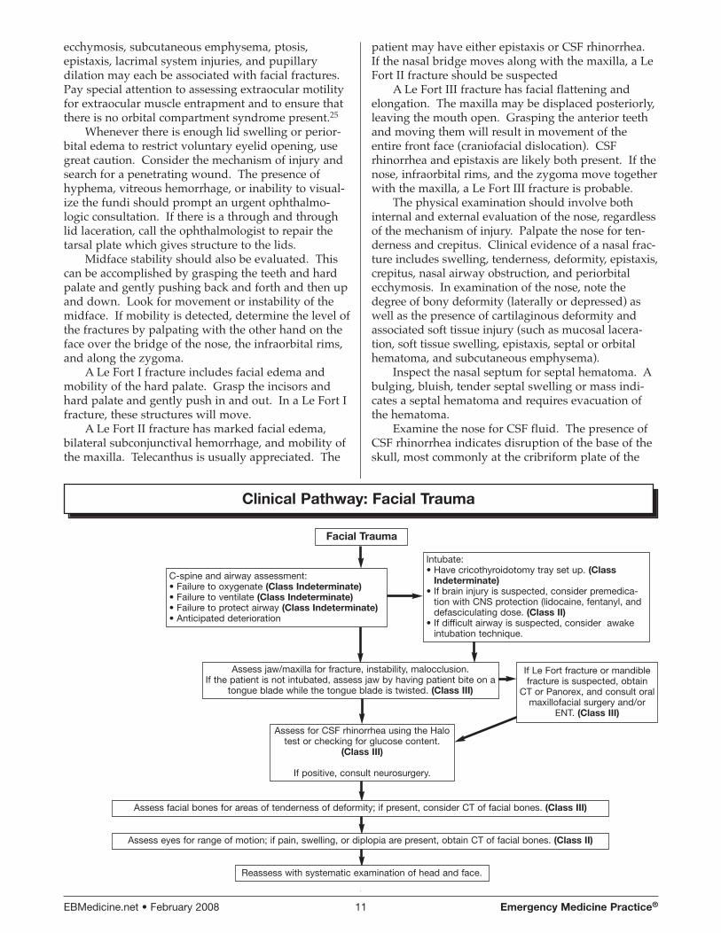
Clinical Pathway: Facial Trauma
Facial Trauma
C-spine and airway assessment:• Failure to oxygenate (Class Indeterminate)• Failure to ventilate (Class Indeterminate)• Failure to protect airway (Class Indeterminate)• Anticipated deterioration
Reassess with systematic examination of head and face.
Intubate: • Have cricothyroidotomy tray set up. (Class
Indeterminate)• If brain injury is suspected, consider premedica-
tion with CNS protection (lidocaine, fentanyl, anddefasciculating dose. (Class II)
• If difficult airway is suspected, consider awakeintubation technique.
Assess jaw/maxilla for fracture, instability, malocclusion. If the patient is not intubated, assess jaw by having patient bite on a
tongue blade while the tongue blade is twisted. (Class III)
If Le Fort fracture or mandiblefracture is suspected, obtain
CT or Panorex, and consult oralmaxillofacial surgery and/or
ENT. (Class III)
Assess facial bones for areas of tenderness of deformity; if present, consider CT of facial bones. (Class III)
Assess eyes for range of motion; if pain, swelling, or diplopia are present, obtain CT of facial bones. (Class II)
Assess for CSF rhinorrhea using the Halotest or checking for glucose content.
(Class III)
If positive, consult neurosurgery.
Emergency Medicine Practice® 12 February 2008 • EBMedicine.net
ethmoid bone (associated with a naso-ethmoidfracture or from disruption of the posterior wall of thefrontal sinus). An alternative site is a basal skullfracture or temporal bone fracture that leaks into themiddle ear and then drains through the eustachiantube into the nose.
Look at the nose for telecanthus and widening ofthe nasal bridge. Widening of the intercanthal dis-tance (> 40 mm) suggests the possibility of a naso-orbito-ethmoid fracture.
A frontal laceration should make the examinerparticularly suspicious of an underlying fracture inthe patient who has been involved in a motor vehiclecollision. The laceration must be carefully examinedfor a bony step-off. The orbits should also be carefullyevaluated. The deformity of a fracture is often hiddenby edema, so the physical examination alone may notbe sufficient in the patient with substantial foreheadlacerations.
Fractures of the mandible present in differentfashions depending on the location and the severity ofthe injury. A fracture can be obvious if it has a largedegree of displacement and has caused lacerations inthe mucosa or skin. During the examination, look forsigns of asymmetry and swelling. The most commonpresenting symptoms of patients with mandibularfractures are pain and malocclusion. Additional signsand symptoms include intraoral bleeding, lower lipand chin hypoesthesia or anesthesia, trismus, devia-tion with jaw movement, swelling or hematoma of thefloor of the mouth, and ecchymosis of the gingiva.43,44
Although a sublingual hematoma is not a consistentfinding, it is strongly suggestive of a mandibularfracture when present. Any of these findings shouldprompt the examiner to obtain further radiographicstudies.
Occasionally, a patient will have a widenedappearance to the face. This may occur when bothcondyles are fractured combined with a fracture of thesymphysis allowing the mandible to open like a book.
For the tongue blade test (TBT), have the awakepatient grip a tongue blade with his/her teeth andthen break it by rotation (twisting).45 If the patient isable to do this on both sides of the jaw, it is unlikelythat a mandibular fracture exists.46 This test has beenshown to be 95.7% sensitive if the patient can breakthe blade without pain.45
The external auditory canal can occasionally bedamaged by a fracture of the mandible, so the canalshould be visualized bilaterally in patients who havehad trauma to the jaw. The temporal-mandibular joint(TMJ) can be felt by placing a finger into the ear canaland pressing forward (anteriorly). This may be moresensitive than palpation of the TMJ in the preauriculararea. If the patient has no pain here, fracture is lesslikely.
In the conscious and cooperative patient, adetailed cranial nerve (CN) examination should beperformed.47 The optic nerve (cranial nerve II) can beassessed by having the patient read and by visual
field acuity. Extraocular movements test the integrityof CN III, IV, and VI.48
Evaluate the supraorbital, infraorbital, inferioralveolar, and mental nerve distributions for hypesthe-sia or anesthesia. Hypoesthesia of the face suggestscranial nerve V injury. Injury of the facial nerve (CNVII) produces paresis or paralysis of the muscles offacial expression.
The cranial nerve examination of the unconsciouspatient is more difficult and relies on the testing ofbrain stem reflexes.47 Assessment of vision can bequite difficult. Pupillary reflexes may remain intact aslong as the efferent pathway of cranial nerve III isintact, even when complete unilateral vision loss ispresent.49 Evaluation of the CN II pathway and theefferent CN III parasympathetic pathway is possiblein the unconscious patient with the swinging flash-light test. Testing patients with unilateral afferent CNII damage reveals bilateral equal papillary constrictionwhen the flashlight is directed towards the eye withremaining vision. When the light is directed towardsthe damaged eye, both pupils will dilate. This is theMarcus-Gunn pupillary reflex.49 See Volume 9 Number9, An Evidence-Based Approach To Abnormal Vision, for acomplete description of this test.
In the unconscious patient, extraocular move-ments can be tested with the doll’s eye reflex (oculo-cephalic reflex). Testing of the gag reflex evaluatesCN IX and CN X. The ice-water caloric test evaluatesthe function of CN VIII. To perform this test, washthe auditory canal and tympanic membrane with icedwater; this will cause the patient to develop nystag-mus. This test should not be performed when there isa rupture of the tympanic membrane. The cornealreflex tests the afferent fibers of CN V and the efferentfibers of CN VII. For this test, touching the corneawith a small wisp of cotton causes the eyelid to close.
Laboratory Analysis
Distinguishing between CSF and serous nasal secre-tions may be difficult. Blood from head injuredpatients may mix with CSF and mask the recognitionof a leak. The simplest tests for CSF fluid are easy toperform but are not very accurate. If a sufficientsample of nasal drainage can be obtained, it can besent to the lab and analyzed. CSF is odorless, salty,and has a specific gravity of 1.006. The protein level ismuch less than nasal fluid, while the chloride level isgreater. More importantly, CSF has a greater concen-tration of glucose than mucus or lacrimal secretions.The quantitative determination of a glucose level innasal fluid not contaminated by blood can be diagnos-tic of CSF rhinorrhea if the nasal fluid contains morethan 30 mg/dL. Negative test results for glucosevirtually eliminate the possibility of CSF. CSF willseparate from blood when the mixture is placed onfilter paper, resulting in a central area of blood withan outer ring or halo. Blood mixed with tap water,saline, and rhinorrhea fluid also produces a ring. The
EBMedicine.net • February 2008 13 Emergency Medicine Practice®
halo sign does occur, but clearly does not clinch thediagnosis. Another simple test involves collectingrhinorrhea on a handkerchief. Nasal secretions willdry and leave a stiff residue, whereas CSF will dryand leave the cloth soft. This test depends on thelower protein level found in CSF. Glucose oxidasepaper or dextrose sticks have historically been used toidentify CSF. However, they have been shown to beunreliable because lacrimal gland secretions and nasalmucus have reducing substances that may cause apositive reaction with glucose concentrations as lowas 5 mg/dL. The gold standard for laboratory diag-nosis of CSF fistulae is beta-2-transferrin. This proteinis found in only three bodily fluids – CSF, perilymph,and vitreous humor. Therefore, production of clearnasal discharge that is positive for beta-2-transferrin ishighly diagnostic for CSF. Unfortunately, this testrequires electrophoresis and often takes about fourdays for results.
Diagnostic Imaging Studies
After the patient has been stabilized and examined,appropriate radiographs and computerized tomo-grams (CTs) can be obtained. A patient with suspectedcranial trauma should have CT scans of the brain aswell as facial bone-specific radiographs and CTs. Ifthere are obvious or suspected facial fractures, an ap-propriate approach would be to obtain axial cuts fromthe top of the skull through the entire cervical spine.Current generation spiral CT scanners can performsuch an examination within minutes. Reconstructionof the films with computer-generated three-dimen-sional imaging allow examination of the entire face.
RadiographsA basic facial series consists of three or four films: aWaters view (PA view with cephalad angulation), aCaldwell view (PA view), a lateral view, and a sub-mentovertex view occasionally. If a nasal fracture issuspected, then a lateral view of the nasal bone withspecial nasal technique may be done. Of these views,the most consistently helpful view in facial trauma isthe Waters view. It tends to show all of the majorfacial structures at least as well and often better thanother radiographic views of the face.
There are some suggestions that help with inter-pretation of the facial bone series.1. Look at the orbits…carefully! Sixty to seventy
percent of all facial fractures involve the orbit insome way. It is important to look carefully at theorbital borders and apex as well as the optic canal.Exceptions include:
a. A localized nasal bone fractureb. A zygomatic arch fracturec. A LeFort I fracture
2. Know the common facial fracture patterns and lookfor them.
a. Zygomaticomaxillary complex fracture (tripodfracture)
b. LeFort I, II, and IIIc. Zygomatic archd. Orbital blow-out fracture
On a Waters view, the examiner may see a softtissue mass on the superior margin of the maxillarysinus, representing the herniated intraorbital softtissues protruding into the maxillary sinus. A trap-door fragment of bone may be seen protruding intothe sinus, often hinged on the ethmoid side. CT of theorbit will show these abnormalities much better.
e. Alveolar process of the maxilla3. Look at both sides of the facial films … bilateral
symmetry is a friend.4. Direct signs of a fracture:
a. Nonanatomic linear lucencyb. Cortical defect or diastatic suturec. Bone fragments overlapping, creating a
radiographic double densityd. Asymmetry of any part of the face (symmetry
is a friend)5. Indirect signs of a fracture:
a. Soft tissue swellingb. Periorbital or intracranial airc. Fluid in a paranasal sinus
Radiographic Views Of The NoseThe use of x-ray to evaluate the patient with simplenasal trauma is of limited value as decisions regardingthe treatment of nasal trauma are based on clinicalfindings.15,50,51 A plain nasal bone view cannot bereliable in exclusion of a naso-ethmoid fracture andCT scan is therefore appropriate with persistentepistaxis, CSF rhinorrhea, or both.
If deformity persists after the resolution of theedema, films may be ordered at follow-up examina-tion to help plan the repair. This would not be theusual responsibility of the emergency physician.Omission of nasal films is cost-effective, since mostnasal fractures will need no reduction.
Although isolated nasal fractures do not generallyrequire radiographic studies, it is appropriate to ordera CT scan after a thorough history and physicalexamination when other facial fractures are suspected.Three-dimensional reconstruction may help theconsultant plan surgical repair.
Radiographic Views Of The OrbitThe degree of orbital floor displacement and thepresence of soft tissue protruding through a fractureare diagnosed accurately with coronal CT scans of theorbit and facial bones. Axial scans are useful but arenot as accurate. Surgical intervention may be indi-cated when there is significant orbital floor disruption,entrapment, enophthalmos, or persistent diplopia.47
Radiographic Views Of The ZygomaZygomatic arch fractures can be seen on the under-exposed submental view (sometimes called a bucket-handle view). In this view, the arches will appear likebucket-handles, hence the nickname. A zygomaticarch fracture can also be seen on Water’s view, a
Emergency Medicine Practice® 14 February 2008 • EBMedicine.net
Towne view, or the facial series.If a tripod fracture is suspected, obtain the
Water’s view, Caldwell view, and the bucket-handleview. The Caldwell view evaluates the zygomati-cofrontal suture and the frontal process of thezygoma.
If at all possible when dealing with a suspectedorbitozygomatic fracture, obtain a CT of the area.Although the Water’s view may show some signs of afracture, plain films are considered inadequate forevaluation of a fracture of the zygoma.56
Radiographic Views Of The MandibleThe mandibular series of radiographs consists of twolateral oblique films, a reverse Towne’s view, and ananterior-posterior projection. The Towne’s view is anAP view with the neck flexed forward. It is best tovisualize condylar regions and the ascending rami ofthe mandible; a PA view is helpful in seeing the man-dibular symphysis. In cases where missing teeth areunaccounted for, perform a chest x-ray to evaluate foraspiration. Cervical spine fractures are present inapproximately 2% of patients with mandibular frac-tures and should be evaluated routinely.28
Condylar and coronoid fractures are more difficultto detect than those in other areas of the mandible.
When the mandible is injured, it behaves as if itwere a complete ring. The ring is rigid and connectedat each end to the skull by a firm joint. If one fractureof the mandible is found in a radiograph, anotherfracture or dislocation is quite likely to be present.Fractures of the angle of the mandible on one side willoften have a fracture of the mandibular condyle onthe other side.
The single most informative plain film radiologicstudy used in diagnosing mandibular fractures is thepanoramic radiograph (Figure 11).52 When the pano-ramic radiograph is compared with simple radio-graphs, it is clear that the panoramic radiograph issuperior to the plain film radiograph for diagnosis ofmandibular fractures.53,54 In one study, the panoramicradiograph was shown to diagnose 92% of fractures of
the mandible.28 The panoramic radiograph providesthe ability to view the entire mandible in one radi-ograph.
The utility and accuracy of CT depends upon thegeneration of the machine and the techniques used. Ifavailable, helical CT scanning may have enhancedimaging quality and equivalent sensitivity in theidentification of mandibular fractures. Spiral scan-ning machines are significantly more accurate thanolder machines and may replace the panoramicradiograph as the diagnostic tool of choice.55 In fact,helical computed tomography was 100% sensitive indiagnosing fractures of the mandible compared with aPanorex, which was 86% sensitive.28 On the otherhand, another study showed that the number andanatomical location of mandible fractures identifiedby helical CT and panoramic tomography were notsignificantly different.70
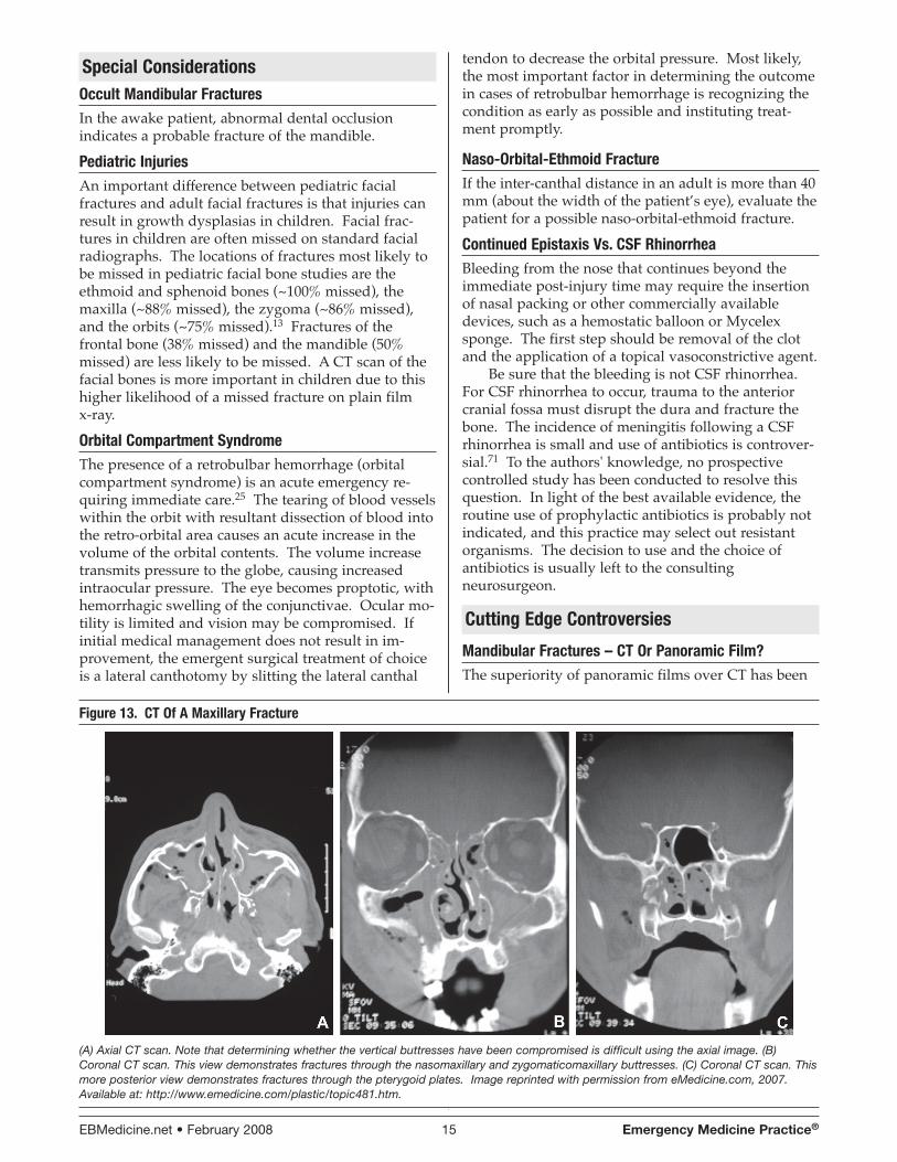
Radiographic Views Of The Maxilla And MidfaceThe midface skeleton is much more difficult to assessusing plain films than is the mandible. The presenceof very thin bones, fluid-filled sinuses (congestion vs.blood), and soft tissues make accurate assessmentproblematic. Diagnosis of all midface fractures hasbeen enhanced by high-resolution CT scanning. Axialand coronal CT scans with thin cuts of the facial bonesare recommended for all of these fractures (Figures 12and 13).
Radiographic Views Of The Frontal BoneA CT of the head is indicated for complete evaluationof a frontal bone injury, since patients can have adisplaced posterior table fracture without palpableanterior fractures. Crepitus may be found when thepatient has multiple fragments of bone that are mo-bile. CT’s not only allow the evaluation of the ante-rior and posterior table of the frontal bone, they allowthe examiner to evaluate for fluid within the sinuses,the presence of intracerebral injuries or air within thecranial cavity, and associated facial fractures.
Figure 12. Axial CT Of The Maxilla
This image through the maxillary sinuses reveals a fracture of theright wall of the right maxillary sinus, the source of the emphysema.Reprinted with permission from Gentili, Amilcare, UCSD Depart-ment of Radiology. Maxillary Sinus Fracture. Available at:http://www.gentili.net/image.asp?ID=917448377&imgid=Ctorbit2.jpg&Fx=Maxillary+Sinus+Fracture. Accessed 19 December 2007.
Figure 11. Panoramic View Of An Unfavorable MandibularFracture Dental
This demonstrates a mandibular fracture with obvious misalignmentdue to the distracting forces of the masseter muscle. Reprintedfrom Amrhein, Edward S. Copyright © 2007 The McGraw-HillCompanies. All rights reserved.
EBMedicine.net • February 2008 15 Emergency Medicine Practice®
Special ConsiderationsOccult Mandibular FracturesIn the awake patient, abnormal dental occlusionindicates a probable fracture of the mandible.
Pediatric InjuriesAn important difference between pediatric facialfractures and adult facial fractures is that injuries canresult in growth dysplasias in children. Facial frac-tures in children are often missed on standard facialradiographs. The locations of fractures most likely tobe missed in pediatric facial bone studies are theethmoid and sphenoid bones (~100% missed), themaxilla (~88% missed), the zygoma (~86% missed),and the orbits (~75% missed).13 Fractures of thefrontal bone (38% missed) and the mandible (50%missed) are less likely to be missed. A CT scan of thefacial bones is more important in children due to thishigher likelihood of a missed fracture on plain film x-ray.
Orbital Compartment SyndromeThe presence of a retrobulbar hemorrhage (orbitalcompartment syndrome) is an acute emergency re-quiring immediate care.25 The tearing of blood vesselswithin the orbit with resultant dissection of blood intothe retro-orbital area causes an acute increase in thevolume of the orbital contents. The volume increasetransmits pressure to the globe, causing increasedintraocular pressure. The eye becomes proptotic, withhemorrhagic swelling of the conjunctivae. Ocular mo-tility is limited and vision may be compromised. Ifinitial medical management does not result in im-provement, the emergent surgical treatment of choiceis a lateral canthotomy by slitting the lateral canthal
tendon to decrease the orbital pressure. Most likely,the most important factor in determining the outcomein cases of retrobulbar hemorrhage is recognizing thecondition as early as possible and instituting treat-ment promptly.
Naso-Orbital-Ethmoid FractureIf the inter-canthal distance in an adult is more than 40mm (about the width of the patient’s eye), evaluate thepatient for a possible naso-orbital-ethmoid fracture.
Continued Epistaxis Vs. CSF RhinorrheaBleeding from the nose that continues beyond theimmediate post-injury time may require the insertionof nasal packing or other commercially availabledevices, such as a hemostatic balloon or Mycelexsponge. The first step should be removal of the clotand the application of a topical vasoconstrictive agent.
Be sure that the bleeding is not CSF rhinorrhea.For CSF rhinorrhea to occur, trauma to the anteriorcranial fossa must disrupt the dura and fracture thebone. The incidence of meningitis following a CSFrhinorrhea is small and use of antibiotics is controver-sial.71 To the authors' knowledge, no prospectivecontrolled study has been conducted to resolve thisquestion. In light of the best available evidence, theroutine use of prophylactic antibiotics is probably notindicated, and this practice may select out resistantorganisms. The decision to use and the choice ofantibiotics is usually left to the consultingneurosurgeon.
Cutting Edge Controversies
Mandibular Fractures – CT Or Panoramic Film?The superiority of panoramic films over CT has been
Figure 13. CT Of A Maxillary Fracture
(A) Axial CT scan. Note that determining whether the vertical buttresses have been compromised is difficult using the axial image. (B)Coronal CT scan. This view demonstrates fractures through the nasomaxillary and zygomaticomaxillary buttresses. (C) Coronal CT scan. Thismore posterior view demonstrates fractures through the pterygoid plates. Image reprinted with permission from eMedicine.com, 2007.Available at: http://www.emedicine.com/plastic/topic481.htm.
Emergency Medicine Practice® 16 February 2008 • EBMedicine.net
questioned, and this may no longer be completelyaccurate for all CT machines.55 The utility of CTdepends upon the generation/techniques used. Spiralscanning machines are significantly more accuratethan older machines and may replace the panoramicradiograph as the diagnostic tool of choice.55 There isno question that fine-cut (1-3 mm) CT scanning willdelineate mandibular fractures. In one study, thehelical computed tomographic scan was 100% sensi-tive in the diagnosis of mandibular fractures, com-pared with a Panorex which was only 86% sensitive.57
The fractures missed by the Panorex were generally inthe posterior mandible. However, this study sug-gested that a dental root fracture may be bettervisualized by Panorex, particularly when the fractureis located in the angle.
A disadvantage of the panoramic radiograph isthat it may not be available in some emergencydepartments. Additionally, a panoramic radiographrequires an upright patient, which renders it unsuit-able for unstable trauma patients.58 It also lacks finedetail in the temporomandibular joint (TMJ),symphysis, and dental/alveolar process regions withresulting missed fracture rates of about 8%.
A maxillofacial CT may be useful in mandibularfractures if the patient has multiple midface injuries, isin a cervical collar, or cannot otherwise undergopanoramic radiography.55 This study will be signifi-cantly more expensive than the panoramic film. A CTof the head is obligatory if the patient has sustained aloss of consciousness due to trauma that results in amandibular fracture. CT scanning with a spiralscanner can yield information in three-dimensionalreconstructions.
The question (and controversy) is which is themost cost-effective and reliable study. Since thestudies comparing the two diagnostic techniques haverelatively low numbers, it is entirely possible that thetwo techniques remain equivalent. In this case, thebest test is suggested by other factors, such as mobil-ity of the patient, associated injuries, and cost.
Clinical Examination Of The Nose Or RadiographicImaging?Simple nasal fractures from isolated nasal traumafrequently require no immediate intervention, unlessthere is profuse epistaxis. Displaced fractures aretreated by open or closed reduction of the fracturedbones and/or the septum into correct anatomicpositions up to 5-10 days after the fracture in adultsand 3-7 days in children.59 A major controversy (notdiscussed in this monograph) among plastic andotolaryngologic surgeons is the utility of open vs.closed reduction of the nose.59,60 The use of x-rays inpatients with simple nasal trauma is common in somecenters but is of limited value.15
In isolated nasal trauma, radiographs have a highnumber of false-negative results and a large butunknown number of false positive results. Multiplestudies have shown that radiographs do not help the
clinician when a nasal bone fracture alone is sus-pected.50,61
History taking and the physical examinationappear to be the best diagnostic tools for diagnosingnasal fractures and determining which nasal fracturesrequire treatment by a consultant. Clinical evidenceof a nasal fracture includes swelling, tenderness,deformity, epistaxis, crepitus, nasal airway obstruc-tion, and periorbital ecchymosis. Nasal bone filmsshould be abandoned as a clinical diagnosis is suffi-cient for accurate treatment.51,62
Should Nasal Intubation Be Used In Suspected FacialFractures?Nasal intubation has several advantages during repairof facial trauma. It will allow approximation ofocclusion and intraoral surgical repair of lacerationsand fractures. It may be tolerated longer than oralintubation in patients with associated trauma.However, controversy has surrounded nasal intuba-tion in the presence of facial trauma for some time.The risk of penetration of the cranial vault has beenthe main concern regarding this procedure. Theoreti-cally, a fracture could allow passage of the tubes intothe cranium, although a review of the literatureshowed this risk to be low.63-65 Marlow et al foundonly two documented cases of intracranial placementof an endotracheal tube.63 While it can’t be routinelyrecommended, it may be useful in select patients.
Intracranial placement of nasogastric tubes hasbeen reported multiple times;65 for this reason,passage of a nasogastric tube should NOT be done.66
There is only one reported occurrence of anintracranial placement of a nasopharyngeal airway.67
Use of a fiberoptic device may prevent possiblepassage into the cranial vault. The fiberoptic broncho-scope/laryngoscope with a tube over the scope mayprevent a disaster and — where available — shouldbe used if nasal intubation is contemplated. Thistechnique is not feasible if visualization is obscuredwith secretions, blood, or swelling.
Patients with CSF rhinorrhea should be presumedto have cribriform plate damage; therefore, they are atincreased risk if any nasal tube is passed, whether anasal endotracheal, nasopharyngeal, or naso-gastricintubation.
If the trauma involves both the mandible and theupper face and the cervical spine is still considered‘at-risk,’ a surgical airway may be more appropriate.If the cervical spine has been adequately evaluated,oral intubation may be more appropriate. Recentanesthesia literature has advocated the “submental”approach for endotracheal intubation in facialtrauma.68 Since this requires oral intubation prior topushing the end of the endotracheal tube through asubmental incision, it would not constitute an emer-gency airway technique.
Should Antibiotics Be Used For Facial Fractures?Mandibular and sinus fractures are essentially open
EBMedicine.net • February 2008 17 Emergency Medicine Practice®
fractures that should be considered contaminated.Fractures of the mandible that are in a tooth-bearingregion are compound fractures, even if non-displaced.Because bacteria from the mouth and saliva bathe thesurfaces of the fracture until the soft issues healenough to seal the wound, most clinicians recommendimmediate prophylactic antibiotics. This may bechanging. A recent prospective study showed nodifference in the rates of wound infection in uncom-plicated mandibular fractures with intraoral extensionwho received antibiotics versus those who receivedplacebo only.34 There is no comparable study forother facial or sinus fractures regarding the use ofantibiotics.
Less controversial indications for peri-operativeantibiotics include heavily contaminated fractures,severely lacerated soft tissues, severely comminutedfractures, and delayed fracture treatments. Prophy-laxis should be considered in patients who havevalvular heart disease or prosthetic implants.
Disposition
Definitive repair of most facial fractures is not a surgi-cal emergency, and treatment is often delayed in thepatient with multiple injuries. A recent study compar-ing patients undergoing repair within three days of a
mandible injury to those repaired after three daysfound no increase in complication rates.32 With theexception of fractures that significantly alter normaldental occlusion or compromise the airway (mandibu-lar fractures and some maxillary fractures), repair offacial fractures may be delayed for as much as twoweeks. After concomitant injuries and comorbid con-ditions are evaluated, treatment planning can begin.The timing of the repair may be left to the consultingsurgeon.
ConsultationAll patients with visual acuity changes associatedwith mid-facial fractures should have a consultationwith ophthalmology,49 as 28% of patients with midfa-cial fractures have moderate to serious eye injuries.69
Minor or transient eye injuries, such as cornealabrasion, mild impairment of visual acuity andaccommodation, and orbital emphysema, were foundin 63% of patients. If there are any significant orquestionable findings in patients with facial fractures,ophthalmologic consultation should be obtained. Ifthe globe is proptotic and tense, a retrobulbarhematoma and subsequent orbital compartmentsyndrome should be suspected.
Patients with complex facial fractures or displacedfractures of the nose, zygoma, frontal, or orbital
1. Did you get a cervical spine film?Although most facial fractures are in fairly thinand fragile bones, significant force may be appliedto the head and transmitted along the cervicalspine. Consider a cervical spine series even whenthe patient is alert and talking.
2. Is alcohol the ONLY reason for the patient’saltered sensorium?Head trauma may be associated with facialtrauma and the patient usually warrants a headCT. If the patient is intoxicated, check for otheretiologies.
3. If there is a frontal sinus fracture, did you checkfor underlying cerebral bleeding?When the thick frontal bone is broken, underlyingcerebral trauma is common.
4. Did you note the diplopia?Diplopia after facial trauma is a complaint thatwarrants a careful neurologic examination andfacial bone CT to examine for orbital fractures,masses, and displacement.
5. Did you check for orbital compartmentsyndrome?After life-saving measures, maintenance of visionis the next most important goal in the care of thepatient with maxillofacial trauma. Expandinghematoma in the orbit can jeopardize vision in theeye with an orbital compartment syndrome. If the
globe is proptotic and tense, a retrobulbarhematoma and subsequent orbital compartmentsyndrome should be suspected. Urgent ophthal-mologic consultation is indicated.
6. Did the patient have a nasal septal hematoma?A septal hematoma can be disfiguring and shouldbe sought/noted in the chart.
7. Is the missing tooth aspirated?A chest x-ray is appropriate when your patienthas a missing tooth. Check an abdominal film toensure that the tooth was swallowed.
8. Is the discharge from the nose epistaxis or CSFrhinorrhea?CSF rhinorrhea is common with a nasal ethmoidfracture. It requires neurosurgical consultation.
9. Did you note the depression of the zygoma andunderlying zygomatic arch and orbital/maxillarycomplex fracture?Facial edema can mask these injuries easily. If thepatient has significant facial edema, be sure to getfacial bone x-rays or CT.
10. Did you look for the fight bite injury to thehand?Remember to look for associated injuries. It is notat all uncommon for the recipient of facial traumato defend him/herself and sustain a tooth-induced laceration to the hand that requiressurgical debridement.
Risk Management Questions For Maxillofacial Trauma
Emergency Medicine Practice® 18 February 2008 • EBMedicine.net
fractures should have prompt consultation with anoral surgeon, otolaryngologic surgeon, or plasticsurgeon that performs this kind of surgery.
Patients with multiple trauma and/or significantco-morbidities should have consultation with thetrauma surgeon.
AdmissionMost patients with complex facial injuries will beadmitted to the hospital. Underlying co-morbidities,ingestion of recreational drugs and/or alcohol, andadditional concomitant injuries and mechanisms ofinjury will drive admission to the hospital for thosepatients with other associated problems. The awakepatient with responsible home care and isolatedmandibular or nasal injuries may be safely discharged.
Follow-upFacial fractures become difficult to move by 7-10 daysand are fixed by 2-3 weeks. This may happen soonerin children. Follow-up for facial fractures shouldoccur within this time.
Discharge InstructionsBe sure to tell the patient to expect swelling of theinjured areas. Advise them that this can best behandled with ice to the area and elevation. Nasalspray or similar medications may be used to decreaseswelling within the nares and to allow better breath-ing. Sleeping in an upright position may alsodecrease swelling. Remind the patient to avoid layingon the injured areas or applying pressure to theseareas.
Case Conclusion
4:10 a.m. Saturday morning: You just finished the evalua-tion of your 19-year-old ‘guest’ with significant facialtrauma from the ‘remedial correction’ process at the bar.The patient’s airway was clear, no CSF was noted, andthere weren’t any septal hematomas. Although you wereprepared to insert a pack after examination of the nares tocontrol the epistaxis, it resolved spontaneously. Theneurological examination showed diplopia which you feltwould be due to a blow-out fracture. Due to the alteredsensorium, you immediately proceeded to a CT scan offacial bones, cervical spine, and head rather than plainfilms. CT of the neck and head were normal. CT of thefacial bones showed that the patient had a mandibularfracture, nasal fracture, a tripod fracture of the left zygomawith orbital involvement, and an alveolar ridge fracturewith a three-tooth fragment. A blowout fracture wasconfirmed on the CT scan.
Since the patient had altered sensorium from alcoholand/or head trauma and abundant facial trauma, youadmitted him to the trauma service for observation andinterval repair of the complex zygomaticomaxillary frac-ture. The ENT surgeon and the ophthalmologic surgeonare going to double team the patient for evaluation of thediplopia.
References
Evidence-based medicine requires a critical appraisalof the literature based upon study methodology andnumber of subjects. Not all references are equallyrobust. The findings of a large, prospective, random-ized, and blinded trial should carry more weight thana case report.
To help the reader judge the strength of eachreference, pertinent information about the study, suchas the type of study and the number of patients in thestudy, will be included in bold type following thereference, where available. In addition, the mostinformative references cited in this paper, as deter-mined by the authors, will be noted by an asterisk (*)next to the number of the reference.
1. Glynn SM, Shetty V, Elliot-Brown K, et al. Chronic post-traumatic stressdisorder after facial injury: A 1-year prospective cohort study. J Trauma.2007;62:410-418. (Prospective study, 336 patients)
2. Rusch MD, Grunert BK, Sanger JR, et al. Psychological adjustment in childrenafter traumatic disfiguring injuries: A 12-month follow-up. Plast ReconstructiveSurg. 2000;106:1451-1458. (Prospective study, 57 patients)
3. Tebble NJ, Adams R, Thomas DW, Price P. Anxiety and self-consciousness inpatients with facial lacerations one week and six months later. Brit J OralMaxillofacial Surgery. 2006;44:520-525. (Prospective case series, 63 patients)
4. Kelly P, Crawford M, Higuera S, Hollier LH. Two hundred ninety-fourconsecutive facial fractures in an urban trauma center: Lessons learned. PlastReconstructive Surg. 2005;116:42e-49e. (Retrospective case report, 198 patientswith 294 fractures)
5. Duma SM, Jernigan MV. The effects of airbags on orbital fracture patterns infrontal automobile crashes. Ophth Plast Reconst Surg. 2003;19(2):107-111.(Retrospective database mining)
*6. Alvi A, Doherty T, Lewen G. Facial fractures and concomitant injuries intrauma patients. Laryngoscope. 2003;113:102-106. (Retrospective review, 151patients)
*7. Hogg N, Stewart TC, Girotti MJ. Epidemiology of maxillofacial injuries attrauma hospitals in Ontario, Canada between 1992 and 1997. J Trauma.2000;49(3):425-432. (Retrospective database mining)
*8. Major MS, MacGregor A, Bumpous JM. Patterns of maxillofacial injuries as afunction of automobile restraint use. Laryngoscope. 2000;110:608-611.(Retrospective database review)
9. Murphy RX, Birmingham KL, Okunski WJ, Wasser T. The influence of airbagand restraining devices on the patterns of facial trauma in motor vehiclecollisions. Plast Reconstructive Surg. 2000;105:516-520. (Retrospective review -database mining: Pennsylvanial Trauma Outcome Study)
10. Bledsoe GH, Li G, Levy F. Injury risk in professional boxing. Southern Med J.2005;98(10):994-998. (Retrospective review)
11. Francis DO, Kaufman R, Yueh B, et al. Air bag-induced orbital blow-outfractures. Laryngoscope. 2006;116:1966-1972. (Case series of 150 orbital fracturesderived from 2739 crashes in CIREN database)
*12. Ferreira PC, Amarante JM, Silva PN, et al. Retrospective study of 1251maxillofacial fractures in children and adolescents. Plast Reconstructive Surg.2005;115:1500-1508. (Retrospective study of 1251 fractures in 912 patients)
13. van As AB, van Loghem AJ, Biermans BFJ, et al. Causes and distribution offacial fractures in a group of South African children and the value of computedtomography in their assessment. Int J Oral Maxillofac Surg. 2006;35:903-906.(Retrospective case report, 107 patients)
14. Bartkiw TP, Pynn BR, Brown DH. Diagnosis and managment of nasal fractures.Int J Trauma Nurs. 1995;1:11-18. (Review article)
15. Mondin V, Rinaldo A, Ferlito A. Management of nasal bone fractures. Am JOtolaryngol. 2005;26(3):181-185. (Review article)
16. Cox AJI. Nasal fractures - The details. Facial Plastic Surg. 2000;16:87-94. (Reviewarticle)
17. Kucik CJ, Clenney T, Phelan J. Management of acute nasal fractures. Am FamPhysician. 2004;70:1315-1320. (Review article)
18. Warwar RE, Bullock JD, Ballal DR, Ballal RD. Mechanisms of orbital floorfractures: A clinical, experimental, and theoretical study. Ophth Plast ReconstSurg. 2000;16(3):188-200. (Cadaver and computer modelling study)
19. Cruz AAV, Eichenberger GCD. Epidemiology and management of orbitalfractures. Curr Opin Ophthalmol. 2004;15:416-421. (Review Article)
20. Harris GJ, Garcia GH, Logani SC, Murphy ML. Correlation of preoperativecomputed tomography and postoperative ocular motility in orbital blowoutfractures. Ophth Plast Reconst Surg. 2000;16(3):179-187. (Retrospective cohortstudy)
*21. Jank S, Schuchter B, Emshoff R, et al. Clinical signs of orbital wall fractures as afunction of anatomic location. Oral Surg Oral Med Oral Pathol Oral Radiol Endod.2003;96:149-153. (Retrospective study of 424 patients)
22. Kreidl KO, Dennis YK, Mansour SE. Prevalence of significant intraocularsequelae in blunt orbital trauma. Am J Emerg Med. 2003;21:525-528. (Retrospec-tive review)
23. Soll SM. Trapdoor orbital fractures. Ophth Plast Reconst Surg. 2000;16(4):306-307. (Letter)
24. Tse R, Allen L, Damir M. The white-eyed medial blowout fracture. PlastReconstructive Surg. 2007;119:277-286. (Retrospective review, 5 cases)
25. Burnstein M. Clinical recommendations for repair of orbital facial fractures.Curr Opin Ophthalmol. 2003;2003(14). (Review)
26. Fulcher TP, Sullivan TJ. Orbital roof fractures. Ophth Plast Reconst Surg.2003;19(5):359-363. (Retrospective case review, 21 patients)
EBMedicine.net • February 2008 19 Emergency Medicine Practice®
27. Azevedo AB, Trent RB, Ellis A. Population-based analysis of 10,766 hospitaliza-tions for mandibular fractures in California, 1991-1993. J Trauma.1998;45(6):1084-1087. (Retrospective datamining study)
28. Stacey DH, Doyle JF, Mount DL, et al. Management of mandible fractures. PlastReconstructive Surg. 2006;117:48e-60e. (Review article)
29. Smartt JM, Low DW, Bartlett SP. The pediatric mandible: II. Management oftraumatic injury or fracture. Plast Reconstructive Surg. 2005;116(2):28e-41e.(Review article)
30. Das UM, Nagarathna C, Viswanath D, et al. Management of facial trauma inchildren: A case report. J Indian Soc Pedod Prev Dent. 2006;Sept:161-163. (Casereport)
31. Schlievert R. Infant Mandibular fractures: Are you considering child abuse? PedEmerg Care. 2006;22(3):181-183. (Case report)
32. Biller JA, Pletcher SD, Goldberg AN, Murr AH. Complications and the time torepair of mandible fractures. Laryngoscope. 2005;115:769-772. (Retrospectivereview 84 patients)
*33. Boole JR, Holtel M, Amoroso P, Yore M. 5196 Mandible fractures among 4381active duty army soldiers, 1980-1998. Laryngoscope. 2001;111(1691-1696).(Retrospective review of 5196 fractures)
34. Abubaker AO, Rollert MK. Postoperative antibiotic prophylaxis in mandibularfractures: A preliminary randomized, double-blind, and placebo-controlledclinical study. Journal of Oral & Maxillofacial Surgery. 2001;59(12):1415-1419. (Prospective double blinded study)
35. Furr AM, Schweinfurth JM, May WL. Factors associated with long-termcomplications after repair of mandibular fractures. Laryngoscope. 2006;116:427-430. (Retrospective review of 64 patients)
36. Whatley WS, Allison DW, Chandra RK, et al. Frontal sinus fractures inchildren. Laryngoscope. 2005;115:1741-1745. (Retrospective review, 120patients- 11 with frontal fractures)
*37. Rice DH. Management of frontal sinus fractures. Curr Opin Otolaryngol HeadNeck Surg. 2004;12:46-48. (Review Article)
38. Wallis A, Donald PJ. Frontal sinus fractures: a review of 72 cases. Laryngoscope.1988;98(6 Pt 1):593-598. (Retrospective review 72 patients)
39. Hackl W, Fink C, Hausberger K, et al. The incidence of combined facial andcervical spine injuries. J Trauma. 2001;50:41-45. (Retrospective review)
40. Shimoyama T, Kaneko T, Horie N. Initial management of massive oral bleedingafter midfacial fracture. J Trauma. 2003;54:332-336. (Case report, 5 cases,literature review)
41. Bynoe RP, Kerwin AJ, Parker HH, et al. Maxillofacial injuries and life-threatening hemorrhage: Treatment with transcatheter arterial embolizations. JTrauma. 2003;55:74-79. (Retrospective review)
42. Schultz RC. Soft tissue injuries of the face. In: Aston SJ, Beasley RW, Thorne C,eds. Plastic Surgery. Philadelphia: Lippincott-Raven, 1997:371-382. (Reference textchapter)
43. Meyers R, Haydel M, Robinson CS. Use of Physical Examination Findings toIdentify Patients with Facial Fractures on Computed Tomography.2005;12(5_suppl_1):14-b-15. (Abstract-only presentation for SAEM)
44. Schwab RA, Genners K, Robinson WA, et al. Clinical predictors of mandibularfractures. American Journal of Emergency Medicine. 1998;16(3):304-305.(Convenience sample of 119 patients)
*45. Alonso LL, Purcell TB. Accuracy of the tongue blade test in patients withsuspected mandibular fracture. Journal of Emergency Medicine. 1995;13(3):297-304. (Consecutive sample of 110 patients)
46. Haydel, Meyers, Mills. Use of the Tongue-Blade Test to Identify Patients withMandible & Maxillary Sinus Fractures. Annals of Emergency Medicine2005;46(3):66-66. (Abstract-only presentation at ACEP)
47. Katzen JT, Jarrahy R, Eby JB, et al. Craniofacial and skull base trauma. JTrauma. 2003;54:1026-1034. (Literature review)
48. Barton FE, Berry WL. Evaluation of the acutely injured orbit. In: Aston SJ,Hornblass A, Meltzer MA, Rees TD, eds. Third International Symposium ofPlastic and Reconstructive Surgery of the Eye and Adnexa. Baltimore: Williamsand Wilkins, 1982:34. (Reference text chapter)
49. Weisman RA, Savino PJ. Management of patients with facial trauma andassociated ocular/orbital injuries. Otolaryngol Clin North Am. 1991;24:37-57.(Review article)
50. Sharp JF, Denholm S. Routine x-rays in nasal trauma: The influence of audit onclinical practice. J R Soc Med. 1994;87:153-154. (Editorial)
51. Logan M, O’Driscoll K, Masterson J. The utility of nasal bone radiographs innasal trauma. Clin Radiol. 1994;49:192-194. (Prospective study, 100 patients)
52. Nair MK, Nair UP. Imaging of mandibular trauma: ROC analysis. AcademicEmerg Med. 2001;8(7):689-695. (Cadaver study, 25 cadavers)
*53. Chayra GA, Meador LR, Laskin DM. Comparison of panoramic and standardradiographs in the diagnosis of mandibular fractures. J Oral Maillofac Surg.1986;44:677-679. (Comparative study, 88 Fractures]
*54. Moilanen A. Primary radiographic diagnosis of fractures in the mandible. Int JOral Surg. 1982;11:299-303. (Retrospective study, 272 fractures)
55. Giovannini U, Goudot P. Radiological evaluation of mandibular anddentoalveolar fractures. Plast Reconstructive Surg. 2002;109(6):2165-2166.(Letter)
56. Hollier LH, Thornton J, Pazmino P, Stal S. The management of orbitozygomaticfractures. Plast Reconstructive Surg. 2003;111:2386-2393. (Review article)
57. Wilson IF, Lokeh A, Benjamin CI, et al. Prospective comparison of panoramictomography (Zonography) and helical computed tomography in the diagnosisand operative management of mandibular fractures. Plast Reconstructive Surg.2001;107:1369-1375. (Prospective study, 73 mandibular fractures in 42consecutive patients)
58. Guss DA, Clark RF, Peitz T, Taub M. Pantomography vs. mandibular series forthe detection of mandibular fractures. Academic Emerg Med. 2000;7(2):141-145.(Prospective, blinded study of 54 patients)
59. Staffel JG. Optimizing treatment of nasal fractures. Laryngoscope.2002;112(10):1709-1719. (Retrospective chart and literature review followedby a prospective, non-randomized sequential trial)
60. Fernandes SV. Nasal fractures: the taming of the shrewd. Laryngoscope.2004;114(3):587-592. (Retrospective review)
61. Li S, Papsin B, Brown DH. Value of nasal radiographs in nasal traumamanagement. J Otolaryngol. 1996;25:162-164. (Questionnaire)
62. Royal College of Radiologists Working Party. Making the best use of adepartment of clinical radiology: guideline for doctors (4th edition). RoyalCollege of Radiologists. London, 1998. (Policy Statement)
63. Marlow TJ, Goltra DDJ, Schabel SI. Intracranial placement of a nasotrachealtube after facial fracture: a rare complication. J Emerg Med. 1997;15(2):187-191.(Case report)
64. Arrowsmith JE, Robertshaw HJ, Boyd JD. Nasotracheal intubation in thepresence of frontobasal skull fracture. Can J Anesth 1998;45(1):71-75. (Casereport)
65. Rosen CL, Wolfe RE, Chew SE, et al. Blind nasotracheal intubation in thepresence of facial trauma. J Emerg Med. 1997;15(2):141-145. (Retrospectivestudy of 311 patients)
66. Goodisson DW, Shaw GM, Snape L. Intracranial intubation in patients withmaxillofacial injuries associated with base of skull fractures. J Trauma.2001;50:363-366. (Literature review)
67. Schade K, Borzotta A, Michaels A. Intracranial malposition of nasopharyngealairway. J Trauma 2000;49(5):967-968. (Case report, review of literature)
68. Caron G, Paquin R, Lessard MR, et al. Submental endotracheal intubation: Analternative to tracheotomy in patients with midfacial and panfacial fractures. JTrauma. 2000;48(2):235-240. (Retrospective review 25 patients)
69. Al-Qurainy IA, Stassen LF, Dutton GN, et al. The characteristics of midfacialfractures and the association with ocular injury: a prospective study. BritishJournal of Oral and Maxillofacial Surgery. 1991;29(5):291-301. (Prospective studyof 363 patients)
70. Roth FS, Kokoska MS, Awwad E. et al. The identification of mandible fracturesby helical computed tomography and panorex tomography. J Cranial FacialSurgery. 2005;16(3):394-399. (Comparative study of physician interpretation)
71. Lindstrom DR, Toohill RJ, Loehrl TA, Smith TL. Management of cerebrospinalfluid rhinorrhea: the Medical College of Wisconsin experience. Laryngoscope.2004;114(6):969-974. (Review article)
CME Questions1. A trauma patient is reporting difficulty biting
down. The most likely fracture involves the:a. Malar eminenceb. Zygomatic archc. Mandibled. Vomere. Glabella
2. If the patient has cerebrospinal fluid drainagefrom the nose after facial trauma, you should:
a. Pack the area with gauzeb. Increase the intravenous infusion ratec. Use a vasoconstrictive agentd. Consult the ophthalmologiste. Consult the neurosurgeon
3. A patient with a blow-out orbital fracturetypically has trouble looking in what direction?
a. Down b. Up c. Rightward gazed. Leftward gazee. Varies
4. In a patient with midfacial fractures, you shouldnot attempt to insert a (an):
a. Orogastric tubeb. Nasogastric tubec. Oral airwayd. Cricothyrotomye. All of the above
5. A major risk seen with severe facial fracturesdue to blunt trauma is:
a. Ocular injuryb. Cervical spine injuryc. Airway obstructiond. Intracranial head injurye. All of the above can be complications/
associations of severe facial trauma6. A laterally directed injury to the nose is most
likely to cause which of the following?a. Cartilaginous disruptionb. Limitation of ocular motionc. Telecanthusd. Fractures with posterior telescopinge. Contralateral displacement of the nose
7. A 22-year-old ROTC drill team member presentsafter hitting his right cheek with his rifle buttduring practice. He reports progressivedifficulty with opening his mouth. Which of thefollowing steps in evaluation is appropriate?
a. Treat the patient with nonsteroidal anti-inflammatory agents
b. Have the patient apply cold compresses.c. Perform imaging studies and have the patient
follow up with the oral surgeon if the zygo-matic arch is fractured.
d. Consult with the dentiste. Treat the patient with a muscle relaxant
8. On examination of a patient with a severe facialfracture, you note that several teeth are missing.Your evaluation of this patient should include:
a. Cervical spine filmsb. Head/brain CTc. Chest X-rayd. All of the above should be obtained
Emergency Medicine Practice® 20 February 2008 • EBMedicine.net
Physician CME InformationDate of Original Release: February 1, 2008. Date of most recent review: January
10, 2008. Termination date: February 1, 2011.
Accreditation: This activity has been planned and implemented in accordancewith the Essentials and Standards of the Accreditation Council for ContinuingMedical Education (ACCME) through the joint sponsorship of Mount SinaiSchool of Medicine and Emergency Medicine Practice. The Mount Sinai Schoolof Medicine is accredited by the ACCME to provide continuing medicaleducation for physicians.
Credit Designation: The Mount Sinai School of Medicine designates thiseducational activity for a maximum of 48 AMA PRA Category 1 Credit(s)™ peryear. Physicians should only claim credit commensurate with the extent of theirparticipation in the activity.
ACEP Accreditation: Emergency Medicine Practice is approved by the AmericanCollege of Emergency Physicians for 48 hours of ACEP Category 1 credit perannual subscription.
AAFP Accreditation: Emergency Medicine Practice has been reviewed and isacceptable for up to 48 Prescribed credits per year by the American Academyof Family Physicians. AAFP Accreditation begins August 1, 2006. Term ofapproval is for two years from this date. Each issue is approved for 4Prescribed credits. Credits may be claimed for two years from the date of thisissue.
AOA Accreditation: Emergency Medicine Practice has been approved for 48Category 2B credit hours per year by the American Osteopathic Association.
Needs Assessment: The need for this educational activity was determined by asurvey of medical staff, including the editorial board of this publication; reviewof morbidity and mortality data from the CDC, AHA, NCHS, and ACEP; andevaluation of prior activities for emergency physicians.
Target Audience: This enduring material is designed for emergency medicinephysicians, physician assistants, nurse practitioners, and residents.
Goals & Objectives: Upon completion of this article, you should be able to: (1)demonstrate medical decision-making based on the strongest clinical evidence;(2) cost-effectively diagnose and treat the most critical ED presentations; and(3) describe the most common medicolegal pitfalls for each topic covered.
Discussion of Investigational Information: As part of the newsletter, faculty maybe presenting investigational information about pharmaceutical products that isoutside Food and Drug Administration approved labeling. Information presentedas part of this activity is intended solely as continuing medical education and isnot intended to promote off-label use of any pharmaceutical product.Disclosure of Off-Label Usage: This issue of Emergency Medicine Practicediscusses no off-label use of any pharmaceutical product.
Faculty Disclosure: It is the policy of Mount Sinai School of Medicine to ensureobjectivity, balance, independence, transparency, and scientific rigor in all CME-sponsored educational activities. All faculty participating in the planning orimplementation of a sponsored activity are expected to disclose to theaudience any relevant financial relationships and to assist in resolving anyconflict of interest that may arise from the relationship. Presenters must alsomake a meaningful disclosure to the audience of their discussions of unlabeledor unapproved drugs or devices.
In compliance with all ACCME Essentials, Standards, and Guidelines, all faculty forthis CME activity were asked to complete a full disclosure statement. Theinformation received is as follows: Dr. Stewart, Dr. Fiechtl, and Dr. Wolf reportno significant financial interest or other relationship with the manufacturer(s) ofany commercial product(s) discussed in this educational presentation.
Method of Participation:
• Print Subscription Semester Program: Paid subscribers with current and validlicenses in the United States who read all CME articles during each EmergencyMedicine Practice six-month testing period, complete the post-test and theCME Evaluation Form distributed with the June and December issues, andreturn it according to the published instructions are eligible for up to 4 hours ofCME credit for each issue. You must complete both the post test and CMEEvaluation Form to receive credit. Results will be kept confidential. CMEcertificates will be delivered to each participant scoring higher than 70%.
• Online Single-Issue Program: Current, paid subscribers with current and validlicenses in the United States who read this Emergency Medicine Practice CMEarticle and complete the online post-test and CME Evaluation Form atEBMedicine.net are eligible for up to 4 hours of Category 1 credit toward theAMA Physician’s Recognition Award (PRA). You must complete both the post-test and CME Evaluation Form to receive credit. Results will be keptconfidential. CME certificates may be printed directly from the Web site to eachparticipant scoring higher than 70%.
Hardware/Software Requirements: You will need a Macintosh or PC to accessthe online archived articles and CME testing. Adobe Reader is required to viewthe PDFs of the archived articles. Adobe Reader is available as a freedownload at www.adobe.com.
CEO: Robert Williford President and Publisher: Stephanie Williford Director of Member Services: Liz Alvarez
Direct all editorial or subscription-related questions to EB Medicine: 1-800-249-5770 • Fax: 1-770-500-1316 • Non-U.S. subscribers, call: 1-678-366-79335550 Triangle Parkway, Suite 150 • Norcross, GA 30092
E-mail: [email protected] • Web Site: EBMedicine.net
Emergency Medicine Practice (ISSN Print: 1524-1971, ISSN Online: 1559-3908) is published monthly (12 times per year) by EB Practice, LLC, 5550 Triangle Parkway, Suite 150,Norcross, GA 30092. Opinions expressed are not necessarily those of this publication. Mention of products or services does not constitute endorsement. This publication is intendedas a general guide and is intended to supplement, rather than substitute, professional judgment. It covers a highly technical and complex subject and should not be used for makingspecific medical decisions. The materials contained herein are not intended to establish policy, procedure, or standard of care. Emergency Medicine Practice is a trademark of EBPractice, LLC. Copyright © 2008 EB Practice, LLC. All rights reserved. No part of this publication may be reproduced in any format without written consent of EB Practice, LLC.Individual subscription price: $299, Institutional subscription price: $899 U.S. funds. (Call for international shipping prices.)
Emergency Medicine Practice is not affiliated with any pharmaceutical firm or medical device manufacturer.
Coming In Future Issues:
JaundiceNarrow Complex Supraventricular Tachycardia
Class Of Evidence DefinitionsEach action in the clinical pathways section of Emergency MedicinePractice receives a score based on the following definitions.
Class I• Always acceptable, safe• Definitely useful• Proven in both efficacy and
effectivenessLevel of Evidence: • One or more large prospective
studies are present (with rareexceptions)
• High-quality meta-analyses• Study results consistently positive
and compellingClass II• Safe, acceptable• Probably usefulLevel of Evidence: • Generally higher levels of evidence• Non-randomized or retrospective
studies: historic, cohort, or case-control studies
• Less robust RCTs• Results consistently positiveClass III• May be acceptable• Possibly useful• Considered optional or alternative
treatmentsLevel of Evidence:• Generally lower or intermediate
levels of evidence
• Case series, animal studies,consensus panels
• Occasionally positive resultsIndeterminate• Continuing area of research• No recommendations until further
researchLevel of Evidence: • Evidence not available• Higher studies in progress• Results inconsistent, contradictory• Results not compellingSignificantly modified from: TheEmergency Cardiovascular CareCommittees of the American HeartAssociation and representativesfrom the resuscitation councils ofILCOR: How to Develop Evidence-Based Guidelines for EmergencyCardiac Care: Quality of Evidenceand Classes of Recommendations;also: Anonymous. Guidelines forcardiopulmonary resuscitation andemergency cardiac care. EmergencyCardiac Care Committee andSubcommittees, American HeartAssociation. Part IX. Ensuringeffectiveness of community-wideemergency cardiac care. JAMA1992;268(16):2289-2295.