Maternal Modulation of Neonatal Immune System Development
10
Maternal Modulation of Neonatal Immune System Development OMAR R. FAGOAGA and SANDRA L. NEHLSEN-CANNARELLA* Departmentof Pathology, Immunology Center, Loma Linda University School of Medicine and Medical Center, 11234 Anderson St. Room 2578, Loma Linda, CA 92354-2870 USA (Submitted August 2000; Accepted 10 April 2001) Changes in programming of neonatal immune development were effected through maternal immune modulation (Leishmania major inoculation). In progeny of these dams, immune profiles in both blood and spleen were changed throughout the neonatal period and were pronounced after weaning. White blood cell (WBC) and lymphocyte counts in blood of 45-day-old progeny were two-fold less than control animals. In blood, proportions of B cells were greater, while T helpers, Tc/s and NK cells were less than in controls. In contrast, proportions of splenic B and NK cells were greater than controls. But, proportions of all T and Tc/s cells on d20 and 45 were lower than controls. In blood, absolute numbers of all T, Th naı ¨ve and Th memory cells were lower than in controls. In contrast, in the spleen, numbers of NK, T and Th naı ¨ve and memory cells were up to 200% greater than in control pups. Cytokine responses of splenic lymphocytes stimulated through CD3 ligation revealed no difference in IL-4 production. In contrast, IL-2 and IFNg were lower on d45 and 5, respectively, in the experimental compared to control mice. These data support the hypothesis that maternal immune events during gestation can modulate the pattern of immune development in offspring. INTRODUCTION Challenges of the maternal immune system during, and even before, pregnancy have been shown to be capable of modulating neonatal immune responses. Recent data suggest that this modulation is mediated by T helper cells and, depending on the antigen, may be expressed as a reduction in IgM and IgG responses to specific immuni- zation in the 6-week-old pup or as an increase in IgM levels in fetal circulation (Fujii and Yamaguchi, 1992). The latter suggests an advancement in isotype switching in the neonate. Perhaps, this temporal advancement is the result of maternal factors crossing the placenta. Furthermore, not only humoral but also cell-mediated immunity has been modulated (Field and Caspary, 1971; Cramer et al., 1974; Russell, 1975). These investigators reported neonatal immunity to antigens recognized by the maternal immune system before conception or during pregnancy and proposed such explanations as transplacental passage of antigen, transfer factor, antibodies and sensitized maternal cells leading to active sensitization of the neonate. The mechanism for fetal sensitization has been proposed to be a result of either maternal cell passage, vertical antigen transmission or maternal transfer of soluble factors. Cell traffic across the placenta has been well documented in humans (Desai and Greger, 1963; Russell, 1975; Pollack et al., 1982) and in the mice (Beer et al., 1977; Piotrowski and Croy, 1996). Maternal lymphoid cells naturally enter the allogeneic fetus and take up residency in liver, spleen and bone marrow of wild type mice (Piotrowski and Croy, 1996). Passage of maternal cells into the fetus could explain the observed B cell unresponsiveness to non-inherited maternal HLA specificities documented in adults that are otherwise highly reactive to alloantigens (Claas et al., 1988). Findings of these studies indicate that maternal cells, transiting the uteroplacental interface, could directly convey information to the fetus about pathogens in the environment. Vertical transmission of antigens, however, appears to be a viable alternative explanation for fetal sensitization to maternal antigens (Stastny, 1965; Horton and Oppenheim, 1976). Still others (Barnetson et al., 1976) have proposed the presence of a soluble lymphocyte factor by demonstration of sensitization to Mycobacterium leprae in newborn of infected mothers. On the other hand, maternally derived cytokines are potential candidates for modulating fetal and, subsequently, neonatal immunity. ISSN 1044-6672 print q 2002 Taylor & Francis Ltd DOI: 10.1080/10446670290030972 *Corresponding author. Tel.: þ 1-313-966-0936. Fax: þ 1-313-966-0934. E-mail: [email protected] Developmental Immunology, 2002 Vol. 9 (1), pp. 9–17
Transcript of Maternal Modulation of Neonatal Immune System Development

Maternal Modulation of Neonatal Immune SystemDevelopment
OMAR R. FAGOAGA and SANDRA L. NEHLSEN-CANNARELLA*
Department of Pathology, Immunology Center, Loma Linda University School of Medicine and Medical Center, 11234 Anderson St. Room 2578,Loma Linda, CA 92354-2870 USA
(Submitted August 2000; Accepted 10 April 2001)
Changes in programming of neonatal immune development were effected through maternal immunemodulation (Leishmania major inoculation). In progeny of these dams, immune profiles in both bloodand spleen were changed throughout the neonatal period and were pronounced after weaning. Whiteblood cell (WBC) and lymphocyte counts in blood of 45-day-old progeny were two-fold less thancontrol animals. In blood, proportions of B cells were greater, while T helpers, Tc/s and NK cells wereless than in controls. In contrast, proportions of splenic B and NK cells were greater than controls. But,proportions of all T and Tc/s cells on d20 and 45 were lower than controls. In blood, absolute numbersof all T, Th naı̈ve and Th memory cells were lower than in controls. In contrast, in the spleen, numbersof NK, T and Th naı̈ve and memory cells were up to 200% greater than in control pups. Cytokineresponses of splenic lymphocytes stimulated through CD3 ligation revealed no difference in IL-4production. In contrast, IL-2 and IFNg were lower on d45 and 5, respectively, in the experimentalcompared to control mice. These data support the hypothesis that maternal immune events duringgestation can modulate the pattern of immune development in offspring.
INTRODUCTION
Challenges of the maternal immune system during, and
even before, pregnancy have been shown to be capable of
modulating neonatal immune responses. Recent data
suggest that this modulation is mediated by T helper cells
and, depending on the antigen, may be expressed as a
reduction in IgM and IgG responses to specific immuni-
zation in the 6-week-old pup or as an increase in IgM levels
in fetal circulation (Fujii and Yamaguchi, 1992). The latter
suggests an advancement in isotype switching in the
neonate. Perhaps, this temporal advancement is the result
of maternal factors crossing the placenta. Furthermore, not
only humoral but also cell-mediated immunity has been
modulated (Field and Caspary, 1971; Cramer et al., 1974;
Russell, 1975). These investigators reported neonatal
immunity to antigens recognized by the maternal immune
system before conception or during pregnancy and
proposed such explanations as transplacental passage of
antigen, transfer factor, antibodies and sensitized maternal
cells leading to active sensitization of the neonate.
The mechanism for fetal sensitization has been
proposed to be a result of either maternal cell passage,
vertical antigen transmission or maternal transfer of
soluble factors. Cell traffic across the placenta has been
well documented in humans (Desai and Greger, 1963;
Russell, 1975; Pollack et al., 1982) and in the mice
(Beer et al., 1977; Piotrowski and Croy, 1996). Maternal
lymphoid cells naturally enter the allogeneic fetus and
take up residency in liver, spleen and bone marrow of wild
type mice (Piotrowski and Croy, 1996). Passage of
maternal cells into the fetus could explain the observed B
cell unresponsiveness to non-inherited maternal HLA
specificities documented in adults that are otherwise
highly reactive to alloantigens (Claas et al., 1988).
Findings of these studies indicate that maternal cells,
transiting the uteroplacental interface, could directly
convey information to the fetus about pathogens in the
environment.
Vertical transmission of antigens, however, appears to
be a viable alternative explanation for fetal sensitization to
maternal antigens (Stastny, 1965; Horton and Oppenheim,
1976). Still others (Barnetson et al., 1976) have proposed
the presence of a soluble lymphocyte factor by
demonstration of sensitization to Mycobacterium leprae
in newborn of infected mothers. On the other hand,
maternally derived cytokines are potential candidates for
modulating fetal and, subsequently, neonatal immunity.
ISSN 1044-6672 print q 2002 Taylor & Francis Ltd
DOI: 10.1080/10446670290030972
*Corresponding author. Tel.: þ1-313-966-0936. Fax: þ1-313-966-0934. E-mail: [email protected]
Developmental Immunology, 2002 Vol. 9 (1), pp. 9–17
The placental barrier to cytokines appears to be selective.
Granulocyte colony-stimulating factor readily passes the
uteroplacental barrier (Medlock et al., 1993) while others
such as IL-8 (Reisenberger et al., 1996) and erythropoietin
(Koury et al., 1988) do not. Although effects of
transplacental passage of cytokines on fetal immune
function are clearly possible, it is known that cytokines do
not convey antigen-specific information that could direct
antigen-specific perinatal host immunity. However, their
transfer across the placenta raises the possibility that the
normal fetal and postnatal immune development may be
changed by maternal immune experiences during
gestation.
Maturational profiles of immune phenotypes (IgþB,
Thy-1, CD4, CD8 cells) during the neonatal period
have, however, been reported to be seemingly
preprogrammed (Fagoaga and Nehlsen-Cannarella,
2000). Furthermore, Harp and Sacco (1996) produced
active infection (Cryptosporidium parvum ) in young
postnatal animals and observed unchanged patterns of
immune cell development; on first examination, these
findings seem to indicate the presence of an internal
clock unaffected by postnatal events. To date, no
evaluation of the impact of maternal immune experi-
ences during gestation on immune development of cell
phenotype and cytokine function in the progeny has
been reported.
The postnatal immune system is defined by the profile
of immune cell phenotypes and by their capacity to
mount appropriate cytokine responses when challenged.
We proposed that these responses could be modulated by
maternal immune activity during gestation. More
specifically, induction of maternal T helper 1 immune
responses during gestation modulate patterns of fetal
immune development (cell phenotypes and cytokine
response capabilities). To test this hypothesis, pregnant
dams received an injection of Leishmania major
extract (protozoa killed by sonication), and the
phenotypic profiles and cytokine response capabilities of
their pups were studied. Specific sensitization of pups was
tested in vitro using a “memory recall” protocol (spleen
cells challenged with L. major extract in vitro ) by
assessing production of cytokines in culture after a
brief incubation. It was anticipated that immune
phenotypes and cytokine profiles of these pups would be
modulated.
RESULTS
Pregnant (gestational d13) CD-1 mice were inoculated
with L. major extract (intraperitoneal). This immuni-
zation, designed to induce maternal inflammatory (Th1)
immune responses, was tested by challenging maternal
spleen lymphocytes (collected on the day of delivery) and
assessing IFNg and IL-2 production. Based on historical
reports, it was hypothesized that progeny of these
immunized dams would be sensitized to L. major antigen
and have an advanced repertoire of immune cell
phenotypes and Th1 functional capacity.
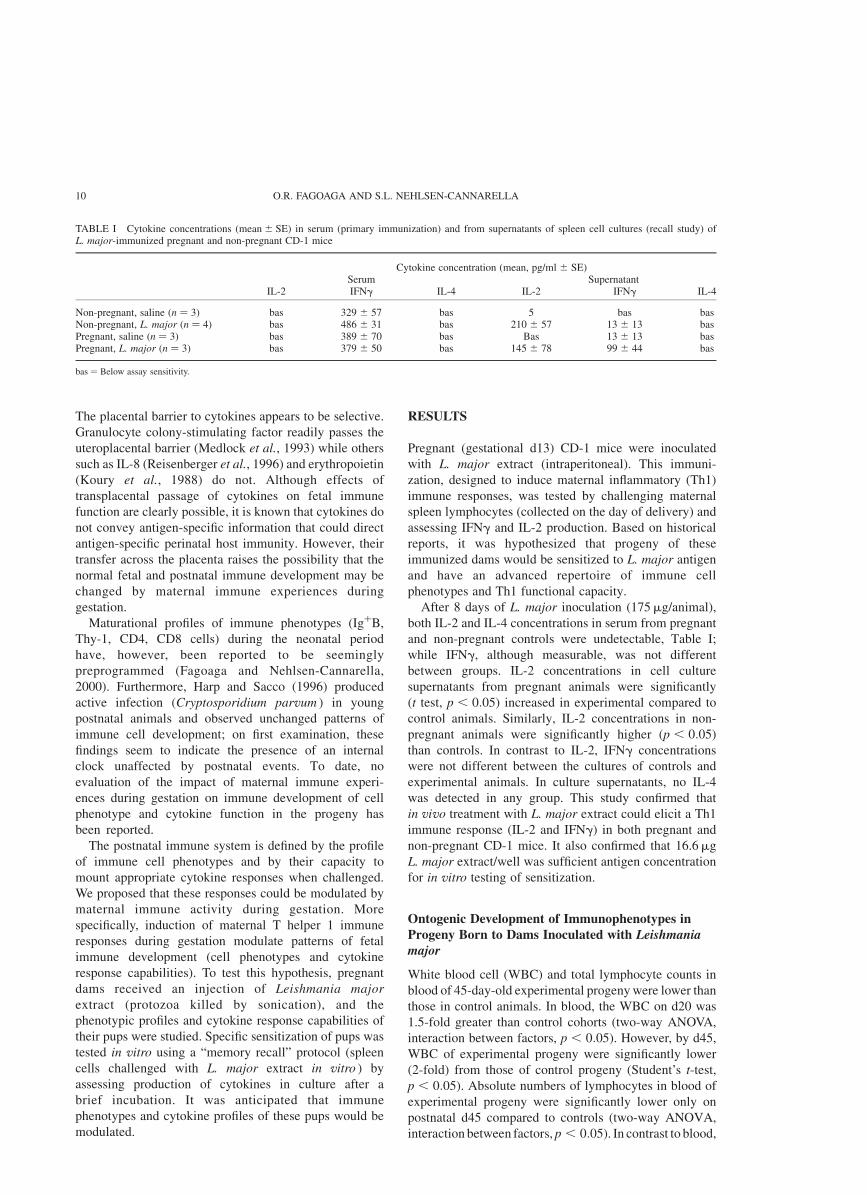
After 8 days of L. major inoculation (175mg/animal),
both IL-2 and IL-4 concentrations in serum from pregnant
and non-pregnant controls were undetectable, Table I;
while IFNg, although measurable, was not different
between groups. IL-2 concentrations in cell culture
supernatants from pregnant animals were significantly
(t test, p , 0:05) increased in experimental compared to
control animals. Similarly, IL-2 concentrations in non-
pregnant animals were significantly higher ðp , 0:05Þ
than controls. In contrast to IL-2, IFNg concentrations
were not different between the cultures of controls and
experimental animals. In culture supernatants, no IL-4
was detected in any group. This study confirmed that
in vivo treatment with L. major extract could elicit a Th1
immune response (IL-2 and IFNg) in both pregnant and
non-pregnant CD-1 mice. It also confirmed that 16.6mg
L. major extract/well was sufficient antigen concentration
for in vitro testing of sensitization.
Ontogenic Development of Immunophenotypes in
Progeny Born to Dams Inoculated with Leishmaniamajor
White blood cell (WBC) and total lymphocyte counts in
blood of 45-day-old experimental progeny were lower than
those in control animals. In blood, the WBC on d20 was
1.5-fold greater than control cohorts (two-way ANOVA,
interaction between factors, p , 0:05). However, by d45,
WBC of experimental progeny were significantly lower
(2-fold) from those of control progeny (Student’s t-test,
p , 0:05). Absolute numbers of lymphocytes in blood of
experimental progeny were significantly lower only on
postnatal d45 compared to controls (two-way ANOVA,
interaction between factors, p , 0:05). In contrast to blood,
TABLE I Cytokine concentrations ðmean ^ SEÞ in serum (primary immunization) and from supernatants of spleen cell cultures (recall study) ofL. major-immunized pregnant and non-pregnant CD-1 mice
Cytokine concentration (mean, pg/ml ^ SE)Serum Supernatant
IL-2 IFNg IL-4 IL-2 IFNg IL-4
Non-pregnant, saline ðn ¼ 3Þ bas 329 ^ 57 bas 5 bas basNon-pregnant, L. major ðn ¼ 4Þ bas 486 ^ 31 bas 210 ^ 57 13 ^ 13 basPregnant, saline ðn ¼ 3Þ bas 389 ^ 70 bas Bas 13 ^ 13 basPregnant, L. major ðn ¼ 3Þ bas 379 ^ 50 bas 145 ^ 78 99 ^ 44 bas
bas ¼ Below assay sensitivity.
O.R. FAGOAGA AND S.L. NEHLSEN-CANNARELLA10

in spleen, treatment effects were observed only in absolute
numbers of lymphocytes; these were 1.3-fold higher on d45
than in control mice (Student’s t-test, p , 0:05). Thus,
treatment effects were observed but they were not apparent
until after weaning. Proportions of lymphocyte subsets in
offspring born to experimental or control dams were then
compared for treatment effects.
Proportions of Immune Cell Phenotypes in Blood
In blood of experimental progeny, proportions of B cells
were greater (Fig. 1, panel A) while T cell subsets and NK
cells were less than those in control progeny (Fig. 1, panel
B). T cell effects were reflected in decreased Tc/s cells
(two-way ANOVA, main effects, treatment vs. control,
p , 0:05), that were lowest on d45 (Student’s
t-test, p , 0:05). Proportions of NK and naı̈ve Th
(CD62posCD44low) in blood were significantly affected,
changes were dependent on age [two-way ANOVA,
interaction between factors (age vs. treatment), p , 0:05].
NK proportions in experimental animals were lower on d3
and higher on d10 (Student’s t-test, p , 0:05), and tended
(t-test, p ¼ 0:07) to be higher on d45 when compared to
control progeny of the same ages. In contrast, proportions
of naı̈ve Th were significantly lower on d4 (Student’s
t-test, p , 0:05) and tended ðp ¼ 0:12Þ to be higher on
d45. Thus, L. major inoculation during pregnancy had a
modulating effect on the proportions of specific immune
cell phenotypes in circulation.
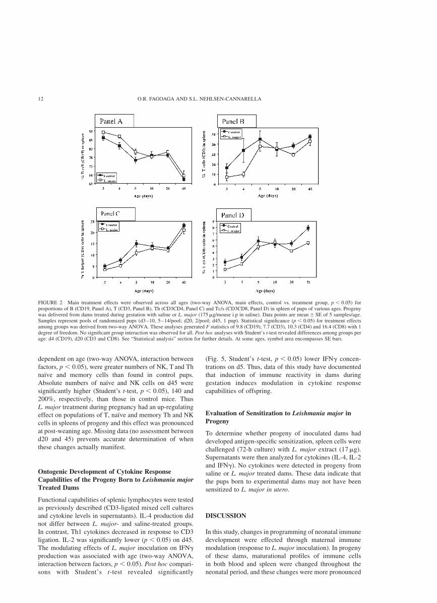
Proportions of Immune Cell Phenotypes in Spleen
Proportions of splenic phenotypes in experimental progeny
were increased compared to control progeny (Fig. 2). Main
treatment effects were observed across all ages (two-way
ANOVA, main effects, treatment vs. control group,
p , 0:05) for CD19, CD3, CD4 and CD8. Overall, B
cells were higher and T cells lower in experimental pups.
Proportions of T cells on d20 and Tc/s cells on d20 and 45
were significantly below normal (Student’s t-test,
p , 0:05). NK cells were strongly affected by treatment,
and were dependent on age (two-way ANOVA, interaction
between factors, p , 0:05). On d10, the NK population
was 1.7-fold higher than controls (Student’s t-test,
p , 0:05). In summary, proportions of B cells in blood
and spleen were higher, while T cells were lower in
experimental progeny compared to control progeny.
Numbers of Immune Cell Phenotypes in Blood
Absolute numbers of blood cells in experimental progeny
were lower than control progeny, including T, naı̈ve Th
(CD62LposCD44low) and memory Th (CD62Lneg
CD44high), (Fig. 3), and was the main treatment effect
(two-way ANOVA, main effects, control vs. treatment
group, p , 0:05). Furthermore, decreases in numbers of
all subsets of T cells were dependent on age (two-way
ANOVA, interaction between factors, p , 0:05). T cells
and T cell subsets were 1.9- to 3.4-fold lower than controls
on d45, but Tc/s rose 1.5-fold higher on d20 (Student’s
t-test, p , 0:05). In general, L. major inoculation during
gestation decreased the number of blood immune cell
phenotypes in progeny, but most of the effect was not
apparent until adulthood. The specific time when the
effects were more noticeable post-weaning could not be
determined because data between d20 and 45 was not
collected.
Numbers of Immune Cell Phenotypes in Spleen
The principal treatment effects observed in spleen cells
were higher NK and naı̈ve Th cells (Fig. 4; two-way
ANOVA, main effects, control vs. experimental progeny,
p , 0:05). Additional effects of treatment, significantly
FIGURE 1 Proportions of B (CD19, Panel A) and Tc/s (CD8, Panel B)cells in blood of pups at various ages delivered from dams treated duringgestation with saline or L. major (175mg/mouse i.p. in saline). Datapoints are mean ^ SE of 5 samples/age, except 6 for d20 saline group;and 5 samples/age for L. major. Samples represent pools of randomizedpups (d3-10, 5–14/pool; d20, 2/pool; d45, 1 pup). Statistical significanceðp , 0:05; df ¼ 1; F ¼ 4:18 for CD19; and F ¼ 4:13 for CD8) fortreatment effects among groups was derived from two-way ANOVA.Post hoc analyses were performed with Student’s t-test to revealdifferences among groups per age (*, p , 0:05Þ: See “StatisticalAnalysis” section for further details. At some ages, symbols encompassarea of SE bars.
MATERNAL IMMUNE MODULATION OF OFFSPRING 11
dependent on age (two-way ANOVA, interaction between
factors, p , 0:05), were greater numbers of NK, T and Th
naı̈ve and memory cells than found in control pups.
Absolute numbers of naı̈ve and NK cells on d45 were
significantly higher (Student’s t-test, p , 0:05), 140 and
200%, respectively, than those in control mice. Thus
L. major treatment during pregnancy had an up-regulating
effect on populations of T, naı̈ve and memory Th and NK
cells in spleens of progeny and this effect was pronounced
at post-weaning age. Missing data (no assessment between
d20 and 45) prevents accurate determination of when
these changes actually manifest.
Ontogenic Development of Cytokine ResponseCapabilities of the Progeny Born to Leishmania majorTreated Dams
Functional capabilities of splenic lymphocytes were tested
as previously described (CD3-ligated mixed cell cultures
and cytokine levels in supernatants). IL-4 production did
not differ between L. major- and saline-treated groups.
In contrast, Th1 cytokines decreased in response to CD3
ligation. IL-2 was significantly lower ðp , 0:05Þ on d45.
The modulating effects of L. major inoculation on IFNg
production was associated with age (two-way ANOVA,
interaction between factors, p , 0:05). Post hoc compari-
sons with Student’s t-test revealed significantly
(Fig. 5, Student’s t-test, p , 0:05) lower IFNg concen-
trations on d5. Thus, data of this study have documented
that induction of immune reactivity in dams during
gestation induces modulation in cytokine response
capabilities of offspring.
Evaluation of Sensitization to Leishmania major in
Progeny
To determine whether progeny of inoculated dams had
developed antigen-specific sensitization, spleen cells were
challenged (72-h culture) with L. major extract (17mg).
Supernatants were then analyzed for cytokines (IL-4, IL-2
and IFNg). No cytokines were detected in progeny from
saline or L. major treated dams. These data indicate that
the pups born to experimental dams may not have been
sensitized to L. major in utero.
DISCUSSION
In this study, changes in programming of neonatal immune
development were effected through maternal immune
modulation (response to L. major inoculation). In progeny
of these dams, maturational profiles of immune cells
in both blood and spleen were changed throughout the
neonatal period, and these changes were more pronounced
FIGURE 2 Main treatment effects were observed across all ages (two-way ANOVA, main effects, control vs. treatment group, p , 0:05) forproportions of B (CD19, Panel A), T (CD3, Panel B), Th (CD3CD4, Panel C) and Tc/s (CD3CD8, Panel D) in spleen of pups of various ages. Progenywas delivered from dams treated during gestation with saline or L. major (175mg/mouse i.p in saline). Data points are mean ^ SE of 5 samples/age.Samples represent pools of randomized pups (d3–10, 5–14/pool; d20, 2/pool; d45, 1 pup). Statistical significance ðp , 0:05Þ for treatment effectsamong groups was derived from two-way ANOVA. These analyses generated F statistics of 9.8 (CD19), 7.7 (CD3), 10.3 (CD4) and 16.4 (CD8) with 1degree of freedom. No significant group interaction was observed for all. Post hoc analyses with Student’s t-test revealed differences among groups perage: d4 (CD19), d20 (CD3 and CD8). See “Statistical analysis” section for further details. At some ages, symbol area encompasses SE bars.
O.R. FAGOAGA AND S.L. NEHLSEN-CANNARELLA12

after weaning. Blood leukocytes, lymphocytes and
specifically T cells of experimental progeny were lower
than in control progeny and this difference was noted to be
marked on d45. In contrast, spleen lymphocytes, T cells,
naı̈ve and memory Th cells and NK cells of experimental
progeny were higher on d45 compared to control progeny,
suggesting an increased homing of circulating lympho-
cytes to splenic compartments. That effects of maternal
immune experience during gestation are most apparent in
progeny reaching adulthood, may be a result of yet further
maternal-mediated immune regulation, perhaps through
passive transfer of immunoglobulins during late gestation
and via milk during the suckling period (Newman, 1995).
Some of these regulatory products in milk include
antibodies, cells (lymphocytes, macrophages and neutro-
phils), lactoferrin, B12 binding protein, bifidus factor,
fibronectin, gamma-interferon, hormones, growth factors
and lysozyme.
Expectations were that this experiment would result in
up-regulation of Th1 function; instead, it appears that Th2
functions may have been induced. Spleen cells of progeny
had decreased production of IL-2 and IFNg compared to
normal controls. It is possible that a state of tolerance rather
than inflammatory reactivity was induced in dams and
pups, evidenced by the decrease in Th1 cytokine
production in response to polyclonal stimulation. The
decrease in Th1 cytokine production can be explained by
taking into account three facts: soluble antigens primarily
induce Th2 reactions (Zinkernagel and Kelly, 1997);
inoculation in mid-gestation can lead to tolerance (Beer
et al., 1977); and down-regulation of Th1-type cytokines is
a consequence of tolerance induction (Chen and Field,
1995). If dams had been tolerized and this immune status
passed to their progeny, a shift in cytokine profile away
from Th1 would be expected. Cytokines production might
have been modified by the abundant immune products that
FIGURE 3 Absolute numbers of Th (CD4, Panel A), naı̈ve Th (CD62posCD44low, Panel B) and memory Th (CD62LnegCD44high, Panel C) cells inblood of pups of various ages delivered from dams treated during gestation with L. major (175mg/mouse i.p.) or saline. Data points are mean ^ SE of 5samples/age, except 6 for d20 in saline group; and 5 sample/age in L. major group. Samples represent pools of randomized pups (d3-10, 5–14/pool; d20,2/pool; d45, 1 pup). Statistical significance (p , 0:05; df ¼ 1; F ¼ 4:9 for Th; 2.4 for naı̈ve and 5.3 for memory) for treatment effects among groups wasderived from two-way ANOVA. Interaction between treatment and age suggest that effects are seen only on d45 (F statistic: na€ıve ¼ 2:4 andmemory ¼ 5:3; df ¼ 5; p , 0:05Þ: Post hoc analyses with Student’s t-test (two-tail) revealed differences among groups per age (*, p , 0:05; df ¼ 8;t statistic of 23.35 (CD4), 23.64 (naı̈ve), and 23.64 (memory). See “Statistical analysis” section for further details. At some ages, symbols obscure SEbars.
MATERNAL IMMUNE MODULATION OF OFFSPRING 13

are delivered through suckling (Newman, 1995), thus,
explaining the observation of more significant differences
being noted post-weaning. However, this conclusion
cannot be validated with the current data and further
experimentation is required to explore the influence of milk
immune products acquired through suckling in this model.
The present study raises the possibility that maternal
immune experiences may equip offspring in utero to
defend against specific pathogens encountered outside
the womb while preventing vertical transmission of
infection. If this had been the case, sensitization of
experimental offspring to L. major antigen would have
been induced. The memory recall experiments, performed
by challenging spleen cells of offspring with L. major
extract, produced no detectable cytokines after three days
of culture. These results indicate lack of sensitization to
L. major in the offspring. Alternatively, absence of
response may be due to challenging neonatal cells with too
high antigen concentration; it has been reported by one
investigative team that neonatal antigen presentation is
100-fold more efficient than adults (Van Tol et al., 1983;
1984). The dose of L. major antigen used in the memory
FIGURE 4 Natural killer (NK, Panel A) and naive Th cells(CD62LposCD44low, Panel B) in spleens of pups of various agesdelivered from dams treated during gestation with saline or L. major(175 ug/mouse i.p. in saline). Data points are mean ^ SE of 5 samples/age,except 6 on d20 in saline group; and 5 sample/age for L. major. Samplesrepresent pools of randomized pups (d3-10, 5–14/pool; d20, 2/pool; d45,1 pup). Statistical significance (p , 0:05; df ¼ 5; F ¼ 8:27 for NK and16.2 for naive) for treatment effects among groups was derived from two-way ANOVA. Post hoc analysis with Student’s t-test (two-tail) to revealdifferences among groups per age (*, p , 0:05; df ¼ 8; t statistic of 2.99(NK) and 5.4 (naı̈ve)). See “Statistical analysis” section for further details.At some ages, symbols obscure SE bars.
FIGURE 5 IFNg (top panel) and IL-2 (bottom panel) production(stimulated) by spleen cells of CD-1 outbred mice (male and female)delivered from dams treated during gestation with saline or L. major(175mg/mouse i.p in saline). Each culture contained 1 £ 106 CD3 þCD4 þ splenocytes and hamster anti-mouse CD3 monoclonal antibodyin complete media. Control cells were cultured in media only. Data areexpressed as mean ^ SE of samples (control: d5 ¼ 4; d10 ¼ 3; d20 ¼10; d45 ¼ 11; experimental: d5 ¼ 7; d10 ¼ 7; d20 ¼ 3; d45 ¼ 5Þ:Samples in stimulated cultures were pools of randomized pups (d5 and10, 10/pool; d20, 3/pool; d45, 1 pup). Statistical significance (p , 0:05;df ¼ 3; F ¼ 3:6) between groups was derived from two-way ANOVA.Post hoc analyses with Student’s t-test (two-tail) revealed differencesamong groups per age (*, p , 0:05; df ¼ 9 (IFNg) and 14 (IL-2);t statistic of 22.63 for IFNg and 22.45 for IL-2). See “Statisticalanalysis” section for further details.
O.R. FAGOAGA AND S.L. NEHLSEN-CANNARELLA14

recall assays had been optimized with adult splenocytes,
a deficiency in this study that needs further
investigation. However, assuming that a prozone effect
had not occurred, these results were unexpected since
Herman et al. (1982) reported sensitization of offspring
from Leishmania donovani-infected hamsters. Lack of
sensitization in our experimental progeny may also be the
result of using a non-pathogenic, purified extract
administered i.p.; Herman et al. (1982) administered live
amastigotes (active intracardiac infection) to their
experimental animals.
Active infection is processed differently than an
injection of soluble antigen from a killed parasite. The
life cycle of this parasite is complex and during the
infection process, promastigotes express abundant gp63
glycoprotein. On the other hand, amastigotes (from which
the extract was made) express very low levels of this
protein (Kurtzhals et al., 1994; Reiner and Locksley,
1995). The cytokine environment during induction of
immune responses to gp63 determines the course of
disease resulting from the type of immune responses
induced (Kurtzhals et al., 1994; Reiner and Locksley,
1995). Humoral responses are primarily induced by this
protein (Kurtzhals et al., 1994) in the early phase of
infection and Kemp et al. (1994) showed that cytokine
response in visceral Leishmaniasis is primarily a Th2
response, whereas cutaneous Leishmaniasis elicits pre-
dominantly Th1 response. This suggests that there are
different cytokine environments in visceral vs. cutaneous
sites, and that response is also dependent on route of
administration (i.p. vs. subcutaneous).
The unexpected result of these experiments may be
rooted not only in the type of antigen used to induce Th
responses during pregnancy, but also the immunization
program (dose, route and timing; Zinkernagel and Kelly,
1997). In this work, L. major extract was injected i.p. and
this may be a factor for lack of sensitization in progeny,
even though maternal spleens tested in vitro tended to a
polarized Th1 response. Time of inoculation during
pregnancy may be another factor; in a different model,
tolerization resulted when dams were inoculated 5–7 days
antepartum (Beer et al., 1977). In the current work,
L. major extract was given 7 days antepartum, a time
chosen as most likely time to avoid fetal loss due to release
of Th1 cytokines (Krishnan et al., 1996). This single
immunizing event may not have been sufficient to induce
sensitization in the offspring, and instead may have
induced tolerance. Sustained chronic immune responses to
live L. donovani over the whole course of pregnancy could
explain sensitizations realized by Herman et al. (1982).
In summary, the data indicate that at maturity, T cells in
blood augmented their homing into lymphoid compart-
ments as evidenced by significant increases in splenic NK
and T cell numbers at postnatal d45 (Fig. 4). Furthermore,
these changes in splenic cellularity produced significant
decreases in IL-2 (d45) and IFNg (d5) production by
activated spleen cells compared to controls. Taken
together, these data support the hypothesis that immune
events experienced by the mother during gestation
can modulate the pattern of immune development
(cell phenotypes and cytokine response capabilities) in
the offspring. Although significant effects in development
were induced during gestation, a complete understanding
of the mechanism must be ascertained before elective
manipulation can be entertained. Timing and mode of
delivering sensitizing or tolerizing immunogens must be
defined, as well as dose and schedule of immunization. Of
considerable importance is the need to determine what
effect concurrent immune events would have on the
intended process since regulatory function promulgated
by APC is subject to prevailing signals dictated by local
neuroendocrine immune products.
MATERIALS AND METHODS
CD-1 mice, a genetically wild-type strain were purchased
from Charles River Laboratories (Wilmington, MA). The
first pregnant CD-1 mice arrived in the vivarium on
gestational d13. All mice were housed individually in
small cages in the same room and maintained in a 12- and
12-hour light–dark cycle. Postpartum females, adult
males and progeny provided the breeding nucleus needed
to generate additional pups for later studies. A “trios”
method of breeding was employed. Briefly, one male was
mated to two females and housed together for 48 h.
Females were then individually housed; a vaginal plug
confirmed that copulation had occurred. The limited
duration of conspecific exposure coordinated the timing
of birth. Body weights of female breeders were
measured daily to confirm the successful progress of
pregnancy.
Collection of Blood and Spleen
Mice, 10 days of age or less were killed by decapitation
and blood was collected into heparinized capillary tubes
from cut surface of the body. For 20- and 45-days old
groups, blood was collected from individual mice by
cardiac puncture following pentobarbital anesthesia
(40 mg/kg body weight); then mice were killed by
decapitation. The spleen was extracted from the peritoneal
cavity of each mouse under sterile procedures, weighed
without mesenteric fat, and placed in sterile RPMI-1640
media (Fisher Scientific, Pittsburgh, PA) at room
temperature. Lymphocytes were harvested within
30 min. Briefly, a spleen cell suspension was prepared
by gently pressing tissue through a fine nylon mesh sieve
and resuspending in complete media (RPMI-1640
containing prescreened 10% fetal calf serum and 1%
penicillin/streptomycin). These reagents were purchased
from Fisher Scientific, Pittsburgh, PA. Leukocytes in all
blood and spleen cell suspensions were counted with a
Unopette blood counting system (Becton Dickinson,
Franklin Lakes, NJ) and a hemocytometer.
MATERNAL IMMUNE MODULATION OF OFFSPRING 15

Pools were created by mixing blood or spleens of
randomly-sorted pups from different litters of the same
age. Each pool contained eight pups each for ages 0, 1 and
2 days. Although adequate sample volumes were obtained
for immunophenotyping, it was impractical to perform
experiments for study of cytokine production in mice
before 2 days of age.
Immunophenotyping
Flow cytometric analysis was performed as previously
described (Fagoaga et al., 2000). Briefly, single cell
preparations from whole blood and spleen ð1 £
105 cells=tubeÞ were stained with 10ml fluorochrome-
conjugated monoclonal antibodies (mAb). A mAb panel
was constructed for two- (fluoroisothiocyanate [FITC] and
phycoerythrin [PE]) and three-color immunophenotyping
(FITC, PE and Cy-Chrome). Two-color analysis was used
to enumerate B vs. T cells (CD19*FITC/CD3*PE), T
helper/inducer cells (CD4*FITC/CD3*PE), T cytotoxic/-
suppressor cells (CD8*FITC/CD3*PE) and natural killer
cells (CD3*FITC/NK1.1*PE). Three-color analysis was
used to assess naı̈ve and memory T helper cells
(CD62L*FITC/CD44*PE/CD4*Cy-Chrome). All rat
anti-mouse mAb were obtained from PharMingen (San
Diego, CA), except CD3*FITC (hamster anti-mouse
antibody) obtained from Caltag Laboratories (San
Francisco, CA). After incubation (15 min, 48C), erythro-
cytes were lysed with 0.8% ammonium chloride solution
and samples centrifuged at 300g for 5 min. Each
supernatant was aspirated and cell pellets resuspended in
300ml phosphate buffered saline (PBS).
Analytical controls included (a) unstained cells and
(b) cells mixed with isotype antibody to evaluate degree of
non-specific staining. Furthermore, an unlabeled, purified
rat anti-mouse CD16/CD32 (FcgIII/II receptor;
PharMingen, San Diego, CA) monoclonal antibody was
used as an agent for blocking non-specific binding through
the FcgIII/II receptor.
Proportions of lymphocyte populations (CD3,
CD3CD4, CD3CD8, NK, CD19) were expressed as
percentages normalized to lymphocyte purity (sum of
lymphocyte subsets; CD19 þ CD4 þ CD8 þ NK). This
normalization excluded debris, other non-lymphoid cells
(mainly monocytes) and Tgd cells (CD3posCD4neg
CD8neg). Normalization was not required for analyzing
T helper cells for CD62L and CD44 expression because
these cells were selectively gated, based on their far red
(CyChrome) fluorochrome characteristic. Absolute num-
bers of each population were generated by multiplying
WBC counts by the sum of proportions of lymphocyte
subsets (i.e.P
CD19 þ CD3 þ NKs) and again by
proportion of each population (CD3, CD3CD4,
CD3CD8, NK and CD19 subsets). Absolute numbers of
T helper cells analyzed for expression of CD62L and
CD44 were similarly generated.
T Cell Stimulation Assay
A monoclonal antibody to CD3 was produced from cell
culture of clone 145-2C11 (PharMingen, San Diego, CA)
as previously described (Fagoaga and Nehlsen-
Cannarella, 2000). Splenocytes were collected from
spleens as described above. Subsequently, cell cultures
were set up in 24-well plates (Fisher Scientific, Pittsburgh,
PA). Cultures consisted of either unsorted single cell
spleen suspensions (SSCS). To obtain a constant number
of CD4 cells in each culture, absolute numbers of CD4
cells in unsorted cell preparations were determined by
flow cytometry. An aliquot containing 1 £ 106 CD3 þ
CD4 þ splenocytes was added to each well with 1 ml of
optimally diluted CD3 monoclonal antibody. Each culture
volume was brought up to 2 ml with complete media.
Cytokine Assays
Commercial cytokine ELISA kits from ENDOGEN
(Woburn, MA) were used to assess IL-2, IL-4 and IFNg
production by murine splenocytes after CD3 ligation and
after in vivo and in vitro L. major challenge. Assay
procedures followed manufacture’s instructions and as
previously described (Fagoaga and Nehlsen-Cannarella,
2000).
Determination of In Vivo and In Vitro Leishmaniamajor Dose
L. major extract of killed protozoa (sonicated) (Antibody
Systems, Hurst, TX) was suspended in saline (1 mg/ml). A
pilot study was conducted with pregnant and non-pregnant
ðn ¼ 6Þ mice to determine optimal in vivo dose to induce a
Th1 response (IL-2 and IFNg production) Table I. Each
animal received 175ml (intraperitoneal injection) of either
L. major extract (pregnant, n ¼ 3; control, n ¼ 3) or saline
(pregnant, n ¼ 3; control, n ¼ 3Þ: This dose was
recommended by Antibody Systems for Th1 induction.
Pregnant animals were inoculated once (gestational d8, 11
or 13); no further injections were given as a precaution to
avoid induction of premature labor and delivery. Spleens
and sera were collected from dams on day of parturition
(gestational d20) and from non-pregnant controls 9 days
post-inoculation.
Successful immunization of these animals was con-
firmed by assessing cytokine concentrations (IL-2, IFNg,
IL-4) in serum and in supernatants from spleen cells in
culture after in vitro challenge with L. major (recall). Sera
for these determinations were obtained by cardiac
exsanguination under sodium pentobarbital anesthesia as
previously described. For the immune memory recall
study, spleens were harvested from exsanguinated mice
and single cell suspensions prepared as described. Cells
were incubated ð1 £ 106 cells=wellÞ in 96-well micro-
culture plates (Fisher Scientific, Pittsburgh, PA) with
16.6mg/well of L. major extract (“starting” concentration
suggested by supplier) in complete media for 72 h in 5%
CO2 in humidified air at 378C. At the end of incubation,
O.R. FAGOAGA AND S.L. NEHLSEN-CANNARELLA16

cell culture supernatants were collected and assayed for
IL-2, IFNg and IL-4.
Statistical Analysis
One-way ANOVA and Duncan’s post hoc test for multiple
comparisons among different ages were calculated. If test
for homosedasticity was significant (greater than the
critical value for F-max test for homogeneity of
variances), data were log transformed. After transfor-
mation, if significant homogeneity of variances persisted,
a Kruskal Wallis ANOVA with multiple comparisons was
performed to assess differences among age groups. Two-
way ANOVA was used to determine treatment effects of
L. major extract inoculations. Furthermore, Duncan’s test
for multiple comparisons and t-tests were used as post hoc
tests to determine group differences. A value of p , 0:05
was considered significant.
References
Barnetson, R.S.T.C., Bjune, G. and Duncan, M.E. (1976) “Evidence for asoluble lymphocyte factor in the transplacental transmission ofT-lymphocyte responses to Mycobacterium leprae”, Nature 260,150–151.
Beer, A.E., Billingham, R.E., Head, J.R. and Parmely, M.J. (1977)“Possible influence of maternal-to-perinatal cell transfer on thedevelopment of host defenses”, In: Cooper, M.D. and Dayton, D.H.,eds, Development of Host Defenses (Raven Press, New York),pp 251–271.
Chen, N. and Field, E.H. (1995) “Enhanced type 2 and diminished type 1cytokines in neonatal tolerance”, Transplantation 59, 933–941.
Claas, F.H., Gijbels, Y., van-der-Velden-de-Munck, J. and van-Rood, J.J.(1988) “Induction of B cell unresponsiveness to noninheritedmaternal HLA antigens during fetal life”, Science 241, 1815–1817.
Cramer, D.V., Kunz, H.W. and Gill, T.J. (1974) “Immunologicsensitization prior to birth”, Am. J. Obstet. Gynecol. 120, 431–439.
Desai, R.G. and Greger, W.P. (1963) “Maternofetal passage of leukocytesand platelets in man”, Blood 21, 665–673.
Fagoaga, O.R. and Nehlsen-Cannarella, S.L. (2000) “Ontogenicdevelopment of Th1 and Th2 function in the mouse”, J. Dev.Immunol., 8(1), 47–60.
Fagoaga, O.R., Yellon, S.M. and Nehlsen-Cannarella, S.L. (2000)“Maturation of lymphocytes immunophenotypes and memory Thelper cell differentiation during development in mice”, J. Dev.Immunol., In press.
Field, E.J. and Caspary, E.A. (1971) “Is maternal lymphocytesensitization passed to the child?”, Lancet, 337–342.
Fujii, Y. and Yamaguchi, N. (1992) “Maternal T cells of immunizedpregnant mice induce immune suppression in their offspring”,Immunology 77, 171–176.
Harp, J.A. and Sacco, R. (1996) “Development of cellular immunefunctions in neonatal to weanling mice: relationship to Cryptospori-dium parvum infection”, J. Parasitol. 82, 245–249.
Herman, R., Nolan, T.J. and Fahey, J.R. (1982) “Sensitization of offspringof Leishmania donovani-infected hamsters to immunization and ofoffspring of immunized hamsters to challenge”, Am. J. Trop. Med.Hyg. 31, 730–739.
Horton, J.E. and Oppenheim, J.J. (1976) “Relationship of transformationof newborn human lymphocytes by dental plaque antigen tothe degree of maternal periodontal disease”, Cell. Imunol. 21,153–160.
Kemp, M., Hey, A.S. and Kurtzhals, J.A.L. (1994) “Dichotomy of thehuman T-cell response to leishmania antigen. I. Th1-like response toLeishmania major promastigote antigens in individuals recoveredfrom cutaneous leishmaniasis”, Clin. Exp. Immunol. 96, 410–415.
Koury, M.J., Bondurant, M.C., Graber, S.E. and Sawyer, S.T. (1988)“Erythropoietin messenger RNA levels in developing mice andtransfer of 125I-erythropoietein by the placenta”, J. Clin. Investig. 82,154–161.
Krishnan, L., Guilbert, L.J., Wegmann, T.G., Belosevic, M. andMosmann, T.R. (1996) “T helper 1 response against Leishmaniamajor in pregnant C57BL/6 mice increases implantation failure andfetal resorptions. Correlation with increased IFN-gamma and TNFand reduced IL-10 production by placental cells”, J. Immunol. 156,653–662.
Kurtzhals, J.A.L., Hey, A.S., Jardim, A., Kemp, M., Schaefer, K.-U.,Odera, E.O., Christensen, C.B.V., Githure, J.I., Olafson, R.W.,Theander, T.G. and Khrazmi, A. (1994) “Dichotomy of the humanT cell response to Leishmania antigens. II. Absent Th2-like responseto gp63 and Th1-like response to lipophosphoglycan-associatedprotein in cells from cured visceral leishmaniasis patients”, Clin. Exp.Immunol. 96, 416–421.
Medlock, E.S., Kaplan, D.L., Cecchini, M., Ulich, T.R., del Castillo, J.and Andresen, J. (1993) “Granulocyte colony-stimulating factorcrosses the placenta and stimulates fetal rat granulopoiesis”, Blood81, 916–922.
Newman, J. (1995) “How breast milk protects newborns”, Sci. Am.(December), 76–79.
Piotrowski, P. and Croy, B.A. (1996) “Maternal cells are widelydistributed in the murine fetuses in utero”, Biol. Reprod. 54,1103–1110.
Pollack, M.S., Kirkpatrick, D., Kapoor, N., Dupont, B. and O’Reilly, R.J.(1982) “Identification by HLA typing of intrauterine-derivedmaternal T cells in four patients with severe combined immuno-deficiency”, N. Engl. J. Med. 307, 662–666.
Reiner, S.L. and Locksley, R.M. (1995) “The regulation of immunity toLeishmania major”, Ann. Rev. Immunol. 13, 151–177.
Reisenberger, K., Egater, C., Volg, S., Sternberger, B., Kiss, H. andHusslein, P. (1996) “The transfer of Interleukin-8 across the humanplacenta perfuded in vitro”, Obstet. Gynecol. 87, 613–616.
Russell, A.S. (1975) “Cell-mediated immunity to microbial antigens inmother and child”, Clin. Exp. Immunol. 22, 457–460.
Stastny, P. (1965) “Accelerated graft rejection in the offspring ofimmunized mothers”, J. Immunol. 95, 929–936.
Van Tol, M.J.D., Zijlstra, J., Heijnen, C.J., Kuis, W., Zegers, B.J.M. andBallieux, R.E. (1983) “Antigen-specific plaque-forming cell responseof human cord blood lymphocytes after in vitro stimulation by T cell-dependent antigens”, Eur. J. Immunol. 13, 390–397.
Van Tol, M.J.D., Zijlstra, J., Zegers, B.J.M. and Ballieux, R.E. (1984)“Distinct role of neonatal and adult monocytes in the regulation ofin vitro antigen-induced plaque-forming cell response in man”,J. Immunol. 134, 1902–1908.
Zinkernagel, R.M. and Kelly, J. (1997) “How antigen influencesimmunity”, Immunologist 5/4, 114–120.
MATERNAL IMMUNE MODULATION OF OFFSPRING 17

Submit your manuscripts athttp://www.hindawi.com
Stem CellsInternational
Hindawi Publishing Corporationhttp://www.hindawi.com Volume 2014
Hindawi Publishing Corporationhttp://www.hindawi.com Volume 2014
MEDIATORSINFLAMMATION
of
Hindawi Publishing Corporationhttp://www.hindawi.com Volume 2014
Behavioural Neurology
EndocrinologyInternational Journal of
Hindawi Publishing Corporationhttp://www.hindawi.com Volume 2014
Hindawi Publishing Corporationhttp://www.hindawi.com Volume 2014
Disease Markers
Hindawi Publishing Corporationhttp://www.hindawi.com Volume 2014
BioMed Research International
OncologyJournal of
Hindawi Publishing Corporationhttp://www.hindawi.com Volume 2014
Hindawi Publishing Corporationhttp://www.hindawi.com Volume 2014
Oxidative Medicine and Cellular Longevity
Hindawi Publishing Corporationhttp://www.hindawi.com Volume 2014
PPAR Research
The Scientific World JournalHindawi Publishing Corporation http://www.hindawi.com Volume 2014
Immunology ResearchHindawi Publishing Corporationhttp://www.hindawi.com Volume 2014
Journal of
ObesityJournal of
Hindawi Publishing Corporationhttp://www.hindawi.com Volume 2014
Hindawi Publishing Corporationhttp://www.hindawi.com Volume 2014
Computational and Mathematical Methods in Medicine
OphthalmologyJournal of
Hindawi Publishing Corporationhttp://www.hindawi.com Volume 2014
Diabetes ResearchJournal of
Hindawi Publishing Corporationhttp://www.hindawi.com Volume 2014
Hindawi Publishing Corporationhttp://www.hindawi.com Volume 2014
Research and TreatmentAIDS
Hindawi Publishing Corporationhttp://www.hindawi.com Volume 2014
Gastroenterology Research and Practice
Hindawi Publishing Corporationhttp://www.hindawi.com Volume 2014
Parkinson’s Disease
Evidence-Based Complementary and Alternative Medicine
Volume 2014Hindawi Publishing Corporationhttp://www.hindawi.com