enhances maternal immune responses promoting survival and ...
Archival Report
Maternal Immune Activation in NonhumanPrimates Alters Social Attention in JuvenileOffspringChristopher J. Machado, Alexander M. Whitaker, Stephen E.P. Smith, Paul H. Patterson, andMelissa D. Bauman
ABSTRACTBACKGROUND: Sickness during pregnancy is associated with an increased risk of offspring neurodevelopmentaldisorders. Rodent models have played a critical role in establishing causal relationships and identifying mechanismsof altered brain and behavior development in pups prenatally exposed to maternal immune activation (MIA). Werecently developed a novel nonhuman primate model to bridge the gap between human epidemiological studies androdent models of prenatal immune challenge. Our initial results demonstrated that rhesus monkeys given the viralmimic synthetic double-stranded RNA (polyinosinic:polycytidylic acid stabilized with poly-l-lysine) during pregnancyproduce offspring with abnormal repetitive behaviors, altered communication, and atypical social interactions.METHODS: We utilized noninvasive infrared eye tracking to further evaluate social processing capabilities in asubset of the first trimester MIA-exposed offspring (n 5 4) and control animals (n 5 4) from our previous study.RESULTS: As juveniles, the MIA offspring differed from control animals on several measures of social attention,particularly when viewing macaque faces depicting the fear grimace facial expression. Compared with controlanimals, MIA offspring had a longer latency before fixating on the eyes, had fewer fixations directed at the eyes, andspent less total time fixating on the eyes of the fear grimace images.CONCLUSIONS: In the rhesus monkey model, exposure to MIA at the end of the first trimester results in abnormalgaze patterns to salient social information. The use of noninvasive eye tracking extends the findings from rodent MIAmodels to more human-like behaviors resembling those in both autism spectrum disorder and schizophrenia.
Keywords: Autism spectrum disorders, Immunology, Macaque, Nonhuman primate, Poly IC, Schizophrenia, Socialattention.
http://dx.doi.org/10.1016/j.biopsych.2014.07.035
The prenatal environment and, in particular, the maternalimmune system may have a profound effect on fetal neuro-development (1–4). In humans, exposure to infections duringpregnancy may increase the risk of giving birth to a child whowill later develop an autism spectrum disorder (ASD) orschizophrenia (SZ) (5–14). Animal models have demonstratedthat exposing pregnant dams to prenatal challenges, such asinfluenza (15–18) or the bacterial endotoxin lipopolysaccharide(19–22), produce offspring with behavioral abnormalities andneuropathology relevant to both SZ and ASD. The diversity ofinfections associated with alterations in neurodevelopmentsuggests that the maternal immune response, rather than aspecific pathogen, drives changes in fetal brain development.The emerging maternal immune activation (MIA) hypothesishas been directly tested in animal models by artificiallyactivating the immune system of pregnant rodents with theviral mimic, synthetic double stranded RNA (polyinosinic:polycytidylic acid [poly IC]), a toll-like receptor-3 agonist thatstimulates an inflammatory response in the absence of aspecific pathogen (23). Rodent pups born to dams treatedwith poly IC at mid gestation demonstrate behavioral abnor-
malities, neuropathology, and altered gene expression relevantto both ASD and SZ [reviewed in (24–28)].
It is important to emphasize that sickness during pregnancyis not uncommon (29,30), and clearly, not all women whoexperience infection during pregnancy have children laterdiagnosed with a neurodevelopmental disorder (31). A numberof factors, including genetic susceptibility, the intensity andtiming of the infection, and exposure to additional postnatalchallenges, all may influence the degree to which the prenatalimmune challenge alters neurobehavioral development (32).Translational animal models provide a powerful tool to system-atically examine how these factors contribute to offspringpathophysiology following MIA. In the rodent MIA model, theeffects of poly IC on brain and behavior development appearto be mediated by the maternal cytokine response, in partic-ular interleukin-6 (33,34), and exacerbated by postnatal envi-ronmental stressors (35).
While rodent models have provided compelling evidence fora causal relationship between MIA and aberrant brain andbehavioral development in the offspring (36–40), there arelimitations in relying solely on rodent models to study complex
& 2014 Society of Biological Psychiatry 1ISSN: 0006-3223 Biological Psychiatry ]]], 2014; ]:]]]–]]] www.sobp.org/journal
BiologicalPsychiatry
SEE COMMENTARY ON PAGE
human brain disorders. Nonhuman primates, such as therhesus macaque (Macaca mulatta), can provide a more directcomparison with human brain and behavior pathologies todetermine the clinical relevance of the MIA model to humanneurodevelopmental disorders (41–43). Previous primate mod-els have documented changes in brain and behavior develop-ment of macaque offspring following third trimester exposureto influenza or lipopolysaccharide (18,19); however, the effectsof MIA at earlier gestational time points have not beenexplored. We have developed a novel, nonhuman primatemodel of maternal immune activation using a modified form ofpoly IC (poly IC stabilized with poly-l-lysine [poly ICLC]) that isrecognized by the primate immune system and induces atransient innate inflammatory response (44,45). Pregnantrhesus monkeys injected with poly ICLC at the end of eitherthe first or second trimester produce offspring with abnormalmotor stereotypies and repetitive behaviors (46). While bothfirst and second trimester MIA offspring produced feweraffiliative vocalizations than control animals, only the firsttrimester MIA offspring showed signs of atypical socialinteractions with unfamiliar peers. Given that both ASD andSZ are characterized by changes in social cognition andemotion (47), as well as altered visual attention devoted tofacial expressions (48,49), we initiated a series of noninvasiveeye-tracking studies to provide further insight into the natureof the social impairments observed in the MIA offspring. Here,we present the initial results from these eye-tracking studiesdemonstrating abnormal patterns of social attention in themacaque offspring exposed to MIA in the first trimester.
METHODS AND MATERIALS
All experimental procedures were developed in collaborationwith the veterinary, animal husbandry, and environmentalenrichment staff at the California National Primate ResearchCenter and approved by the University of California, DavisInstitutional Animal Care and Use Committee. All attempts weremade (in terms of social housing, enriched diet, use of positivereinforcement strategies, and minimizing the duration of dailytraining/testing sessions) to promote the psychological well-being of the animals that participated in this research. Addi-tional methodological details are provided in Supplement 1.
Subjects and Living Conditions
Noninvasive eye tracking was used to evaluate social attentionin a subset of juvenile macaque monkeys from our previousstudy (46) before puberty, when the animals were approx-imately 2.5 years of age. All control (CON) (n 5 4) and firsttrimester MIA exposed male animals (n 5 5) were selected toparticipate in the current series of experiments to follow-up onthe social behavior differences described in our earlier report.One of the MIA animals did not habituate to the testingprocedures and was therefore dropped from the study,yielding a final sample size of n 5 4 for the MIA group. TheMIA offspring were born to dams injected with .25 mg/kgsynthetic double-stranded RNA (poly ICLC; Oncovir, Inc.,Washington, DC) via intravenous injection while temporarilyrestrained by trained technicians at the end of the first trimesteron gestational days 43, 44, and 46. CON offspring were born todams injected with saline at these same time points or at the end
of the second trimester (gestational days 100, 101, and 103) orhad no manipulation at all during pregnancy. Preliminary analy-ses revealed that the behavioral profiles of the male saline-treated control monkeys (n 5 1 first trimester, n 5 2 secondtrimester) and the male untreated control monkey (n 5 1) werevery similar and thus pooled to form a single control group. Whilewe detected no differences in first trimester, second trimester,and untreated control animals in our previous report (46), it isimportant to note that repeated, daily prenatal stress has beenshown to alter neurobehavioral development in nonhumanprimates (50–54). To minimize the influence of prenatal stress,MIA and saline-control dams were preselected for their willing-ness to receive intravenous injections and followed identicalprocedures for routine ultrasounds, housing, and prenatal care.
Noninvasive Eye Tracking
All data collection occurred while the animals sat in a modifiedprimate chair with a slanted top (Crist Instrument Co., Inc.,Damascus, Maryland). Visual stimuli were presented to eachmonkey using a personal computer running the Eprime 2.0Professional software package (Psychology Software Tools,Pittsburgh, Pennsylvania). All gaze data were collected usingthe Eye-Trac 6 .NET User Interface program (Applied ScienceLaboratories Bedford, Massachusetts) on a separate personalcomputer.
Experiment 1: Facial Expressions
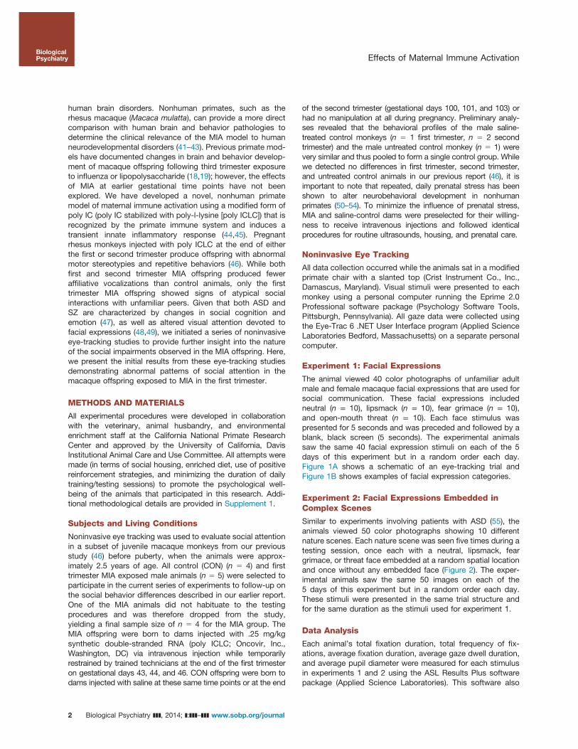
The animal viewed 40 color photographs of unfamiliar adultmale and female macaque facial expressions that are used forsocial communication. These facial expressions includedneutral (n = 10), lipsmack (n = 10), fear grimace (n = 10),and open-mouth threat (n = 10). Each face stimulus waspresented for 5 seconds and was preceded and followed by ablank, black screen (5 seconds). The experimental animalssaw the same 40 facial expression stimuli on each of the 5days of this experiment but in a random order each day.Figure 1A shows a schematic of an eye-tracking trial andFigure 1B shows examples of facial expression categories.
Experiment 2: Facial Expressions Embedded inComplex Scenes
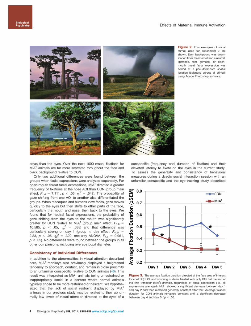
Similar to experiments involving patients with ASD (55), theanimals viewed 50 color photographs showing 10 differentnature scenes. Each nature scene was seen five times during atesting session, once each with a neutral, lipsmack, feargrimace, or threat face embedded at a random spatial locationand once without any embedded face (Figure 2). The exper-imental animals saw the same 50 images on each of the5 days of this experiment but in a random order each day.These stimuli were presented in the same trial structure andfor the same duration as the stimuli used for experiment 1.
Data Analysis
Each animal’s total fixation duration, total frequency of fix-ations, average fixation duration, average gaze dwell duration,and average pupil diameter were measured for each stimulusin experiments 1 and 2 using the ASL Results Plus softwarepackage (Applied Science Laboratories). This software also
Effects of Maternal Immune Activation
2 Biological Psychiatry ]]], 2014; ]:]]]–]]] www.sobp.org/journal
BiologicalPsychiatry
calculated the conditional probability of a fixation shifting fromone area of interest (AOI) within an image to another. Forexperiment 1, eye-tracking parameters were computed forrectangular AOIs that encompassed the entire face or theeyes, nose, and mouth separately. For experiment 2, resultswere generated for AOIs that encompassed the smallembedded face and the entire scene background.
Each dependent variable was summarized as an averageacross all four face categories or with each category consid-ered separately. The data were first examined with Shapiro-Wilk tests and found to be largely not normally distributed. Allmeasures were therefore log10(X 1 1) transformed beforeparametric statistical analyses, but nontransformed valueswere used for all figures. Data were analyzed using anaylsesof variance (ANOVAs) with groups as a between-subjectsfactor and day as within-subjects factors with repeatedmeasures. A Huynh-Feldt correction was used to adjust thedegrees of freedom if the group variance did not remain equal
across days. Post hoc tests for significant group 3 dayinteractions included one-way ANOVAs and paired-sample ttests. Because the number of post hoc comparisons was fiveor fewer for each variable, we did not correct p values formultiple comparisons. For all analyses, alpha was set atp , .05. Because main effects of day did not indicatedifferences between the two experimental groups, discussionof those effects has been omitted from the following sections.
RESULTS
Experiment 1: Facial Expressions
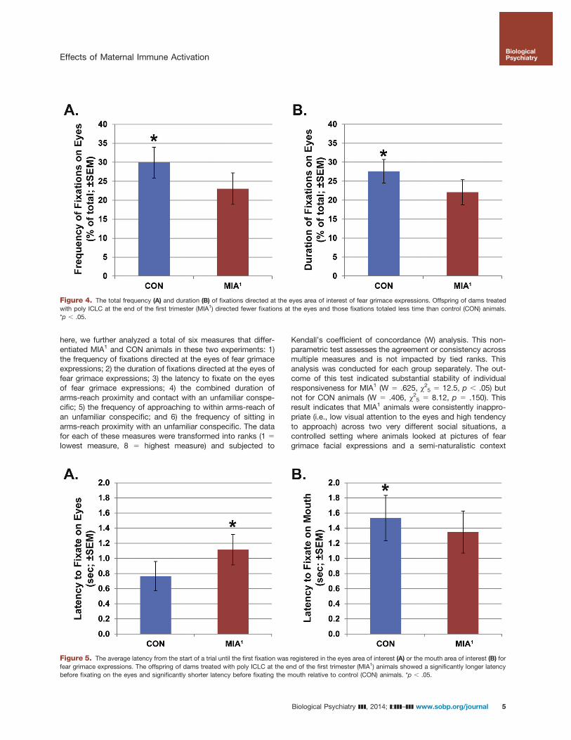
When data were averaged across all facial expressions, onlyone difference between CON and offspring of dams treatedwith poly ICLC at the end of the first trimester (MIA1) wasfound. There was a significant group 3 day interaction foraverage fixation duration when animals looked at an AOI thatencompassed the entire face (F1,6 5 2.892, p , .05, ηp2 5.325; Figure 3). The two groups did not differ significantly onany given day, but MIA1 showed a significant decrease inaverage fixation duration between day 1 and day 2 (t3 5 3.952;p , .05) with additional, but nonsignificant, decreases untilday 4. By contrast, CON showed largely consistent levels ofaverage fixation duration on the faces until a significantdecrease was detected between day 4 and day 5 (t3 53.077; p 5 .05).
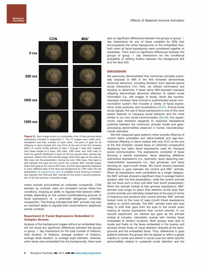
Face categories were then analyzed separately. A majorityof the differences between the groups were found for feargrimace expressions, particularly with visual attention directedtoward the eyes AOI. CON directed a greater frequency (groupmain effect; F1,6 5 7.839, p , .05, ηp2 5 .566; Figure 4A) andduration (group main effect; F1,6 5 10.117, p , .05, ηp2 5 .628;Figure 4B) of fixations at the eyes of conspecifics displayingfear grimaces than MIA1. The average dwell duration on theeyes AOI for CON also increased significantly between day 1and day 2, but such values for MIA1 did not change (group 3day effect; F4,24 5 3.111, p , .05, ηp2 5 .341; day 1 vs. day 2,t3 5 3.169, p 5 .05). In fact, by day 4, the average dwellduration directed at the eyes AOI for CON was significantlygreater than MIA (one-way ANOVA; F1,6 5 9.135, p , .05).
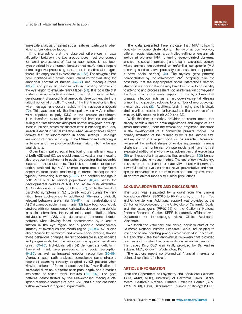
The temporal sequence of fixations also differentiated thegroups. The latency from the beginning of a trial until a fixationwas directed at the eyes AOI was also shorter for CON relativeto MIA1 (group main effect; F1,6 5 7.994, p , .05, ηp2 5 .571;Figure 5A). By contrast, MIA1 had a shorter latency until afixation was directed at the mouth AOI relative to CON (groupmain effect; F1,6 5 6.442, p , .05, ηp2 5 .517; Figure 5B).A supplementary video is available online showing the differ-ence in fixations directed at a composite image of all 10 feargrimace stimuli by CON and MIA1 animals. Still frames from thefirst 1500 msec (at 500 msec intervals) are shown in Figure 6 foreach group. It was during this brief time frame at the beginningof a stimulus presentation when the groups seemed to differmost in visual attention to fear grimaces. Fixations for CONanimals quickly converge on the face and around the eyes inparticular within the first 500 msec and remain largely clusteredthere and on the mouth for the subsequent 1000 msec. Bycontrast, fixations for MIA1 animals also converge quickly onthe face but are directed more quickly at the nose and mouth
Figure 1. Schematic of a typical testing trial (A). A 5-second black screenpreceded and followed each face stimulus. Animals were also required tofixate a center and peripheral pulsating star target for .500 msec to receive ajuice reward and proceed to the next trial. Examples of the four face stimuluscategories (neutral, lipsmack, fear grimace, and open-mouth threat) are alsoshown (B).
Effects of Maternal Immune Activation
Biological Psychiatry ]]], 2014; ]:]]]–]]] www.sobp.org/journal 3
BiologicalPsychiatry
areas than the eyes. Over the next 1000 msec, fixations forMIA1 animals are far more scattered throughout the face andblack background relative to CON.
Only two additional differences were found between thegroups when facial expressions were analyzed separately. Foropen-mouth threat facial expressions, MIA1 directed a greaterfrequency of fixations at the nose AOI than CON (group maineffect; F1,6 5 7.111, p , .05, ηp2 5 .542). The probability ofgaze shifting from one AOI to another also differentiated thegroups. When macaques and humans view faces, gaze movesquickly to the eyes but then shifts to other parts of the face,particularly the mouth and nose, then back to the eyes. Wefound that for neutral facial expressions, the probability ofgaze shifting from the eyes to the mouth was significantlygreater for CON relative to MIA1 (group main effect; F1,6 510.585, p , .05, ηp2 5 .638) and that difference wasparticularly strong on day 1 (group 3 day effect; F4,24 52.83, p , .05, ηp2 5 .320; one-way ANOVA, F1,6 5 9.961,p , .05). No differences were found between the groups in allother comparisons, including average pupil diameter.
Consistency of Individual Differences
In addition to the abnormalities in visual attention describedhere, MIA1 monkeys also previously displayed a heightenedtendency to approach, contact, and remain in close proximityto an unfamiliar conspecific relative to CON animals (46). Thisresult was interpreted as MIA1 animals being unrestrained orinappropriately social in a context where normal animalstypically chose to be more restrained or hesitant. We hypothe-sized that the lack of social restraint displayed by MIA1
animals in our previous study may be related to their abnor-mally low levels of visual attention directed at the eyes of a
conspecific (frequency and duration of fixation) and theirelevated latency to fixate on the eyes in the current study.To assess the generality and consistency of behavioralmeasures during a dyadic social interaction session with anunfamiliar conspecific and the eye-tracking study described
Figure 2. Four examples of visualstimuli used for experiment 2 areshown. Each background was down-loaded from the internet and a neutral,lipsmack, fear grimace, or open-mouth threat facial expression wasadded at a pseudorandom spatiallocation (balanced across all stimuli)using Adobe Photoshop software.
Figure 3. The average fixation duration directed at the face area of interestfor control (CON) and offspring of dams treated with poly ICLC at the end ofthe first trimester (MIA1) animals, regardless of facial expression (i.e., allexpressions averaged). MIA1 showed a significant decrease between day 1and day 2 and then remained generally constant after that. Average fixationduration for CON animals remained constant until a significant decreasebetween day 4 and day 5. *p , .05.
Effects of Maternal Immune Activation
4 Biological Psychiatry ]]], 2014; ]:]]]–]]] www.sobp.org/journal
BiologicalPsychiatry
here, we further analyzed a total of six measures that differ-entiated MIA1 and CON animals in these two experiments: 1)the frequency of fixations directed at the eyes of fear grimaceexpressions; 2) the duration of fixations directed at the eyes offear grimace expressions; 3) the latency to fixate on the eyesof fear grimace expressions; 4) the combined duration ofarms-reach proximity and contact with an unfamiliar conspe-cific; 5) the frequency of approaching to within arms-reach ofan unfamiliar conspecific; and 6) the frequency of sitting inarms-reach proximity with an unfamiliar conspecific. The datafor each of these measures were transformed into ranks (1 5lowest measure, 8 5 highest measure) and subjected to
Kendall’s coefficient of concordance (W) analysis. This non-parametric test assesses the agreement or consistency acrossmultiple measures and is not impacted by tied ranks. Thisanalysis was conducted for each group separately. The out-come of this test indicated substantial stability of individualresponsiveness for MIA1 (W 5 .625, χ25 5 12.5, p , .05) butnot for CON animals (W 5 .406, χ25 5 8.12, p 5 .150). Thisresult indicates that MIA1 animals were consistently inappro-priate (i.e., low visual attention to the eyes and high tendencyto approach) across two very different social situations, acontrolled setting where animals looked at pictures of feargrimace facial expressions and a semi-naturalistic context
Figure 4. The total frequency (A) and duration (B) of fixations directed at the eyes area of interest of fear grimace expressions. Offspring of dams treatedwith poly ICLC at the end of the first trimester (MIA1) directed fewer fixations at the eyes and those fixations totaled less time than control (CON) animals.*p , .05.
Figure 5. The average latency from the start of a trial until the first fixation was registered in the eyes area of interest (A) or the mouth area of interest (B) forfear grimace expressions. The offspring of dams treated with poly ICLC at the end of the first trimester (MIA1) animals showed a significantly longer latencybefore fixating on the eyes and significantly shorter latency before fixating the mouth relative to control (CON) animals. *p , .05.
Effects of Maternal Immune Activation
Biological Psychiatry ]]], 2014; ]:]]]–]]] www.sobp.org/journal 5
BiologicalPsychiatry
where animals encountered an unfamiliar conspecific. CONanimals, by contrast, were not consistent across these twoconditions, implying an ability to regulate their behavior differ-entially depending on the context (nonthreatening pictures offacial expressions vs. a potentially dangerous, unfamiliarconspecific). This finding indicates that MIA1 animals may lackan important ability to regulate their social behavior adaptivelyacross contexts.
Experiment 2: Facial Expressions Embedded inComplex Scenes
Analysis of the background images without an embedded facedid not reveal any significant differences between the groupsor group 3 day interactions for the total number of fixations,total duration of fixations, average duration of fixations,average dwell duration, or average pupil diameter. Likewise,when faces were embedded into the backgrounds, there were
also no significant differences between the groups or group 3day interactions for any of these variables for AOIs thatencompassed the entire background or the embedded face,both when all facial expressions were considered together orseparately. There were no significant differences between thegroups or group 3 day interactions for the conditionalprobability of shifting fixation between the background AOIand the face AOI.
DISCUSSION
We previously demonstrated that nonhuman primates prena-tally exposed to MIA in the first trimester demonstrateabnormal behaviors, including deviation from species-typicalsocial interactions (46). Here, we utilized noninvasive eyetracking to determine if these same MIA-exposed macaqueoffspring demonstrate abnormal attention to salient socialinformation (i.e., still images of faces). Much like humans,macaque monkeys have evolved a sophisticated social com-munication system that includes a variety of facial expres-sions, body postures, and vocalizations (56,57). Among thesesocial signals, the use of facial expressions is one of the mostsalient features of macaque social behavior and the mostsimilar to our own social communication (58–60). Our experi-ments were therefore designed to maximize translationalpotential between the nonhuman primate model and gaze-processing abnormalities observed in human neurodevelop-mental disorders.
We first measured gaze patterns when juvenile offspring ofcontrol dams (untreated and saline-treated) and age-/sex-matched offspring of dams treated with poly ICLC at the endof the first trimester viewed faces of unfamiliar conspecificsdisplaying four basic facial expressions used for macaquesocial communication. The expressions consisted of facesshowing a neutral expression, faces depicting affiliative/submissive expressions (i.e., lipsmack), faces depicting sub-missive/fearful expressions (i.e., fear grimaces), and facesshowing an open-mouth threat. We found several importantdifferences in gaze between the control and MIA1 animals.When all expressions were considered as a single category,the MIA1 animals showed a significant drop in average fixationduration after the first presentation, while the control animalsdid not show such a drop until after their fourth presentation.When the animals looked at fear grimace expressions, MIA1
animals took longer to direct their attention at the eyes thancontrol animals and ultimately looked less at the eyes (in termsof frequency and duration) than control animals. MIA1 animalslooked more at the nose of open-mouth threat expressionsrelative to control animals. The MIA1 animals were also lesslikely to shift their gaze from the eyes to the mouth whenlooking at neutral expressions than control animals. In oursecond experiment, we tracked eye gaze as the animalslooked at complex, naturalistic scenes with monkey facesembedded at random locations. Both groups were able tolocate and fixate on the faces embedded in the scenes andshowed similar levels of visual attention directed at the back-grounds and the embedded faces. Thus, differences in gazepatterns between the groups did not appear to reflect a globalinability to locate and attend to social cues but rather specificabnormalities related to sustained social attention and the
Figure 6. Each image shown is a composite of the 10 fear grimace facialexpressions included in experiment 1. The 10 images were made semi-transparent and then overlaid on each other. The point of gaze for alloffspring of dams treated with poly ICLC at the end of the first trimester(MIA1) or control (CON) animals on days 1 through 5 were then mappedonto these images at 0 msec, 500 msec, 1000 msec, and 1500 msec toshow the general distribution of gaze for the two groups when viewing feargrimaces. Notice that CON animals largely direct their gaze at the eyes by500 msec into the presentation. During the next 1000 msec, their gaze issplit between the eyes and the mouth. By contrast, MIA1 animals largelydirect their gaze at the nose at 500 msec, and their gaze generally dispersesto many different parts of the face and background by 1500 msec into thepresentation. A supplemental video is available online showing a compar-ison between the CON and MIA1 animals for the entire 5-second presenta-tion of the fear grimace composite image.
Effects of Maternal Immune Activation
6 Biological Psychiatry ]]], 2014; ]:]]]–]]] www.sobp.org/journal
BiologicalPsychiatry
fine-scale analysis of salient social features, particularly whenviewing fear grimace faces.
It is interesting that the observed differences in gazeallocation between the two groups were most pronouncedfor facial expressions of fear or submission. It has beenhypothesized in the human literature that fearful faces requiremore cognitive processing than other faces that also signalthreat, like angry facial expressions (61–63). The amygdala hasbeen identified as a critical neural structure for evaluating theemotional content of human (64–68) and macaque faces(69,70) and plays an essential role in directing attention tothe eye region to evaluate fearful faces (71). It is possible thatmaternal immune activation during the first trimester of fetaldevelopment disrupted fetal amygdala development during acritical period of growth. The end of the first trimester is a timewhen neurogenesis occurs rapidly in the macaque amygdala(72). This was precisely the time point when MIA1 motherswere exposed to poly ICLC in the present experiment.It is therefore plausible that maternal immune activationduring the first trimester disrupted normal amygdala develop-ment in fetuses, which manifested later in offspring as a fairlyselective deficit in visual attention when viewing faces used toconvey fear or subordination in social settings. Histologicevaluation of brain pathology in the MIA-exposed offspring isunderway and may provide additional insight into the behav-ioral deficits.
Given that impaired social functioning is a hallmark featureof both ASD and SZ, we would expect a valid animal model toalso produce impairments in social processing that resemblefeatures of these disorders. The lack of attention to the eyeregion exhibited by MIA1 animals represents a significantdeparture from social processing in normal macaques andtypically developing humans (73–76) and parallels findings inboth ASD and SZ clinical populations (48,49). While thedevelopmental courses of ASD and SZ are quite different—ASD is diagnosed in early childhood (77), while the onset ofpsychotic symptoms in SZ typically occurs during the tran-sition from adolescence to adulthood (78)—many clinicallyrelevant behaviors are similar (79–81). The manifestations ofASD diagnostic social impairments (82) have been extensivelystudied, with numerous empirical studies documenting deficitsin social interaction, theory of mind, and imitation. Manyindividuals with ASD also demonstrate abnormal fixationpatterns when viewing faces, characterized by a lack offixation in the eye region and a possible compensatorystrategy of fixating on the mouth region (83–88). SZ is alsocharacterized by persistent and severe social deficits, thoughthese behavioral changes are first observable in adolescenceand progressively become worse as one approaches illnessonset (89–93). Individuals with SZ demonstrate deficits intheory of mind, face processing, and social perception(94,95), as well as impaired emotion recognition (96–99).Moreover, scan path analyses consistently demonstrate arestricted scanning strategy adopted by SZ patients whenviewing pictures of faces, characterized by fewer fixations ofincreased duration, a shorter scan path length, and a markedavoidance of salient facial features (100–104). The gazepatterns demonstrated by the MIA-exposed macaque off-spring resemble features of both ASD and SZ and are beingfurther explored in ongoing experiments.
The data presented here indicate that MIA1 offspringconsistently demonstrate aberrant behavior across two verydifferent social situations: a controlled setting where animalslooked at pictures (MIA1 offspring demonstrated abnormalattention to social information) and a semi-naturalistic contextwhere animals encountered an unfamiliar conspecific (MIAoffspring failed to show species-typical hesitation to approacha novel social partner) (46). The atypical gaze patternsdemonstrated by the adolescent MIA1 offspring raise thepossibility that the inappropriate social interactions demon-strated in our earlier studies may have been due to an inabilityto attend to and process salient social information conveyed inthe face. This study lends support to the hypothesis thatprenatal infection acts as a neurodevelopmental diseaseprimer that is possibly relevant to a number of neurodevelop-mental disorders (32). Additional brain imaging and histologicstudies will be needed to further evaluate the relevance of themonkey MIA model to both ASD and SZ.
While the rhesus monkey provides an animal model thatclosely parallels human brain organization and cognitive andsocial functioning, there are ethical and pragmatic limitationsin the development of a nonhuman primate model. Theprimary limitation of the current study is the sample size,and replication in a larger cohort will be essential. Moreover,we are at the earliest stages of evaluating prenatal immunechallenge in the nonhuman primate model and have not yetevaluated additional environmental adversities that exacerbate(35) or therapeutic interventions that ameliorate (105) behav-ioral pathologies in mouse models. The use of noninvasive eyetracking in the nonhuman primate MIA model will provide apowerful tool to evaluate these novel preventative and ther-apeutic interventions in future studies and can improve trans-lation from animal models to clinical populations.
ACKNOWLEDGMENTS AND DISCLOSURES
This work was supported by a grant from the SimonsFoundation (SFARI 9900060 to PHP) and by a gift from Tedand Ginger Jenkins. Additional support was provided by theCenter for Neuroscience at the University of California, Davis,and the base grant (RR00169) of the California NationalPrimate Research Center. SEPS is currently affiliated withDepartment of Immunology, Mayo Clinic, Rochester,Minnesota.
We thank the veterinary and animal services staff of theCalifornia National Primate Research Center for helping torefine the animal handling procedures described in this article.We also thank the four anonymous reviewers that providedpositive and constructive comments on an earlier version ofthis paper. Poly-ICLC was kindly provided by Dr. AndresSalazar, M.D., Oncovir, Washington, DC.
The authors report no biomedical financial interests orpotential conflicts of interest.
ARTICLE INFORMATION
From the Department of Psychiatry and Behavioral Sciences(CJM, AMW, MDB), University of California, Davis, Sacra-mento; California National Primate Research Center (CJM,AMW, MDB), Davis, Sacramento; Division of Biology (SEPS,
Effects of Maternal Immune Activation
Biological Psychiatry ]]], 2014; ]:]]]–]]] www.sobp.org/journal 7
BiologicalPsychiatry
PHP), California Institute of Technology, Pasadena; and TheMIND Institute (MDB), University of California, Davis, Sacra-mento, California.
Deceased (PHP).Address correspondence to Melissa D. Bauman, Ph.D.,
University of California, Davis, The MIND Institute, 2825 50thStreet #1416, Sacramento, CA 95817; E-mail: [email protected].
Received Apr 22, 2014; revised Jul 4, 2014; accepted Jul24, 2014.
Supplementary material cited in this article is availableonline at http://dx.doi.org/10.1016/j.biopsych.2014.07.035.
REFERENCES1. Brown AS (2012): Epidemiologic studies of exposure to prenatal
infection and risk of schizophrenia and autism. Dev Neurobiol 72:1272–1276.
2. Hallmayer J, Cleveland S, Torres A, Phillips J, Cohen B, Torigoe T,et al. (2011): Genetic heritability and shared environmental factorsamong twin pairs with autism. Arch Gen Psychiatry 68:1095–1102.
3. Hagberg H, Gressens P, Mallard C (2012): Inflammation during fetaland neonatal life: Implications for neurologic and neuropsychiatricdisease in children and adults. Ann Neurol 71:444–457.
4. Rosenberg RE, Law JK, Yenokyan G, McGready J, Kaufmann WE,Law PA (2009): Characteristics and concordance of autism spectrumdisorders among 277 twin pairs. Arch Pediatr Adolesc Med 163:907–914.
5. Brown AS, Schaefer CA, Quesenberry CP Jr, Liu L, Babulas VP,Susser ES (2005): Maternal exposure to toxoplasmosis and risk ofschizophrenia in adult offspring. Am J Psychiatry 162:767–773.
6. Brown AS, Schaefer CA, Wyatt RJ, Goetz R, Begg MD, Gorman JM,Susser ES (2000): Maternal exposure to respiratory infections andadult schizophrenia spectrum disorders: A prospective birth cohortstudy. Schizophr Bull 26:287–295.
7. Sorensen HJ, Mortensen EL, Reinisch JM, Mednick SA (2009):Association between prenatal exposure to bacterial infection andrisk of schizophrenia. Schizophr Bull 35:631–637.
8. Babulas V, Factor-Litvak P, Goetz R, Schaefer CA, Brown AS (2006):Prenatal exposure to maternal genital and reproductive infectionsand adult schizophrenia. Am J Psychiatry 163:927–929.
9. Buka SL, Cannon TD, Torrey EF, Yolken RH (2008): Maternalexposure to herpes simplex virus and risk of psychosis among adultoffspring. Biol Psychiatry 63:809–815.
10. Brown AS, Begg MD, Gravenstein S, Schaefer CA, Wyatt RJ,Bresnahan M, et al. (2004): Serologic evidence of prenatal influenzain the etiology of schizophrenia. Arch Gen Psychiatry 61:774–780.
11. Brown AS, Sourander A, Hinkka-Yli-Salomaki S, McKeague IW,Sundvall J, Surcel HM (2014): Elevated maternal C-reactive proteinand autism in a national birth cohort. Mol Psychiatry 19:259–264.
12. Abdallah MW, Larsen N, Mortensen EL, Atladottir HO, Norgaard-Pedersen B, Bonefeld-Jorgensen EC, et al. (2012): Neonatal levels ofcytokines and risk of autism spectrum disorders: An exploratoryregister-based historic birth cohort study utilizing the Danish New-born Screening Biobank. J Neuroimmunol 252:75–82.
13. Atladottir HO, Thorsen P, Ostergaard L, Schendel DE, Lemcke S,Abdallah M, Parner ET (2010): Maternal infection requiring hospital-ization during pregnancy and autism spectrum disorders. J AutismDev Disord 40:1423–1430.
14. Goines PE, Croen LA, Braunschweig D, Yoshida CK, Grether J,Hansen R, et al. (2011): Increased midgestational IFN-gamma, IL-4and IL-5 in women bearing a child with autism: A case-control study.Mol Autism 2:13.
15. Fatemi SH, Emamian ES, Sidwell RW, Kist DA, Stary JM, Earle JA,Thuras P (2002): Human influenza viral infection in utero alters glialfibrillary acidic protein immunoreactivity in the developing brains ofneonatal mice. Mol Psychiatry 7:633–640.
16. Fatemi SH, Sidwell R, Kist D, Akhter P, Meltzer HY, Bailey K, et al.(1998): Differential expression of synaptosome-associated protein25 kDa [SNAP-25] in hippocampi of neonatal mice followingexposure to human influenza virus in utero. Brain Res 800:1–9.
17. Fatemi SH, Reutiman TJ, Folsom TD, Huang H, Oishi K, Mori S, et al.(2008): Maternal infection leads to abnormal gene regulation andbrain atrophy in mouse offspring: Implications for genesis of neuro-developmental disorders. Schizophr Res 99:56–70.
18. Short SJ, Lubach GR, Karasin AI, Olsen CW, Styner M, KnickmeyerRC, et al. (2010): Maternal influenza infection during pregnancyimpacts postnatal brain development in the rhesus monkey. BiolPsychiatry 67:965–973.
19. Willette AA, Lubach GR, Knickmeyer RC, Short SJ, Styner M,Gilmore JH, Coe CL (2011): Brain enlargement and increasedbehavioral and cytokine reactivity in infant monkeys following acuteprenatal endotoxemia. Behav Brain Res 219:108–115.
20. Coyle P, Tran N, Fung JN, Summers BL, Rofe AM (2009): Maternaldietary zinc supplementation prevents aberrant behaviour in anobject recognition task in mice offspring exposed to LPS in earlypregnancy. Behav Brain Res 197:210–218.
21. Fortier ME, Joober R, Luheshi GN, Boksa P (2004): Maternalexposure to bacterial endotoxin during pregnancy enhancesamphetamine-induced locomotion and startle responses in adultrat offspring. J Psychiatr Res 38:335–345.
22. Fortier ME, Luheshi GN, Boksa P (2007): Effects of prenatal infectionon prepulse inhibition in the rat depend on the nature of the infectiousagent and the stage of pregnancy. Behav Brain Res 181:270–277.
23. Traynor TR, Majde JA, Bohnet SG, Krueger JM (2004): Intratrachealdouble-stranded RNA plus interferon-gamma: A model for analysisof the acute phase response to respiratory viral infections. Life Sci74:2563–2576.
24. Boksa P (2010): Effects of prenatal infection on brain developmentand behavior: A review of findings from animal models. Brain BehavImmun 24:881–897.
25. Meyer U, Feldon J (2012): To poly(I:C) or not to poly(I:C): Advancingpreclinical schizophrenia research through the use of prenatalimmune activation models. Neuropharmacology 62:1308–1321.
26. Meyer U, Feldon J, Dammann O (2011): Schizophrenia and autism:Both shared and disorder-specific pathogenesis via perinatal inflam-mation? Pediatr Res 69:26R–33R.
27. Patterson PH (2009): Immune involvement in schizophrenia andautism: Etiology, pathology and animal models. Behav Brain Res204:313–321.
28. Meyer U, Feldon J (2009): Neural basis of psychosis-relatedbehaviour in the infection model of schizophrenia. Behav BrainRes 204:322–334.
29. Laibl VR, Sheffield JS (2005): Influenza and pneumonia in pregnancy.Clin Perinatol 32:727–738.
30. Longman RE, Johnson TR (2007): Viral respiratory disease inpregnancy. Curr Opin Obstet Gynecol 19:120–125.
31. Selten JP, Frissen A, Lensvelt-Mulders G, Morgan VA (2010):Schizophrenia and 1957 pandemic of influenza: Meta-analysis.Schizophr Bull 36:219–228.
32. Meyer U (2014): Prenatal poly(i:C) exposure and other developmentalimmune activation models in rodent systems. Biol Psychiatry 75:307–315.
33. Gallagher D, Norman AA, Woodard CL, Yang G, Gauthier-Fisher A,Fujitani M, et al. (2013): Transient maternal IL-6 mediates long-lasting changes in neural stem cell pools by deregulating anendogenous self-renewal pathway. Cell Stem Cell 13:564–576.
34. Smith SE, Li J, Garbett K, Mirnics K, Patterson PH (2007): Maternalimmune activation alters fetal brain development through interleukin-6. J Neurosci 27:10695–10702.
35. Giovanoli S, Engler H, Engler A, Richetto J, Voget M, Willi R, et al.(2013): Stress in puberty unmasks latent neuropathological consequen-ces of prenatal immune activation in mice. Science 339:1095–1099.
36. Piontkewitz Y, Arad M, Weiner I (2011): Abnormal trajectories ofneurodevelopment and behavior following in utero insult in the rat.Biol Psychiatry 70:842–851.
Effects of Maternal Immune Activation
8 Biological Psychiatry ]]], 2014; ]:]]]–]]] www.sobp.org/journal
BiologicalPsychiatry
37. Garay PA, Hsiao EY, Patterson PH, McAllister AK (2013): Maternalimmune activation causes age- and region-specific changes in braincytokines in offspring throughout development. Brain Behav Immun31:54–68.
38. Malkova NV, Yu CZ, Hsiao EY, Moore MJ, Patterson PH (2012):Maternal immune activation yields offspring displaying mouseversions of the three core symptoms of autism. Brain Behav Immun26:607–616.
39. Shi L, Fatemi SH, Sidwell RW, Patterson PH (2003): Maternalinfluenza infection causes marked behavioral and pharmacologicalchanges in the offspring. J Neurosci 23:297–302.
40. Garbett KA, Hsiao EY, Kalman S, Patterson PH, Mirnics K (2012):Effects of maternal immune activation on gene expression patternsin the fetal brain. Transl Psychiatry 2:e98.
41. Watson KK, Platt ML (2012): Of mice and monkeys: Using non-human primate models to bridge mouse- and human-based inves-tigations of autism spectrum disorders. J Neurodev Disord 4:21.
42. Capitanio JP, Emborg ME (2008): Contributions of non-humanprimates to neuroscience research. Lancet 371:1126–1135.
43. Phillips KA, Bales KL, Capitanio JP, Conley A, Czoty PW, t Hart BA,et al. (2014): Why primate models matter. Am J Primatol 76:801–827.
44. Levy HB, Baer G, Baron S, Buckler CE, Gibbs CJ, Iadarola MJ, et al.(1975): A modified polyriboinosinic-polyribocytidylic acid complexthat induces interferon in primates. J Infect Dis 132:434–439.
45. Caskey M, Lefebvre F, Filali-Mouhim A, Cameron MJ, Goulet JP,Haddad EK, et al. (2011): Synthetic double-stranded RNA inducesinnate immune responses similar to a live viral vaccine in humans.J Exp Med 208:2357–2366.
46. Bauman MD, Iosif AM, Smith SE, Bregere C, Amaral DG, PattersonPH (2014): Activation of the maternal immune system duringpregnancy alters behavioral development of rhesus monkey off-spring. Biol Psychiatry 75:332–341.
47. King BH, Lord C (2011): Is schizophrenia on the autism spectrum?Brain Res 1380:34–41.
48. Pelphrey KA, Sasson NJ, Reznick JS, Paul G, Goldman BD, Piven J(2002): Visual scanning of faces in autism. J Autism Dev Disord 32:249–261.
49. Toh WL, Rossell SL, Castle DJ (2011): Current visual scanpathresearch: A review of investigations into the psychotic, anxiety, andmood disorders. Compr Psychiatry 52:567–579.
50. Schneider ML, Roughton EC, Koehler AJ, Lubach GR (1999): Growthand development following prenatal stress exposure in primates: Anexamination of ontogenetic vulnerability. Child Dev 70:263–274.
51. Schneider ML, Moore CF, Roberts AD, Dejesus O (2001): Prenatalstress alters early neurobehavior, stress reactivity and learning innon-human primates: A brief review. Stress 4:183–193.
52. Schneider ML, Moore CF, Kraemer GW (2004): Moderate levelalcohol during pregnancy, prenatal stress, or both and limbic-hypothalamic-pituitary-adrenocortical axis response to stress inrhesus monkeys. Child Dev 75:96–109.
53. Roberts AD, Moore CF, DeJesus OT, Barnhart TE, Larson JA,Mukherjee J, et al. (2004): Prenatal stress, moderate fetal alcohol,and dopamine system function in rhesus monkeys. NeurotoxicolTeratol 26:169–178.
54. Converse AK, Moore CF, Moirano JM, Ahlers EO, Larson JA, Engle JW,et al. (2013): Prenatal stress induces increased striatal dopamine trans-porter binding in adult nonhuman primates. Biol Psychiatry 74:502–510.
55. Riby DM, Hancock PJ (2009): Do faces capture the attention ofindividuals with Williams syndrome or autism? Evidence fromtracking eye movements. J Autism Dev Disord 39:421–431.
56. Tomasello M, Call J (1997): Social knowledge and interaction. In:Tomasello M, Call J, editors. Primate Cognition. New York: OxfordUniversity Press, 191–230.
57. Chang SW, Brent LJ, Adams GK, Klein JT, Pearson JM, Watson KK,Platt ML (2013): Neuroethology of primate social behavior. Proc NatlAcad Sci U S A 110(suppl 2):10387–10394.
58. Deaner RO, Khera AV, Platt ML (2005): Monkeys pay per view:Adaptive valuation of social images by rhesus macaques. Curr Biol15:543–548.
59. Bower S, Suomi SJ, Paukner A (2012): Evidence for kinshipinformation contained in the rhesus macaque (Macaca mulatta) face.J Comp Psychol 126:318–323.
60. Ferrari PF, Paukner A, Ionica C, Suomi SJ (2009): Reciprocal face-to-face communication between rhesus macaque mothers and theirnewborn infants. Curr Biol 19:1768–1772.
61. Davis M, Whalen PJ (2001): The amygdala: Vigilance and emotion.Mol Psychiatry 6:13–34.
62. Bannerman RL, Milders M, de Gelder B, Sahraie A (2009): Orientingto threat: Faster localization of fearful facial expressions and bodypostures revealed by saccadic eye movements. Proc Biol Sci 276:1635–1641.
63. Whalen PJ (1998): Fear, vigilance, and ambiguity: Initial neuroimagingstudies of the human amygdala. Curr Dir Psychol Sci 7:177–188.
64. Whalen PJ, Rauch SL, Etcoff NL, McInerney SC, Lee MB, Jenike MA(1998): Masked presentations of emotional facial expressions mod-ulate amygdala activity without explicit knowledge. J Neurosci 18:411–418.
65. Adolphs R, Tranel D, Damasio H, Damasio A (1994): Impairedrecognition of emotion in facial expressions following bilateraldamage to the human amygdala. Nature 372:669–672.
66. Adolphs R, Tranel D, Damasio H, Damasio AR (1995): Fear and thehuman amygdala. J Neurosci 15:5879–5891.
67. Adolphs R, Tranel D, Hamann S, Young AW, Calder AJ, Phelps EA,et al. (1999): Recognition of facial emotion in nine individuals withbilateral amygdala damage. Neuropsychologia 37:1111–1117.
68. Spezio ML, Huang PY, Castelli F, Adolphs R (2007): Amygdaladamage impairs eye contact during conversations with real people.J Neurosci 27:3994–3997.
69. Gothard KM, Battaglia FP, Erickson CA, Spitler KM, Amaral DG(2007): Neural responses to facial expression and face identity in themonkey amygdala. J Neurophysiol 97:1671–1683.
70. Hoffman KL, Gothard KM, Schmid MC, Logothetis NK (2007): Facial-expression and gaze-selective responses in the monkey amygdala.Curr Biol 17:766–772.
71. Adolphs R, Gosselin F, Buchanan TW, Tranel D, Schyns P, DamasioAR (2005): A mechanism for impaired fear recognition after amygdaladamage. Nature 433:68–72.
72. Kordower JH, Piecinski P, Rakic P (1992): Neurogenesis of theamygdaloid nuclear complex in the rhesus monkey. Brain Res DevBrain Res 68:9–15.
73. Walker-Smith GJ, Gale AG, Findlay JM (1977): Eye movementstrategies involved in face perception. Perception 6:313–326.
74. Ekman P, Friesen WV (1975): Unmasking the Face: A Guide toRecognizing Emotions from Facial Cues. Oxford, UK: Prentice-Hall.
75. Machado CJ, Bliss-Moreau E, Platt ML, Amaral DG (2011): Socialand nonsocial content differentially modulates visual attention andautonomic arousal in rhesus macaques. PloS One 6:e26598.
76. Leonard TK, Blumenthal G, Gothard KM, Hoffman KL (2012): Howmacaques view familiarity and gaze in conspecific faces. BehavNeurosci 126:781–791.
77. Mandell DS, Novak MM, Zubritsky CD (2005): Factors associatedwith age of diagnosis among children with autism spectrumdisorders. Pediatrics 116:1480–1486.
78. Ziermans TB, Schothorst PF, Sprong M, van Engeland H (2011):Transition and remission in adolescents at ultra-high risk forpsychosis. Schizophr Res 126:58–64.
79. Couture SM, Penn DL, Losh M, Adolphs R, Hurley R, Piven J (2010):Comparison of social cognitive functioning in schizophrenia andhigh functioning autism: More convergence than divergence. Psy-chol Med 40:569–579.
80. Sasson NJ, Pinkham AE, Carpenter KL, Belger A (2011): The benefitof directly comparing autism and schizophrenia for revealing mech-anisms of social cognitive impairment. J Neurodev Disord 3:87–100.
81. Solomon M, Olsen E, Niendam T, Ragland JD, Yoon J, MinzenbergM, Carter CS (2011): From lumping to splitting and back again:Atypical social and language development in individuals withclinical-high-risk for psychosis, first episode schizophrenia, andautism spectrum disorders. Schizophr Res 131:146–151.
Effects of Maternal Immune Activation
Biological Psychiatry ]]], 2014; ]:]]]–]]] www.sobp.org/journal 9
BiologicalPsychiatry
82. American Psychiatric Association (2013): Diagnostic and StatisticalManual of Mental Disorders. Arlington, VA: American PsychiatricPublishing.
83. Dalton KM, Nacewicz BM, Johnstone T, Schaefer HS, GernsbacherMA, Goldsmith HH, et al. (2005): Gaze fixation and the neuralcircuitry of face processing in autism. Nat Neurosci 8:519–526.
84. Kliemann D, Dziobek I, Hatri A, Steimke R, Heekeren HR (2010):Atypical reflexive gaze patterns on emotional faces in autismspectrum disorders. J Neurosci 30:12281–12287.
85. Klin A, Jones W, Schultz R, Volkmar F, Cohen D (2002): Visualfixation patterns during viewing of naturalistic social situations aspredictors of social competence in individuals with autism. Arch GenPsychiatry 59:809–816.
86. Neumann D, Spezio ML, Piven J, Adolphs R (2006): Looking you inthe mouth: Abnormal gaze in autism resulting from impaired top-downmodulation of visual attention. Soc Cogn Affect Neurosci 1:194–202.
87. Spezio ML, Adolphs R, Hurley RS, Piven J (2007): Abnormal use offacial information in high-functioning autism. J Autism Dev Disord37:929–939.
88. Kennedy DP, Adolphs R (2012): Perception of emotions from facialexpressions in high-functioning adults with autism. Neuropsycholo-gia 50:3313–3319.
89. Allen DN, Frantom LV, Strauss GP, van Kammen DP (2005): Differ-ential patterns of premorbid academic and social deterioration inpatients with schizophrenia. Schizophr Res 75:389–397.
90. Ballon JS, Kaur T, Marks II, Cadenhead KS (2007): Social functioning inyoung people at risk for schizophrenia. Psychiatry Res 151:29–35.
91. Chudleigh C, Naismith SL, Blaszczynski A, Hermens DF, Hodge MA,Hickie IB (2011): How does social functioning in the early stages ofpsychosis relate to depression and social anxiety? Early IntervPsychiatry 5:224–232.
92. Cornblatt BA, Auther AM, Niendam T, Smith CW, Zinberg J, BeardenCE, Cannon TD (2007): Preliminary findings for two new measures ofsocial and role functioning in the prodromal phase of schizophrenia.Schizophr Bull 33:688–702.
93. Amminger GP, Schafer MR, Klier CM, Schlogelhofer M, MossahebN, Thompson A, et al. (2012): Facial and vocal affect perception inpeople at ultra-high risk of psychosis, first-episode schizophreniaand healthy controls. Early Interv Psychiatry 6:450–454.
94. Bora E, Pantelis C (2013): Theory of mind impairments in first-episode psychosis, individuals at ultra-high risk for psychosis and infirst-degree relatives of schizophrenia: Systematic review and meta-analysis. Schizophr Res 144:31–36.
95. Ventura J, Wood RC, Jimenez AM, Hellemann GS (2013): Neuro-cognition and symptoms identify links between facial recognitionand emotion processing in schizophrenia: Meta-analytic findings.Schizophr Res 151:78–84.
96. Pinkham AE, Gur RE, Gur RC (2007): Affect recognition deficits inschizophrenia: Neural substrates and psychopharmacological impli-cations. Expert Rev Neurother 7:807–816.
97. Edwards J, Jackson HJ, Pattison PE (2002): Emotion recognition viafacial expression and affective prosody in schizophrenia: A meth-odological review. Clin Psychol Rev 22:789–832.
98. Kohler CG, Walker JB, Martin EA, Healey KM, Moberg PJ (2010):Facial emotion perception in schizophrenia: A meta-analytic review.Schizophr Bull 36:1009–1019.
99. Hoekert M, Kahn RS, Pijnenborg M, Aleman A (2007): Impairedrecognition and expression of emotional prosody in schizophrenia:Review and meta-analysis. Schizophr Res 96:135–145.
100. Loughland CM, Williams LM, Harris AW (2004): Visual scanpathdysfunction in first-degree relatives of schizophrenia probands:Evidence for a vulnerability marker? Schizophr Res 67:11–21.
101. Williams LM, Loughland CM, Green MJ, Harris AW, Gordon E (2003):Emotion perception in schizophrenia: An eye movement studycomparing the effectiveness of risperidone vs. haloperidol. Psychia-try Res 120:13–27.
102. Loughland CM, Williams LM, Gordon E (2002): Schizophrenia andaffective disorder show different visual scanning behavior for faces:A trait versus state-based distinction? Biol Psychiatry 52:338–348.
103. Loughland CM, Williams LM, Gordon E (2002): Visual scanpaths topositive and negative facial emotions in an outpatient schizophreniasample. Schizophr Res 55:159–170.
104. Williams LM, Loughland CM, Gordon E, Davidson D (1999): Visualscanpaths in schizophrenia: Is there a deficit in face recognition?Schizophr Res 40:189–199.
105. Hsiao EY, McBride SW, Hsien S, Sharon G, Hyde ER, McCue T, et al.(2013): Microbiota modulate behavioral and physiological abnormalitiesassociated with neurodevelopmental disorders. Cell 155:1451–1463.
Effects of Maternal Immune Activation
10 Biological Psychiatry ]]], 2014; ]:]]]–]]] www.sobp.org/journal
BiologicalPsychiatry