Maternal Carriage in Late-Onset Group B Streptococcus ...
9
Maternal Carriage in Late-Onset Group B Streptococcus Disease, Italy Alberto Berardi, Caterina Spada, Roberta Creti, Cinzia Auriti, Lucia Gambini, Vittoria Rizzo, Mariagrazia Capretti, Nicola Laforgia, Irene Papa, Anna Tarocco, Angela Lanzoni, Giacomo Biasucci, Giancarlo Piccinini, Giovanna Nardella, Giuseppe Latorre, Daniele Merazzi, Laura Travan, Maria Letizia Bacchi Reggiani, Lorenza Baroni, Matilde Ciccia, Laura Lucaccioni, Lorenzo Iughetti, Licia Lugli Emerging Infectious Diseases • www.cdc.gov/eid • Vol. 27, No. 9, September 2021 2279 RESEARCH In support of improving patient care, this activity has been planned and implemented by Medscape, LLC and Emerging Infectious Diseases. Medscape, LLC is jointly accredited by the Accreditation Council for Continuing Medical Education (ACCME), the Accreditation Council for Pharmacy Education (ACPE), and the American Nurses Credentialing Center (ANCC), to provide continuing education for the healthcare team. Medscape, LLC designates this Journal-based CME activity for a maximum of 1.00 AMA PRA Category 1 Credit(s)™. Physicians should claim only the credit commensurate with the extent of their participation in the activity. Successful completion of this CME activity, which includes participation in the evaluation component, enables the participant to earn up to 1.0 MOC points in the American Board of Internal Medicine's (ABIM) Maintenance of Certification (MOC) program. Participants will earn MOC points equivalent to the amount of CME credits claimed for the activity. It is the CME activity provider's responsibility to submit participant completion information to ACCME for the purpose of granting ABIM MOC credit. All other clinicians completing this activity will be issued a certificate of participation. To participate in this journal CME activity: (1) review the learning objectives and author disclosures; (2) study the education content; (3) take the post-test with a 75% minimum passing score and complete the evaluation at http://www.medscape.org/journal/eid; and (4) view/print certificate. For CME questions, see page XXX. Release date: August 19, 2021; Expiration date: August 19, 2022 Learning Objectives Upon completion of this activity, participants will be able to: • Describe the dynamics of group B Streptococcus (GBS) mother-to-infant transmission according to maternal vaginal-rectal colonization at prenatal screening and at time of late-onset disease (LOD) onset and on additional maternal urine and breast milk cultures collected at LOD onset • Determine molecular typing and antibiotic resistance in mother-to-infant transmission of GBS • Identify clinical implications of the dynamics of GBS mother-to-infant transmission CME Editor Dana C. Dolan, BS, Copyeditor, Emerging Infectious Diseases. Disclosure: Dana C. Dolan, BS, has disclosed no relevant financial relationships. CME Author Laurie Barclay, MD, freelance writer and reviewer, Medscape, LLC. Disclosure: Laurie Barclay, MD, has disclosed no relevant financial relationships. Authors Disclosures: Alberto Berardi, MD; Caterina Spada, MD; Roberta Creti, PhD; Cinzia Auriti, MD; Lucia Gambini, MD, PhD; Vittoria Rizzo, MD; Maria Grazia Capretti, MD, PhD; Nicola Laforgia, MD; Irene Papa, MD, PhD; Anna Tarocco, MD, PhD; Angela Lanzoni, MD; Giacomo Biasucci, MD; Giancarlo Piccinini, MD; Giovanna Nardella, MD; Giuseppe Latorre, MD; Daniele Merazzi, MD; Laura Travan, MD, PhD; Maria Letizia Bacchi Reggiani, MD; Lorenza Baroni, MD; Matilde Maria Ciccia, MD; Laura Lucaccioni, MD; and Licia Lugli, MD, have disclosed no relevant financial relationships. Lorenzo Iughetti, PhD, has disclosed the following relevant financial relationships: served as an advisor or consultant for Eli Lilly and Company; Novo Nordisk; Springer; served as a speaker or a member of a speakers bureau for Eli Lilly and Company; Novo Nordisk; Sandoz; received grants for clinical research from AstraZeneca Pharmaceuticals LP; Eli Lilly and Company; Novartis Pharmaceuticals Corporation; Sandoz. Author affiliations: Azienda Ospedaliero–Universitaria Policlinico, Modena, Italy (A. Berardi, C. Spada, L. Lucaccioni, L. Iughetti, L. Lugli); Istituto Superiore di Sanità, Rome, Italy (R. Creti); Ospedale Pediatrico Bambino Gesù, Rome (C. Auriti); Azienda Ospedaliero-Universitaria Policlinico, Parma, Italy (L. Gambini); Ospedale Civile M. Bufalini, Cesena, Italy (V. Rizzo); Azienda Ospedaliero–Universitaria S. Orsola–Mal- pighi, Bologna, Italy (M. Capretti, M.L. Bacchi Reggiani); Ospedale Policlinico, Bari, Italy (N. Laforgia); Ospedale Infermi, Rimini, Italy (I. Papa); Azienda Ospedaliero–Universitaria S. Anna, Ferrara, Italy (A. Tarocco); Ospedale Santa Maria della Scaletta, Imola, Italy (A. Lanzoni); Ospe- dale G. da Saliceto, Piacenza, Italy (G. Biasucci); Ospedale Santa Maria Delle Croci, Ravenna, Italy (G. Piccinini); Azienda Ospedaliero–Uni- versitaria Ospedali Riuniti, Foggia, Italy (G. Nardella); Ospedale F. Miulli, Acquaviva delle Fonti, Italy (G. Latorre); Ospedale Valduce, Como, Italy (D. Merazzi); IRCCS Materno Infantile Burlo Garofolo, Trieste, Italy (L. Travan); Arcispedale Santa Maria Nuova, Reggio Emilia, Italy (L. Baroni); Ospedale Maggiore, Bologna (M. Ciccia) DOI: https://doi.org/10.3201/eid2709.210049
Transcript of Maternal Carriage in Late-Onset Group B Streptococcus ...
Maternal Carriage in Late-Onset Group B
Streptococcus Disease, ItalyAlberto Berardi, Caterina Spada, Roberta Creti, Cinzia Auriti, Lucia Gambini, Vittoria Rizzo,
Mariagrazia Capretti, Nicola Laforgia, Irene Papa, Anna Tarocco, Angela Lanzoni, Giacomo Biasucci, Giancarlo Piccinini, Giovanna Nardella, Giuseppe Latorre, Daniele Merazzi, Laura Travan, Maria Letizia
Bacchi Reggiani, Lorenza Baroni, Matilde Ciccia, Laura Lucaccioni, Lorenzo Iughetti, Licia Lugli
Emerging Infectious Diseases • www.cdc.gov/eid • Vol. 27, No. 9, September 2021 2279
RESEARCH
Page 1 of 1
In support of improving patient care, this activity has been planned and implemented by Medscape, LLC and Emerging Infectious Diseases. Medscape, LLC is jointly accredited by the Accreditation Council for Continuing Medical Education (ACCME), the Accreditation Council for Pharmacy Education (ACPE), and the American Nurses Credentialing Center (ANCC), to provide continuing education for the healthcare team.
Medscape, LLC designates this Journal-based CME activity for a maximum of 1.00 AMA PRA Category 1 Credit(s)™. Physicians should claim only the credit commensurate with the extent of their participation in the activity.
Successful completion of this CME activity, which includes participation in the evaluation component, enables the participant to earn up to 1.0 MOC points in the American Board of Internal Medicine's (ABIM) Maintenance of Certification (MOC) program. Participants will earn MOC points equivalent to the amount of CME credits claimed for the activity. It is the CME activity provider's responsibility to submit participant completion information to ACCME for the purpose of granting ABIM MOC credit.
All other clinicians completing this activity will be issued a certificate of participation. To participate in this journal CME activity: (1) review the learning objectives and author disclosures; (2) study the education content; (3) take the post-test with a 75% minimum passing score and complete the evaluation at http://www.medscape.org/journal/eid; and (4) view/print certificate. For CME questions, see page XXX.
Release date: August 19, 2021; Expiration date: August 19, 2022 Learning Objectives
Upon completion of this activity, participants will be able to:
• Describe the dynamics of group B Streptococcus (GBS) mother-to-infant transmission according to maternal vaginal-rectal colonization at prenatal screening and at time of late-onset disease (LOD) onset and on additional maternal urine and breast milk cultures collected at LOD onset
• Determine molecular typing and antibiotic resistance in mother-to-infant transmission of GBS
• Identify clinical implications of the dynamics of GBS mother-to-infant transmission
CME Editor
Dana C. Dolan, BS, Copyeditor, Emerging Infectious Diseases. Disclosure: Dana C. Dolan, BS, has disclosed no relevant financial relationships.
CME Author
Laurie Barclay, MD, freelance writer and reviewer, Medscape, LLC. Disclosure: Laurie Barclay, MD, has disclosed no relevant financial relationships.
Authors
Disclosures: Alberto Berardi, MD; Caterina Spada, MD; Roberta Creti, PhD; Cinzia Auriti, MD; Lucia Gambini, MD, PhD; Vittoria Rizzo, MD; Maria Grazia Capretti, MD, PhD; Nicola Laforgia, MD; Irene Papa, MD, PhD; Anna Tarocco, MD, PhD; Angela Lanzoni, MD; Giacomo Biasucci, MD; Giancarlo Piccinini, MD; Giovanna Nardella, MD; Giuseppe Latorre, MD; Daniele Merazzi, MD; Laura Travan, MD, PhD; Maria Letizia Bacchi Reggiani, MD; Lorenza Baroni, MD; Matilde Maria Ciccia, MD; Laura Lucaccioni, MD; and Licia Lugli, MD, have disclosed no relevant financial relationships. Lorenzo Iughetti, PhD, has disclosed the following relevant financial relationships: served as an advisor or consultant for Eli Lilly and Company; Novo Nordisk; Springer; served as a speaker or a member of a speakers bureau for Eli Lilly and Company; Novo Nordisk; Sandoz; received grants for clinical research from AstraZeneca Pharmaceuticals LP; Eli Lilly and Company; Novartis Pharmaceuticals Corporation; Sandoz.
Author affiliations: Azienda Ospedaliero–Universitaria Policlinico, Modena, Italy (A. Berardi, C. Spada, L. Lucaccioni, L. Iughetti, L. Lugli); Istituto Superiore di Sanità, Rome, Italy (R. Creti); Ospedale Pediatrico Bambino Gesù, Rome (C. Auriti); Azienda Ospedaliero-Universitaria Policlinico, Parma, Italy (L. Gambini); Ospedale Civile M. Bufalini, Cesena, Italy (V. Rizzo); Azienda Ospedaliero–Universitaria S. Orsola–Mal-pighi, Bologna, Italy (M. Capretti, M.L. Bacchi Reggiani); Ospedale Policlinico, Bari, Italy (N. Laforgia); Ospedale Infermi, Rimini, Italy (I. Papa); Azienda Ospedaliero–Universitaria S. Anna, Ferrara, Italy (A. Tarocco); Ospedale Santa Maria della Scaletta, Imola, Italy (A. Lanzoni); Ospe-dale G. da Saliceto, Piacenza, Italy (G. Biasucci); Ospedale Santa Maria Delle Croci, Ravenna, Italy (G. Piccinini); Azienda Ospedaliero–Uni-versitaria Ospedali Riuniti, Foggia, Italy (G. Nardella); Ospedale F. Miulli, Acquaviva delle Fonti, Italy (G. Latorre); Ospedale Valduce, Como, Italy (D. Merazzi); IRCCS Materno Infantile Burlo Garofolo, Trieste, Italy (L. Travan); Arcispedale Santa Maria Nuova, Reggio Emilia, Italy (L. Baroni); Ospedale Maggiore, Bologna (M. Ciccia)
DOI: https://doi.org/10.3201/eid2709.210049
RESEARCH
Group B Streptococcus (GBS; Streptococcus agalac-tiae) is a notable cause of sepsis and meningitis
in infancy (1). Intrapartum antimicrobial prophylaxis (IAP) has substantially reduced the rates of early-on-set disease (EOD; onset on day 0–6 postpartum) (2,3) but does not prevent late-onset disease (LOD; onset on day 7–89 postpartum) (4). Thus, in some settings, LOD has become the most common manifestation of neonatal GBS disease (2,3).
Prevention efforts are hampered by poor knowl-edge of both the pathogenesis of LOD and the rel-evance of any mode of GBS transmission. GBS can be transmitted from a mother to the neonate during pas-sage through the birth canal or from sources other than delivery (2). A controversial issue concerns the trans-mission of GBS from a mother to the neonate in the postpartum period (5,6); because IAP does not eradi-cate maternal colonization (5,7), the mother remains a possible source of GBS transmission to the infant. The transmission of LOD GBS has been poorly investigat-ed. Mothers of neonates with LOD show prenatal vagi-nal/rectal (VR) colonization ranging from 30% to 38% (8,9). However, also knowing the maternal VR status at the time of disease onset can help defi ne the maternal carriage more precisely (10); this status may vary over time or, in some cases, be falsely negative at the time of screening (11). Breast milk has been suggested as a possible source of LOD, but its role remains controver-sial (10,12–14). It is not yet clear whether breast milk leads to LOD through repeated GBS transmission and persistent intestinal colonization (13) or is a marker for high levels of neonatal nasopharyngeal GBS coloniza-tion (5). Establishing the role of breast milk is necessary because ending breast-feeding can have long-term con-
sequences. The literature concerning breast milk–asso-ciated cases of LOD is based almost exclusively on case reports, and we found no studies in large populations that provide stronger evidence. Finally, quantifying the burden of LOD transmitted from mothers can help in predicting the effects of future strategies, because a GBS vaccine might reduce maternal carriage (15).
To clarify the dynamics of GBS mother-to-infant transmission, we defi ned maternal carriage on the ba-sis of VR status assessed both at the prenatal screening and at the time of disease onset with full assessment of maternal carriage. We used additional maternal cultures collected from urine and breast milk at dis-ease onset to investigate further possible associations with neonatal LOD.
MethodsWe retrospectively analyzed data from a network of hospitals in Italy. Episodes of LOD GBS are anony-mously reported on a monthly basis to the coordinat-ing center, Azienda Ospedaliero—Universitaria of Modena (Modena, Italy) (10). Hospitals participating in the network follow US Centers for Disease Control and Prevention (CDC) guidelines regarding antenatal GBS screening and IAP administration to women who are GBS-colonized (11). During January 1, 2007–De-cember 31, 2018, we received notifi cation of 175 cases of LOD, of which 98 had a full assessment of maternal carriage (see defi nitions in Appendix, https://ww-wnc.cdc.gov/EID/article/27/9/21-0049-App1.pdf). We used a special form for surveillance, designed for both EOD and LOD reporting, that included pa-tient demographics, mode of delivery, risk factors for EOD, and IAP administration. Surveillance offi cers extracted all clinical information from the labor and delivery records using this standardized form; they obtained any missing data by telephone from the co-ordinating center. To maintain patient confi dentiality, spreadsheets submitted to the principal investigator were anonymous and did not include any identifi able data of patients or caregivers. The case reporting and isolate collection were determined to be non-research public health surveillance. The local ethical committee of Azienda Ospedaliero–Universitaria of Modena ap-proved the study (no. 423/2019). We obtained a waiver of informed consent for each of the patients included.
Microbiological MethodsWe processed vaginal and rectal samples according to CDC recommendations: growth in preenrichment broth and isolation in selected media. We collected and cultured breast milk samples as previously described (10). We processed blood, cerebrospinal fl uid, and
2280 Emerging Infectious Diseases • www.cdc.gov/eid • Vol. 27,No. 9, September 2021
We retrospectively investigated mother-to-infant transmis-sion of group B Streptococcus (GBS) in 98 cases of late-onset disease reported during 2007–2018 by a network in Italy. Mothers with full assessment of vaginal/rectal carriage tested at prenatal screening (APS) and at time of late onset (ATLO) were included. Thirty-three moth-ers (33.7%) were never GBS colonized; 65 (66.3%) were vaginal/rectal colonized, of which 36 (36.7%) were persis-tently colonized. Mothers with vaginal/rectal colonization ATLO had high rates of GBS bacteriuria (33.9%) and posi-tive breast milk culture (27.5%). GBS strains from moth-er–infant pairs were serotype III and possessed the sur-face protein antigen Rib. All but 1 strain belonged to clonal complex 17. GBS strains from 4 mother–infant pairs were indistinguishable through pulsed-fi eld gel electrophoresis. At least two thirds of late-onset cases are transmitted from mothers, who often have vaginal/rectal carriage, positive breast milk culture, or GBS bacteriuria, which suggests heavy maternal colonization.
Late-Onset Group B Streptococcus Disease
urine cultures with automated systems, Bactec 9240 (Becton Dickinson, https://www.bd.com) and Bac-talert (bioMérieux, https://www.biomerieux.com).
We sent the maternal and infant LOD GBS strains isolated at the time of onset to the National Reference Center for Streptococci at Istituto Superiore di Sanità (Rome, Italy). We performed species confirmation by determining group B Lancefield surface antigen us-ing the Streptococcal Grouping kit (Oxoid, https://www.oxoid.com). We based serotyping on a commer-cial latex agglutination test, ImmuLex Streptococcus Group Kit (SSI Diagnostica, https://www.ssidiagnos-tica.com). We performed molecular typing of capsular types Ia-IX using a multiplex PCR assay (16); we iden-tified surface protein antigens belonging to the α-like family by a multiplex PCR (17). We assessed bacterial genetic population structure by multilocus sequence typing (MLST) and, for selected strains, by pulsed-field gel electrophoresis (PFGE). We assessed antimicrobial susceptibility profile to erythromycin, clindamycin, and tetracycline as previously described (18,19). We identified pilus island gene content using PCR (20).
Maternal CulturesWe tested GBS carriage at the vaginal and rectal sites both at the prenatal screening and at the time of LOD onset in a full assessment of maternal carriage. We collected additional breast milk and urine cultures from mothers at LOD onset. We conducted molecular analyses on the available maternal GBS strains col-lected at the time of LOD onset.
Statistical AnalysesWe used Stata/SE version 14.2 (StataCorp, https://www.stata.com) and MedCalc version 9.3 (MedCalc
Software, https://www.medcalc.org). Continuous vari-ables are expressed as mean +SD or median and inter-quartile range (IQR), and categorical data are expressed as numbers (percentages). We compared categorical and continuous variables across patient groups using a χ2 test, Fisher exact test, Student t-test, or Mann-Whitney test, as appropriate. All p values refer to 2-tailed tests of significance; p<0.05 was considered significant.
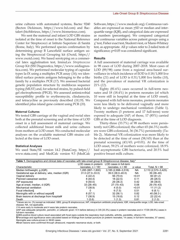
ResultsA full assessment of maternal carriage was available in 98 cases of LOD during 2007–2018. Most cases of LOD (89/98) came from a regional area-based sur-veillance in which incidence of EOD is 0.18/1,000 live births (21) and of LOD is 0.31/1,000 live births (10), and the prevalence of maternal VR colonization is 21% (22).
Eighty (81.6%) cases occurred in full-term neo-nates and 18 (18.4%) in preterm neonates (of which 10 were still in hospital at the time of LOD onset). Compared with full-term neonates, preterm neonates were less likely to be delivered vaginally and more likely to undergo mechanical ventilation (Table 1). Twenty mothers (3 preterm and 17 full-term) were exposed to adequate IAP; of those, 17 (85%) carried GBS at the time of LOD diagnosis.
Thirty-three (33.7%) of 98 mothers were persis-tently not GBS-colonized; the other 65 (66.3%) moth-ers were GBS-colonized, 36 (36.7%) persistently (Ta-ble 2).. Maternal VR colonization was more likely to be detected at the time of onset (58/65) than at the prenatal screening (43/65; p<0.01). At the time of LOD onset, 59.2% of mothers were colonized, 18.9% had asymptomatic GBS bacteriuria, and 20.5% had positive breast milk culture.
Emerging Infectious Diseases • www.cdc.gov/eid • Vol. 27, No. 9, September 2021 2281
Table 1. Demographics and clinical data of neonates with late-onset group B Streptococcus disease, Italy*
Characteristic LOD cases in preterm
neonates, n = 18† LOD cases in full-term
neonates, n = 80 p value Total, N = 98 Median birthweight, g (IQR) 1,285 (987–1,800) 3,185 (2,898–3,518) NA 3,110 (2,570–3,425) Gestational age at delivery, wks, median (IQR) 31.0 (27.0–33.0) 39.0 (38.0–40.0) NA 39 (38–40) Vaginal delivery 4 (22.2) 56 (70.0) <0.01 60 (61.2) Planned caesarean section 8 (44.4) 18 (22.5) 0.11 26 (26.5) IAP exposure‡ 9 (50.0) 29 (36.3) 0.42 38 (38.8) Age at onset, median, d (IQR) 33 (26–45) 27 (15–43) 0.08 29 (16–43) Mechanical ventilation 7 (38.9) 4 (5.0) <0.01 11 (11.2) Focal infection§ 0 6 (7.5) 0.51 6 (6.1) Meningitis with or without sepsis¶ 8 (57.1) 32 (56.1) 0.82 40 (56.3·) Brain lesions at discharge from hospital# 7 (38.9) 15 (18.8) 0.12 22 (22.4) Death 1 (5.6) 1 (1.3) 0.81 2 (1.0) *Values are no. (%) except as indicated. GBS, group B streptococcus; IAP, intrapartum antibiotic prophylaxis; IQR, interquartile range; LOD, late-onset disease; NA, not applicable. †14 were early to moderate and 4 were late preterm neonates. ‡IAP was adequate (ampicillin, penicillin, or cefazolin given >4 h before delivery) in 3/9 (33.3%) cases in preterm neonates and in 17/29 (58.6%) cases in full-term neonates. §GBS-positive blood culture result associated with focal signs outside the respiratory tract (cellulitis, arthritis, parotiditis, others) (10). ¶Percentage and significance were calculated based on findings from lumbar puncture (in preterm neonates, 14 cases; in full-term neonates, 57 cases). Meningitis was culture-proven in 36/40 cases. #Brain lesions were confirmed through ultrasound, magnetic resonance study, or both.
RESEARCH
All mothers with asymptomatic GBS bacteriuria also carried GBS at the VR site. Median urinary bacterial count was 40,000 CFU/mL (interquartile range [IQR] 10,000–100,000 CFU/mL; range 1,000–1 million CFU/mL). GBS bacteriuria was significantly more likely to be detected at the time of LOD onset (17/90 tested) rather than during pregnancy (2/92 tested; p<0.01).
Among 17 women with a positive breast milk cul-ture, 1 mother had mastitis (1 million CFUs/mL) and 16 had no mastitis (median bacterial count 300,000 CFU/mL; IQR 100,000–725,000 CFU/mL; range 9,000–6,400,000 CFU/mL). Fourteen (82.4%) of the 17 mothers were GBS colonized at the VR site at the pre-
natal screening or at the time of onset, or both, but the other 3 (17.6%) were persistently not colonized.
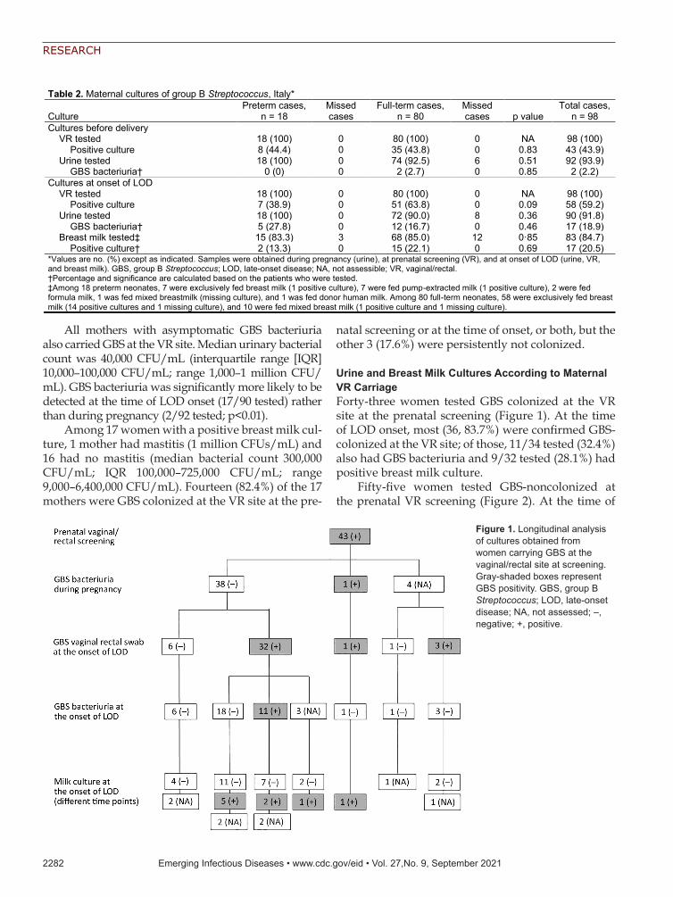
Urine and Breast Milk Cultures According to Maternal VR CarriageForty-three women tested GBS colonized at the VR site at the prenatal screening (Figure 1). At the time of LOD onset, most (36, 83.7%) were confirmed GBS-colonized at the VR site; of those, 11/34 tested (32.4%) also had GBS bacteriuria and 9/32 tested (28.1%) had positive breast milk culture.
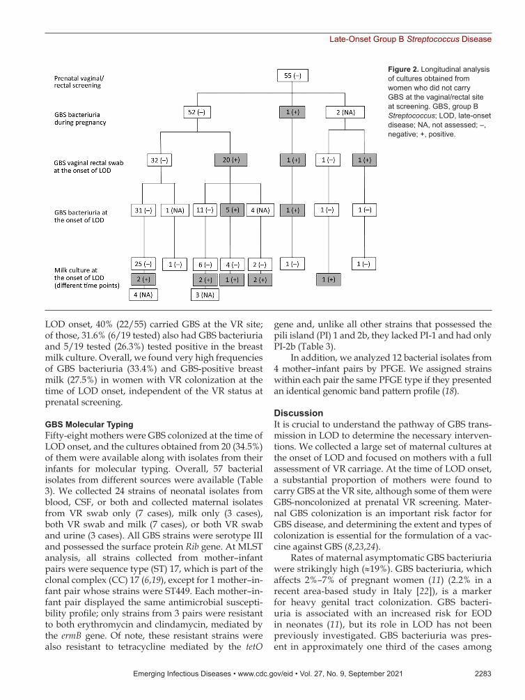
Fifty-five women tested GBS-noncolonized at the prenatal VR screening (Figure 2). At the time of
2282 Emerging Infectious Diseases • www.cdc.gov/eid • Vol. 27,No. 9, September 2021
Figure 1. Longitudinal analysis of cultures obtained from women carrying GBS at the vaginal/rectal site at screening. Gray-shaded boxes represent GBS positivity. GBS, group B Streptococcus; LOD, late-onset disease; NA, not assessed; –, negative; +, positive.
Table 2. Maternal cultures of group B Streptococcus, Italy*
Culture Preterm cases,
n = 18 Missed cases
Full-term cases, n = 80
Missed cases p value
Total cases, n = 98
Cultures before delivery VR tested 18 (100) 0 80 (100) 0 NA 98 (100) Positive culture 8 (44.4) 0 35 (43.8) 0 0.83 43 (43.9) Urine tested 18 (100) 0 74 (92.5) 6 0.51 92 (93.9) GBS bacteriuria† 0 (0) 0 2 (2.7) 0 0.85 2 (2.2) Cultures at onset of LOD VR tested 18 (100) 0 80 (100) 0 NA 98 (100) Positive culture 7 (38.9) 0 51 (63.8) 0 0.09 58 (59.2) Urine tested 18 (100) 0 72 (90.0) 8 0.36 90 (91.8) GBS bacteriuria† 5 (27.8) 0 12 (16.7) 0 0.46 17 (18.9) Breast milk tested‡ 15 (83.3) 3 68 (85.0) 12 0·85 83 (84.7) Positive culture† 2 (13.3) 0 15 (22.1) 0 0.69 17 (20.5) *Values are no. (%) except as indicated. Samples were obtained during pregnancy (urine), at prenatal screening (VR), and at onset of LOD (urine, VR, and breast milk). GBS, group B Streptococcus; LOD, late-onset disease; NA, not assessible; VR, vaginal/rectal. †Percentage and significance are calculated based on the patients who were tested. ‡Among 18 preterm neonates, 7 were exclusively fed breast milk (1 positive culture), 7 were fed pump-extracted milk (1 positive culture), 2 were fed formula milk, 1 was fed mixed breastmilk (missing culture), and 1 was fed donor human milk. Among 80 full-term neonates, 58 were exclusively fed breast milk (14 positive cultures and 1 missing culture), and 10 were fed mixed breast milk (1 positive culture and 1 missing culture).
Late-Onset Group B Streptococcus Disease
LOD onset, 40% (22/55) carried GBS at the VR site; of those, 31.6% (6/19 tested) also had GBS bacteriuria and 5/19 tested (26.3%) tested positive in the breast milk culture. Overall, we found very high frequencies of GBS bacteriuria (33.4%) and GBS-positive breast milk (27.5%) in women with VR colonization at the time of LOD onset, independent of the VR status at prenatal screening.
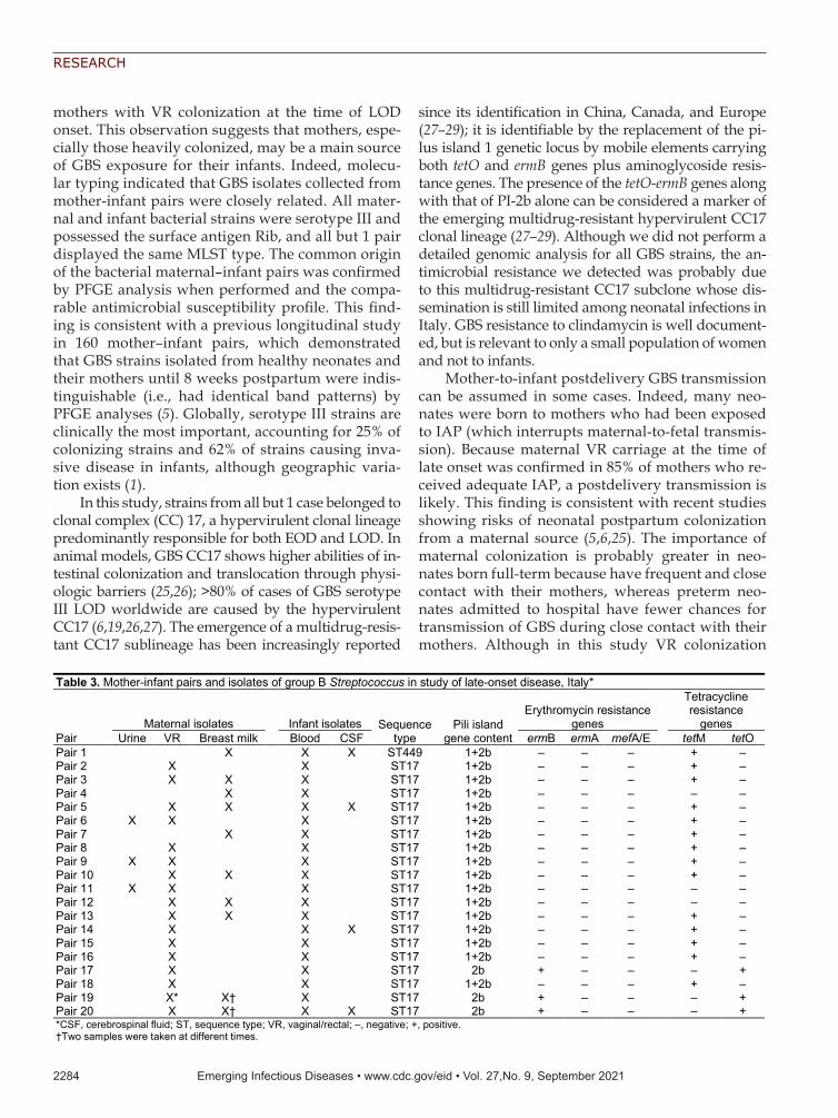
GBS Molecular TypingFifty-eight mothers were GBS colonized at the time of LOD onset, and the cultures obtained from 20 (34.5%) of them were available along with isolates from their infants for molecular typing. Overall, 57 bacterial isolates from different sources were available (Table 3). We collected 24 strains of neonatal isolates from blood, CSF, or both and collected maternal isolates from VR swab only (7 cases), milk only (3 cases), both VR swab and milk (7 cases), or both VR swab and urine (3 cases). All GBS strains were serotype III and possessed the surface protein Rib gene. At MLST analysis, all strains collected from mother–infant pairs were sequence type (ST) 17, which is part of the clonal complex (CC) 17 (6,19), except for 1 mother–in-fant pair whose strains were ST449. Each mother–in-fant pair displayed the same antimicrobial suscepti-bility profile; only strains from 3 pairs were resistant to both erythromycin and clindamycin, mediated by the ermB gene. Of note, these resistant strains were also resistant to tetracycline mediated by the tetO
gene and, unlike all other strains that possessed the pili island (PI) 1 and 2b, they lacked PI-1 and had only PI-2b (Table 3).
In addition, we analyzed 12 bacterial isolates from 4 mother–infant pairs by PFGE. We assigned strains within each pair the same PFGE type if they presented an identical genomic band pattern profile (18).
DiscussionIt is crucial to understand the pathway of GBS trans-mission in LOD to determine the necessary interven-tions. We collected a large set of maternal cultures at the onset of LOD and focused on mothers with a full assessment of VR carriage. At the time of LOD onset, a substantial proportion of mothers were found to carry GBS at the VR site, although some of them were GBS-noncolonized at prenatal VR screening. Mater-nal GBS colonization is an important risk factor for GBS disease, and determining the extent and types of colonization is essential for the formulation of a vac-cine against GBS (8,23,24).
Rates of maternal asymptomatic GBS bacteriuria were strikingly high (≈19%). GBS bacteriuria, which affects 2%–7% of pregnant women (11) (2.2% in a recent area-based study in Italy [22]), is a marker for heavy genital tract colonization. GBS bacteri-uria is associated with an increased risk for EOD in neonates (11), but its role in LOD has not been previously investigated. GBS bacteriuria was pres-ent in approximately one third of the cases among
Emerging Infectious Diseases • www.cdc.gov/eid • Vol. 27, No. 9, September 2021 2283
Figure 2. Longitudinal analysis of cultures obtained from women who did not carry GBS at the vaginal/rectal site at screening. GBS, group B Streptococcus; LOD, late-onset disease; NA, not assessed; –, negative; +, positive.
RESEARCH
mothers with VR colonization at the time of LOD onset. This observation suggests that mothers, espe-cially those heavily colonized, may be a main source of GBS exposure for their infants. Indeed, molecu-lar typing indicated that GBS isolates collected from mother-infant pairs were closely related. All mater-nal and infant bacterial strains were serotype III and possessed the surface antigen Rib, and all but 1 pair displayed the same MLST type. The common origin of the bacterial maternal–infant pairs was confirmed by PFGE analysis when performed and the compa-rable antimicrobial susceptibility profile. This find-ing is consistent with a previous longitudinal study in 160 mother–infant pairs, which demonstrated that GBS strains isolated from healthy neonates and their mothers until 8 weeks postpartum were indis-tinguishable (i.e., had identical band patterns) by PFGE analyses (5). Globally, serotype III strains are clinically the most important, accounting for 25% of colonizing strains and 62% of strains causing inva-sive disease in infants, although geographic varia-tion exists (1).
In this study, strains from all but 1 case belonged to clonal complex (CC) 17, a hypervirulent clonal lineage predominantly responsible for both EOD and LOD. In animal models, GBS CC17 shows higher abilities of in-testinal colonization and translocation through physi-ologic barriers (25,26); >80% of cases of GBS serotype III LOD worldwide are caused by the hypervirulent CC17 (6,19,26,27). The emergence of a multidrug-resis-tant CC17 sublineage has been increasingly reported
since its identification in China, Canada, and Europe (27–29); it is identifiable by the replacement of the pi-lus island 1 genetic locus by mobile elements carrying both tetO and ermB genes plus aminoglycoside resis-tance genes. The presence of the tetO-ermB genes along with that of PI-2b alone can be considered a marker of the emerging multidrug-resistant hypervirulent CC17 clonal lineage (27–29). Although we did not perform a detailed genomic analysis for all GBS strains, the an-timicrobial resistance we detected was probably due to this multidrug-resistant CC17 subclone whose dis-semination is still limited among neonatal infections in Italy. GBS resistance to clindamycin is well document-ed, but is relevant to only a small population of women and not to infants.
Mother-to-infant postdelivery GBS transmission can be assumed in some cases. Indeed, many neo-nates were born to mothers who had been exposed to IAP (which interrupts maternal-to-fetal transmis-sion). Because maternal VR carriage at the time of late onset was confirmed in 85% of mothers who re-ceived adequate IAP, a postdelivery transmission is likely. This finding is consistent with recent studies showing risks of neonatal postpartum colonization from a maternal source (5,6,25). The importance of maternal colonization is probably greater in neo-nates born full-term because have frequent and close contact with their mothers, whereas preterm neo-nates admitted to hospital have fewer chances for transmission of GBS during close contact with their mothers. Although in this study VR colonization
2284 Emerging Infectious Diseases • www.cdc.gov/eid • Vol. 27,No. 9, September 2021
Table 3. Mother-infant pairs and isolates of group B Streptococcus in study of late-onset disease, Italy*
Pair Maternal isolates
Infant isolates Sequence
type Pili island
gene content
Erythromycin resistance genes
Tetracycline resistance
genes Urine VR Breast milk Blood CSF ermB ermA mefA/E tetM tetO
Pair 1
X X X ST449 1+2b – – – + – Pair 2
X
X
ST17 1+2b – – – + –
Pair 3
X X X
ST17 1+2b – – – + – Pair 4
X X
ST17 1+2b – – – – –
Pair 5
X X X X ST17 1+2b – – – + – Pair 6 X X
X
ST17 1+2b – – – + –
Pair 7
X X
ST17 1+2b – – – + – Pair 8
X
X
ST17 1+2b – – – + –
Pair 9 X X
X
ST17 1+2b – – – + – Pair 10
X X X
ST17 1+2b – – – + –
Pair 11 X X
X
ST17 1+2b – – – – – Pair 12
X X X
ST17 1+2b – – – – –
Pair 13
X X X
ST17 1+2b – – – + – Pair 14
X
X X ST17 1+2b – – – + –
Pair 15
X
X
ST17 1+2b – – – + – Pair 16
X
X
ST17 1+2b – – – + –
Pair 17
X
X
ST17 2b + – – – + Pair 18
X
X
ST17 1+2b – – – + –
Pair 19
X* X† X
ST17 2b + – – – + Pair 20
X X† X X ST17 2b + – – – +
*CSF, cerebrospinal fluid; ST, sequence type; VR, vaginal/rectal; –, negative; +, positive. †Two samples were taken at different times.
Late-Onset Group B Streptococcus Disease
rates at the time of LOD onset were higher in full-term mothers (64% vs. 39% in preterm mothers), the difference was not significant, perhaps because of the small sample size.
In this study, we found GBS in ≈20% of breast-feeding mothers. Mastitis in LOD was infrequent; mothers with positive breast milk culture were more often asymptomatic, although their milk bac-terial counts were sometimes high. This finding suggests a silent maternal duct colonization, and it is consistent with case reports of GBS breast milk–associated LOD, in which most mothers have no sign of mastitis (13).
Furthermore, in our study most mothers with positive breast milk culture carried GBS at the VR site, which was often heavily colonized. Maternal VR car-riage would appear to be associated with GBS trans-mission into breast milk, perhaps in some cases after translocation from the gastrointestinal tract through the lymphatic system to the mammary glands (30). In contrast, only 3 mothers who had positive breast milk culture were persistently GBS-noncolonized at the VR site. In such cases, a circular mechanism of GBS transmission to neonates could be implicated. The retrograde theory assumes that GBS, present in the infant’s throat, colonizes the mammary ducts during breast-feeding; GBS load increases in the milk, and, in turn, the infant is infected during breast-feeding. Our data do not suggest that breast milk itself is a risk factor for LOD. Breast milk is known to contain im-munomodulatory and antimicrobial components (12) (i.e., sIgA and cytokines) that may protect from LOD, and the lack of these components seems to increase the risk of persistent neonatal colonization (31) and LOD (32).
Taken all together, these results show that moth-ers are largely the predominant source of GBS in cas-es of LOD, both during childbirth (especially if IAP is not given) and in the postpartum period. GBS-pos-itive breast milk is one of the ways by which heavily colonized mothers expose their infants to GBS.
The first limitation of our investigation is that it was an observational study without a control group. Therefore, the relevance of a positive breast milk in LOD could not be clearly assessed, because we do not know how many breastfeeding GBS-colonized mothers with healthy neonates have GBS-positive breast milk. However, Berardi et al. (5) found much lower rates (≈7%) of GBS-positive breast milk in a cohort of breastfeeding mothers (GBS-colonized at the VR site) who had healthy neonates. In addi-tion, we cannot rule out an accidental contamina-tion of some breast milk samples during collection,
although we had provided instructions for collec-tion. Second, although we proposed doing so, we did not systematically perform full assessment of VR culture both at prenatal screening and at the time of disease onset; just over half of the mothers had the full assessment during surveillance. In fact, not all of them had prenatal screening; furthermore, the collection of cultures at the time of LOD diag-nosis and then shipping the maternal strains were challenging to organize. Third, the PFGE analysis was available only in a few mother-infant pairs. However, previous studies demonstrate that the concordance of GBS strains collected from mothers and their own neonates at delivery or in the fol-lowing weeks is very high, reaching ≈100% of cases (5,6,18). Finally, maternal colonization rates could be higher than we found as a result of inherent in-sufficient sensitivity of maternal VR cultures (11), which may lead to false-negative culture results. We did not investigate the possible role of the fa-ther in the transmission of GBS.
In conclusion, this study suggests that most cases of LOD are strictly associated with maternal VR colonization and that CC17 is the predominant clonal lineage. Rates of asymptomatic GBS bacteri-uria at the time of LOD onset are strikingly high, and this finding suggests heavy maternal coloniza-tion. Positive breast milk culture is relatively com-mon among asymptomatic breastfeeding mothers of neonates with LOD, especially if they carry GBS at the VR site. However, the causal role of breast-feeding remains uncertain, and our data do not lead to definitive conclusions. Mother-to-infant trans-mission may occur after delivery. Our findings call attention to maternal transmission after delivery as an underestimated source of neonatal LOD and may assist in predicting the impact of maternal GBS vaccination.
AcknowledgmentsThe authors thank Monica Imperi for contributing to the molecular typing experiments.
This work was partially supported by the Agenzia Sanitaria Regionale (Emilia-Romagna) Piano Regionale della Prevenzione (2015–2018) C.U.P. n. E43G17000680002.
About the Author Dr. Berardi is the head of the Unità Operativa di Terapia Intensiva Neonatale, Azienda Ospedaliero—Universitaria Policlinico di Modena, Italy. His primary research interests are group B Streptococcus, early- and late-onset sepsis, and neonatal infections.
Emerging Infectious Diseases • www.cdc.gov/eid • Vol. 27, No. 9, September 2021 2285
RESEARCH
References 1. Madrid L, Seale AC, Kohli-Lynch M, Edmond KM,
Lawn JE, Heath PT, et al.; Infant GBS Disease Investigator Group. Infant group B streptococcal disease incidence and serotypes worldwide: systematic review and meta-analyses. Clin Infect Dis. 2017;65(suppl_2):S160–72. https://doi.org/ 10.1093/cid/cix656
2. Puopolo KM, Lynfield R, Cummings JJ; Committee on Fetus and Newborn; Committee on Infectious Diseases. Management of infants at risk for group B streptococcal disease. Pediatrics. 2019;144:e20191881. https://doi.org/ 10.1542/peds.2019-1881
3. Nanduri SA, Petit S, Smelser C, Apostol M, Alden NB, Harrison LH, et al. Epidemiology of invasive early-onset and late-onset group B streptococcal disease in the United States, 2006 to 2015: multistate laboratory and population-based surveillance. JAMA Pediatr. 2019;173:224–33. https://doi.org/10.1001/jamapediatrics.2018.4826
4. Seale AC, Bianchi-Jassir F, Russell NJ, Kohli-Lynch M, Tann CJ, Hall J, et al. Estimates of the burden of group B streptococcal disease worldwide for pregnant women, stillbirths, and children. Clin Infect Dis. 2017;65:suppl_ 2:S200–S219. https://doi.org/10.1093/cid/cix664
5. Berardi A, Rossi C, Creti R, China M, Gherardi G, Venturelli C, et al. Group B streptococcal colonization in 160 mother-baby pairs: a prospective cohort study. J Pediatr. 2013;163:1099–104.e1. https://doi.org/10.1016/ j.jpeds.2013.05.064
6. Tazi A, Plainvert C, Anselem O, Ballon M, Marcou V, Seco A, et al. Risk factors for infant colonization by hypervirulent CC17 group B Streptococcus: toward the understanding of late-onset disease. Clin Infect Dis. 2019;69:1740–8. https://doi.org/10.1093/cid/ciz033
7. Manning SD, Lewis MA, Springman AC, Lehotzky E, Whittam TS, Davies HD. Genotypic diversity and serotype distribution of group B Streptococcus isolated from women before and after delivery. Clin Infect Dis. 2008;46:1829–37. https://doi.org/10.1086/588296
8. Lin FY, Weisman LE, Troendle J, Adams K. Prematurity is the major risk factor for late-onset group B Streptococcus disease. J Infect Dis. 2003;188:267–71. https://doi.org/ 10.1086/376457
9. Joubrel C, Tazi A, Six A, Dmytruk N, Touak G, Bidet P, et al. Group B Streptococcus neonatal invasive infections, France 2007–2012. Clin Microbiol Infect. 2015;21:910–6. https://doi.org/10.1016/j.cmi.2015.05.039
10. Berardi A, Rossi C, Lugli L, Creti R, Bacchi Reggiani ML, Lanari M, et al.; GBS Prevention Working Group, Emilia-Romagna. Group B Streptococcus late-onset disease: 2003–2010. Pediatrics. 2013;131:e361–8. https://doi.org/10.1542/peds.2012-1231
11. Verani JR, McGee L, Schrag SJ; Division of Bacterial Diseases, National Center for Immunization and Respiratory Diseases, Centers for Disease Control and Prevention (CDC). Prevention of perinatal group B streptococcal disease— revised guidelines from CDC, 2010. MMWR Recomm Rep. 2010;59(RR-10):1–36.
12. Le Doare K, Kampmann B. Breast milk and group B streptococcal infection: vector of transmission or vehicle for protection? Vaccine. 2014;32:3128–32. https://doi.org/ 10.1016/j.vaccine.2014.04.020
13. Filleron A, Lombard F, Jacquot A, Jumas-Bilak E, Rodière M, Cambonie G, et al. Group B streptococci in milk and late neonatal infections: an analysis of cases in the literature. Arch Dis Child Fetal Neonatal Ed. 2014;99:F41–7. https://doi.org/10.1136/archdischild-2013-304362
14. Zimmermann P, Gwee A, Curtis N. The controversial role of breast milk in GBS late-onset disease. J Infect. 2017;74(Suppl 1):S34–40. https://doi.org/10.1016/ S0163-4453(17)30189-5
15. Le Doare K, Kampmann B, Vekemans J, Heath PT, Goldblatt D, Nahm MH, et al. Serocorrelates of protection against infant group B Streptococcus disease. Lancet Infect Dis. 2019;19:e162–71. https://doi.org/10.1016/ S1473-3099(18)30659-5
16. Imperi M, Pataracchia M, Alfarone G, Baldassarri L, Orefici G, Creti R. A multiplex PCR assay for the direct identification of the capsular type (Ia to IX) of Streptococcus agalactiae. J Microbiol Methods. 2010;80:212–4. https://doi.org/10.1016/j.mimet.2009.11.010
17. Creti R, Fabretti F, Orefici G, von Hunolstein C. Multiplex PCR assay for direct identification of group B streptococcal alpha-protein-like protein genes. J Clin Microbiol. 2004;42:1326–9. https://doi.org/10.1128/JCM.42.3.1326-1329.2004
18. Imperi M, Gherardi G, Berardi A, Baldassarri L, Pataracchia M, Dicuonzo G, et al. Invasive neonatal GBS infections from an area-based surveillance study in Italy. Clin Microbiol Infect. 2011;17:1834–9. https://doi.org/ 10.1111/j.1469-0691.2011.03479.x
19. Creti R, Imperi M, Berardi A, Pataracchia M, Recchia S, Alfarone G, et al.; Italian Neonatal GBS Infections Working Group. Neonatal group B Streptococcus infections. Pediatr Infect Dis J. 2017;36:256–62. https://doi.org/10.1097/INF.0000000000001414
20. Springman AC, Lacher DW, Waymire EA, Wengert SL, Singh P, Zadoks RN, et al. Pilus distribution among lineages of group B Streptococcus: an evolutionary and clinical perspective. BMC Microbiol. 2014;14:159. https://doi.org/10.1186/1471-2180-14-159
21. Berardi A, Baroni L, Bacchi Reggiani ML, Ambretti S, Biasucci G, Bolognesi S, et al.; GBS Prevention Working Group Emilia-Romagna. The burden of early-onset sepsis in Emilia-Romagna (Italy): a 4-year, population-based study. J Matern Fetal Neonatal Med. 2016;29:3126–31. https://doi.org/10.3109/14767058.2015.1114093
22. Berardi A, Rossi C, Bacchi Reggiani ML, Bastelli A, Capretti MG, Chiossi C, et al. An area-based study on intrapartum antibiotic prophylaxis for preventing group B streptococcus early-onset disease: advances and limitations. J Matern Fetal Neonatal Med. 2017;30:1739–44. https://doi.org/10.1080/14767058.2016.1224832
23. Khatami A, Randis TM, Tavares L, Gegick M, Suzman E, Ratner AJ. Vaginal co-colonization with multiple group B Streptococcus serotypes. Vaccine. 2019;37:409–11. https://doi.org/10.1016/j.vaccine.2018.12.001
24. Carreras-Abad C, Cochet M, Hall T, Ramkhelawon L, Khalil A, Peregrine E, et al. Developing a serocorrelate of protection against invasive group B streptococcus disease in pregnant women: a feasibility study. Health Technol Assess. 2019;23:1–40. https://doi.org/10.3310/hta23670
25. Ratner AJ. Enhanced postnatal acquisition of hypervirulent group B Streptococcus. Clin Infect Dis. 2019;69:1749–51. https://doi.org/10.1093/cid/ciz035
26. Tazi A, Disson O, Bellais S, Bouaboud A, Dmytruk N, Dramsi S, et al. The surface protein HvgA mediates group B Streptococcus hypervirulence and meningeal tropism in neonates. J Exp Med. 2010;207:2313–22. https://doi.org/ 10.1084/jem.20092594
27. Campisi E, Rosini R, Ji W, Guidotti S, Rojas-López M, Geng G, et al. Genomic analysis reveals multi-drug resistance clusters in group B Streptococcus CC17 hypervirulent isolates
2286 Emerging Infectious Diseases • www.cdc.gov/eid • Vol. 27,No. 9, September 2021
Late-Onset Group B Streptococcus Disease
causing neonatal invasive disease in southern mainland China. Front Microbiol. 2016;7:1265. https://doi.org/ 10.3389/fmicb.2016.01265
28. Martins ER, Pedroso-Roussado C, Melo-Cristino J, Ramirez M; Portuguese Group for the Study of Strepto-coccal Infections. Streptococcus agalactiae causing neonatal infections in Portugal (2005–2015): diversification and emergence of a CC17/PI-2b multidrug resistant sublineage. Front Microbiol. 2017;8:499. https://doi.org/10.3389/fmicb.2017.00499
29. Teatero S, Ramoutar E, McGeer A, Li A, Melano RG, Wasserscheid J, et al. Clonal complex 17 group B Streptococcus strains causing invasive disease in neonates and adults originate from the same genetic pool. Sci Rep. 2016;6:20047. https://doi.org/10.1038/srep20047
30. Perez PF, Doré J, Leclerc M, Levenez F, Benyacoub J, Serrant P, et al. Bacterial imprinting of the neonatal
immune system: lessons from maternal cells? Pediatrics. 2007;119:e724–32. https://doi.org/10.1542/peds.2006-1649
31. Le Doare K, Bellis K, Faal A, Birt J, Munblit D, Humphries H, et al. SIgA, TGF-β1, IL-10, and TNFα in colostrum are associated with infant group B Streptococcus colonization. Front Immunol. 2017;8:1269–79. https://doi.org/10.3389/fimmu.2017.01269
32. Dangor Z, Khan M, Kwatra G, Izu A, Nakwa F, Ramdin T, et al. The association between breast milk group B streptococcal capsular antibody levels and late-onset disease in young infants. Clin Infect Dis. 2020;70:1110–4.
Address for correspondence: Alberto Berardi, Unità Operativa di Terapia Intensiva Neonatale, Azienda Ospedaliero-Universitaria Policlinico di Modena, Via del Pozzo, 71-41124 Modena, Italy; email: [email protected]
Emerging Infectious Diseases • www.cdc.gov/eid • Vol. 27, No. 9, September 2021 2287