Materials Research Lab - Research Internships in Science ... · PDF fileBiomineralization of...
31
Materials Research Lab - Research Internships in Science and Engineering http://www.mrl.ucsb.edu/mrl/outreach/educational/RISE/interns01.html[5/10/12 10:02:52 AM] Education Undergraduate Opportunities K-12 Science Activities For Teachers Education Contacts News Research Internships in Science and Engineering Research Interns in Science and Engineering (RISE) Summer 2005 - Student Projects Student Major/School Mentor Faculty Sponsor Department Student Project Edgar Algarin Chemistry UCSB Brian Thibeault Evelyn Hu Electrical & computer engineeringCE Nano-imprint lithography of nanostructures David Ando Physics UCSB Rob Knobel Andrew Cleland Physics Nanolithography nanomatching nanoelectronics Michael Bjorndal Physics Cornell --- Deborah Fygenson Physics DNA melthing and hairpin formation in the presence of intercalating dyes Krista Ehrenclou Chemistry UCSB Cara Evans Joseph Zasadzinski Chemical Engineering Encapsulation of Taxol for use in vesicle based drug delivery systems Edouard Fonck Chemical Engineering UCSB Ray Tu Matt Tirrell Chemical Engineering Specific drug delivery Wesley Francillon Engineering SUNY Stony Brook Rafael Leckie Carlos Levi Materials Study of new thermal barrier coating materials by synthesis of rare earth Oxide-ZrO 2 -Al 2 O 3 Heather Hoff Materials Engineering Calpoly, SLO Alex Small David Pine Chemical Engineering Processing of macroporous materials using ordered polystyrene or silica spheres as templates Kathleen Kolstad Neuroscience Bates College Michelle Staples Lincoln Johnson Neuroscience Role of Matrix metalleproteinases(MMP) and their inhibitors (TIMP) in age related macular degeneration Information and Safety Research Facilities Education People News & Events Webmail
-
Upload
hoangkhanh -
Category
Documents
-
view
217 -
download
0
Transcript of Materials Research Lab - Research Internships in Science ... · PDF fileBiomineralization of...

Materials Research Lab - Research Internships in Science and Engineering
http://www.mrl.ucsb.edu/mrl/outreach/educational/RISE/interns01.html[5/10/12 10:02:52 AM]
Education
UndergraduateOpportunities
K-12 Science Activities
For Teachers
Education Contacts
News
Research Internships in Science and Engineering
Research Interns in Science and Engineering (RISE)Summer 2005 - Student Projects
Student Major/School Mentor Faculty
Sponsor Department Student Project
EdgarAlgarinChemistryUCSB
BrianThibeault
Evelyn HuElectrical &computerengineeringCE
Nano-imprint lithography ofnanostructures
David AndoPhysicsUCSB
RobKnobel
AndrewCleland
PhysicsNanolithography nanomatchingnanoelectronics
MichaelBjorndalPhysicsCornell
---DeborahFygenson
PhysicsDNA melthing and hairpinformation in the presence ofintercalating dyes
KristaEhrenclouChemistryUCSB
CaraEvans
JosephZasadzinski
ChemicalEngineering
Encapsulation of Taxol for usein vesicle based drug deliverysystems
EdouardFonckChemicalEngineeringUCSB
Ray Tu Matt TirrellChemicalEngineering
Specific drug delivery
WesleyFrancillonEngineeringSUNY StonyBrook
RafaelLeckie
Carlos Levi MaterialsStudy of new thermal barriercoating materials by synthesisof rare earth Oxide-ZrO2-Al2O3
HeatherHoffMaterialsEngineeringCalpoly, SLO
AlexSmall
David PineChemicalEngineering
Processing of macroporousmaterials using orderedpolystyrene or silica spheresas templates
KathleenKolstadNeuroscienceBatesCollege
MichelleStaples
LincolnJohnson
Neuroscience
Role of Matrixmetalleproteinases(MMP) andtheir inhibitors (TIMP) in agerelated macular degeneration
Information andSafety
Research Facilities Education People News & Events Webmail

Materials Research Lab - Research Internships in Science and Engineering
http://www.mrl.ucsb.edu/mrl/outreach/educational/RISE/interns01.html[5/10/12 10:02:52 AM]
Penny LettsBiologyQueen'sUniversity
AhmetTezel
SamirMitragotri
ChemicalEngineering
Sonophoresis of skin withfluorescent polystyrenemicrospheres
XerxesLopez-YglesiasPhysicsUCSB
TomKingMattDoty
MarkSherwin
PhysicsAntennae creating fortertahertz radiation sensors
JaclynMartinezBiochemistryUCSB
LukeSherlin
JohnPerona
ChemistryPurification and crystallizationof GLN-tRNAGln proteinCOMPLEXES
WorawatMeevasanaPhysicsUCSB
---GuenterAhlers
PhysicsThe ring patterns in theconvection of isoproponal
AnthonyMorfaBiochemistryUCSB
LarkenEuliss
GalenStucky
ChemistryBiomineralization of CalciumCarbonate
Sachin PatilPhysicsUCSB
MattDoty
MarkSherwin
PhysicsTerahertz tecnologies:radiation detectors for the farinfra red
SaraPetrellaMath&PhysicsSan DiegoStateUniversity
DavidAndeen
Fred Lange Materials X-ray analysis on Zinc Oxide
AshleySensChemistrySantaBarbara CityCollege
AhmetTezel
SamirMitragotri
Electrical &ChemicalEngineering
Low-frequency sonophoretictransdermal transport ofhydrophilic permeants:determination of pore sizewithin porcine skin
AlysonWhitneyChemistryUCSB
KhalidHanif
GeoffreyStrouse
ChemistryDoping CdSe quantum dotswith Vanadium(II)
AaronWilliamsElectricalEngineeringUCSB
WenhuaZhang
Kim TurnerMechanicalEngineering
Porous silicon formation withapplications to biosensingmicroelectomechanical systems

Materials Research Lab - Research Internships in Science and Engineering
http://www.mrl.ucsb.edu/mrl/outreach/educational/RISE/interns01.html[5/10/12 10:02:52 AM]
Return to the RISE homepage
Site Map // Webmail // Site Privacy Notification Guidelines // National Science Foundation // UCSB

Materials Research Lab - Edgar's Project Page
http://www.mrl.ucsb.edu/mrl/outreach/educational/RISE/interns01/EdgarA.html[5/10/12 10:02:59 AM]
Education
UndergraduateOpportunities
K-12 Science Activities
For Teachers
Education Contacts
News
Edgar's Project Page
Intern: Edgar Algarín Mentor: Dr. Brian Thibeault Faculty Supervisor: Professor Evelyn L. Hu Department: Electrical Engineering
NANO-IMPRINT LITHOGARPHY OF NANOSTRUCTURES
Nano-imprint lithography is a useful and cheap method to replicate nanostructures and showspotential to replicate structures under the 100 nm scale. Current processes such as e-beamlithography and UV-lithography can be expensive, time consuming, and difficult to reproduceunder the 100 nm scale. With imprint lithography, a large number of replicas can be madefrom one exposed master, insuring research and production throughput.
Nano-imprinting is the molding of a polymer from a prefabricated master. The master can beproduced using e-beam or UV lithography to define nm-scale patterns. A durable non-sticklayer that can withstand high pressure and high temperature is then applied to the master.This coating is usually composed of hydrocarbon monomers and is important in order toprevent breakage and pull-off of the polymer. In this work the Si master is patterned by UVlithography and reactive ion etching and is coated with a self-assembled layer ofoctadecyltrichlorosaline for the non-stick layer. Once the master is produced (figure A), it isimprinted onto a host substrate bearing a polymer. The polymer used in this work is poly(methyl methacrylate) or PMMA. Depending on the temperature, pressure, and time, thepolymer will flow and fill in the intended structures on the master. After the polymer flow isdone, the sample is cooled and then the host is separated from the master to reveal a copy(figure B). In this work, key parameters for process the Si master and PMMA based imprintwere determined.
Return to the RISE project list
Site Map // Webmail // Site Privacy Notification Guidelines // National Science Foundation // UCSB
Information andSafety
Research Facilities Education People News & Events Webmail

Materials Research Lab - David's Project Page
http://www.mrl.ucsb.edu/mrl/outreach/educational/RISE/interns01/DavidA.html[5/10/12 10:03:06 AM]
Education
UndergraduateOpportunities
K-12 Science Activities
For Teachers
Education Contacts
News
David's Project Page
Intern: David AndoMentor: Rob KnobelFaculty Supervisor: Professor Andrew ClelandDepartment: Physics
Nanolithography Nanomachining Nanoelectronics
My goal this summer was to detect the vibrations of a freestanding nanometer sized rod, whichis made out of silicon. The freestanding nanometer sized rods are made using advancednanolithography nanomachining techniques in the University of California at Santa Barbaraclean room. My approach to the problem of detecting the nano motion was to take an oldprototype atomic force microscope and modify it to measure the motion of a nanometer sizedrod.
Detecting this nano motion allows access to new experimental regimes at the nanoscale. Thisnano size brings out new physical properties as the size approaches the length scales set byquantum mechanics. These new physical properties are under intense study by physicists. Theimmediate benefits of detecting this motion are the ability to characterize the motion ofnanoelectromechanical systems built by the Cleland research group. Characterization involveschecking that the nanoelectromechanical systems work, what quality factor they have, andwhat frequency they resonate at.
Although I have not completed my goal of detecting the nano motion, I have completed mymodified atomic force microscope. The modifications include a photodiode that can detectreflections of the atomic force microscope laser off the nanomachines at a frequency of 500million cycles per second. My other major modification was to remove the atomic forcemicroscope head cantilever and to replace it with a sample holder, which is connected toinstrumentation. The sample holder holds the nanomachines and is designed to be magneticallypositioned in place to allow for greater degrees of freedom in positioning the samples and sothat many nanomachines, which have to be on the same sample, can be looked atsimultaneously.
This image shows my modified atomic force microscope on the left with associated electronicson the right. At the top of my modified atomic force microscope is the laser and on the bottomis my magnetic sample holder.
Information andSafety
Research Facilities Education People News & Events Webmail

Materials Research Lab - David's Project Page
http://www.mrl.ucsb.edu/mrl/outreach/educational/RISE/interns01/DavidA.html[5/10/12 10:03:06 AM]
Return to the RISE project list
Site Map // Webmail // Site Privacy Notification Guidelines // National Science Foundation // UCSB

Materials Research Lab - Michael's Project Page
http://www.mrl.ucsb.edu/mrl/outreach/educational/RISE/interns01/MichaelB.html[5/10/12 10:03:13 AM]
Education
UndergraduateOpportunities
K-12 Science Activities
For Teachers
Education Contacts
News
Michael's Project Page
Intern: Michael BjorndalMentor/Faculty Supervisor: Professor Deborah FygensonDepartment: Physics
DNA MELTING AND HAIRPIN FORMATION IN THE PRESENCE OF INTERCALATING DYES
The effect of intercalating dyes on the melting transition of DNA was seen through the use of anew gel electrophoresis assay. The technique studies the duplex to hairpin transition usingperfect palindromic oligomers. After raising the temperature of the annealed duplexes and thenquenching quickly in a cold water bath, percentages of duplexes and folded hairpins can beseen. One interpretation states that the hairpins represent oligomers that were completelymelted at the temperature of interest and then folded quickly during quenching. By comparisonto UV-visual absorption melting curves, it is possible to determine the order of the transition.Work is being done to determine if the formation of hairpins has another interpretation: theDNA is not completely melted but can stay in the hairpin state favorably at certaintemperatures. Various concentrations of the dyes YoYo and YoPro were used to see their effecton the DNA. The dyes clearly raise the overall melting temperature, with YoYo having aboutthe same effect as twice the concentration of YoPro. This has been of use for people usingthese dyes to watch DNA at and around its melting temperature. One other chemicalcommonly used with DNA, Betamercaptoethanol (BME), obscures the UV-visual absorptioncurves. It is the hope that this new assay will also give a clear estimate of the effect BME hason DNA melting.
Information andSafety
Research Facilities Education People News & Events Webmail

Materials Research Lab - Michael's Project Page
http://www.mrl.ucsb.edu/mrl/outreach/educational/RISE/interns01/MichaelB.html[5/10/12 10:03:13 AM]
Return to the RISE project list
Site Map // Webmail // Site Privacy Notification Guidelines // National Science Foundation // UCSB

Materials Research Lab - Krista's Project Page
http://www.mrl.ucsb.edu/mrl/outreach/educational/RISE/interns01/KristaE.html[5/10/12 10:03:20 AM]
Education
UndergraduateOpportunities
K-12 Science Activities
For Teachers
Education Contacts
News
Krista's Project Page
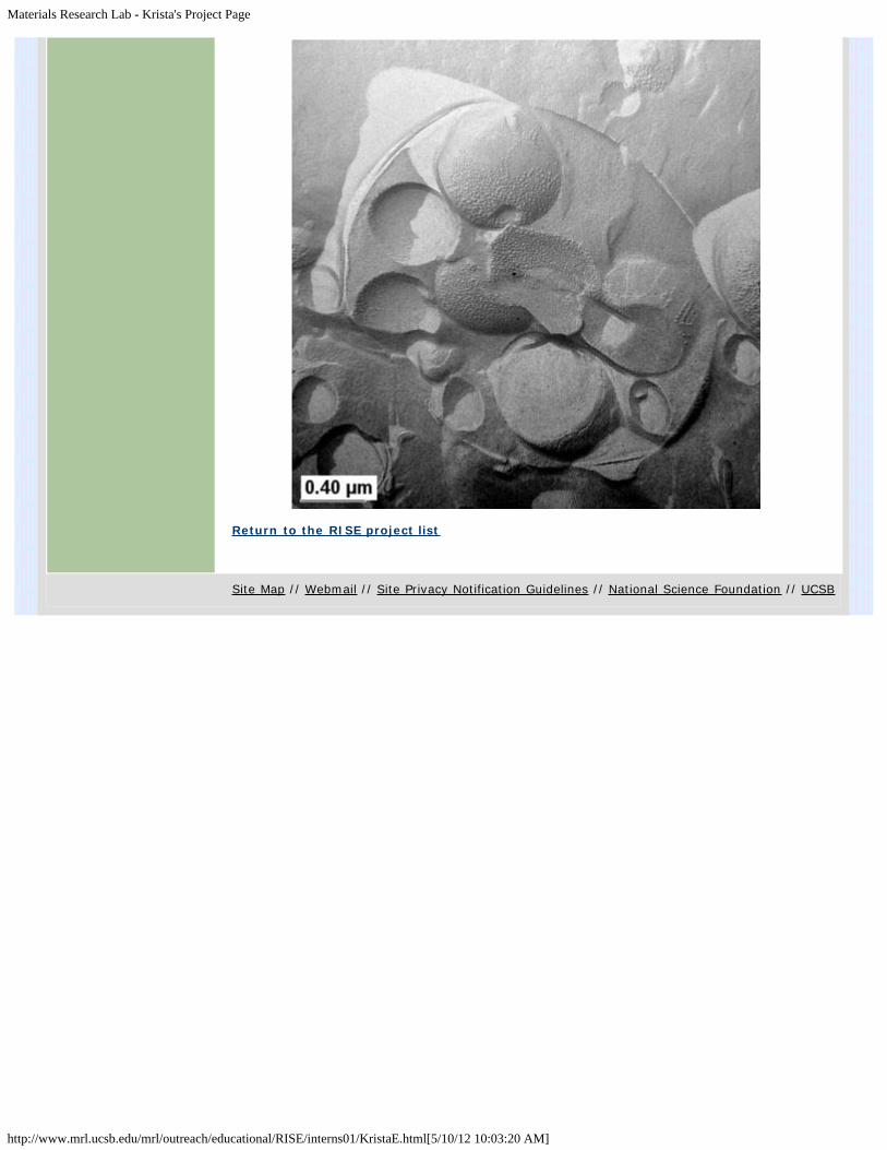
Intern: Krista EhrenclouMentor: Dr. Cara EvansFaculty Supervisor: Professor Joe ZasadzinskiDepartment: Chemical Engineering
ENCAPSULATION OF TAXOL FOR USE IN VESICLE BASED DRUG DELIVERY SYSTEMS
Background: Taxol is an important drug used in the treatment of breast, ovarian, lung, headand neck cancers. However, the drug?s hydrophobic nature has made this drug very difficult toadminister. Currently, Taxol is delivered intravenously in a toxic mixture ofCremophore/Ethanol. In addition to harmful side effects, this method of drug delivery has poordrug circulation time and allows uncontrolled circulation of the drug throughout the body.
Encapsulation of Taxol in a phospholipid bilayer vesicle has the potential to greatly improve thecurrent methods of drug administration. The bilayer membrane would eliminate the use of theCremophore/Ethanol cocktail, provide a protective barrier between the drug and the bodyincreasing drug circulation time and decreasing drug interaction with healthy cells, and providea vehicle capable of cell targeting and controlled release.
Methods: A solution of Taxol in 100% ethanol was added to L-alpha-Dipalmitoylphosphatidylcholine (DPPC) then dialyzed and heated past the melting temperature of DPPC.Freeze fracture replicas of the dialyzed product were imaged by a transmission electronmicroscope for evidence of vesicle formation. Chromatography(SEC), fluorescence and UV/VISspectroscopy were used to isolate and determine the location of free taxol, encapsulated taxoland encapsulation efficiency.
Results: Freeze fracture images revealed vesicle formation in the presence of Taxol. A rippledphase was observed in vesicles formed from the solution with Taxol. The rippled phase isevidence of a change in lipid phase behavior, possibly due to an interaction with Taxol. Vesicleswere isolated from the solution using size-exclusion chromatography (SEC) and identified usingfluorescence spectroscopy. UV/VIS spectroscopic data is currently being collected. UV/VIS willbe performed on fractions collected by SEC to determine the amount of free taxol. Vesiclecontaining fractions will then be treated with detergent to liberate any encapsulated taxol andwill be measured by UV/VIS.
Future Projects: Future projects will include: the completion of the UV/VIS portion of theexperiment, improvement of separation techniques to collect free Taxol, and the refinement ofthe dialysis method to optimize Taxol encapsulation.
Information andSafety
Research Facilities Education People News & Events Webmail
Materials Research Lab - Krista's Project Page
http://www.mrl.ucsb.edu/mrl/outreach/educational/RISE/interns01/KristaE.html[5/10/12 10:03:20 AM]
Return to the RISE project list
Site Map // Webmail // Site Privacy Notification Guidelines // National Science Foundation // UCSB

Materials Research Lab - Edouard's Project Page
http://www.mrl.ucsb.edu/mrl/outreach/educational/RISE/interns01/EdouardF.html[5/10/12 10:03:28 AM]
Education
UndergraduateOpportunities
K-12 Science Activities
For Teachers
Education Contacts
News
Edouard's Project Page
Intern: Edouard FonckMentor: Raymond TuFaculty Supervisor: Professor Matthew TirrellDepartment: Chemical Engineering
SPECIFIC DRUG DELIVERY
In recent studies, peptides amphiphiles have been shown to be a potential tool for specificdrug delivery. Our peptide amphiphile is composed of a hydrophobic tail and a peptide headgroup. The tail is the hydrophobic part made of two saturated alkyl chains, which promotesinclusion into the lipid membrane of the liposomes. The head group is attached to the tails witha glutanic acid linker and a C2 spacer. The head is derived from a region of type IV collagenthat is known to bind to melanoma.
The ability of the peptide amphiphile to specifically bind cells along with their amphiphiliccharacter make them a very good candidate for drug delivery in liposomes (see fig. 1).Liposomes, which are characterized by their spherical geometry and their lipid bi-layers, areable to encapsulate hydrophilic as well as lipophilic molecules. In order to develop a bettersynthetic peptide amphiphile targeted vesicle, we propose to study the effectiveness of variouscompositions on vesicles formation. The success of vesicle formation depends on distinctfactors such as concentration of DMPC, DMPE, PEG and concentrations of peptide amphiphile,temperature and time.
Our goal is to obtain the specific data to optimize the best possible self-assembled structure. Astructure that will not allow the liposomes to aggregate to one to another and that will targetthe specific cell. We will be able to determine those data by conducting experiments at variousmixtures of lipids, molecular weight of PEG and molecular weights of peptide amphiphile. Inorder to determine if the data obtained are satisfactory (formation of liposome and noaggregation), we will run dynamic light scattering to measure the hydrodynamic diameter ofthe liposomes formed. Then, we will load the liposome with Texas Red DHPE;concentration<0.1%, a fluorescent dye-lipid, to help show the outcome of the liposomescomplexed with mouse fibroblast cells.
Information andSafety
Research Facilities Education People News & Events Webmail

Materials Research Lab - Edouard's Project Page
http://www.mrl.ucsb.edu/mrl/outreach/educational/RISE/interns01/EdouardF.html[5/10/12 10:03:28 AM]
Fig. 1: pictures a & c DIL images of the fibroblast cells (those express similar integrins as ofmelanoma cells). Picture b represents the fibroblast cells after addition of liposomes withoutthe peptide amphiphile. We see through fluorescent that only few liposomes attachedthemselves to the fibroblast cells. Picture d shows a high concentration of the liposomes withthe peptide amphiphile bind to the melanoma cells.
Pictures made available by Raymond Tu.
Return to the RISE project list
Site Map // Webmail // Site Privacy Notification Guidelines // National Science Foundation // UCSB

Materials Research Lab - Wesley's Project Page
http://www.mrl.ucsb.edu/mrl/outreach/educational/RISE/interns01/WesleyF.html[5/10/12 10:03:34 AM]
Education
UndergraduateOpportunities
K-12 Science Activities
For Teachers
Education Contacts
News
Wesley's Project Page
Intern: Wesley FrancillonMentor: Rafael LeckieFaculty Supervisor: Professor Carlos G. LeviDepartment: Materials
THE STUDY OF NEW THERMAL BARRIER COATING MATERIALS BY THE SYNTHESIS OF RAREEARTH OXIDE-ZrO2-Al2O3
Thermal Barrier Coatings (TBCs) are a critical component in today?s jet engines. By insulatingthe turbine blades and other hot zone components from the hot combustion gases TBC?sincrease the operating temperature of the engine and extend the life of the components. Thisimproves both the efficiency and reliability of the engines. Key parameters for TBC materialsare low thermal conductivity and the ability to operate at high temperatures. The coatingsprovide an increase in fuel efficiency of the engines of approximately 1% resulting in savingsthat can extend to the 100s of million dollars.
However, the durability of the coatings is an issue under increasing operating temperatures andextended exposure times. One of the crucial parameters for the design and life-prediction ofTBCs is the thermal conductivity. Exposure to the high combustion gas temperatures leads to adecrease in the insulation value of current TBC materials (7 wt. % yttria stabilized zirconia)because of a process called sintering. This increases the oxidation of the metal on the surfaceof the blade, leading to TBC failure and shorter blade life.
The purpose of this research is to study a class of potential new TBC materials, Rare EarthOxide-Zirconia ceramics, that show lower thermal conductivity and possibly greater sinteringresistance than yttria stabilized zirconia. Specifically I will be doing phase diagram studieslooking at lanthanum oxide-zirconia mixtures and their interaction with aluminum oxide.Aluminum oxide is grown as an oxygen barrier on the surface of the turbine blade, underneaththe TBC. Any reactions between the TBC and this protective coating are consideredundesirable. Virtually no data is available in the literature regarding these ternary systems.Following the synthesis of La2O3-ZrO2-Al2O3 compositions, the crystal phases present after
different heat treatments will be established using primarily X-Ray Diffraction.
The second portion of this research project involves a kinetic study of two different stabilizersincluding yittra for comparison at seven weight percent. These stabilizers are Lanthanum andGadolinium. The kinetic study will give an understanding of the temperature at which phase
Information andSafety
Research Facilities Education People News & Events Webmail

Materials Research Lab - Wesley's Project Page
http://www.mrl.ucsb.edu/mrl/outreach/educational/RISE/interns01/WesleyF.html[5/10/12 10:03:34 AM]
segregation occurs that in general unwanted due to ceramic structural changes.
Return to the RISE project list
Site Map // Webmail // Site Privacy Notification Guidelines // National Science Foundation // UCSB

Materials Research Lab - Heather's Project Page
http://www.mrl.ucsb.edu/mrl/outreach/educational/RISE/interns01/HeatherH.html[5/10/12 10:03:41 AM]
Education
UndergraduateOpportunities
K-12 Science Activities
For Teachers
Education Contacts
News
Heather's Project Page
Intern: Heather HoffMentor: Alex Small Faculty Advisor: David Pine Department: UCSB Chemical Engineering
PROCESSING OF MACROPOROUS MATERIALS USING ORDERED POLYSTYRENE OR SILICASPHERES AS TEMPLATES
Two different types of macroporous materials were synthesized for use in electrophoreticapplications. The first was a silica matrix templated with polystyrene spheres, the second wasa PMMA (poly methylmethacrylate) matrix with silica spheres as templates. Monodispersepolystyrene spheres were synthesized via emulsion polymerisation and dispersed in water.Silica spheres were synthesized using the Stober Process and also dispersed in water.Polystyrene spheres were centrifuged for about 15 minutes at 5,000 RPM inside a .4 x 4 mmcapillary tube to achieve ordering of the spheres (close packing). Silica spheres werecentrifuged under the same conditions except at a lower speed (2,000 RPM). Samples weredried for 1-3 days in air and 1 day in a furnace at 60C which yielded minimal cracking. Anothermethod to reduce cracking in polystyrene samples was to disperse the polystyrene spheres inmethanol before centrifuging. Polystyrene templates were infiltrated with a mixture of TMOS(tetra-methyl-ortho-silicate), water and HCl (catalyst). Gelation of TMOS occurred inatmosphere after 40 hours. Following gelation, the polystyrene spheres were removed bycalcination at 525C. The remaining structure was a periodic three-dimensional array ofmacropores in silica. Sizes can range from 300 nm to 1.5 microns based on the size of thepolystyrene spheres used in templating. This method of processing worked moderately well,and final samples were analyzed with SEM. The structure was ordered as well as monodispersewith the expected pore size. Ordered silica spheres were infiltrated with a PMMA precursorrather than TMOS. The precursor was exposed to long range UV light for 2-4 hours topolymerize the PMMA. The silica was etched away (along with the glass capillary tube) inconcentrated HF leaving a porous PMMA structure. Difficulties were encountered while etchingand recovering PMMA material and analysis of the resulting structure is ongoing work.
Information andSafety
Research Facilities Education People News & Events Webmail

Materials Research Lab - Heather's Project Page
http://www.mrl.ucsb.edu/mrl/outreach/educational/RISE/interns01/HeatherH.html[5/10/12 10:03:41 AM]
Return to the RISE project list
Site Map // Webmail // Site Privacy Notification Guidelines // National Science Foundation // UCSB

Materials Research Lab - Kathleen's Project Page
http://www.mrl.ucsb.edu/mrl/outreach/educational/RISE/interns01/KathleenK.html[5/10/12 10:03:48 AM]
Education
UndergraduateOpportunities
K-12 Science Activities
For Teachers
Education Contacts
News
Kathleen's Project Page
Intern: Kathleen KolstadMentor: Michelle StaplesFaculty Supervisor: Professor L.V. JohnsonDepartment: Center for the Study of MacularDegeneration Neuroscience Research Institute
THE ROLE OF MATRIX METALLEOPROTEINASES (MMP) AND THEIR INHIBITORS (TIMP) IN AGERELATED MACULAR DEGENERATION
Age Related Macular Degeneration (AMD) is the most common cause of blindness in adults over60. Drusen, a plaque like deposit found between Bruch's membrane (the inner layer of thechoroid) and the Retinal Pigment Epithelium (RPE) is thought to contribute to the damage ofphotoreceptors in the macula, thus causing degeneration of eyesight. The purpose of thisexperiment is to explore the expression of matrix metalleoproteinases (MMPs) and tissueinhibitors of metalleoproteinases (TIMPs) in RPE cells. MMPs are enzymes that break down theExtra Cellular Matrix (ECM), promote angiogenisis (the formations of new blood vessels) andconnective tissue turnover. TIMPs regulate ECM turnover and neovascularization by inhibitingMMPs. Under normal conditions TIMPs and MMPs are functionally balanced to maintain an idealrate of ECM turnover and angiogenisis. When the functions of MMPs and TIMPs becomeunbalanced (an event thought to occur during the aging process) the result can be excess ECMdigestion and neovascularization, resulting in a build up of ECM deposits.
In the present study, we exposed human fetal RPE to oxidative stress (incubation in 100mMhydrogen peroxide) in order to simulate aspects of the aging process. The expression of MMP1,-2,-3,-7,-9 and TIMPimmunohistochemistry. Each TIMP and MMP was labeled with a primary antibody and aflourescently labeled secondary antibody. The results were analyzed under a confocalmicroscope. Out of all the proteins analyzed, TIMP -1,-2,-3,-4 and MMP -7 and -9 possessedvisible and comparable staining in both the control and the H202 cells. All visibly labeled TIMPs
and MMPs exhibited slightly less staining under oxidative stress. These results wereinconsistent with previously collected Array data (except for TIMP-4, which showed a decreaseunder oxidative stress). Because of this inconsistency the next step in the project was to alterthe incubation H202 periods to 6, 10, 24, and 48 hours. The stressed cells and growth media
will then be analyzed by immunohistochemistry and the ELISA method (Enzyme-LinkedImmunosorbent Assays). The ELISA will detect the proteins that have been excreted into themedia, and therefore may not of appeared in the initial immunohistochemistry experiments.With this information we will be able to determine more precisely the cellular expression andsecretion of TIMP?s and MMP?s under varying levels of oxidative stress.
Information andSafety
Research Facilities Education People News & Events Webmail

Materials Research Lab - Kathleen's Project Page
http://www.mrl.ucsb.edu/mrl/outreach/educational/RISE/interns01/KathleenK.html[5/10/12 10:03:48 AM]
Return to the RISE project list
Site Map // Webmail // Site Privacy Notification Guidelines // National Science Foundation // UCSB

Materials Research Lab - Penny's Project Page
http://www.mrl.ucsb.edu/mrl/outreach/educational/RISE/interns01/PennyL.html[5/10/12 10:03:55 AM]
Education
UndergraduateOpportunities
K-12 Science Activities
For Teachers
Education Contacts
News
Penny's Project Page
Intern: Penny LettsMentor: Ahmet TezelFaculty Supervisor: Professor Samir MitragotriDepartment: Chemical Engineering
SONOPHORESIS OF SKIN WITH FLUORESCENT POLYSTYRENE MICROSPHERES
Modern methods of drug delivery have many disadvantages. Intravenous drug delivery ofteninvolves patient fear and non-compliance. Long-term oral delivery often results in liver damagesince, to overcome first-pass metabolism, oral doses are taken in very high amounts. Oral drugdelivery, even for the few drugs that survive gastrointestinal degradation, is not well-controlledbecause fluctuations in the absorption rates of the digestive system vary the amount of drugactually entering the blood stream. An appealing alternative to intravenous and oral drugdelivery is transdermal drug delivery, a painless method with added benefits of possiblesustained controlled release and extraction of fluid for samples. Transdermal drug delivery ishindered by the impermeability of the stratum corneum, the outermost layer of the skin.Different methods to overcome this barrier are currently being studied. One of the mostpromising is sonophoresis, the application of ultrasound to the skin. Sonophoresis enhancesskin permeability for a prolonged period of time with no apparent harmful side effects.Unfortunately, its mechanisms are not well understood. The primary sonophoresis mechanismbelieved to increase skin permeability is transient cavitation. Pressure from ultrasound and theskin forces cavitation bubbles to collapse in on themselves, forming microjets that shoot intothe skin like needles, disrupting the stratum corneum and delivering drugs from the liquidmedium. Another possible mechanism is physical impact of the particles with the ultrasoundtransducer, which vibrates at a speed of O(100)m/s. The particles are accelerated and shotinto the skin at high velocities. My project involved sonicating skin with fluorescent polystyrenemicrospheres (~2um and 5um diameters) and determining the number of particles to reachvarious depths in the stratum corneum by standard tape stripping of the skin and counting theparticles under a fluorescent microscope. Comparisons of these results may help in theanalysis of sonophoresis mechanisms.
Transient cavitation should deliver smaller particles most efficiently because more particleswould become trapped in the microjet. Particle delivery from impact with the transducer shouldpeak since larger particles encounter more resistance from the skin but smaller particles haveless momentum. Experiments showed that 2mm microspheres entered the stratum corneum athigher percentages, at all depths, than the 5mm microspheres. While this may suggest thatmicrojets are more responsible for the rise in skin permeability than the impact of thetransducer, more particle sizes must be studied before the results can be reliably analyzed.
Information andSafety
Research Facilities Education People News & Events Webmail

Materials Research Lab - Penny's Project Page
http://www.mrl.ucsb.edu/mrl/outreach/educational/RISE/interns01/PennyL.html[5/10/12 10:03:55 AM]
Return to the RISE project list
Site Map // Webmail // Site Privacy Notification Guidelines // National Science Foundation // UCSB
Materials Research Lab - Xerxes's Project Page
http://www.mrl.ucsb.edu/mrl/outreach/educational/RISE/interns01/XerxesLY.html[5/10/12 10:04:02 AM]
Education
UndergraduateOpportunities
K-12 Science Activities
For Teachers
Education Contacts
News
Xerxes's Project Page
Intern: Xerxes Lopez-YglesiasMentors: Tom King, Matt DotyFaculty Supervisor: Profesor Mark SherwinDepartment: Physics
ANTENNAE CREATION FOR TERAHERTZ RADIATION SENSORS
The aim of our project was to test the sensitivity of a terahertz radiation detector using theUCSB free electron laser as a radiation source. Unfortunately, many problems arose in theprocess of preparing the detector for use. The largest problem was creating antennas with asharp enough tip to contact the detector. For the length of our internship, we focused on waysto chemically etch a sharp point on the tip of a gold wire to be used as our antennae.
Return to the RISE project list
Site Map // Webmail // Site Privacy Notification Guidelines // National Science Foundation // UCSB
Information andSafety
Research Facilities Education People News & Events Webmail

Materials Research Lab - Jaclyn's Project Page
http://www.mrl.ucsb.edu/mrl/outreach/educational/RISE/interns01/JaclynM.html[5/10/12 10:04:08 AM]
Education
UndergraduateOpportunities
K-12 Science Activities
For Teachers
Education Contacts
News
Jaclyn's Project Page
Intern: Jaclyn MartinezMentor: Luke SherlinFaculty Supervisor: Professor John J. PeronaDepartment :Chemistry and Biochemistry
PURIFICATION AND CRSTYALLIZATION OF GLN-tRNAGln PROTEIN COMPLEXES
During protein synthesis, the genetic code embedded in DNA is transcribed and translated tocreate a protein chain. Messenger RNA (mRNA) contains the genetic code in a series oftrinucleotide units or codons. Transfer RNA (tRNA) provides a link between the mRNA sequenceand the protein chain by carrying an amino acid that corresponds with the complementarycodon of the tRNA. Some structures of tRNA are known but no structures of human tRNA have
been determined. Through my research, the kinetic parameters of the human tRNAGln will becalculated from aminoacylation assays. The tRNA construct will then be crystallized, and x-raycrystallography will be used to determine the molecular structure. The synthesis andpurification of tRNA constructs involves first a DNA polymerase, which is used to construct aDNA template from which human tRNA is translated using a Bacteriophage T7 RNA polymerase.
Once the correct product is verified, the tRNAGln is purified through a variety of techniques anddialysized to ensure that the tRNA stays at a low salt concentration and at a constant pH. Thetechniques have been successful and produced a valid product. In order to evaluate the abilityof E. coli glutaminyl-tRNA synthetase in acting on the human tRNA construct, aminoacylationassays will be run to determine the rate of aminoacylation. After which the tRNA will becrystallized for further study.
Return to the RISE project list
Site Map // Webmail // Site Privacy Notification Guidelines // National Science Foundation // UCSB
Information andSafety
Research Facilities Education People News & Events Webmail

Materials Research Lab - Worawats's Project Page
http://www.mrl.ucsb.edu/mrl/outreach/educational/RISE/interns01/WorawatM.html[5/10/12 10:04:15 AM]
Education
UndergraduateOpportunities
K-12 Science Activities
For Teachers
Education Contacts
News
Worawats's Project Page
Intern: Worawat MeevasanaMentor and Faculty Supervisor: Professor GuenterAhlersDepartment: Physics
THE RING PATTERNS IN THE CONVECTION OF ISOPROPONAL
The purpose of this experiment is to study the ring patterns in the convection of isoproponal.We are interested in the pattern because we wish to experimentally verify a recently proposedtheory describing pattern behavior above onset. In this experiment, we attempted to obtainring patterns in the circular cell by using a sidewall heater. We applied a constant power to thesidewall heater while increasing the temperature difference between the top and bottom platesto above onset. As a result, the ring pattern occured below onset; however, the ring patternstarted to disappear close to onset while the square and roll patterns started occurring. We failto obtain the ring pattern above onset. The failure may be caused by the impurity of theisoproponal which is indicated by the occurrence of the square pattern.
Return to the RISE project list
Information andSafety
Research Facilities Education People News & Events Webmail

Materials Research Lab - Anthony's Project Page
http://www.mrl.ucsb.edu/mrl/outreach/educational/RISE/interns01/ANthonyM.html[5/10/12 10:04:21 AM]
Education
UndergraduateOpportunities
K-12 Science Activities
For Teachers
Education Contacts
News
Anthony's Project Page
Intern: Anthony Morfa Mentor: Larken Euliss Faculty Supervisor: Professor Galen Stucky Department: Chemistry
BIOMINERALIZATION OF CALCIUM CARBONATE
Nature has a profound ability to control crystalline structure in biological systems. Calciumcarbonate is being studied because it composes the primary mineral found in the shells ofmollusks. The proteins that assemble the shell are able to control the crystalline growth on thenanoscale. In this way the protein activity has become a model for controlling crystallinegrowth. Utilizing this model for crystalline growth, we are currently organizing calciumcarbonate on different length scales by using AB block copolypeptides, which can both nucleateand self-assemble the individual crystals. The primary block used in the block copolypeptides is
aspartic acid because it has been found to bind with Ca2+ which will help control the waycalcium carbonate will form. This research has already found that the addition of the blockcoploypeptides containing aspartic acid can change the morphology of the crystalline material.It is our hope that by changing the stereochemistry of the amino acids we can cause apolymorph transition in our material. Another goal of this research is to apply this method toother crystalline materials.
Return to the RISE project list
Site Map // Webmail // Site Privacy Notification Guidelines // National Science Foundation // UCSB
Information andSafety
Research Facilities Education People News & Events Webmail

Materials Research Lab - Sachin's Project Page
http://www.mrl.ucsb.edu/mrl/outreach/educational/RISE/interns01/SachinP.html[5/10/12 10:04:27 AM]
Education
UndergraduateOpportunities
K-12 Science Activities
For Teachers
Education Contacts
News
Sachin's Project Page
Intern: Sachin PatilMentor: Matt DotyFaculty Supervisor: Professor Mark SherwinDepartment: Physics
TERAHERTZ TECHNOLOGIES: RADIATION DETECTORS FOR THE FAR INFRA RED
Technologies in the Terahertz frequency range (1-10 THz, or 300-30 µm wavelengths) havelagged technologies in the nearby microwave and infrared frequency ranges. Among the manydevices now being developed and improved are radiation detectors. Current ones operate bymeans of thermal excitation and require cooling to temperatures below 4K - a task which canbe very time consuming. At present, the Sherwin Group possesses a protype THz detector thatdoes not require any cooling. Our task thus far has been to learn more about the detector andget it fully operational. Over the last few weeks, we have focused primarily on buildingantennas for this detector. More specifically, we have been using an etchant consisting ofpotassium ferro-cyanide, sodium cyanide, and de-ionized water to chemically sharpen the goldwires that will serve as the antennas for this detector.
Return to the RISE project list
Site Map // Webmail // Site Privacy Notification Guidelines // National Science Foundation // UCSB
Information andSafety
Research Facilities Education People News & Events Webmail

Materials Research Lab - Sara's Project Page
http://www.mrl.ucsb.edu/mrl/outreach/educational/RISE/interns01/SaraP.html[5/10/12 10:04:34 AM]
Education
UndergraduateOpportunities
K-12 Science Activities
For Teachers
Education Contacts
News
Sara's Project Page
Intern: Sara PetrellaMentor: David AndeenFaculty Supervisor: Professor Fred LangeDepartment: Materials
X-RAY ANALYSIS ON ZINC OXIDE
X-ray diffraction was performed on hydrothermally deposited zinc oxide films in order toanalyze the quality of the film. Methods of analysis employed include the Warren-Averbachmethod and the Integral-Breadth method. Warren-Averbach analysis requires a Fourier seriesfit of a diffraction peak to obtain information on grain size and strain for different lengthscales. The Integral-Breadth Method strictly takes into account width of a diffraction peak. Thetwo methods should agree with each other in order to reinforce the results. Assuming agaussian peak, we have fit curves to our data by means of a Fourier series typical of Warren-Averbach. In addition we have developed an algorithm for a goodness of fit of our data withthe Fourier series.
Information andSafety
Research Facilities Education People News & Events Webmail

Materials Research Lab - Sara's Project Page
http://www.mrl.ucsb.edu/mrl/outreach/educational/RISE/interns01/SaraP.html[5/10/12 10:04:34 AM]
Return to the RISE project list
Site Map // Webmail // Site Privacy Notification Guidelines // National Science Foundation // UCSB

Materials Research Lab - Ashley's Project Page
http://www.mrl.ucsb.edu/mrl/outreach/educational/RISE/interns01/AshleyS.html[5/10/12 10:04:42 AM]
Education
UndergraduateOpportunities
K-12 Science Activities
For Teachers
Education Contacts
News
Ashley's Project Page
Intern: Ashley Sens Mentor and Faculty Supervisor: Professor SamirMitragotri Department: Chemical Engineering
TRANSDERMAL DRUG DELIVERY (TDD) REPRESENTS A NONINVASIVE AND PAINLESS METHODOF ADMINISTERING THERAPEUTIC MOLECULES THROUGH THE SKIN
Transdermal drug delivery (TDD) represents a noninvasive and painless method ofadministering therapeutic molecules through the skin. However, in the absence of TDDenhancers, the stratum corneum is nearly impermeable to large (>300 Da) and/or hydrophilicdrug molecules, such as proteins. The application of low-frequency sonophoresis (LFS) is anenhancement technique that exponentially improves the delivery of these moleculestransdermally. The mechanisms by which sonophoresis induces greater skin permeability andincreases the flux of drug molecules have only recently begun to be investigated. This researchattempts to partially address these mechanistic questions as they relate to the microscopicphysical alterations that occur within the stratum corneum. Application of LFS is thought toalter the stratum corneum by three possible mechanisms: (i) increasing pore radii, (ii)increasing the number of pores and/or (iii) decreasing the pore tortuousity. These mechanismswere explored experimentally using full-thickness pig skin and three model drugs of varyingmolecular weights: water, mannitol and inulin. Permeabilities for these drugs were determinedas a result of sonophoresis at an applied frequency of 76 kHz. The steady state permeabilitieswere calculated over a 29 hour period. Based on these permabilities and drug concentrationthe flux for each model drug was then determined. Finally, the experimentally determinedpermeabilities were incorporated into a theoretical model of the transport of hydrophilicpermeants during LFS in order to calculate the pore radii within the stratum corneum.
Information andSafety
Research Facilities Education People News & Events Webmail

Materials Research Lab - Alyson's Project Page
http://www.mrl.ucsb.edu/mrl/outreach/educational/RISE/interns01/AlysonW.html[5/10/12 10:04:50 AM]
Education
UndergraduateOpportunities
K-12 Science Activities
For Teachers
Education Contacts
News
Alyson's Project Page
Intern: Alyson WhitneyMentor: Khalid HanifFaculty Supervisor: Professor Geoff StrouseDepartment: Chemistry
DOPING CdSe QUANTUM DOTS WITH VANADIUM(II)
Quantum dots have optical properties that are based upon their size. By controlling the size ofthe quantum dots one can control their optical properties. Doping the nano particles withdifferent metal clusters not only enhances the optical properties of the quantum dot but canalso add magnetic properties to the material as well.
During the summer I doped CdSe quantum dots with V(II). Vanadium was used because it hasa tetrahedral configuration and contains three d orbital electrons. Because the energy gapbetween the orbitals is small there is more of a chance that and electron could move betweenthe levels, which promotes the idea that the material would act like a switch and could be usedfor memory.
In order to make CdSe quantum dots I had to first make the precursor cluster,Li2[Cd4(SPh)10], which was then combined with selenium to form Li4[Se4Cd10(SPh)16]. The
Li4[Se4Cd10(SPh)16] was then placed into melted hexadecylamine (HDA) and heated. The HDA
served to passivate the surface of the CdSe so that the product did not become a bulkmaterial. To ensure that the proper size nano particle was obtained, absorptions had to betaken. Since the optical properties of quantum dots are based on their size one can takeabsorbances of the quantum dots during their growth to determine how large they havebecome. The CdSe quantum dots that I worked with absorbed at about six hundrednanometers. The CdSe was to be used as a reference to compare with the vanadium dopedCdSe.
The V(II) doped CdSe was achieved by combining Li4[Se4Cd10(SPh)16] with different amounts
of vanadium(II)chloride and then heated in the presence of HDA. Absorbances were againtaken so that a uniform absorption of six hundred nanometers could be obtained. Multiplesamples of the same size CdSe quantum dot were obtained, each with different concentrationsof vanadium. X-ray diffraction was used to determine if the CdSe had actually been doped or ifthe vanadium was only on the surface of the particle.
Information andSafety
Research Facilities Education People News & Events Webmail

Materials Research Lab - Alyson's Project Page
http://www.mrl.ucsb.edu/mrl/outreach/educational/RISE/interns01/AlysonW.html[5/10/12 10:04:50 AM]
Return to the RISE project list
Site Map // Webmail // Site Privacy Notification Guidelines // National Science Foundation // UCSB

Materials Research Lab - Aaron's Project Page
http://www.mrl.ucsb.edu/mrl/outreach/educational/RISE/interns01/AaronW.html[5/10/12 10:04:56 AM]
Education
UndergraduateOpportunities
K-12 Science Activities
For Teachers
Education Contacts
News
Aaron's Project Page
Intern: Aaron WilliamsLab Mentors: Raji Baskaran, Wenhua ZhangFaculty Supervisor: Professor Kimberly TurnerDepartment: Mechanical Engineering
POROUS SILICON FORMATION WITH APPLICATIONS TO BIOSENSINGMICROELECTROMECHANICAL SYSTEMS
Formation of porous silicon (PSi) by electrochemical processing was investigated with futureplans to use the PSi as part of biosensing MEMS devices. An electrolytic cell was designed andmanufactured to suit our specific needs. The electrochemical process we used had fourvariables, these being; wafer resistivity, concentration of chemical solution used as electrolyte,current density, and process time. These variables were manipulated such that we experiencedeverything from electro-polishing to the formation of PSi to our only roughing the surface ofour samples. Processing was done with a 50/50 (volume) mixture of 50% Hydrofluoric Acidand 90% Ethanol. We used p-type Boron doped wafers with risistivties ranging from .02-.1 cm
up to 10-15 cm. Current densities from 2.4 mA/cm2 up to 50 mA/cm2 were used duringprocessing of PSi. Processing times ranged from 10-30 minutes. Future plans include using PSion biosensing devices where the material would be used to attach analyte particles to itssurface. PSi would be used specifically because of it extremely high specific area which canreach several hundred square meters per cubic centimeter.
Return to the RISE project list
Information andSafety
Research Facilities Education People News & Events Webmail