MATERIALS AND METHODS - Shodhgangashodhganga.inflibnet.ac.in/bitstream/10603/68552/7... ·...
26
MATERIALS AND METHODS MATERIALS Adult nematodes were collected by dissecting the gut of freshly kllled hosts. Ascaridia galli was recovered from the small intestine of country - breed fowl (Gallus domesticus), Bunostomum trigonocephalum from the small intestine of goat (Capra capra) and Strongyl uris bengal ens is from the rectum of garden lizard (Calotes versicolor). Developing s. bengalensis which are also available from coelomic cavity were not taken into consideration. METHODS . . A. Scanning electron microscopy (SEM) Specimens for SEM were maintained in Hank's balanced salt solution (HBSS) for 3 hours (h) and thoroughly washed by passive agitation of the medium to I get rid of the adhering debris and mucus and finally washed by double- distilled water. Cleaned nematodes were killed by pouring hot fixative on them in a watch glass and fixed in 2.5% glutaraldehyde in 0.1 M sodium- cacodylate buffer (pH 7.2 or 7.3) for 4 h. After 1 h of incubation in the I fixative they were placed on a clean slide and cut 'into small pieces by
Transcript of MATERIALS AND METHODS - Shodhgangashodhganga.inflibnet.ac.in/bitstream/10603/68552/7... ·...

MATERIALS AND METHODS
MATERIALS
Adult nematodes were collected by dissecting the gut of freshly kllled
hosts. Ascaridia galli was recovered from the small intestine of country
- breed fowl (Gallus domesticus), Bunostomum trigonocephalum
from the small intestine of goat (Capra capra) and Strongyl uris
bengal ens is from the rectum of garden lizard (Calotes versicolor).
Developing s. bengalensis which are also available from coelomic cavity
were not taken into consideration.
METHODS . .
A. Scanning electron microscopy (SEM)
Specimens for SEM were maintained in Hank's balanced salt solution (HBSS)
for 3 hours (h) and thoroughly washed by passive agitation of the medium to I
get rid of the adhering debris and mucus and finally washed by double-
distilled water. Cleaned nematodes were killed by pouring hot fixative on
them in a watch glass and fixed in 2.5% glutaraldehyde in 0.1 M sodium
cacodylate buffer (pH 7.2 or 7.3) for 4 h. After 1 h of incubation in the
I fixative they were placed on a clean slide and cut 'into small pieces by

30
' chopping with a sharp razor blade and placed again in the fixative. This
procedure ensures better fixation of structures and prevents extrusion of
body contents which takes place when living nematodes are cut open (Bird,
1971). The pieces were briefly washed by two rinses of 0.1 M sodium -
cacodylate buffer solution and post-fixed in 2% osmium tetroxide in the
same buffer (pH 7.2) for 3 h. The worms were washed repeatedly in double
distilled water and dehydrated through ascending grades of ethanol (in 30%
ethanol 10 minutes (min), 50% ethanol : 10 min, 70% ethanol : 10 min, 90%
ethanol 2 changes of 1 h duration) followed by ethanol : iso-amyl acetate
mixtures (absolute ethanol : iso-amyl acetate, 3:1 for 30 min; 1:1 for 30
min ; 1:3 for 30 min and 0:1 for 1 h). The specimens were critical point
dried in a Hitachi critical point dryer : Type HCP-2 (Hitachi Koki Co.
Ltd.; Tokyo, Japan), fixed on metal stubs by synthetic glue and coated with
gold to an ion thickness of 20 nm in vacuo in a IB 2 Ion coater (Eiko.
Engineering Ltd., Japan) and examined using a Hitachi S-530 scanning
electron microscope at the accelerating voltage of I0-20 kV.
For studies on the effects of anthelmintic drug on surface ultrastructure
(after Fairweather et al., 1987)
Mebendazole used was obtained from Indian Drugs and·Pharmaceuticals Ltd.,
Naini tal (A Government of India Unadertaking) as 100 mg tablets. The
compound was powdered in a mortar and pestle, was p~sted in a few drops of
ethanol and a stock solution was prepared by adding appropriate volume of

31
double-distilled water. Concentration was reduced through serial dilutions
and finally l ml of drug solution was added to 9 ml of culture medium to
get the required concentration of drug. Culture medium used in this study
was Eagle's minimum essential medium (MEM) with Hank's salts. After the I
lapse of definite time periods the worms were transferred to fresh drug
free culture. medium, washed by passive agitation of the medium and
processed further as for the SEM study. A control series of specimens
were maintained in drug free culture medium for the similar time
periods.
B. Histochemistry
Live specimens were washed for 30 min in HBSS and fixed either in 10%
neutral formalin of Lillie or Carnoy's fixative for 48 h and 2 h
respectively. After incubating for l h in the fixatives the nematodes were
cut. into small pieces of a few rnm length and placed again in the same
fixative for appropriate period. Formalin fixed materials were placed in
cold (4°C) distilled water for 24 h for removing the unbound fixatives and
dehydrated in a graded series of ethanol for 30 min each as follows : 30,
SO, 70, 80, 90% and finally 2 changes of 30 min each in absolute ethanol.
Materials fixed in.Carnoy's fixative were placed directly in 90% ethanol (5
min) and finally i~ ~bsolute ethanol for 1 h. They were placed in ethanol:
xylene (1: 1) mixture for 30 min, in pure xylene for 30 min at room
temperature (temp.), in xylene : wax (1:1) mixture for 30 min at 60°C and
infiltrated in molten wax for 3-4 h and finally embedded in \vax.

32
Following histochemical staining procedures were adopted.
Mallory's triple stain (after Bird, 1971)
* Formalin fixed paraffin sections were brought down to water and
mordanted in.saturated HgC12 ·in 5% acetic acid for 10 min. r---- ~-------'-
~·~ Rinsed in distilled water.
* Stained in 1% .aqueous acid fuchsin for 5 seconds (sec).
* Differentiated in distilled water.
* Mordanted in 0.1% phosphomolybdic acid for 10 sec.
* Washed for 10 sec in distilled water.
"~~ Sections were stained in Mallory's stain (aniline blue WS, 500 ·mg ;
orange G, 2 g ; oxalic acid, 2 g distilled water, 100 ml) for 25 sec.
~·~ Drained and wiped back of slide.
* Differentiated and dehydrated in 95% ethanol 'followed by further
dehydration in absolute ethanol.
~·~ Cleared and mounted in D.P .X.
Mercury-bromophenol blue (MBB) method for proteins (after Pearse, 1968)
* Formalin fixed paraffin sections were brought 'down to water.
. .
Stained in NBB solution (1% HgCl2 and 0.05% bromophenol blue in 2%
aqueous acetic acid) for 2 h at room temp. • .

33
* Rinsed in 0.5% acetic acid for 5 min.
* Sections were transferred directly into tertiary butyl alcohol.
* Cleared in xylene and mounted in D.P.X.
Mallory's phosphottmgstic acid haematoxylin. (PrAll) method (after Pearse, '
1968)
;':
Formalin fixed paraffin sections were brought down to water.
Post chromed for 30 min in a mixture of 3 parts of 3% aqueous K2cr2o7
and 1 part of 10% HCl.
~·~ Washed in water.
~·~ Differentiated for 1 min in acid permanganate (47 .5 ml 0.5% aqueous
* Washed in water.
* Bleached in 1% oxalic acid until white.
1~ Rinsed in water and the slides were stained in PTAH (Haernatoxylin,
100 rrg ; ~t::urgsti.c oc.id, 2 g ; distillEd \-Bter, 100 ml. Dissolved separately
and mixed. Ripened for several months) for 12-24 h.
Excess stain was removed by briefly rinsing in 95% ethanol (water
removes the red component rapidly).
·'..
·'· ..
Dehydrated in 99% ethanol (this diff~rentiates the blue component).
Cleared and mounted in D.P.X.
Van Gieson' s stain (after Bancroft and Stevens, 1977)
* Formalin fixed paraffin sections were brought down to water.
34
* Stained in Weigert's iron haematoxylin (Solution A : haematoxylin, 1 g;
absolute ethanol, 100 ml ; , prepared fresh. ' Solution B : 30% FeC13
solution, 4 ml ; cone. HCl, 1 ml and distilled water, 95 ml. Solutions •
A and B were mixed and used immediately) for 5-10 min.
* Washed thoroughly in running tap water.
* Rinsed in distilled water.
~·: Sections were dipped in van Gieson' s stain (saturated aqueous picric
acid solution, 50 ml ; 1% aqueous acid fuchsin solution, 9 ml ;
distilled water, 50 ml) for 2-5 min.
* Dehydrated in 95% ethanol, cleared in xylene and mounted in D.P.X.
Verhoeff's method for elastic fibres (after Bancroft and Stevens, 1977)
* Paraffin sections were brought dowt1 to water.
"1: Sections \vere stained in freshly prepared Verhoef£' s stain [1 g
haematoxylin was dissolved in 20 ml of absolute ethanol. To this

35
solution added in order : 8 ml of 10% FeCl3 and 8 ml of strong iodine
solution (iodine,2 g ; potassium iodide, 4 g ; distilled ~ater, 100
ml)] for 15-60 min until the sections become uniformly black.
~·: Rinsed in water and differentiated inl 2% FeCl3 solution.
Differentiation was controlled microscopically till the stain was
removed from collagen and muscle. •
* Washed thoroughly in tap water.
~·: Iodine staining of section background was removed by treatment with 95%
ethanol for 5 min.
-;': Washed in running tap water.
* Dehydrated ~n 95% and absolute ethanol, cleared in xylene and mounted
in D.P.X.
Millon reaction (after Pearse, 1968)
* Formalin fixed paraffin sections were brought to water.
* Sections were placed in a small beaker contain~ng the reagent (10 g of
Hgso4 was aded to 100 ml of 10% H2so4 and 'heated until dissolved.
Volume was made upto 200 ml by distilled water. I 5 ml of 0.25% NaN02 was
added to 50 ml of above solution) and boiled gently.
~·: After bringing to room temp. the sections were washed three times in I
distilled water (2 rrdilll each wash). I
* Dehydrated, cleared in xylene and mounted in D.P.X.
36
Ninhydrin-Schiff method for protein-bound NH2 (Pearse, 1968) I
* Carnoy fixed paraffin sections were brought down to water.
* Sections were treated with 0.5% ninhydrin in absolute ethanol for 16-20
hat 3rc.
* Washed gently in running water for 2-5 min.
,'( Sections were irrnnersed in Schiff's reagent (full strength de Tomasi
.variant) for 15-25 min.
* Washed in running water for 10 min.
* Dehydrated, cleared and mounted in D.P.X.
Periodic acid-Schiff (PAS) technique (modified from Pearse, 1968 and Bird,
1971)
<': Formalin fixed paraffin sections were brought to water.
'': A set of control slides were treated \vith salivary amylase for 3 h at
3rc and the experimental slides were left in distilled water for a
similar time period.
<': Oxidised for 10 min in 1% periodic acid.
* Washed in running water for 3 min and rinsed in distilled water.
* Sections were placed in Schiff's reagent for 10-~0 min.

37
~·: Rinsed in three changes of sulphite v1ash (10% potassium metabisulphite,
5 ml ; distilled water, 90 ml and l N HCl, 5 ml) 2 min in each.
~·: Dehydrated in ethanol series , cleared in xylene and moun ted in D.P. X.
Acetone-Sudan Black (ASB) method for bound lipids (after Pearse, 1968)
* Formalin fixed paraffin sections were brought down to water and washed
in running tap water for overnight.
* Rinsed in absolute acetone.
~·: Stained in 2% Sudan black B in acetone at 37°C for l-24 h.
* Washed in xylene till no further stain came away (5-10 min) ..
* Mounted in D.P.X.
Histochemical detection of effect of laser radiation on the cuticular
surface of nematode :
Adult A. galli were collected from freshly autopsied hosts. The worms
were loosely tied to a glass slide by adhesive tape. Laser beam from a He
-Ne laser (Model NEo-3M Japan and X= 632.8 nm) operating in the. TEMoo
mode having a beam diameter 1"V l mm and beam divergence "'l : mm rad is
made incident on a fixed area of the worm surface (at room temp.) from a
distance of 25 em from the radiation source. The worm surface exposed to
continuous laser radiation (cw laser) for 30 and 45 min respectively were
38
' processed for histochemical preparations. Mallory's triple strain was used
to probe the possible injury. A set of control slides were prepared
simultaneously.
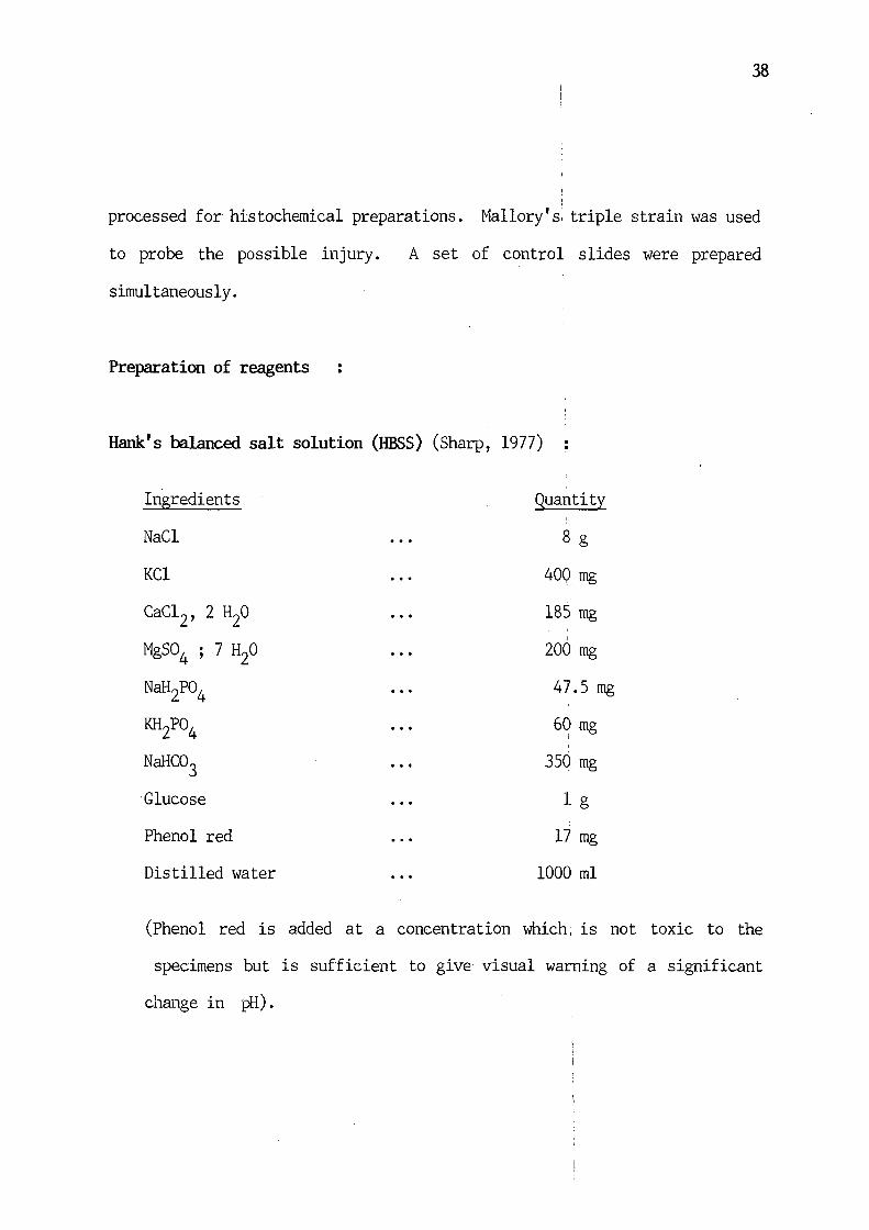
Preparation of reagents
Hank's balanced salt solution (HESS) (Sharp, 1977)
Ingredients Quantity
NaCl 8 g
KCl 400 mg
cac12, 2 H20 185 mg
' MgS04 ; 7 H20 200 mg
NaH2Po4 47.5 mg
KH2Po4 60 mg i
NaHC03 350 mg
Glucose 1 g
Phenol red 17 mg
Distilled water 1000 ml
(Phenol red is added at a concentration which, is not toxic to the ' specimens but is sufficient to give visual warning of a significant
change in pH).

Eagle's min~ essential medium (MEM) (Sharp, 1977)
Ingredients
L-arginine HCl
L-cystine
L-glutamine
L-histidine HCl.H20
L-isoleucine
L-lysine HCl
L-methionine
L-phenylalanine
L-threonine
L-tryptophan
L-tyrosine
L-valine
D-Ca pantothenate
Choline chloride·
Folic acid
i-inositol
Nicotinamide
Pyridoxal. HCl
Riboflavin
Thiamin. HCl
Hank's balanced salt solution
·, . . . .
Quantity
126.40 mg
24.02 mg
292.30 mg I
41.90 mg
52.50 mg
73.06 mg
14.90 mg
33.02 mg
47.64 mg
10.20 mg
36.22 mg
46.90 mg
1.00 mg
1.00 mg
1.00 mg
2.00 mg
1.00 mg
1.00 mg
0.10 mg
1.00 mg
1000 ml
39

'
Buffered 10% neutral formalin of Lillie (after Pearse, 1968)
40 % formaldehyde
Distilled water
NaH2Po4, H20
Na2 HP04 (anhydrous)
lOO·rnl
900 ml
4 g
6.5 g
40
· Camoy' s fixative : I
Ethanol (absolute) 60 ml
Chloroform 30 ml
Glacial acetic acid 10 ml I
!Schiff's reagent (de Tomasi variant) (after Pearse, 1968) I
ll g of basic fuchsin was dissolved in 200 ml of boiling distilled water.
~Shaken for 5 min and cooled to exactly 50°C. The solution was filtered and
·i 20 rnl of N HCl was added to the filtrate. Cooled to 25°C and 1 g sodium
fmetabisulphite (Na2s2o5) was added. The solution was left in dark for 24 h.
'2 g activated charcoal (norite) was added and shaken for 1 min. Filtered
!and the filtrate was stored in the dark at 0°' -4°s C. Reagent was brought to
iroom temp. before use.
I
.C. Biochemistry :
1For biochemical experiments mature specimens of both sexes were maintained I 1in HBSS for 1 h and thoroughly washed to get rid of the adhering debris
I

41
and mucus. The worms were incised and the interior of the body wall was I
gently scraped to isolate the cuticle. Two extremities of cuticle were
discarded. T11e dissection was carried out in cold condition. Wet weight of
the cuticle was recorded and processed further for different biochemical I
analyses.
Estimation of total protein (after Lowry e t al. , 1951)
Extraction
. .
Previously weighed cuticles were homogenised with known volume of 0.1 N NaOH
in cold condition and centrifuged at 12 000 rpm\ for 15 min. The clear !
supernatant was decanted in a clean dry test tube.
Estimation
Known volume of supernatant was taken in a test tube and the volume was made
upto 0.5 ml by adding double-distilled water. 5 ml of protein reagent was
added and thoroughly mixed. Solution was incubated for 10 min at room
temp.. 0.5 ml of diluted Folin-Ciocalteau reagent was added rapidly with
immediate mixing. After 30 min the optical density of the blue coloured
solution was read at 660 ~ against an appropriate blank solution in a
photoelectric colorimeter (Model AE II, Tokyo Erma Optical Works Ltd.,
Japan). Protein concentration was calculated from a standard curve prepared
by using standard BSA solution.

42
Reagents
·k 0.1 N NaOH 400 mg of NaOH dissolved in 100 ml of double-distilled
water.
Protein reagent : 100 ml of 4% Na2co3 was prepared to 0.1 N NaOH. 1 ml
of 4% potassium tartarate was added to it and mixed well. 1 ml of 2%
Cuso4 solution was cdila:l drop by drop \vith continuous agitation to avoid
turbidity. This reagent was freshly prepared. i
* Folin-Ciocalteau reagent : Commercial reagent (BDH) was diluted with an
equal volume of double-distilled water before use.
* Standard BSA solution : 10 mg of bovine serum albumin was dissolved in
10 ml double-distilled \vater.
Estimation of total carbohydrate (after Umbreit
Estraction
et al., i
1958)
Previously weighed cuticles were homogenised in known volume of double-
distilled water and centrifuged for 15 min at 10 000 rpm. The clear
supernatant was decanted in a clean dry test tube.
Estimation
A known· volume of supernatant \vas taken in a test tube and the vohnne v1as
made upto 3 ml by adding double-distilled water. The tube was placed in a
beaker containing ice and 6 ml of anthrone reagent was added slowly along
the wall of the tube. After thoroughly mixing I
the tube was heated in a

43
boiling water both for 5 min. The solution was cooled and after development
,of colour the optical density of test solution was read at 620 nm against a
, reagent blank in a photoelectric colorimeter (Model AE II, Tokyo Erma l !
:optical Works Ltd., Japan). Carbohydrate concentration was calculated from
1a standard curve·prepared by using standard glucose solution.
Reagent
\* Anthrone reagent 400 mg anthrone dissolved in 20 ml 68.4 M H2so4 (190 I
I mLof cone. H2so4 was added to 100 ml of double-distilled water).
'~'( Standard glucose solution : 20 mg glucose was dissolved in 100 ml
I double-distilled water.
[Estimation of monosaccharides I
(pentose and hexose)
!
!Extraction (after Oser, 1965)
I
\Previously weighed cuticle was homogenised with known volume of double-
;distilled water and centrifuged for 10 min at 10 000 rpm. The supernatant i !was treated with equal volume of ethyl ether in separating funnel and the
ether layer was rejected. The ethyl ether treatment was repeated thrice,
1thus disrupting any binding of carbohydrates and proteins with lipoidal
baterials. The lipid free extract was treated with equal volume of cold 10% I i
ITCA and kept in ice for 10 min for complete precipitation of protein. The
extract \vas centrifuged for lO min at 3 000 rpm and the supernatant was i I ~eady for estimation.
44
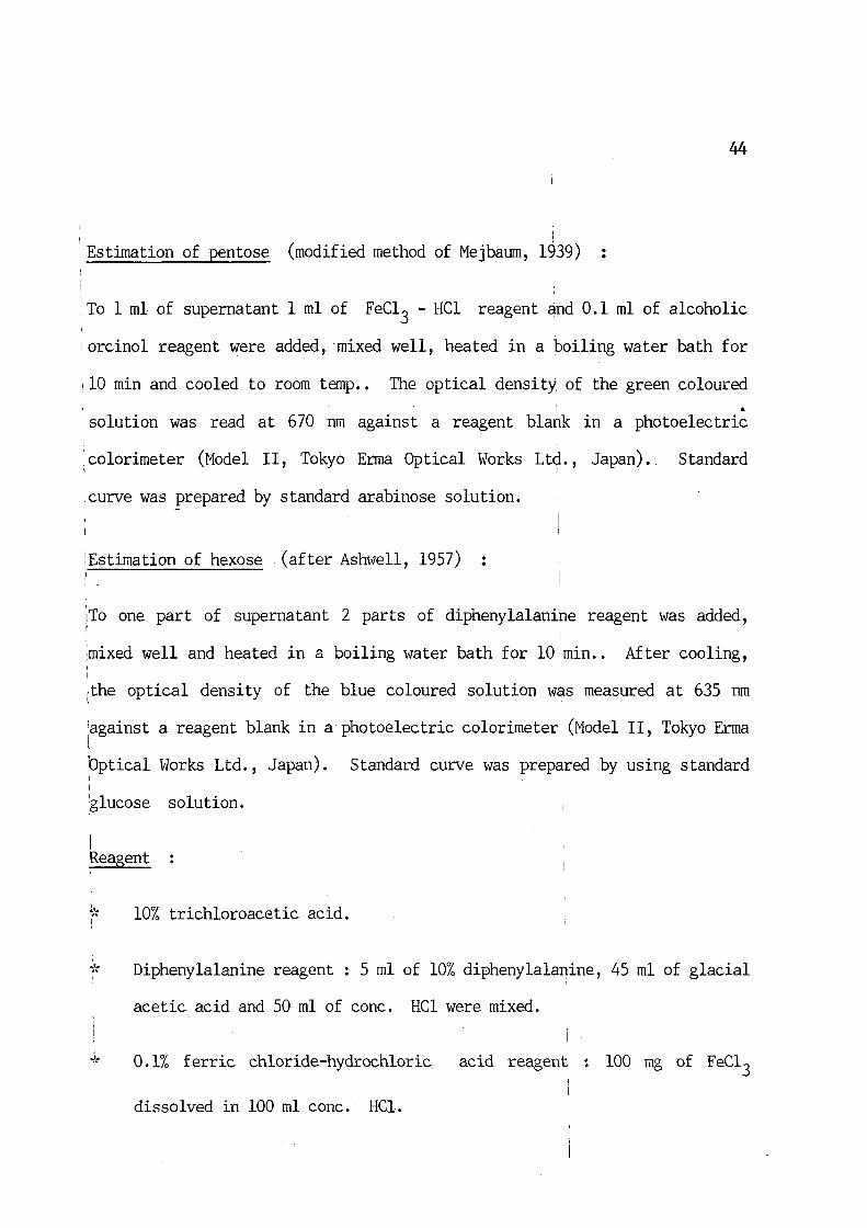
Estimation of pentose (modified method of Mejbaum, 1939)
To 1 ml of supernatant 1 ml of FeC13 - HCl reagent and 0.1 ml of alcoholic
1 orcinol reagent were added, mixed well, heated in a boiling water bath for
1 10 min and cooled to room temp .. The optical density of the green coloured
• solution was read at 670 run against a reagent blank in a photoelectric
I
;colorimeter (Model II, Tokyo Erma Optical Works Ltd., Japan). Standard
,curve was prepared by standard arabinose solution.
!Estimation of hexose . (after Ash\vell, 1957)
!To one part of supernatant 2 parts of diphenylalanihe reagent was added,
mixed well and heated in a boiling water bath for 10 min .. After cooling, I
I
~the optical density of the blue coloured solution was measured at 635 nm
:against a reagent blank in a photoelectric colorimeter (Model II, Tokyo Erma l Optical Works Ltd., Japan). Standard curve was prepared by using standard I I
glucose solution.
I ~eagent
I
~·: 10% trichloroacetic acid.
* Diphenylalanine reagent : 5 ml of 10% diphenylalanine, 45 ml of glacial
acetic acid and 50 ml of cone. HCl were mixed.
0.1% ferric chloride-hydrochloric acid reagent 100 mg of FeC13
dissolved in 100 ml cone. HCl.

45
* Orcinol reagent 100 mg of orcinol dissolved in 1 ml 95% ethanol.
·'· "
Standard glucose solution
double-distilled water.
Standard arabinose solution
double distilled water.
20 mg of glucose dissolved in 100 ml
10 mg of arabinose dissolved in 100 ml
Estimation of trehalose (after Duchateau and Florkin', 1957)
Extraction
Previously weighed cuticle was homogenised in a· known volume of double-
distilled water and centrifuged for 5 min at 2500 rpm. One volume (vol.) of
supernatant was taken in a test tube to which 3-4 vols. of 40% potassium
hydroxide solution was added and kept in a boiling water bath for 1 h.
After proper cooling 1-2 vols. of 95% ethanol was added to precipitate
glycogen. The supernatant was decanted off.
Estimation
1 A test tube containing 1 ml of supernatant was kept in an ice bath and 6 ml
! of anthrone reagent was slowly added to it. After thorough mixing the tube
J was heated in a boiling water bath for exactly 13 min. After cooling the
1 optical density of the test solution was read at 622 nm in a photoelectric
colorimeter (Model AE II, Tokyo Erma Optical 1 Works Ltd., Japan). I
i Percentage of trehalose was calculated from a standard curve prepared by l !using standard trehalose solution.

46
Reagents
* 40% potassium hydroxide solution.
~·: 95% ethanol.
* Anthrone reagent 75 mg anthrone and 1 g thiou~ea dissolved in 100 ml
70% H2so4
.'!
'1: Standard trehalose solution 20 mg of trehalose dissolved in 100 ml of
double-distilled water.
Eastimation of glycogen (modified anthrone method of; Klicpera et al., 1957)
Extraction
50-100 mg of cuticle was transferred to a centrifuge tube containing 1 ml of I
30% potassium hydroxide solution. The sample was subjected to hydrolysis I
for 1 h at 100°C. The tube was cooled and 0.2 ml 1 of 2% sodium sulphate
solution and 6 ml of absolute ethanol were added to it and mixed well. The
tube was tightly plugged and left overnight in a refrigerator for complete
precipitation of glycogen. The precipitated glycogen was collected by
' centrifugation at 3 000 rpm for 15 min and was redissolved in a known volume
of double-distilled water.
Estimation
0.5 ml of the above solution was taken in a test tube. The tube was kept in
an ice bath and 6 ml of anthrone reagent was slowly aded to it. After
thorough mixing the tube was heated in a boiling ~ater bath for 10 min.

47
After proper cooling the optical density of the test solution was read at
610 nm against a reagent blank in a photoelectric colorimeter (Model AE II,
Tokyo Erma Optical Works Ltd. , Japan). Percentage of glycogen was
calculated from a standard curve prepared by using standard glucose '
solution.
Reagents
~·: 30% potassium hydroxide solution.
'1: 2% sodium sulphate solution.
Anthrone reagent 160 mg of anthrone dissolved ~n 100 ml of cone.
·'· " Standard glucose solution
double-distilled water.
100 mg glucose dissolved in 100 ml of
Estimation of total lipid (after Folch et al., 1957)
Extraction
Previously weighed cuticle was homogenised with a known volume of chloroform
~methanol (2:1) mixture and centrifuged for 15 min at.3 000 rpn. The clear '' i
; supen1atant was taken for estimation.
I Estimation
I
I 1
·To 0.2 ml of supernatant 4.8 ml of cone. H2so4 was added, thoroughly mixed

48
and heated:.ina boiling water bath for 10 min. After. cooling to room temp.,
0. 2 ml of mixture was taken in a tube to which 3. 8 ml of orthophosphoric
acid and 1 ml of vanillin solution was added and mixed by shaking. The
solution was kept at room temp. for 20 min and the optical density· of the
pink colour developed was read at 530 nrn against a reagent blank in
photoelectric e:olorimeter (Model AE II, Tokyo Erma Optical Works Ltd.,
Japan). Standard curve was prepared by using standard palmitic acid
solution.
Reagents
·'· .. Vanillin solution 600 mg of vanillin dissolved in 100 ml double-
distilled water.
* Standard palmitic acid solution : 5 rng of palmitic acid dissolved in 10
ml of chloroform : methanol (2:1) mixture.
Estimation of cholesterol (after Roy et al., 1955)'
Extraction
Previously weighed cuticle was homogenised with appropriate volume of
glacial acetic acid along with glass powder and centrifuged for 10 min at·
3 000 rpm~ The clear supernatanL was taken for estimation.
Estimation
0.15 ml of supernatant was taken in a test tube and 5.85 ml of ghtcial I
acetic acid was added slmvly along the wall of the itube. To this solution I

49
2 ml of FaC13 reagent was added, thoroughly mixed' and kept in dark at room
temp. for 30 min. The pink colour developed was read at 560 nm against a
reagent blank in a photoelectric colorimeter (Model AE II, Tokyo Erma
Optical Works Ltd., Japan). Standard cholesterol solution was used for the
preparation of standard curve.
Reagents
Ferric chloride reagent : 1 g FeC13 was dissolved in 10 ml glacial
acetic acid. 1 ml of this solution was taken in, a flask and 99 ml cone.
H2so4 \vas added and mixed.
* Standard cholesterol solution : 5 mg of cholesterol dissolved in 10 ml
of chloroform : methanol (2:1) mixture.
Estimation of ascorbic acid (after Roe and Kuether, 1943)
Extraction
Previously weighed cuticle was homogenised with a kno~ volume of 6% TCA and
a small amount of norite was added to it. The mixture was shaken
1 vigorously for a minute and filtered. The filtrate was ready for ascorbic
', acid estimation.
t
I I Estimation
I i '1To 0.5 ml of filtrate 3.5 ml of 6% TCA, 1 drop of 10% thiourea solution and
i I
l1 ml of 2,4-dinitrophenylhydrazine reagent were added. The solution was 1

50
mixed well and heated in· a boiling water bath for 10 min. After cooling in
an ice bath, 5 ml of 85% H2so4 was added to the, solution and allowed to
stand at room temp. for 30 min. The optical density of the solution \vas
read at 540 nm against a reagent blank in a photoelectric colorimeter (Model
AE II, Tokyo Erma Optical Works Ltd., Japan). Standard curve was prepared
from standard ascorbic acid solution.
Reagents
·'· "
·'· "
2,4-dinitrophenylhydrazine reagent : 2 g of1
2,4-DNPH dissolved in
100 ml of 9 N H2so4. The mixture was allowed to stand overnight and
filtered. The reagent should be freshly prepared.
85% sulphuric acid :To 10 ml double-distilled.water 90 ml cone. H2so4
was added.
~·: 10% thiourea solution : 10 g of thiourea dissolved in 50 ml absolute
ethanol and the volume was made upto 100 ml by qouble-distilled water.
~·: 6% trichloroacetic acid :. 6 g of TCA dissolved in 100 ml double-
·'· "
distilled water.
Standard ascorbic acid solution
100 ml double-distilled water.
10 mg of ascorbic acid dissolved in
Estimation of alkaline phosphatase activity [E.C. 3.1. 3.1 (after Bessey
e t a 1 . , 1946)]

51
Extraction
Previously weighed cuticle was homogenised with known volume of 0. 25% M
sucrose solution in cold condition. The homog~~ate was centrifuged in a
refrigerated centrifuge (0° -4° C) for 10 min at 5 000 rpm. The supernatant
was taken for estimation.
Estimation
Four test tubes marked T, C, B and S (T - test, C ;.. control, B -blank and
S - standard) were taken. To T and C test tubes l ml, and to B and C 1.1
ml of buffer solution was taken. In T and C test tubes l ml of
pheny lphosphate solution was added. To the S . test tube l ml of working
standard solution and to B test tube l ml of double-distilled water \vas
added. The test tube marked T was incubated for 5-6 min at 3rc and then
0 .l ml of supernatant was added to it. Afterwards all the test tubes were
incubated at 3rc for 15 min and then 0.8 ml of 0.5 N NaOH solution and
1.2 ml of 0.5 N NaHC03 solution was added to all the test tubes. In the C
test tube 0.1 ml of supernatant was added and thoroughly mixed. Then 1 ml
of 4-aminoantipyrene solution and 1 ml of potassium ferricyanide solution
' was added to all the test tubes and mixed by shaking.. The optical densities
of all the solutions were measured at 520 nm in a photoelectric colorimeter
(Model AE II, Tokyo Enna Optical Works Ltd., Japan). ,
Calculation
, King-Armstrong units per 100 ml of supernatant,

= T - C X concentration of standard X 100 = T - C S - B concentration of test S - B
= T - C X 10 S - B
Reagents
X O.Ol X 100 CIT
52
1~ Carbonate-bicarbonate buffer solution (0.1 M ; pH 10.4 at Jrc) : 6.36g
of pure anhydrous Na2co3 and 3.36 g of NaHC03 ~as dissolved in 1 litre
of double-distilled water.
~·~ Disodium phenylphosphate solution (substrate 0.02 M) : 218 mg of
anhydrous disodium phenyl phosphate was dissolved in double-distilled
water. The volume was made upto 100 ml and stored at 4°C. I
* Stock phenol standard solution (1 ,rng/ml) : 100 mg of pure crystalline
phenol was dissolved in 100 ml of 0.1 N HCl and stored at 4°C.
* Working standard solution : To 1 ml of stock solution double-distilled
water was added to make the volume 100 ml.
* 0.5 ml N sodium hydroxide solution : To 2 g of NaOH double-distilled
water was added to make the volume 100 ml.
0.5 N sodium bicarbonate solution : To 4.2 g of pure dry NaHC03 double
distilled water was added to make the volume 100 ml.
* 4-amino antipyrene solution : To 600 mg of 4-amino antipyrene doublei
distilled water was added to make the volume 100 ml.

53
* Potassium ferricyanide solution : To 2.4 g of potassium ferricyanide
double-distilled water was added to make the volume 100 ml.
Emission spectroscopic analysis of inorganic elements (after Majumdar etal.,
1986)
Adult A. ~·alli were collected from freshly killed hosts. The cuticle
was dissected out by using only plastic equiprnents, thus ruducing
contamination of the sample. The contamination was further avoided by using
borosilicate glass wares which were rinsed with cone. nitric acid followed
by thorough rinsing in tap and double-distilled water. Weighed cuticle was
ashed in a muffle furnace in a platinum crucible at 350°C for 6 h. The ash
sample was analysed by Ernssion Spectroscopy following 1 the method of Hilger's
spectroscopy D C arcing with 9 amp current strength by an excitation of
1 graphite anode electrodes where 2 500-3 500 A0 was employed.
1 Study of glucose uptake by liquid scintillation counter (after Court et al.,
I 11988)
i I
:Adult A. galli collected from freshly killed hosts \vere maintained in I 1
1 glucose free Eagle's MEM at 3rc for l h. The parasites were transferred
fto 20 ml of Eagle's MEN containing 25 LICi of 14c-glucose maintained at Jrc • . tNon-radioactive glucose was omitted from Eagle's MEMl After specific time i
1intervals (15 min, 30 min, 45 min, 1 h, l h 30 min and 2 h) the worms were ' I
\transferred to fresh radioisotope-free Eagle's MEM, briefly rinsed to get I I

54
rid of the radioisotope adhering to the cuticle surface and were blotted on
absorbent paper. The parasites were placed on a previously weighed cavity
slide, were cut at the middle and the inner contents were scraped out of
the cuticle with a clean scalpel. Weights of cutic+e and rest of the body
were determined. The materials \vere homogenised with 50% TCA · and
centrifuged at 3 000 rpm for 10 min. 0.1 ml of supernatants were taken in
scintillation vials to which 5 ml of scintillation fluid (cocktail 'T') was
added. The activity \vas then counted in a liquid scintillation counter
(Beckman L S 5 000 T D, USA) and counts per minute (cprn) were recorded.
Reagents
* 50% trichloroacetic acid.
* Cocktail 'T' for liquid scintillation (Spectrochem Pvt. ltd., Bombay)
Toluene
Triton X-100
PPO
PO POP
666 ml/L
332 ml/L
5 g /L
0.15 g /L