Massively parallel manipulation of single cells and microparticles using optical images Pei Yu...
16
Massively parallel manipulation of single cells and microparticles using optical images Pei Yu Chiou, Aaron T. Ohta & Ming C. Wu Nature, Vol. 436, 370-372, 2005 Ayca Yalcin
-
date post
20-Dec-2015 -
Category
Documents
-
view
214 -
download
1
Transcript of Massively parallel manipulation of single cells and microparticles using optical images Pei Yu...
Massively parallel manipulation of single cells and microparticles using optical
images
Pei Yu Chiou, Aaron T. Ohta & Ming C. Wu
Nature, Vol. 436, 370-372, 2005
Ayca Yalcin
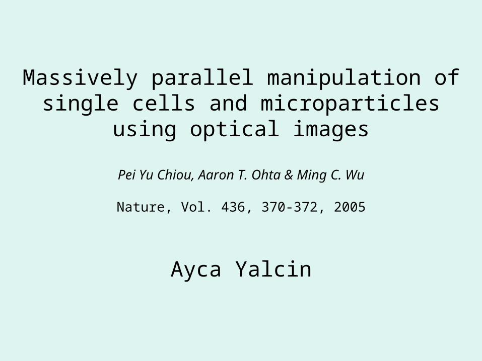
Motivation: Need for ability to manipulate cells and particles with high resolution and high throughput for biological and colloidal science applications.
Proposed Method: Optical image driven dielectrophoresis.
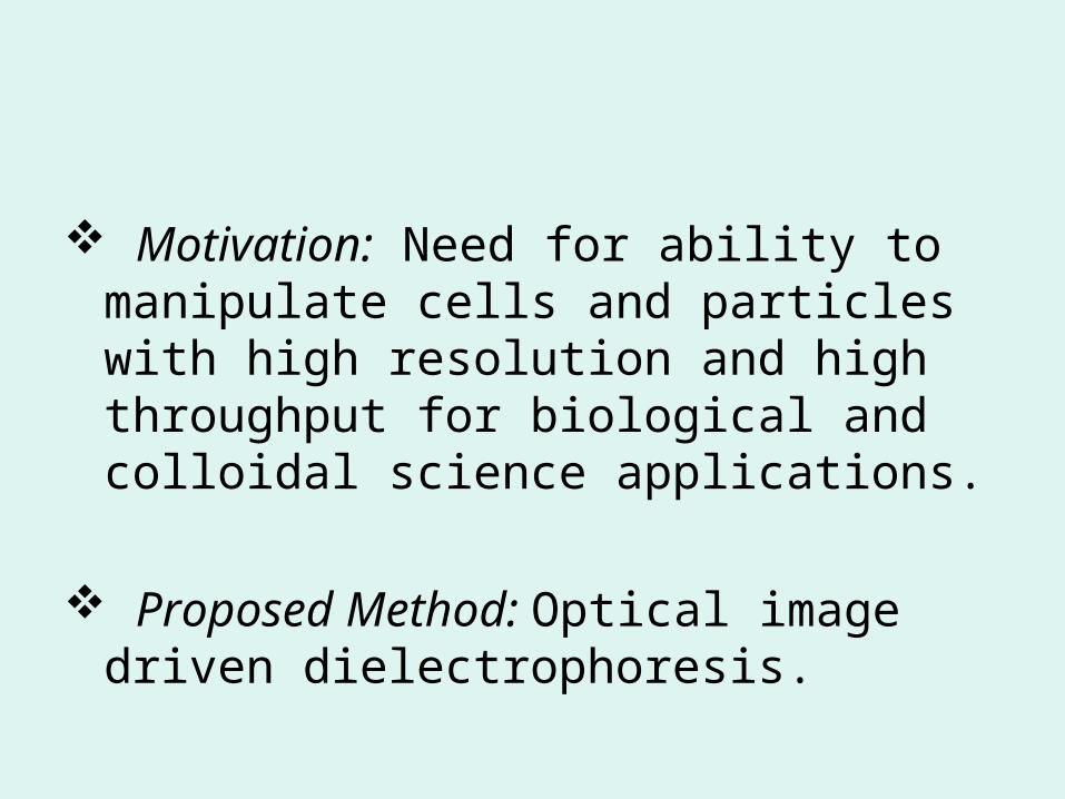
OUTLINE
Optical tweezers and dielectrophoresis Xerography Optoelectronic tweezers (OET) Device structure Demonstration of throughput and
resolution Comparison to other techniques Previous work Conclusion
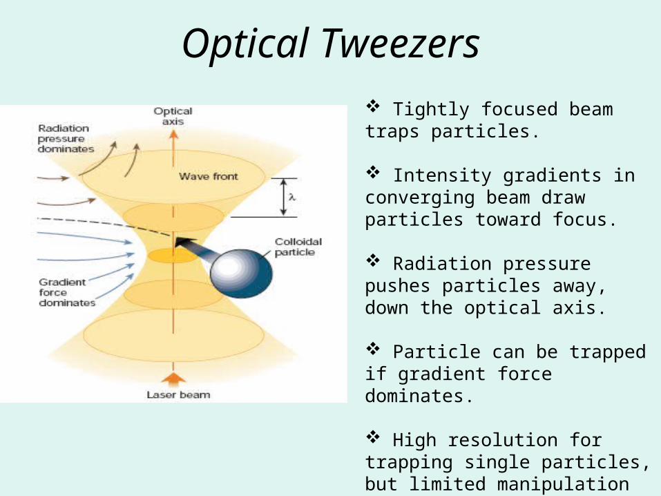
Optical Tweezers Tightly focused beam traps particles.
Intensity gradients in converging beam draw particles toward focus.
Radiation pressure pushes particles away, down the optical axis.
Particle can be trapped if gradient force dominates.
High resolution for trapping single particles, but limited manipulation area.
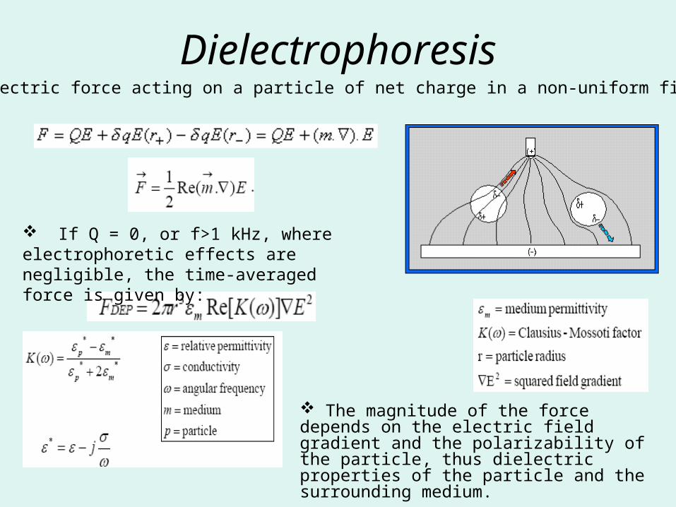
Dielectrophoresis
The magnitude of the force depends on the electric field gradient and the polarizability of the particle, thus dielectric properties of the particle and the surrounding medium.
Total electric force acting on a particle of net charge in a non-uniform field:
If Q = 0, or f>1 kHz, where electrophoretic effects are negligible, the time-averaged force is given by:
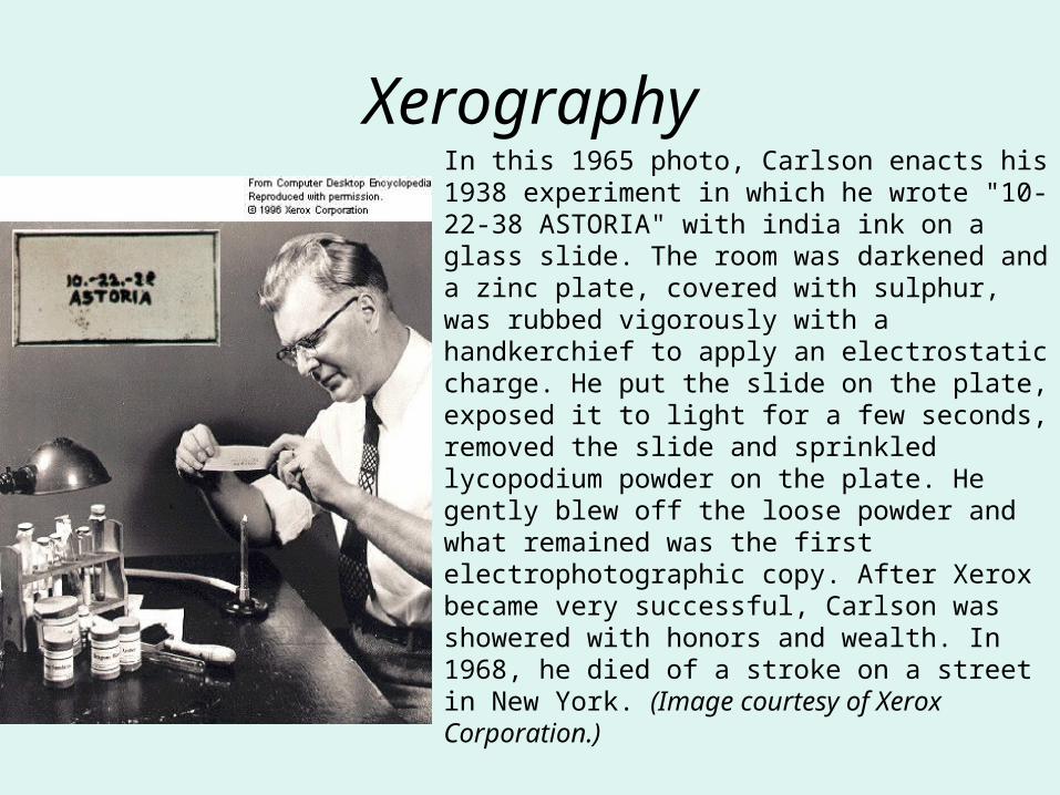
XerographyIn this 1965 photo, Carlson enacts his 1938 experiment in which he wrote "10-22-38 ASTORIA" with india ink on a glass slide. The room was darkened and a zinc plate, covered with sulphur, was rubbed vigorously with a handkerchief to apply an electrostatic charge. He put the slide on the plate, exposed it to light for a few seconds, removed the slide and sprinkled lycopodium powder on the plate. He gently blew off the loose powder and what remained was the first electrophotographic copy. After Xerox became very successful, Carlson was showered with honors and wealth. In 1968, he died of a stroke on a street in New York. (Image courtesy of Xerox Corporation.)
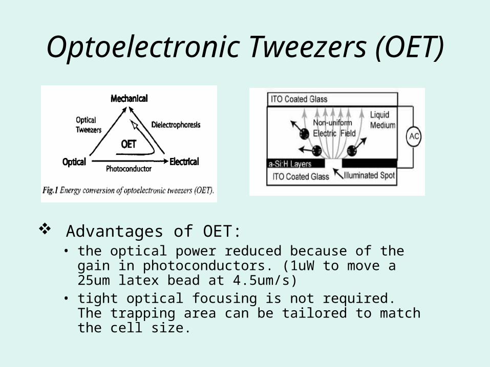
Optoelectronic Tweezers (OET)
Advantages of OET:• the optical power reduced because of the gain in
photoconductors. (1uW to move a 25um latex bead at 4.5um/s)
• tight optical focusing is not required. The trapping area can be tailored to match the cell size.
Device Structure
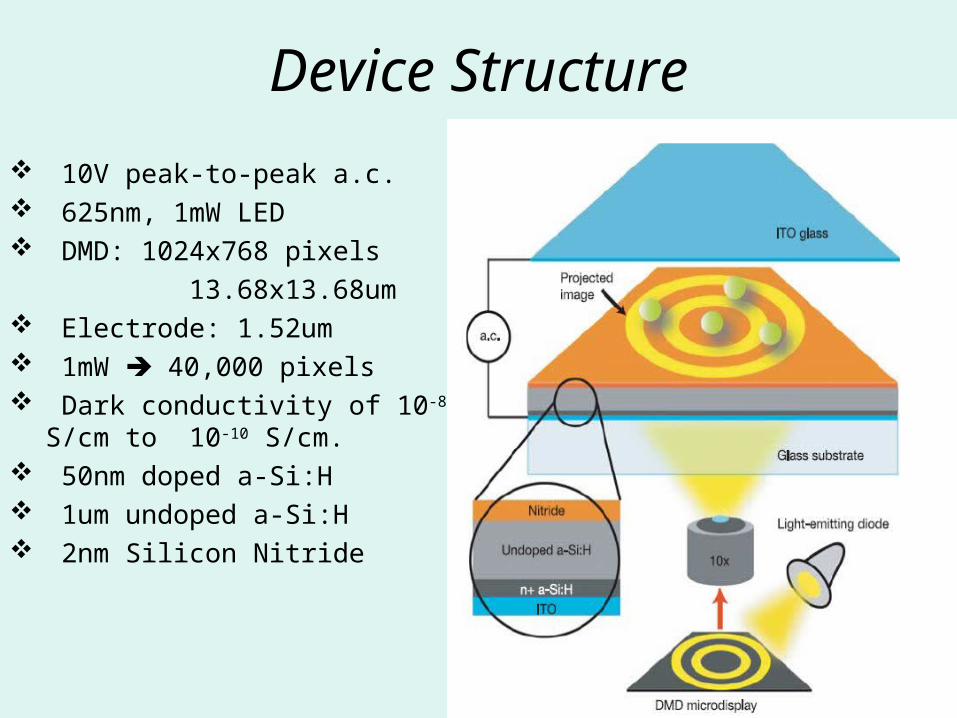
10V peak-to-peak a.c. 625nm, 1mW LED DMD: 1024x768 pixels
13.68x13.68um Electrode: 1.52um 1mW 40,000 pixels Dark conductivity of 10-8
S/cm to 10-10 S/cm. 50nm doped a-Si:H 1um undoped a-Si:H 2nm Silicon Nitride
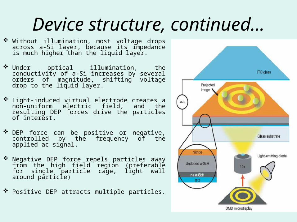
Device structure, continued… Without illumination, most voltage drops
across a-Si layer, because its impedance is much higher than the liquid layer.
Under optical illumination, the conductivity
of a-Si increases by several orders of magnitude, shifting voltage drop to the liquid layer.
Light-induced virtual electrode creates a non-uniform electric field, and the resulting DEP forces drive the particles of interest.
DEP force can be positive or negative, controlled by the frequency of the applied ac signal.
Negative DEP force repels particles away from the high field region (preferable for single particle cage, light wall around particle)
Positive DEP attracts multiple particles.
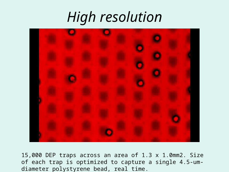
High resolution
15,000 DEP traps across an area of 1.3 x 1.0mm2. Size of each trap is optimized to capture a single 4.5-um-diameter polystyrene bead, real time.
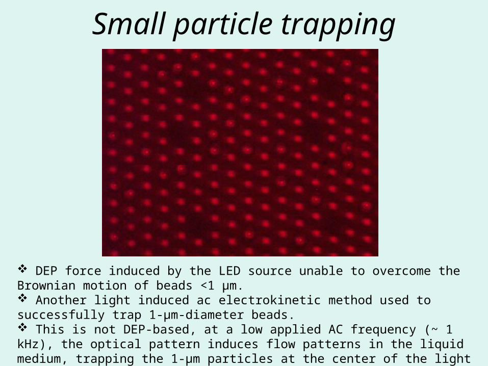
Small particle trapping
DEP force induced by the LED source unable to overcome the Brownian motion of beads <1 μm. Another light induced ac electrokinetic method used to successfully trap 1-μm-diameter beads. This is not DEP-based, at a low applied AC frequency (~ 1 kHz), the optical pattern induces flow patterns in the liquid medium, trapping the 1-μm particles at the center of the light spots.
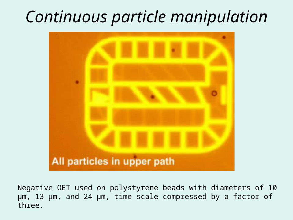
Continuous particle manipulation
Negative OET used on polystyrene beads with diameters of 10 μm, 13 μm, and 24 μm, time scale compressed by a factor of three.
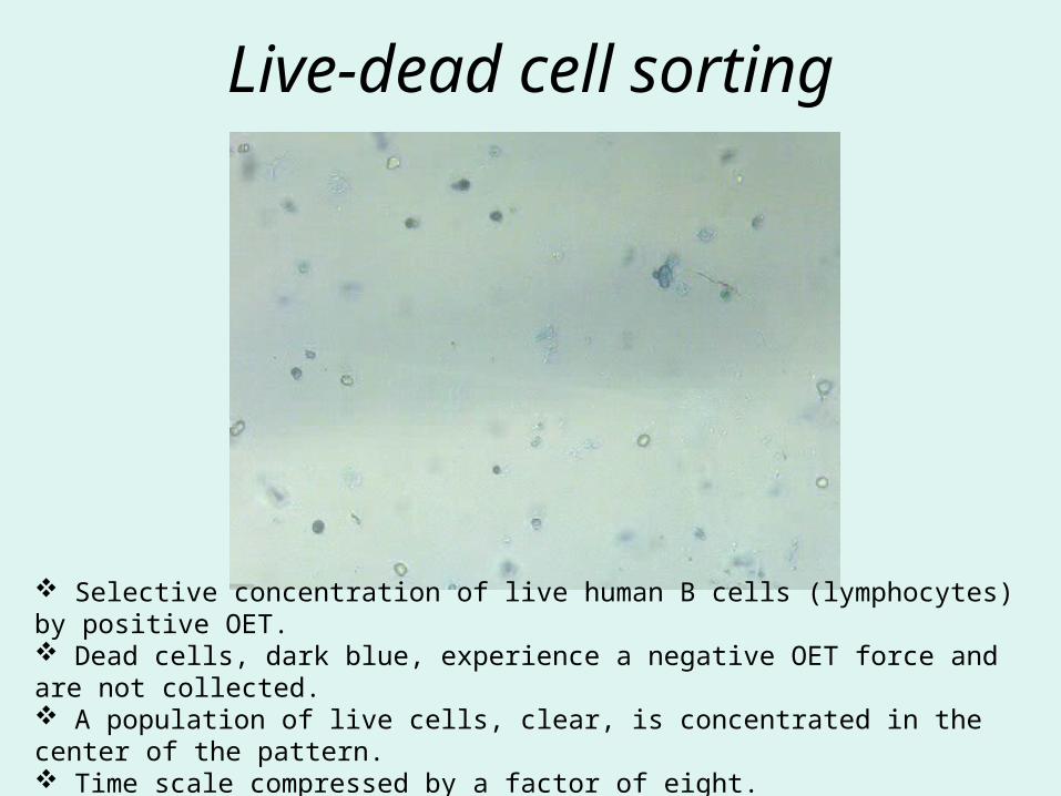
Live-dead cell sorting
Selective concentration of live human B cells (lymphocytes) by positive OET. Dead cells, dark blue, experience a negative OET force and are not collected. A population of live cells, clear, is concentrated in the center of the pattern. Time scale compressed by a factor of eight.
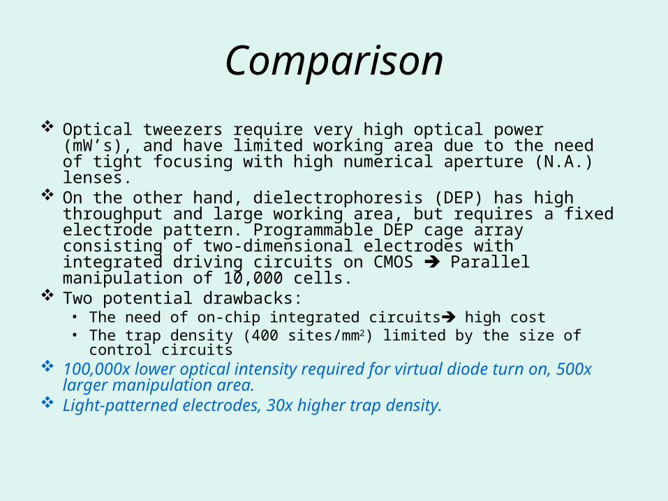
Comparison
Optical tweezers require very high optical power (mW’s), and have limited working area due to the need of tight focusing with high numerical aperture (N.A.) lenses.
On the other hand, dielectrophoresis (DEP) has high throughput and large working area, but requires a fixed electrode pattern. Programmable DEP cage array consisting of two-dimensional electrodes with integrated driving circuits on CMOS Parallel manipulation of 10,000 cells.
Two potential drawbacks: • The need of on-chip integrated circuits high cost• The trap density (400 sites/mm2) limited by the size of control
circuits 100,000x lower optical intensity required for virtual diode
turn on, 500x larger manipulation area. Light-patterned electrodes, 30x higher trap density.
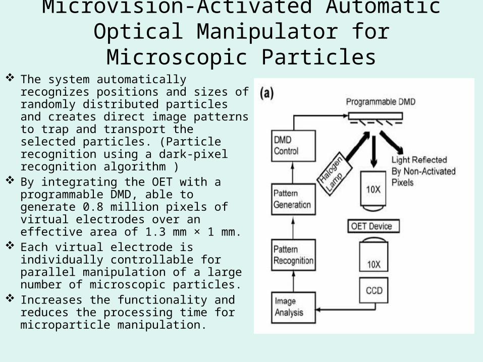
The system automatically recognizes positions and sizes of randomly distributed particles and creates direct image patterns to trap and transport the selected particles. (Particle recognition using a dark-pixel recognition algorithm )
By integrating the OET with a programmable DMD, able to generate 0.8 million pixels of virtual electrodes over an effective area of 1.3 mm × 1 mm.
Each virtual electrode is individually controllable for parallel manipulation of a large number of microscopic particles.
Increases the functionality and reduces the processing time for microparticle manipulation.
Microvision-Activated Automatic Optical Manipulator for Microscopic
Particles
Conclusion
Multiple manipulation functions combined to achieve complex, multi-step manipulation protocols.
Single cell analysis spectrum of response of each individual cell.