![arXiv:1212.4837v2 [astro-ph.HE] 19 Mar 2013 · form by a collapse of a massive cloud (Begelman et al. 2006) or a massive star (Fryer et al. 2001; Madau & Rees 2001; Schneider et al.](https://static.fdocuments.in/doc/165x107/605a419bd7353d0b0801dc90/arxiv12124837v2-astro-phhe-19-mar-2013-form-by-a-collapse-of-a-massive-cloud.jpg)
Massive collapse of the lung with acute respiratory infection
5
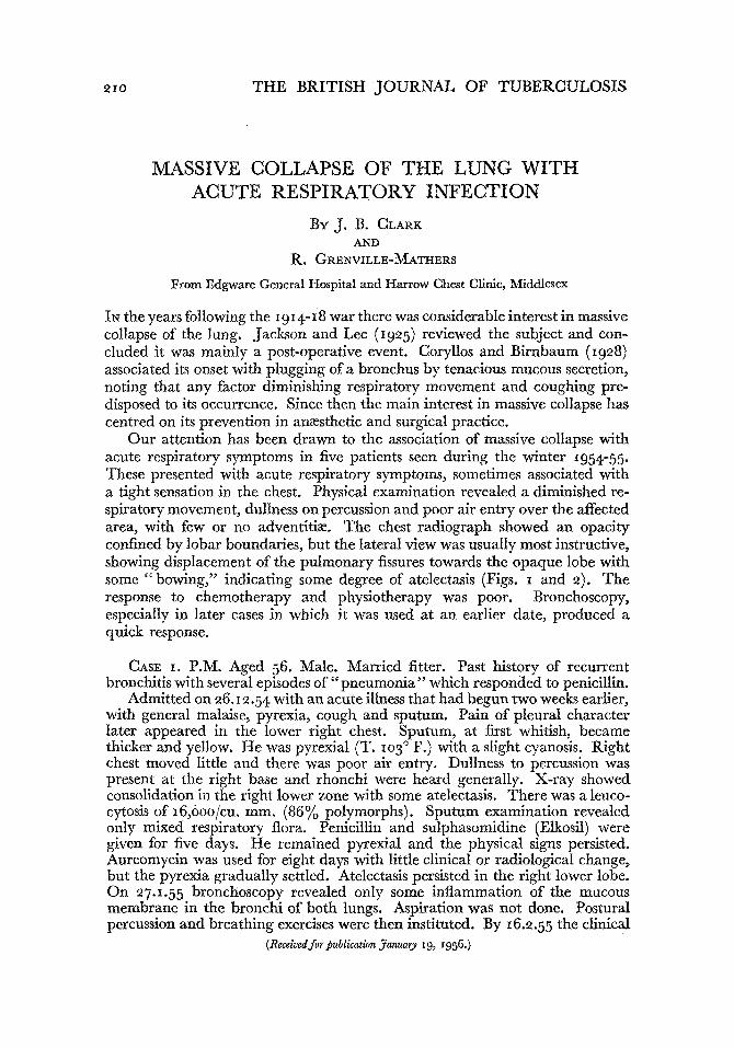
~Io THE BRITISH JOURNAL OF TUBERCULOSIS MASSIVE COLLAPSE OF THE LUNG WITH ACUTE RESPIRATORY INFECTION BY J. B. CLARK AND R. GRENVILLE-~[ATHERS From Edgware General Hospital and Harrow Chest Clinic, Middlesex IN the years following the 1914-18 war there was considerable interest in massive collapse of the lung. Jackson and Lee (I925) reviewed the subject and con- cluded it was mainly a post-operative event. Coryllos and Birnbaum (I928) associated its onset with plugging of a bronchus by tenacious mucous secretion, noting that any factor diminishing respiratory movement and coughing pre- disposed to its occurrence. Since then the main interest in massive collapse has centred on its prevention in anaesthetic and surgical practice. Our attention has been drawn to the association of massive collapse with acute respiratory symptoms in five patients seen during the winter I954-55. These presented with acute respiratory symptoms, sometimes associated with a tight sensation in the chest. Physical examination revealed a diminished re- spiratory movement, dullness on percussion and poor air entry over the affected area, with few or no adventiti~e. The chest radiograph showed an opacity confined by lobar boundaries, but the lateral view was nsually most instructive, showing displacement of the pulmonary fissures towards the opaque lobe with some "bowing," indicating some degree of atelectasis (Figs. i and 2). The response to chemotherapy and physiotherapy was poor. Bronchoscopy, especially in later cases in which it was used at an earlier date, produced a quick response. CAS~. I. P.M. Aged 56. Male. Married fitter. Past history of recurrent bronchitis with several episodes of" pneumonia" which responded to penicillin. Admitted on 26.12.54 with an acute illness that had begun two weeks earlier, with general malaise, pyrexia, cough and sputum. Pain of pleuraI character later appeared in the lower right chest. Sputum, at first whitish, became thicker and yellow. He was pyrexial (T. Io3 ° F.) with a slight cyanosis. Right chest moved little and there was poor air entry. Dullness to percussion was present at the right base and rhonchi were heard generally. X-ray showed consolidation in the right lower zone with some atelectasis. There was a leuco- cytosis of I6,6oo/eu. ram. (86% polymorphs). Sputum examination revealed only mixed respiratory flora. Penicillin and sulphasomidine (Elkosil) were given for five days. He remained pyrexial and the physical signs persisted. Aureomycin was used for eight days with little clinical or radiological change, but the pyrexia gradually settled. Atelectasis persisted in the right lower lobe. On 27.I.55 bronchoscopy revealed only some inflammation of the mucous membrane in the bronchi of both lungs. Aspiration was not done. Postural percussion and breathing exercises were then instituted. By 16.2.55 the clinical (Received for publicationoTanuary I9, I956. )
Transcript of Massive collapse of the lung with acute respiratory infection
~Io THE BRITISH J O U R N A L OF TUBERCULOSIS
MASSIVE COLLAPSE OF THE LUNG WITH ACUTE RESPIRATORY INFECTION
BY J. B. CLARK AND
R. GRENVILLE-~[ATHERS
From Edgware General Hospital and Harrow Chest Clinic, Middlesex
IN the years following the 1914-18 war there was considerable interest in massive collapse of the lung. Jackson and Lee (I925) reviewed the subject and con- cluded it was mainly a post-operative event. Coryllos and Birnbaum (I928) associated its onset with plugging of a bronchus by tenacious mucous secretion, noting that any factor diminishing respiratory movement and coughing pre- disposed to its occurrence. Since then the main interest in massive collapse has centred on its prevention in anaesthetic and surgical practice.
Our attention has been drawn to the association of massive collapse with acute respiratory symptoms in five patients seen during the winter I954-55. These presented with acute respiratory symptoms, sometimes associated with a tight sensation in the chest. Physical examination revealed a diminished re- spiratory movement, dullness on percussion and poor air entry over the affected area, with few or no adventiti~e. The chest radiograph showed an opacity confined by lobar boundaries, but the lateral view was nsually most instructive, showing displacement of the pulmonary fissures towards the opaque lobe with some "bowing," indicating some degree of atelectasis (Figs. i and 2). The response to chemotherapy and physiotherapy was poor. Bronchoscopy, especially in later cases in which it was used at an earlier date, produced a quick response.
CAS~. I. P.M. Aged 56. Male. Married fitter. Past history of recurrent bronchitis with several episodes o f " pneumonia" which responded to penicillin.
Admitted on 26.12.54 with an acute illness that had begun two weeks earlier, with general malaise, pyrexia, cough and sputum. Pain of pleuraI character later appeared in the lower right chest. Sputum, at first whitish, became thicker and yellow. He was pyrexial (T. Io3 ° F.) with a slight cyanosis. Right chest moved little and there was poor air entry. Dullness to percussion was present at the right base and rhonchi were heard generally. X-ray showed consolidation in the right lower zone with some atelectasis. There was a leuco- cytosis of I6,6oo/eu. ram. (86% polymorphs). Sputum examination revealed only mixed respiratory flora. Penicillin and sulphasomidine (Elkosil) were given for five days. He remained pyrexial and the physical signs persisted. Aureomycin was used for eight days with little clinical or radiological change, but the pyrexia gradually settled. Atelectasis persisted in the right lower lobe. On 27.I.55 bronchoscopy revealed only some inflammation of the mucous membrane in the bronchi of both lungs. Aspiration was not done. Postural percussion and breathing exercises were then instituted. By 16.2.55 the clinical
(Received for publication oTanuary I9, I956. )
AND DISEASES OF THE CHEST 2II
signs were lessening, but the X-ray remained unchanged until on ~2.2.55 a film showed increasing aeration of the lower lobe. He was discharged to con- valescence on 5.3.55.
CASE 2. B.W. Aged 29. Housewife. In late October I954 an illness began with coryza, and respiratory symptoms developed with a temperature of Io5 ° F. Cough was present with little sputum, and dyspncea was noticeable. Penicillin produced no effect. Admitted on 6.I 1.54 when the temperature was Io2 ° F. She appeared alert. There was diminished movement of the chest at both bases where the percussion note was impaired and air entry diminshed. Crepi- tations were present at the right base.
White blood cells were io,4oo/cu, mm. (68% polymorphs). Sputum con- tained mixed respiratory flora. Chest X-ray showed consolidation and atelec- tasis of the posterior segments of the right lower lobe. Aureomycin for five days produced no change. Bronchoscopy on i I. i 1.54 revealed "some inflammation of the basal bronchi, particularly the right posterobasal segment, with thick purulent discharge which was sucked out. No other abnormality seen." Next day the air entry had increased at the right base and clinical improvement was uninterrupted. On ~2.I i .54 an X-ray showed only a few linear shadows at the right base. She was discharged on 28.I 1.54. A final X-ray on 6.1.55 was clear.
CAsE 3. G.B. Male. Aged 28. Machine tool fitter. Past history of right pneu- monia, i948. On 22.I2.54 he felt shivery and not quite well. He was sent home from work with headache, discomfort in the right chest and pain on deep inspiration. The temperature was Io3 ° F. Vomiting occurred mid-morn- ing and again in the evening. On 24.i2.54 a productive dough appeared and he was admitted to hospital. He appeared flushed. Temperature IoI ° F., P. 94, R. 26. The right side of the chest moved little. There was dullness to percussion at the right base where breath sounds were distant. White blood ceils numbered Io,2oo/cu. mm. (88% polymorphs). He was treated with penicillin, but the pyrexia continued.
X-ray on 28.12.54 showed a dense opacity in the right lower lobe with some atelectasis. Another on 3.1.55 showed the right middie lobe also to be opaque.
Postural percussion and expectorants were employed with little effect. The pyrexia slowly settled and by i o. 1.55 the clinical signs of consolidation in the right lower lobe were still present but a little less marked. Chest X-ray showed a little clearing of the lung shadows.
Bronchoscopy on t3.I.55 showed considerable narrowing in the region of the right segmental bronchi. This appeared to be mainly due to inflamed or cedematous mucous membrane which bled easily when touched. Biopsy was taken and the histological findings were "Partial ulceration, the submucosa is congested and infiltrated with polymorphs, lymphocytes and histiocytes. There are some areas of distortion where the cells are not identifiable. The picture is that of an acute inflammation."
Next day air entry had improved considerably and an X-ray on i7.I.55 showed only scanty basal shadows, while on 24.I.55 it appeared clear. He was discharged on 28.I.55.
C*s~ 4. T.J. Aged 17. Schoolgirl. About i5.I2.54 she developed a febrile illness with pharyngitis and was treated with sulphathiazole. By i9.i2.54 there was also a cough and a "noisy chest." Penicillin therapy was begun that day and sulphonamides ceased. On 23.i2.54 she was seen by one of us for the first
212 THE BRITISH J O U R N A L OF TUBERCULOSIS
time. She was febrile and there were signs of consolidation in the lower right chest. X-ray showed atelectasis of the apical and posterior segments of the right lower lobe. Penicillin was continued and she was encouraged to cough, the mother being instructed in postural percussion. The pyrexia settled and she brought up a little whitish sputum, but signs of consolidation persisted and an X-ray on 28.12.54 showed only slightly less extensive shadows. In view of this bronchoscopy was done on 6.1.55. While inserting the bronchoscope a plug of mucus was coughed up. The orifice of the middle lobe was reddened and flattened. Suction produced no further mucus. The clinical signs then dis- appeared and X-ray on Io.I.55 showed scanty residual shadows over the apex of the right lower lobe and she was discharged home. X-ray on I5.2.55 clear.
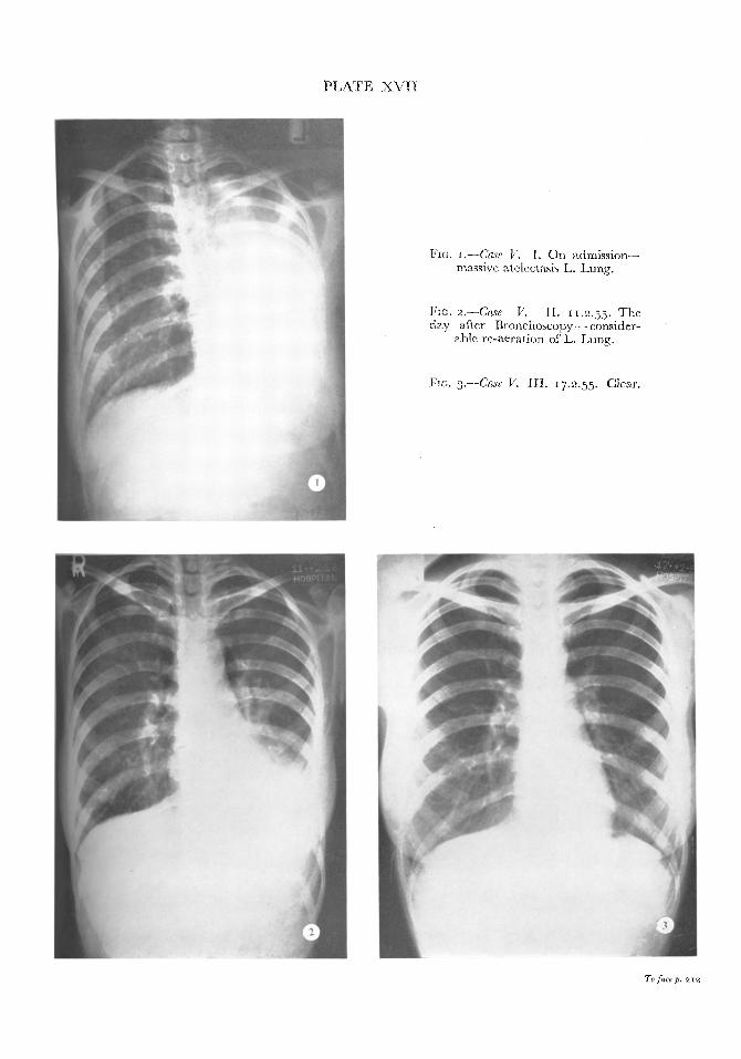
CAsE 5. S.B. Female. Aged 22. Single. Past history: Asthma in earlier life. On 1.2.55 a cough began but there was little upset until on 6.2.55 she awoke with a tight sensation over the lower sternum and this slowly became painful, involving the right and left shoulders, and by 8.2.55 there was continuous pain over the lower chest, worse on deep breathing. On admission to hospital she appeared flushed. Temperature was 99"6 ° F., P. 92, R. 20. Trachea and medi- astinum were displaced to the left. The left side of the chest moved little and air entry was poor. An X-ray on the following day showed an opaque left lung and white blood cells were 22,2oo/cu. ram. (84% polymorphs). Sputum grew mixed respiratory flora. Penicillin and sulphasomidine were started and the next day bronchoscopy showed the left main bronchus to be completely blocked by thick purulent material which was aspirated.
On the following day she appeared better, trachea was central, movement of the left chest had increased, and a chest X-ray showed only partial atelec- tasis of the posterior basal segments of the left lower lobe. Improvement continued and an X-ray on I7.2.55 was clear (Figs. I, 2, 3). She was discharged on 19.2.55.
Discuss ion
In patients of 4 ° or over unresolved pneumonia is always suspect as being possibly due to an underlying neoplastic obstruction. In lobar pneumonia massive collapse is not expected frequently since the affected portion of lung is usually solid. Reiman (1938) pointed out that "Minor degrees of atelectasis probably occur at various stages in most cases of pneumonia and are probably for the most part of no importance. In the occasional case, however, massive atelectasis may serve to explain the puzzling suppression of breath sounds and the misinterpreted signs of thickened pleura or pleural effusion which occasion- ally confront the examiner."
A review of the literature has revealed some cases similar to our series. Tidy and Phillips (I914) described a case with slowly resolving clinical signs of atelectasis in a young man. X-ray six weeks after the onset was normal. Mainzer (1931) described two cases. The first a man of 3 o, was considered to have lobar pneumonia but was becoming weaker and more dyspnoeic. Chest X-ray showed an opaque right chest and bronchoscopy revealed obstruc- tion of the right main bronchus with gelatinous material, some of which was removed by suction with dramatic improvement. The second case was a man of 43 with acute respiratory symptoms and a radiological opacity at the left base. Bronchoscopy revealed obstruction in the left main bronchus.
P L A T E X V I I
Fic, I.--Case V. I. On admission-- massive atelectasis L. Lung.
Fro. 2.--(Tase V. i I . 11.2.55. The day after Bronchoscopy---consider-
able re-aeration of L. Lung.
FIO. 3.--Cm~ K III . 17.2.55. Clear.
To J~ce p. ~2
AND DISEASES OF THE CHEST 2i 3
Aspiration failed but a sponge passed down the bronchoscope and twisted on itself withdrew gelatinous material. Recovery was then rapid. Miller (i95o) described a woman with pyrexia and cough who had consolidation at the right base. Treatment with penicillin made her apyrexial but the right lower lobe remained opaque radiologically. At bronchoscopy a month later a tenacious, gelatinous plug was removed on the sucker with subjective improvement. The physical and radiological signs settled during the succeeding five weeks.
In younger people it appears that physical signs of unresolved pneumonia arise from bronchial obstruction due to tenacious, gelatinous exudation. There seems little benefit to be gained by continuing courses of chemotherapy in such patients, and we advocate the more frequent use of bronchoscopy in these people, not only for diagnostic purposes but also on therapeutic grounds. Such procedure should include the passage of a sucker down the affected bronchi even if the visible bronchi appear patent. In this series the course of the diseases has been considerably shortened by this procedure. Physiotherapy appears to be ineffectual and may well be responsible for sucking of the ob- structing plugs further down the affected bronchi after expulsion of the air by percussion.
Summary Five cases of pulmonary aelectasis occurring in acute respiratory infection ~,
are reported. Chemotherapy and physiotherapy produced little improvement and the more frequent use of bronchoscopy is advocated in this type of case.
We wish to thank Dr. G. H. Jennings for permission to report cases I and V, Mr. R. Laird and Mr. T. W. Stephens for hronchoscopies, the several General Practitioners who referred these patients, and Dr. H.J. Trenchard for criticisms.
REFERENCES CORYLLOS, P. N., and BmNBAUM, G. L. (I928): Arch. Surg. (Chicago), 16, 5oi. JACKSON, C., and LEE, W. E. (~9=5): Ann. Surg., 82, 364. MAmZ~R, F. S. (I93I): Amer. 07. Surg., 12, 4ao. MmL~R, A. (I95o): Brit. reed. 07., 2, 764- R~IMAN, H. A. (I938): "The Pneumonias." Philadelphia. TIDY, H. L., and PHILLIPS, E. (I914): Lancet, 1, i=45.