Marieb ch13a
44
ELAINE N. MARIEB EIGHTH EDITION 13 Copyright © 2006 Pearson Education, Inc., publishing as Benjamin Cummings PowerPoint ® Lecture Slide Presentation by Jerry L. Cook, Sam Houston University ESSENTIALS OF HUMAN ANATOMY & PHYSIOLOGY PART A The Respiratory System
-
Upload
elaine-briosos -
Category
Science
-
view
366 -
download
12
Transcript of Marieb ch13a
ELAINE N. MARIEB
EIGHTH EDITION
13
Copyright © 2006 Pearson Education, Inc., publishing as Benjamin Cummings
PowerPoint® Lecture Slide Presentation by Jerry L. Cook, Sam Houston University
ESSENTIALSOF HUMANANATOMY
& PHYSIOLOGY
PART A
The Respiratory System
Copyright © 2006 Pearson Education, Inc., publishing as Benjamin Cummings
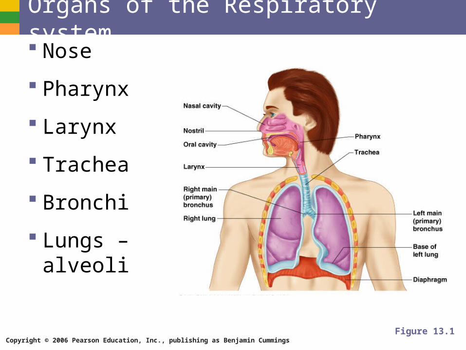
Organs of the Respiratory system Nose
Pharynx
Larynx
Trachea
Bronchi
Lungs – alveoli
Figure 13.1
Copyright © 2006 Pearson Education, Inc., publishing as Benjamin Cummings
Function of the Respiratory System Oversees gas exchanges between the blood
and external environment
Exchange of gasses takes place within the lungs in the alveoli
Passageways to the lungs purify, warm, and humidify the incoming air
Copyright © 2006 Pearson Education, Inc., publishing as Benjamin Cummings
The Nose The only externally visible part of the
respiratory system
Air enters the nose through the external nares (nostrils)
The interior of the nose consists of a nasal cavity divided by a nasal septum
Copyright © 2006 Pearson Education, Inc., publishing as Benjamin Cummings
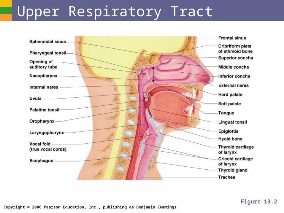
Upper Respiratory Tract
Figure 13.2
Copyright © 2006 Pearson Education, Inc., publishing as Benjamin Cummings
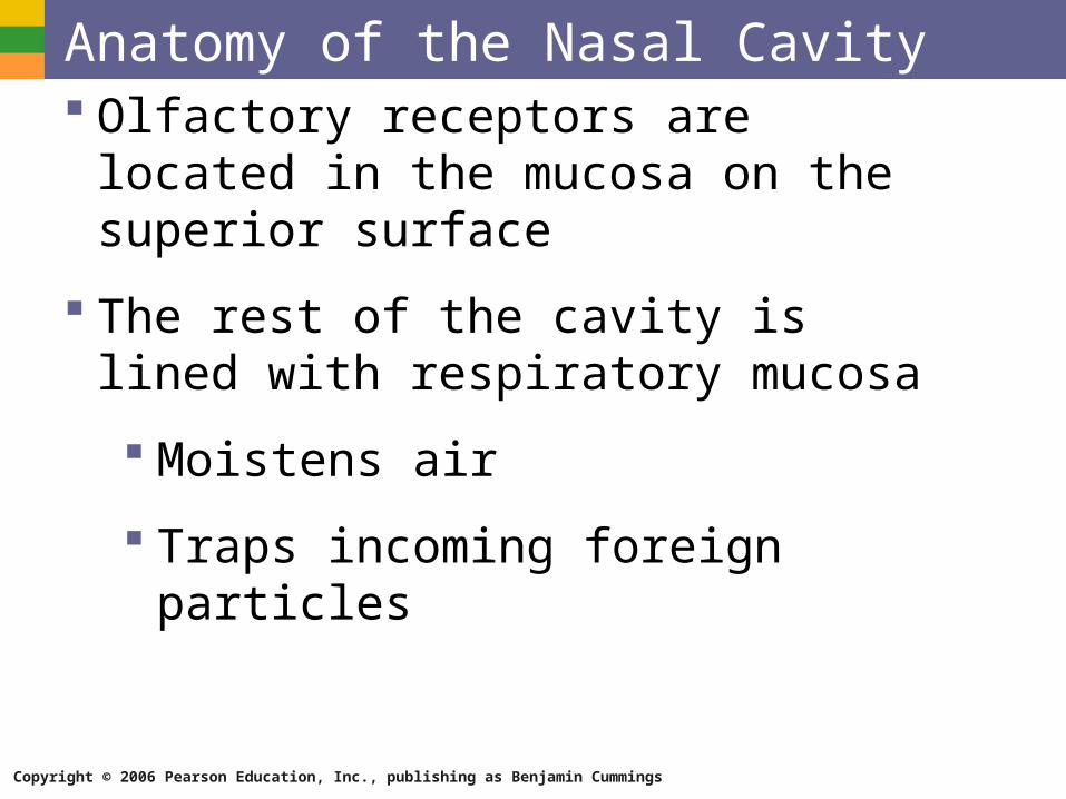
Anatomy of the Nasal Cavity Olfactory receptors are located in the mucosa
on the superior surface
The rest of the cavity is lined with respiratory mucosa
Moistens air
Traps incoming foreign particles

Copyright © 2006 Pearson Education, Inc., publishing as Benjamin Cummings
Anatomy of the Nasal Cavity Lateral walls have projections called conchae
Increases surface area
Increases air turbulence within the nasal cavity
The nasal cavity is separated from the oral cavity by the palate
Anterior hard palate (bone)
Posterior soft palate (muscle)
Copyright © 2006 Pearson Education, Inc., publishing as Benjamin Cummings
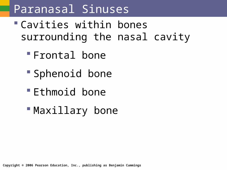
Paranasal Sinuses Cavities within bones surrounding the nasal
cavity
Frontal bone
Sphenoid bone
Ethmoid bone
Maxillary bone
Copyright © 2006 Pearson Education, Inc., publishing as Benjamin Cummings
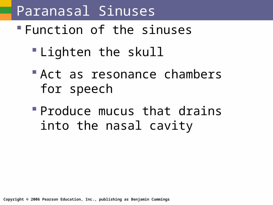
Paranasal Sinuses Function of the sinuses
Lighten the skull
Act as resonance chambers for speech
Produce mucus that drains into the nasal cavity
Copyright © 2006 Pearson Education, Inc., publishing as Benjamin Cummings
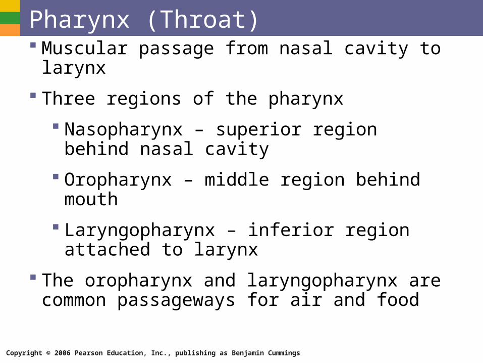
Pharynx (Throat) Muscular passage from nasal cavity to larynx
Three regions of the pharynx
Nasopharynx – superior region behind nasal cavity
Oropharynx – middle region behind mouth
Laryngopharynx – inferior region attached to larynx
The oropharynx and laryngopharynx are common passageways for air and food
Copyright © 2006 Pearson Education, Inc., publishing as Benjamin Cummings
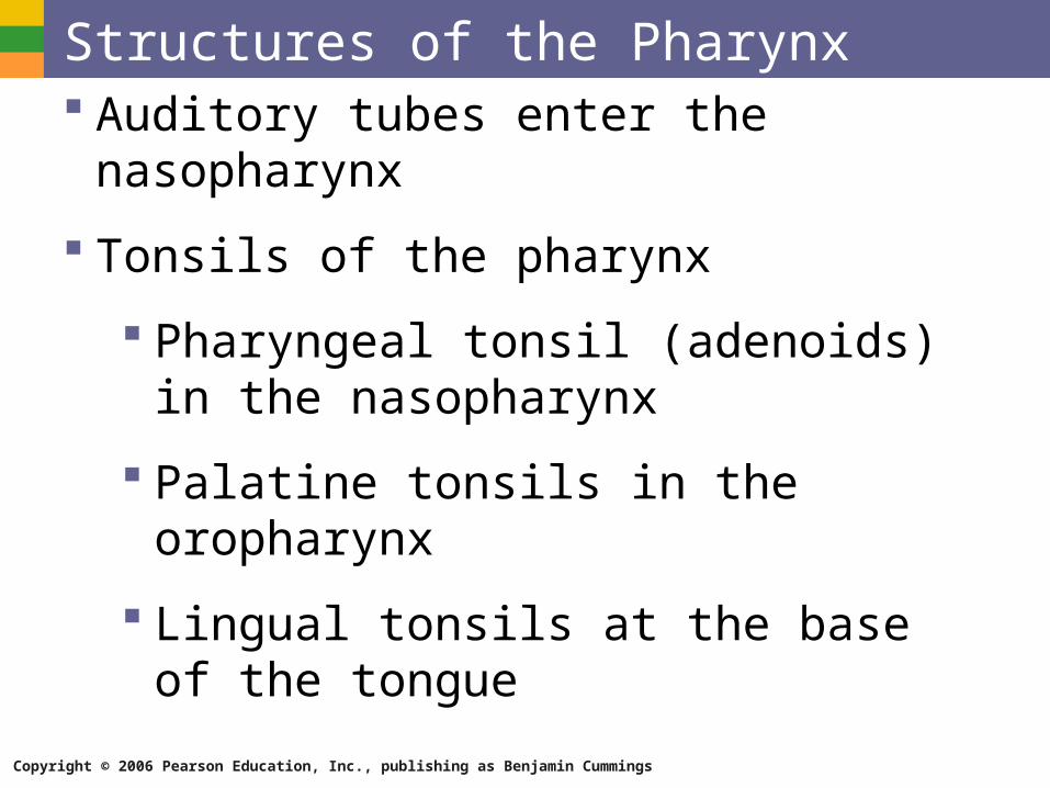
Structures of the Pharynx Auditory tubes enter the nasopharynx
Tonsils of the pharynx
Pharyngeal tonsil (adenoids) in the nasopharynx
Palatine tonsils in the oropharynx
Lingual tonsils at the base of the tongue
Copyright © 2006 Pearson Education, Inc., publishing as Benjamin Cummings
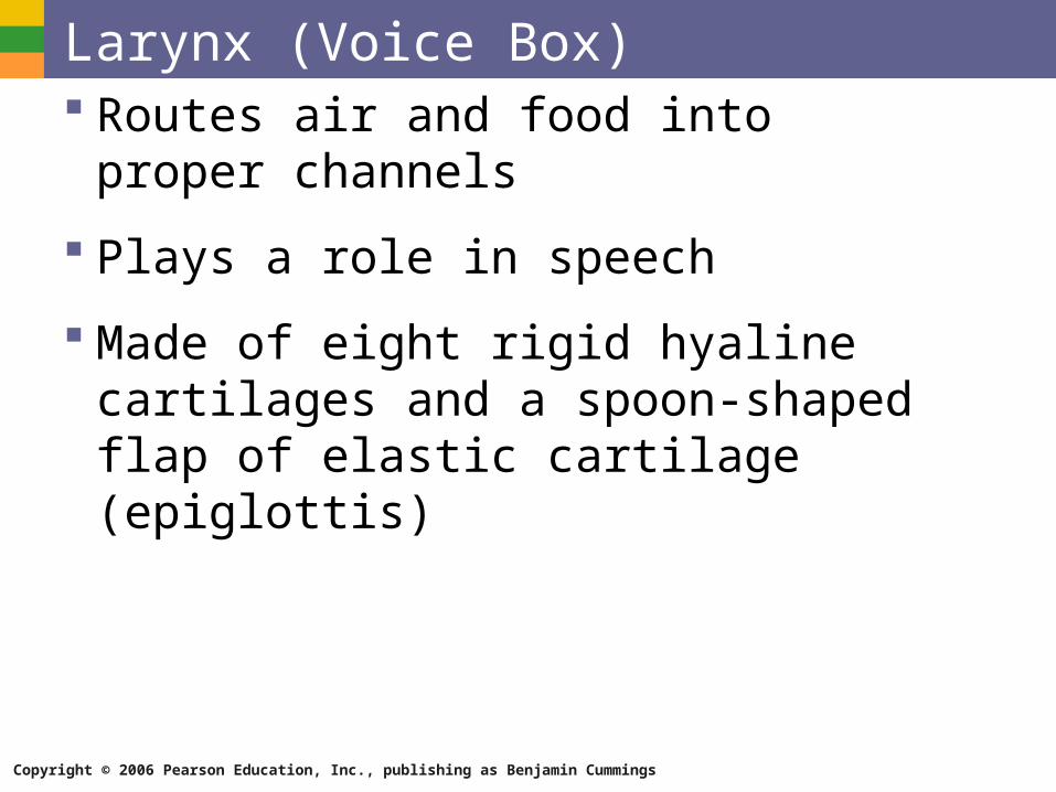
Larynx (Voice Box) Routes air and food into proper channels
Plays a role in speech
Made of eight rigid hyaline cartilages and a spoon-shaped flap of elastic cartilage (epiglottis)
Copyright © 2006 Pearson Education, Inc., publishing as Benjamin Cummings
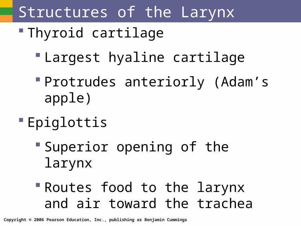
Structures of the Larynx Thyroid cartilage
Largest hyaline cartilage
Protrudes anteriorly (Adam’s apple)
Epiglottis
Superior opening of the larynx
Routes food to the larynx and air toward the trachea
Copyright © 2006 Pearson Education, Inc., publishing as Benjamin Cummings
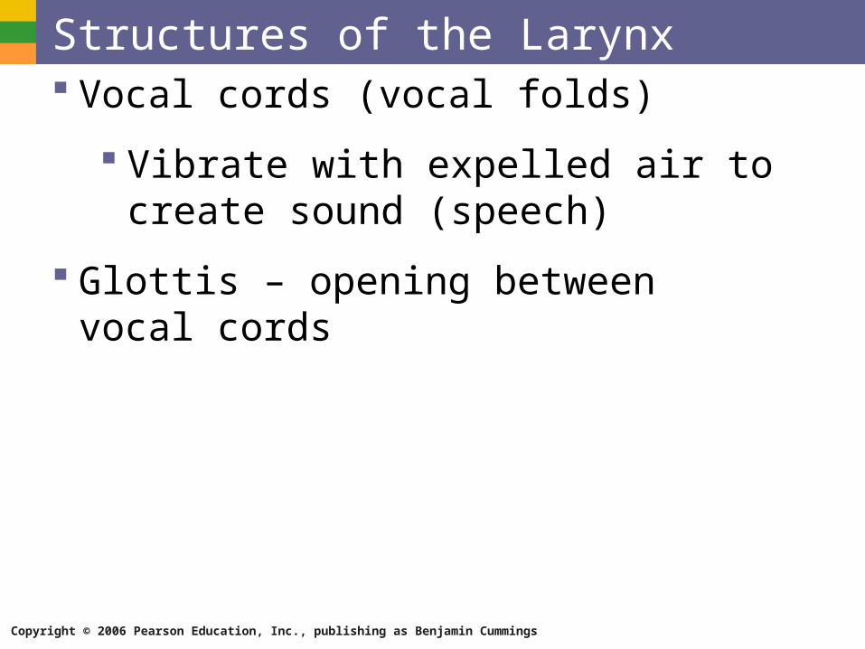
Structures of the Larynx Vocal cords (vocal folds)
Vibrate with expelled air to create sound (speech)
Glottis – opening between vocal cords
Copyright © 2006 Pearson Education, Inc., publishing as Benjamin Cummings
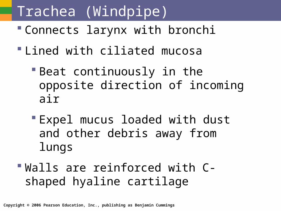
Trachea (Windpipe) Connects larynx with bronchi
Lined with ciliated mucosa
Beat continuously in the opposite direction of incoming air
Expel mucus loaded with dust and other debris away from lungs
Walls are reinforced with C-shaped hyaline cartilage
Copyright © 2006 Pearson Education, Inc., publishing as Benjamin Cummings
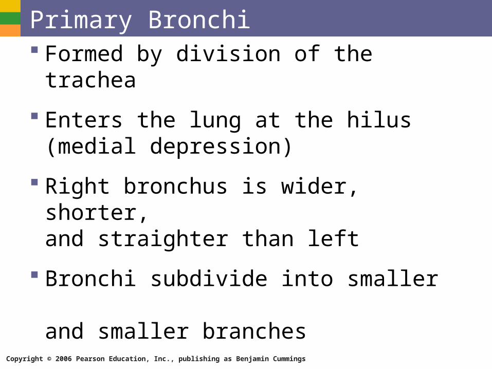
Primary Bronchi Formed by division of the trachea
Enters the lung at the hilus (medial depression)
Right bronchus is wider, shorter, and straighter than left
Bronchi subdivide into smaller and smaller branches
Copyright © 2006 Pearson Education, Inc., publishing as Benjamin Cummings
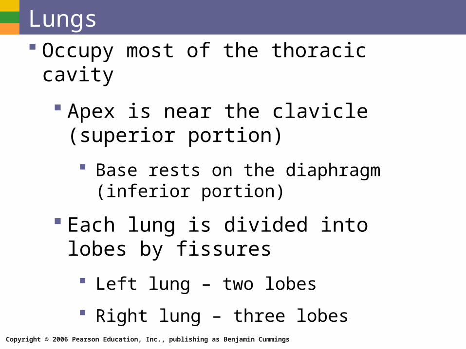
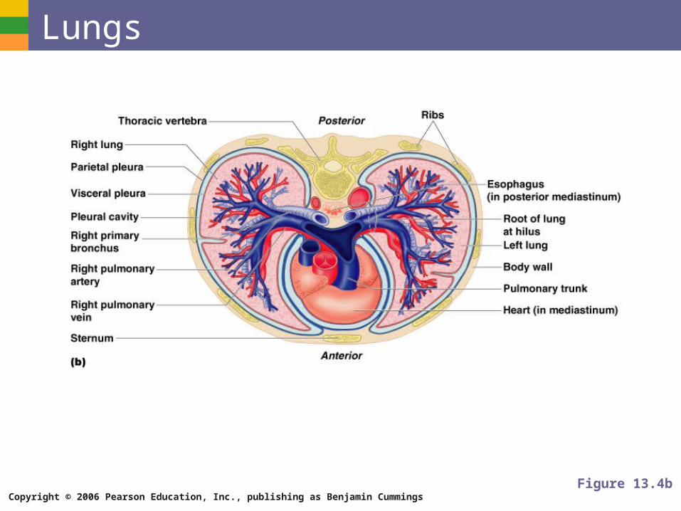
Lungs Occupy most of the thoracic cavity
Apex is near the clavicle (superior portion)
Base rests on the diaphragm (inferior portion)
Each lung is divided into lobes by fissures
Left lung – two lobes
Right lung – three lobes
Copyright © 2006 Pearson Education, Inc., publishing as Benjamin Cummings
Lungs
Figure 13.4b

Copyright © 2006 Pearson Education, Inc., publishing as Benjamin Cummings
Coverings of the Lungs Pulmonary (visceral) pleura covers the lung
surface
Parietal pleura lines the walls of the thoracic cavity
Pleural fluid fills the area between layers of pleura to allow gliding
Copyright © 2006 Pearson Education, Inc., publishing as Benjamin Cummings
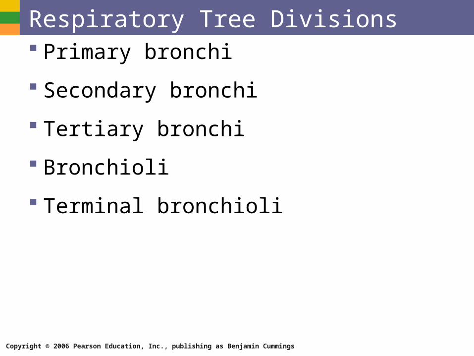
Respiratory Tree Divisions Primary bronchi
Secondary bronchi
Tertiary bronchi
Bronchioli
Terminal bronchioli
Copyright © 2006 Pearson Education, Inc., publishing as Benjamin Cummings
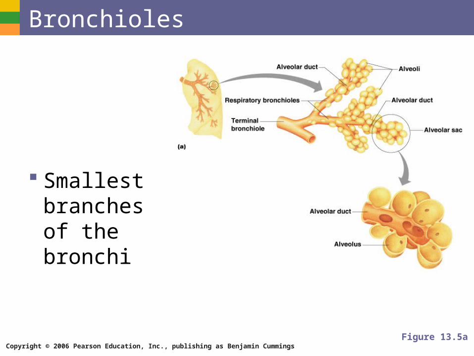
Bronchioles
Smallest branches of the bronchi
Figure 13.5a
Copyright © 2006 Pearson Education, Inc., publishing as Benjamin Cummings
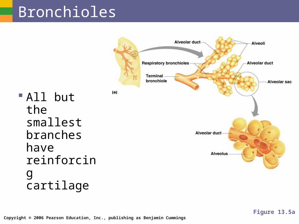
Bronchioles
All but the smallest branches have reinforcing cartilage
Figure 13.5a
Copyright © 2006 Pearson Education, Inc., publishing as Benjamin Cummings
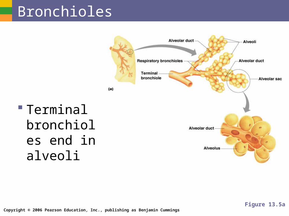
Bronchioles
Terminal bronchioles end in alveoli
Figure 13.5a
Copyright © 2006 Pearson Education, Inc., publishing as Benjamin Cummings
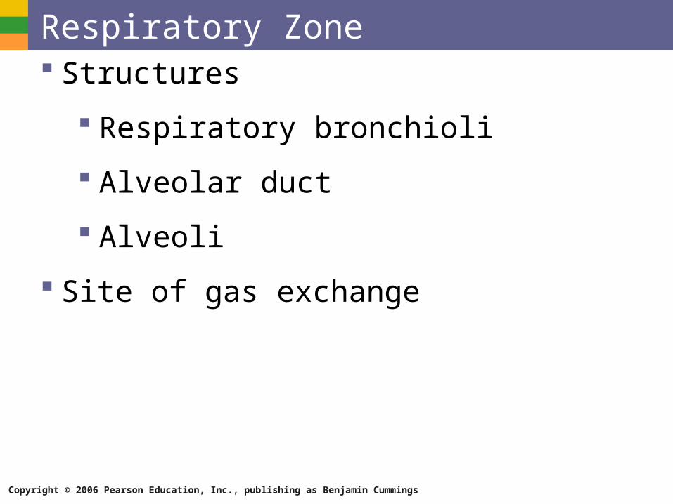
Respiratory Zone Structures
Respiratory bronchioli
Alveolar duct
Alveoli
Site of gas exchange
Copyright © 2006 Pearson Education, Inc., publishing as Benjamin Cummings
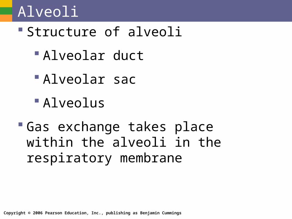
Alveoli Structure of alveoli
Alveolar duct
Alveolar sac
Alveolus
Gas exchange takes place within the alveoli in the respiratory membrane
Copyright © 2006 Pearson Education, Inc., publishing as Benjamin Cummings
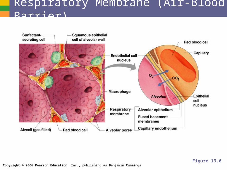
Respiratory Membrane (Air-Blood Barrier) Thin squamous epithelial layer lining alveolar
walls
Pulmonary capillaries cover external surfaces of alveoli
Copyright © 2006 Pearson Education, Inc., publishing as Benjamin Cummings
Respiratory Membrane (Air-Blood Barrier)
Figure 13.6
Copyright © 2006 Pearson Education, Inc., publishing as Benjamin Cummings
Gas Exchange Gas crosses the respiratory membrane by
diffusion
Oxygen enters the blood
Carbon dioxide enters the alveoli
Macrophages add protection
Surfactant coats gas-exposed alveolar surfaces
Copyright © 2006 Pearson Education, Inc., publishing as Benjamin Cummings
Events of Respiration Pulmonary ventilation – moving air in and
out of the lungs
External respiration – gas exchange between pulmonary blood and alveoli
Copyright © 2006 Pearson Education, Inc., publishing as Benjamin Cummings
Events of Respiration Respiratory gas transport – transport of
oxygen and carbon dioxide via the bloodstream
Internal respiration – gas exchange between blood and tissue cells in systemic capillaries
Copyright © 2006 Pearson Education, Inc., publishing as Benjamin Cummings
Mechanics of Breathing (Pulmonary Ventilation) Completely mechanical process
Depends on volume changes in the thoracic cavity
Volume changes lead to pressure changes, which lead to the flow of gases to equalize pressure
Copyright © 2006 Pearson Education, Inc., publishing as Benjamin Cummings
Mechanics of Breathing (Pulmonary Ventilation) Two phases
Inspiration – flow of air into lung
Expiration – air leaving lung
Copyright © 2006 Pearson Education, Inc., publishing as Benjamin Cummings
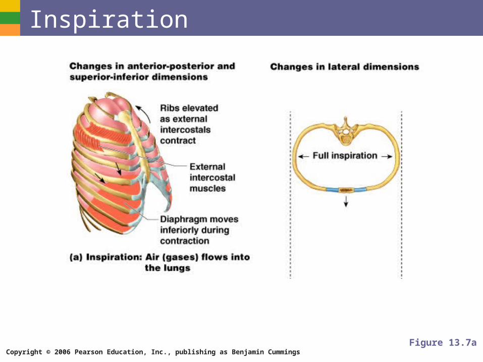
Inspiration Diaphragm and intercostal muscles contract
The size of the thoracic cavity increases
External air is pulled into the lungs due to an increase in intrapulmonary volume
Copyright © 2006 Pearson Education, Inc., publishing as Benjamin Cummings
Inspiration
Figure 13.7a
Copyright © 2006 Pearson Education, Inc., publishing as Benjamin Cummings
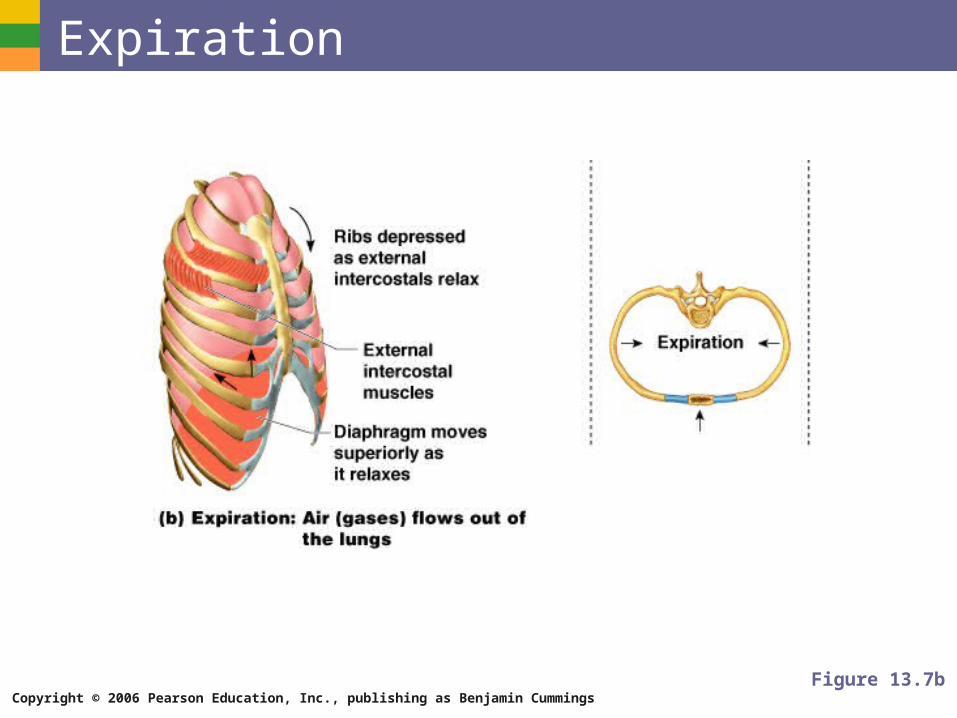
Expiration Largely a passive process which depends on
natural lung elasticity
As muscles relax, air is pushed out of the lungs
Forced expiration can occur mostly by contracting internal intercostal muscles to depress the rib cage
Copyright © 2006 Pearson Education, Inc., publishing as Benjamin Cummings
Expiration
Figure 13.7b
Copyright © 2006 Pearson Education, Inc., publishing as Benjamin Cummings
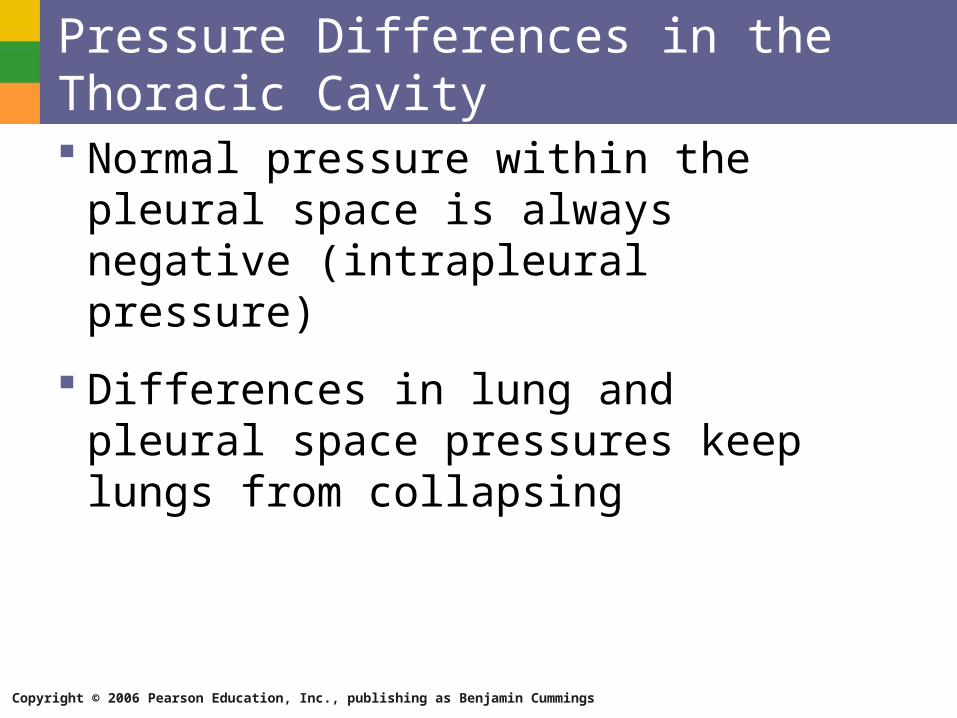
Pressure Differences in the Thoracic Cavity Normal pressure within the pleural space is
always negative (intrapleural pressure)
Differences in lung and pleural space pressures keep lungs from collapsing
Copyright © 2006 Pearson Education, Inc., publishing as Benjamin Cummings
Nonrespiratory Air Movements Can be caused by reflexes or voluntary
actions
Examples
Cough and sneeze – clears lungs of debris
Laughing
Crying
Yawn
Hiccup
Copyright © 2006 Pearson Education, Inc., publishing as Benjamin Cummings
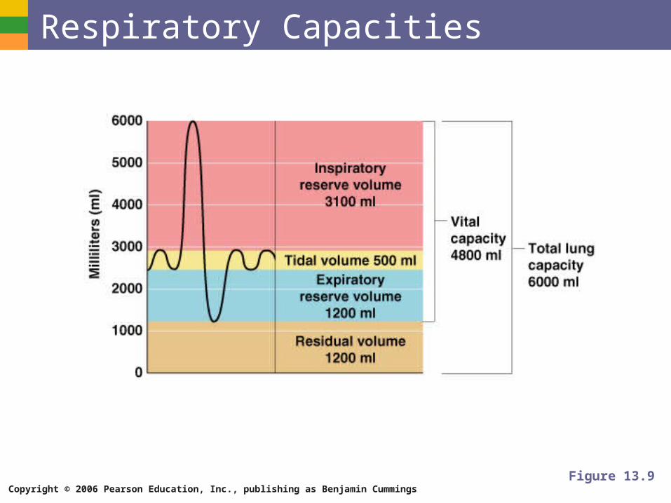
Respiratory Volumes and Capacities Normal breathing moves about 500 ml of air with
each breath (tidal volume [TV])
Many factors that affect respiratory capacity
A person’s size
Sex
Age
Physical condition
Residual volume of air – after exhalation, about 1200 ml of air remains in the lungs
Copyright © 2006 Pearson Education, Inc., publishing as Benjamin Cummings
Respiratory Volumes and Capacities Inspiratory reserve volume (IRV)
Amount of air that can be taken in forcibly over the tidal volume
Usually between 2100 and 3200 ml
Expiratory reserve volume (ERV)
Amount of air that can be forcibly exhaled
Approximately 1200 ml
Copyright © 2006 Pearson Education, Inc., publishing as Benjamin Cummings
Respiratory Volumes and Capacities Residual volume
Air remaining in lung after expiration
About 1200 ml

Copyright © 2006 Pearson Education, Inc., publishing as Benjamin Cummings
Respiratory Volumes and Capacities Vital capacity
The total amount of exchangeable air
Vital capacity = TV + IRV + ERV
Dead space volume
Air that remains in conducting zone and never reaches alveoli
About 150 ml
Copyright © 2006 Pearson Education, Inc., publishing as Benjamin Cummings
Respiratory Volumes and Capacities Functional volume
Air that actually reaches the respiratory zone
Usually about 350 ml
Respiratory capacities are measured with a spirometer
Copyright © 2006 Pearson Education, Inc., publishing as Benjamin Cummings
Respiratory Capacities
Figure 13.9