Manual of Standard Operating Procedures - UW … Flow Cytometry Facility SOP University of...
16
Pathology Flow Cytometry Facility SOP University of Washington Page 1 Updated 8/7/2017 Flow Cytometry Core Facility Department of Pathology University of Washington Manual of Standard Operating Procedures User Registration and Safe Working Practices BD FACSARIA III Cell Sorting in H-581C
Transcript of Manual of Standard Operating Procedures - UW … Flow Cytometry Facility SOP University of...

Pathology Flow Cytometry Facility SOP
University of Washington Page 1
Updated 8/7/2017
Flow Cytometry Core Facility Department of Pathology University of Washington
Manual of Standard Operating Procedures User Registration and Safe Working Practices BD FACSARIA III Cell Sorting in H-581C

Pathology Flow Cytometry Facility SOP
University of Washington Page 2
Table of Contents
I. Purpose
II. General Facility Information
III. Approval to Sort
IV. Pre-Sort Procedures
V. Post-Sort Procedures
VI. Unexpected Stream Shutoff During Sort
VII. Spill Procedures
VIII. Exposure Incident
IX. Waste Management
X. Aerosol Management System
XI. Quality Control and Maintenance
XII. Influx Operator Training and Experience
XIII. Resources & References
Attachments:
1. Exposure Control Plan: Emergency Information for posting
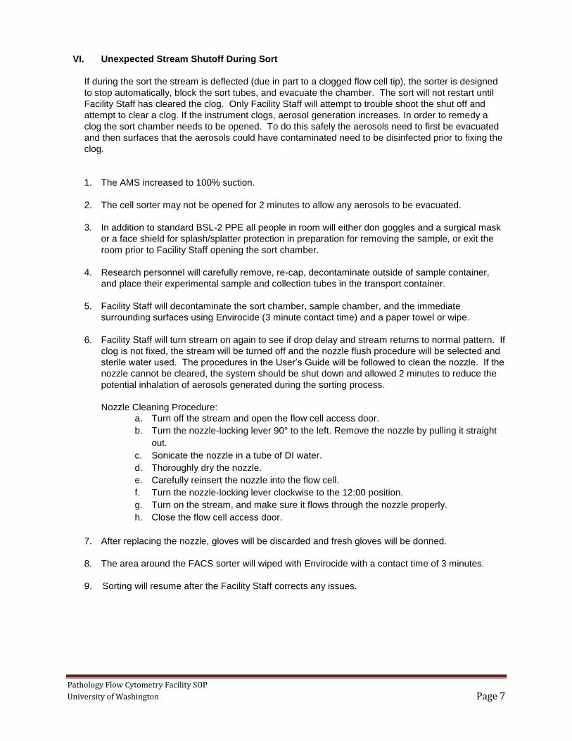
2. Biohazard Door Sign
3. Phone List
4. Path Flow Facility Biological Spill Log
5. Facility Orientation and Training Record
I. Purpose
The purpose of this standard operating procedure (SOP) is to outline the Biosafety plan for the
preparation, transport, and use of samples using a Becton Dickinson (BD) Biosciences FACS Aria III
cell sorter with aerosol management system (AMS) for work in the Flow Cytometry Facility. The
procedures outlined in this SOP are a supplement to general laboratory safety procedures outlined in
the UW Biosafety Manual.
High-speed cell sorting can produce aerosols that may present a health hazard to workers. The
procedures outlined in this SOP are to help ensure the safety of staff, proper use and maintenance of
the equipment, and to avoid cross-contamination of samples between users.
II. General Facility Information
The Pathology Flow Cytometry Core Facility is located on 5th floor of the Health Sciences Building, H-
581C. The facility is overseen by Dr. Peter Rabinovitch and maintained by Donna Prunkard,
manager of the facility.
The cell sorting room H-581C is approved for work at Biosafety Level 1 and 2 (BSL-1 and BSL-2).
Room H-581C is maintained under negative pressure relative to the larger lab space. All cells
exposed to or infected with bacterial or viral agents must be approved for cell sorting by
Environmental Health & Safety (EH&S) and the Institutional Biosafety Committee (IBC) on a case by
case basis. (See http://www.ehs.washington.edu/rbsresplan/bua.shtm for more information).

Pathology Flow Cytometry Facility SOP
University of Washington Page 3
The following table from EH&S outlines the Biosafety levels and the associated requirements that
must be met for sorting in the facility.
EH&S Summary of Biosafety Practices for Cell Sorting in H-581C
Type of cell/ procedure
Exempt BSL-1 BSL-1 BSL-2
Examples of Cells Fixed human cells, unfixed wild type cells from
murine or other non-human / non-primate species that
have NOT been exposed to any microbial agent (e.g.,
viral, bacterial, fungal, protozoan, parasitic) and
have not been genetically modified
or Cells determined by EH&S
and the IBC to be recombinant NIH-exempt
BSL-1.
Unfixed cells from murine or other non-human / non-primate species that have NOT been exposed to
any microbial agent but have been genetically modified using
non-viral methods (e.g. cells from transgenic animals or cells treated
with nucleic acids). or
Cells determined by EH&S and the IBC to be approved as non-
recombinant BSL-1 or recombinant BSL-1.
or nuclei released from human cells
following detergent treatment.
Unfixed cells from human or non-human primates
or Unfixed cells that have been
genetically modified using viral methods
or Cells exposed to microbial agents
(e.g., viral, bacterial, fungal, protozoan, parasitic)
AND Have been approved by EH&S and the IBC for BSL-2 containment and
sorting
IBC approval with current BUA to sort
in the Facility
Not required. Required. Required
Biosafety Training Required Required Required
blood borne Pathogen (BBP)
Training
Not required. Not required. Initial and annual renewal required for work with human cells and other
cells exposed to blood borne pathogens.
Sign-up Required. Include name, phone number, sample
description
Required. Include name, phone number, sample description and a
clear BSL-1 notation.
Required. Include name, phone
number, sample description and a clear BSL-2 notation.
Sample Transport No requirement Leak-proof secondary container. Leak-proof secondary container.
Room Restriction None. None. YES – door to H-581C must be closed during sort and the BSL-2
Biohazard sign posted on the outside.
Personal Protective Equipment (PPE)
Closed toe shoes. When manipulating samples (i.e.
loading/unloading samples), gloves, lab coat, and face
protection are recommended.
Closed toe shoes. When manipulating samples (i.e.
loading/unloading samples), gloves, lab coat, and face protection are strongly
recommended.
Spills: Lab coat, nitrile gloves, goggles/face shield required.
Closed toe shoes, lab coat and gloves required at all times. When
manipulating samples (i.e. loading/unloading samples), face
protection is also required.
Spills: Lab coat, nitrile gloves, goggles/faceshield required.
Waste Contaminated materials must be decontaminated prior to
disposal.
Gloves and other waste must be disposed of as biohazardous
waste. See SOP.
Gloves and other waste must be disposed of as biohazardous. See
CAF SOP.
The Biological Use Authorization (BUA) issued by EH&S will define what sample material can be
sorted in the Pathology Flow Cytometry Core Facility and designate what Biosafety Level applies.
Based on the Biosafety Level, the appropriate Sort SOP will be followed. Refer to sections IV & V.
Radioactive samples are prohibited in the Flow Cytometry Facility.
The facility may be used by researchers within and outside the University of Washington after completing
the prerequisite requirements specified in this SOP.

Pathology Flow Cytometry Facility SOP
University of Washington Page 4
The facility is available for sorting Monday – Friday from 10 am to 5:30. After hours usage may be
approved for users who have received relevant instruction from the facility staff, are self-sufficient with the
instrument software, and are trained on the equipment. If there is unexpected stream shutoff during after-
hours usage the sort must be terminated and instrument shut down procedures carried out.
III. Approval to Sort
The following are required from each user:
1. Completion of facility orientation and training, familiarity with contents of this SOP, and signature
on Pathology Flow Cytometry Orientation and Training Form.
Pathology Flow Cytometry facility training will be provided by the facility manager or assistant. It
will include an overview of the calendar system, safety requirements, and training on this SOP.
Safety training will be included in the first cytometer appointment. This training will be
documented on the Flow Cytometry Orientation and Training Form. The training records will be
maintained by the flow facility staff.
2. Biological Use Authorization (BUA) from Environmental Health & Safety (EH&S) for samples that
are not listed as exempt from the “EH&S Summary of Biosafety Practices for Cell Sorting in H-
581C” table in Section II.
EH&S Biological Use Authorization (BUA). The National Institutes of Health Office of
Biotechnology Activities (NIH OBA) mandates that the Institutional Biosafety Committee (IBC)
review and approve all research that involves recombinant DNA. The UW IBC reviews and
approves all research involving infectious agents. Staff safety for work involving blood and other
potentially infectious materials that may contain blood borne pathogens (BBP’s) is mandated by
the State of Washington Department of Safety and Health and enforced by the Department of
Labor and Industries (WAC-296-823). All researchers and facilities at the UW must comply with
these regulations.
PI's are responsible for obtaining BUA's, which apply to all members of their research groups.
Contact EH&S Research and Occupational Safety Office (RBSO) via email at
[email protected], via UW mailbox at 357165, or via fax at 206-221-3068. For a BUA
application, see: http://www.ehs.washington.edu/rbsresplan/bua.shtm.
Each BUA includes information on cell lines and infectious agents used by each laboratory, their
appropriate level of containment, and sites at which they can be used.
3. Biosafety Training
All users are required to have completed the UW online Biosafety Training module. http://www.ehs.washington.edu/psotrain/corsdesc.shtm
3. Blood borne Pathogens (BBP) Training Documentation
Employees who have potential for exposure to human blood or other potentially infected materials
which includes human cells and cell lines must be enrolled in the UW BBP Program. The
Program includes offering of the Hepatitis B vaccine, initial and annual training, and a written site-
specific Exposure Control Plan (ECP). The Path Flow Facility's ECP is covered in this document.
http://www.ehs.washington.edu/psotrain/corsdesc.shtm
The Facility Manager has the right to deny sorting of any cells that do not have prior approval by
EH&S to sort in the facility or that have not been clearly disclosed by the user at the time of sign-up.

Pathology Flow Cytometry Facility SOP
University of Washington Page 5
IV. Pre-Sort Procedures
After the steps in Section III are complete and approval to use the facility is granted, the following
procedures must be followed by facility users prior to sorting.
1. Sign-up: Each user is responsible for signing up using the online scheduling system
(https://www.pathology.washington.edu/research/flow/). Users sign up for sorting time in 15
minute increments. Fifteen minutes should be left unscheduled between all appointments to
allow for decontamination and cleaning and for calibration for the next user.
a. Sign-up information MUST include: user name, sample description, BSL level, budget
number, collection tube and nozzle size.
b. Each entry is approved after the Facility Staff has looked at the request and made sure
that there will be appropriate staff and all the required information is provided.
c. All BSL-2 sorts must include a distinct BSL-2 notation as well as additional notation
further clarifying the sample description (e.g. ES cells, patient samples, transfected cells,
infected cells etc).
2. Sample Preparation: Samples must be completely prepped (washed in saline, filtered, and
placed on ice) prior to arrival to the Cell Analysis Facility. Users are responsible for contacting
the facility staff to discuss proper sample preparation prior to sorting. This includes single cell
suspensions, cell washing, and nozzle tip considerations.
3. Sample Transport: All BSL-2 and Recombinant DNA samples must be transported to and from
the facility in an appropriate container: i.e. - a leak-proof, puncture-resistant plastic carrier with a
secure lid, which would be able to contain the specimen in case of breakage of the primary
container.
4. Biohazard Warning Sign: The door to H581c (the Aria sort room) shall be closed and the
Biohazard door sign displayed (attached) when any BSL-2 sample is in the facility. Users are
required to verify that the Biohazard warning sign is displayed upon entry with a BSL-2 sample.
5. Personal Protective Equipment (PPE) requirements for user: Upon entering the Aria sort room,
each individual must sanitize their hands and put on PPE as specified by EH&S appropriate to
the level of containment necessary for the samples being sorted. See the table EH&S Summary
of Biosafety Requirements for Cell Sorting in H-581C (section II).
6. Mixed Biosafety Level Procedures. Cells requiring different Biosafety levels (e.g. BSL-1 and BSL-
2) may be present in H581C for sorting or analyzing at the same time. It is the responsibility of
users with BSL-2 cells to notify all others present in the room to don the appropriate BSL-2 PPE.
It is the responsibility of BSL-1 users to check the door sign for BSL-2 work in progress.
7. Sample Handling: Samples may only be uncapped immediately prior to placement in the Aria
loading chamber. Operations other than loading that require uncapping of samples must take
place prior to entry into the Cell Analysis Facility, or behind the biohazard splash shield in the sort
room. Except for exempt samples although it is still recommended.
8. All use of needles requires pre-approval. If a sample needs to be re-filtered to remove clumps,
the users must transport sample to a Biosafety Cabinet in an approved container and return to
the facility with the filtered sample.

Pathology Flow Cytometry Facility SOP
University of Washington Page 6
The facility staff will perform these following procedures prior to the sort:
a. Start the flow cytometer system.
b. Check waste container, empty if necessary and add bleach to 10% tank volume.
c. Ensure that the ULPA filter is completely seated against the bottom of the evacuator filter well
and that the tubing is securely attached to the filter and instrument manifold.
d. Ensure that air filter is installed in the sort collection chamber door.
e. Close sort collection chamber door.
f. Turn on main power on back of aerosol management system evacuator. Set the suction
control rate to 20% and no higher. Higher rates can affect stability of the side streams. Verify
that the filter flow gauge reads less than 2.4 inches of water. If gauge reads 2.4 or higher,
the filter and tubing needs to be replaced prior to sort.
g. Make sure that the sheath tank is full.
h. QC the instrument with manufacturer recommended alignment bead to make sure that
laser timing and area scaling are within recommended tolerances.
i. Test the stability of the sort side streams and droplet break-off with the aid of the AccuDrop
management system.
j. Start sort and monitor the sort performance using the AccuDrop camera.
V. Post-Sort Procedures
The following procedures are required after sorts. Daily equipment shutdown procedures specified in
the user’s guide will be performed by the facility staff or trained after-hours user.
1. Trained user will stop the flow of sample into sort collection chamber.
2. Trained user will press up arrow to increase suction to 100% on the AMS.
3. Wait 1 minute to allow any aerosols to be evacuated.
4. Trained user will carefully remove, re-cap, decontaminate outside of sample containers, and
place their experimental sample and collection tubes in the transport container.
5. Trained user will run 10 % bleach for 5 minutes followed by a one minute backflush of the sample
line.
6. Facility Staff or trained user will decontaminate the sort chamber, sample chamber, immediate
surrounding surfaces, and any additional surfaces which were used (i.e.- computer workspace,
keyboard, mouse, or lab bench/splash shield area) with Envirocide (air dry)
7. Facility Staff will empty the waste tank after the waste liquid has been exposed to 10% bleach for
at least 30 minutes. The sheath waste tank will be emptied as needed and 1000 ml of bleach will
be added to the empty tank.
8. Users will remove PPE upon exiting in the following order:
a. Discard gloves in the biohazardous waste containers.
b. Remove lab coat.
c. Remove eye protection and decontaminate by wiping with decontamination wipes. Dispose of
waste wipe in the red biohazard container. Replace eye protection in holder.
9. Sanitize hands.
10. Exit the sort room and IMMEDIATELY thoroughly wash hands at nearest sink.
Pathology Flow Cytometry Facility SOP
University of Washington Page 7
VI. Unexpected Stream Shutoff During Sort
If during the sort the stream is deflected (due in part to a clogged flow cell tip), the sorter is designed
to stop automatically, block the sort tubes, and evacuate the chamber. The sort will not restart until
Facility Staff has cleared the clog. Only Facility Staff will attempt to trouble shoot the shut off and
attempt to clear a clog. If the instrument clogs, aerosol generation increases. In order to remedy a
clog the sort chamber needs to be opened. To do this safely the aerosols need to first be evacuated
and then surfaces that the aerosols could have contaminated need to be disinfected prior to fixing the
clog.
1. The AMS increased to 100% suction.
2. The cell sorter may not be opened for 2 minutes to allow any aerosols to be evacuated.
3. In addition to standard BSL-2 PPE all people in room will either don goggles and a surgical mask
or a face shield for splash/splatter protection in preparation for removing the sample, or exit the
room prior to Facility Staff opening the sort chamber.
4. Research personnel will carefully remove, re-cap, decontaminate outside of sample container,
and place their experimental sample and collection tubes in the transport container.
5. Facility Staff will decontaminate the sort chamber, sample chamber, and the immediate
surrounding surfaces using Envirocide (3 minute contact time) and a paper towel or wipe.
6. Facility Staff will turn stream on again to see if drop delay and stream returns to normal pattern. If
clog is not fixed, the stream will be turned off and the nozzle flush procedure will be selected and
sterile water used. The procedures in the User’s Guide will be followed to clean the nozzle. If the
nozzle cannot be cleared, the system should be shut down and allowed 2 minutes to reduce the
potential inhalation of aerosols generated during the sorting process.
Nozzle Cleaning Procedure:
a. Turn off the stream and open the flow cell access door.
b. Turn the nozzle-locking lever 90° to the left. Remove the nozzle by pulling it straight
out.
c. Sonicate the nozzle in a tube of DI water.
d. Thoroughly dry the nozzle.
e. Carefully reinsert the nozzle into the flow cell.
f. Turn the nozzle-locking lever clockwise to the 12:00 position.
g. Turn on the stream, and make sure it flows through the nozzle properly.
h. Close the flow cell access door.
7. After replacing the nozzle, gloves will be discarded and fresh gloves will be donned.
8. The area around the FACS sorter will wiped with Envirocide with a contact time of 3 minutes.
9. Sorting will resume after the Facility Staff corrects any issues.

Pathology Flow Cytometry Facility SOP
University of Washington Page 8
VII. Spill Procedures
In the event of a spill inside the cell sorter, notify the flow facility manager to clean spill. Flow facility
staff will decontaminate the sort chamber, sample chamber, immediate surrounding surfaces, and
any additional surfaces which were affected with 10% bleach with a 20 minute contact time.
In the event of a spill outside the cell sorter, alert others in the room to avoid the spill area. Place
paper towels directly on top of the spill. Gently pour a 10% bleach/water solution on the outside
edges and corners of the paper towel and allow it to wick into the spill area. After a 20 minute contact
time, dispose of the paper towel into a biohazard waste container. Notify Cell Analysis Facility Staff
and record in the spill log.
After cleaning spill, change gloves and any other soiled PPE.
VIII. Exposure Incident
An exposure incident is defined as eye, mouth, mucous membrane, non-intact skin, or parenteral
contact with blood or potentially infectious materials or chemicals. Procedures to follow after an
exposure incident are posted in the facility for quick reference (also attached to this SOP). See
http://www.ehs.washington.edu/forms/pso/exposureresponseposter.pdf
For a medical emergency, go to the nearest emergency room or call 911.
In the event of an exposure incident, it is essential that first aid procedures be initiated immediately.
Immediately wash the exposed area with soap and water, or flush mucous membranes with water for
15 minutes. The eye wash is located next to sink in H581.
The Facility Manager will assist the exposed worker in seeking the necessary and immediate medical
evaluation and consultation following an exposure incident.
Where and how to seek care: Monday through Friday 9:00 a.m. to 5:00 p.m. contact the Employee
Health Center-UW (EHC-UW) at 685-1026. After hours or if the clinic staff is not available, go to the
UWMC Emergency Department (NE-207 UWMC). Be prepared to give the healthcare providers
information about the exposure. Tell them that you are a UW employee and provide information
about the agent or cells involved in the accident. Information such as agent, route of exposure, dose
or concentration, unusual characteristics of the agent, animal infection, cell line, and PI contact
information.
All work related exposures, incidents, and near misses must be reported on the UW Online Accident
Reporting System. Here is the link for report: http://www.ehs.washington.edu/ohsoars/index.shtm
IX. Waste Management
Decontamination and disposal of waste is the responsibility of the Facility Staff.
Liquid Waste: Small volumes of 1ml or less can be left in the sample tube and disposed of in the
sharps container. Larger volumes must be diluted with fresh bleach while still in the sorter room to a
final bleach concentration of 10% (0.5% sodium hypochlorite) for at least 30 minutes prior to disposal
in the H581sink. Flush the sink with water for 1 minute. Samples containing mutagenic dyes must be
disposed of in charcoal waste bottle.
Waste tank: The waste tank will be emptied as needed. The tank will be disconnected from the wet
cart before emptying. The waste tank cap must not be wetted. If wet, the filter in the cap will cause

Pathology Flow Cytometry Facility SOP
University of Washington Page 9
the tank to malfunction; to keep the cap dry, it will be placed on the bench, label side up, when it is
not on the tank. If liquid is seen in the waste cap trap, the drain plug will be removed and liquid fully
drained before replacing the plug.
Solid Waste: All solid waste shall be disposed of as biohazardous waste and will be collected in either
a red sharps container or in a biohazardous waste bag contained inside a rigid, leak-proof container.
This container must be labeled or clearly display the bag’s biohazard symbol. Solid waste includes
FACS tubes, pipettes and soiled tissues or towels, and gloves. Filled containers will be transported to
the autoclave room located in Lab Services, T-276. Containers will be tagged with temperature
indicator tape which will be monitored after autoclaving.
X. Aerosol Management System
This section applies only to facility staff
The BD™ aerosol management system (AMS) is a device that promotes the containment of aerosols
by evacuating the sort collection chamber in the BD FACSAria™ flow cytometer. The option uses an
attached vacuum source to rapidly evacuate aerosolized particles through an ultra-low penetrating air
filter during routine sorting or analysis. The AMS is handled and maintained by the facility manager.
The AMS includes the following:
1. Evacuator to generate negative pressure
2. Ultra-low penetrating air (ULPA) filter to trap particles, with attached tubing that connects the
evacuator to the instrument replaced every 180 hours of usage. Tracking will take place in a
log book.
3. Air filter for the sort collection chamber door changed every 30 days. Tracking will take place
in a log book.
The AMS evacuates the waste air from the sort collection chamber using the evacuator. The
waste air filtered through the external AMS Ultra Low Particle Aerosol (ULPA) filter (0.12 micron).
The ULPA filter has 99.9999% filter efficiency down to and including particles 0.1 microns in size.
For particles of this size, 1 out of 1,000,000 is expected to pass through the ULPA filter. The
evacuator will be turned on before sorting and turned off after sorting. Generated sorter aerosols
are 0.2microns in size and are effectively captured by the AMS when working properly.
The AMS ULPA filter (0.12 micron) and tubing will be replaced according to the Manual. It will be
replaced when the flow gauge indicator is >2.4 at a 20% flow setting, when the filter life gauge
falls to 20% or when the red filter life indicator LED is blinking.. PPE required lab coat, gloves,
eye protection.
Changing the ULPA filter procedure
The filter will be changed according to the manual instructions. Only the Facility Staff member
changing the filter will be in the room when the filter is being changed.
1. Close the door and display the Biohazard sign.
2. Turn the AMS on and to 100% flow setting.
3. Spray Envirocide into the sort collection chamber so that it is pulled into and evacuated
through the AMS tubing to the ULPA filter.
4. Let run for 10 minutes.
5. Turn off evacuator main power and disconnect the electrical plug.
6. In addition to the required PPE a N95 disposable, filtering face piece respirator will be worn
while removing and disposing of the filter and tubing.
7. Disconnect tubing from the manifold and cover the tube end.
8. Remove spring-loaded filter.
9. Lift off the ULPA filter and attached tubing from the evacuator and dispose of both the filter
and tubing as biohazardous waste.
10. Spray the AMS and surrounding area with Envirocide and let sit for three minutes.

Pathology Flow Cytometry Facility SOP
University of Washington Page 10
11. Insert new ULPA filter into the evacuator filter well.
12. Push down on the filter to ensure that it is seated against the bottom of the filter chamber.
13. Lift the spring loaded filter hold-down and place it on top of the filter.
14. Connect the evacuator power plug to the power source.
15. Press and hold the filter Life restart button on the membrane panel for 5-10 seconds.
16. Connect one end of the replacement tubing to the ULPA filter and the other end to the tubing
manifold.
17. Perform the Glo Germ aerosol containment procedure.
Glo Germ aerosol containment procedure
Every time the ULPA filter is changed, the AMS will be tested for proper functioning. The AMS
will be tested under three conditions: aerosol generation without AMS(positive control), aerosol
generation with AMS (worst-case), and proper instrument function with AMS. The AMS must be
tested under simulated worst-case failure mode. In this mode the instrument is set to 70 psi and
50,000 particles/second, with the waste catcher blocked to create large aerosol. This is done by
covering waste catcher with a cut off piece of sample tubing.
The Glo Germ Test, described below will be performed each time the filter is changed under each
of the three conditions.
1. Measurements are taken using glass slides inside an Aero Tech air sampler directly on top of
the chamber and approximately 2 feet away to simulate operator positioning.
2. Measurements will be taken at 5 minute time points to allow for Glo Germ particles to
accumulate on microscope slides.
3. Less than 1 particle per slide, under the condition of proper instrument function with AMS, will
be considered as verification of proper working AMS.
The flow cytometer must pass all tolerances of aerosol containment as described in this
procedure. If these tolerances are not met, BSL-2 sorting is not permitted until the issue is
resolved
XI. Quality Control and Maintenance
The FACSAria II is under an annual service contract and receives two quality control service calls per
year. All manufacturer recommended procedures in Chapter 6 of the User’s guide will be followed.
Fluidic lines are check daily for leaks or crimping. The manufacturer will be contacted before making
any instrument modifications. Testing must also be done whenever changes are made to the cell
sorter that may affect escape of aerosols, e.g., installation of a new drive head or flow cell,
replacement of the sort chamber door, or alterations in the aerosol management system.
Quality control beads are run daily to make sure the stream, break-off, and lasers fall within
recommended tolerances.
Room Pressurization Check: Weekly smoke tests will be performed to ensure that Room H581C
is under negative pressure relative to the adjacent laboratory space.
Prior to each sort, the sorter will be carefully checked for wet areas indicating leaks in the tubing.
Inspect tubing for cracks and signs of stress, particularly around the fittings and valve junctions.
Inspect sheath lines and waste lines, and replace any leaking tubing.
Failure to pass any of the above QC tests which cannot be immediately corrected requires that all
BSL-2 sorts must cease until the issue is resolved.

Pathology Flow Cytometry Facility SOP
University of Washington Page 11
XII. Influx Operator Training and Experience
Only experienced flow cytometry operators are permitted to operate the Influx. The operator will be
trained carefully in the proper instrument operation and all relevant safety procedures, including
aerosol containment testing. Initial training using BSL1, or fixed material should be mastered before
sorting BSL2 samples by any operator.
XIII. Resources and References
a) EH&S Research & Occupational Safety Office 206-221-7770
a. Ask for a Biosafety Officer
b) UW BioSafety Manual: http://www.ehs.washington.edu/rbsbiosafe/bsmanualindex.shtm
c) Washington State Bloodborne Pathogen Regulations:
http://www.lni.wa.gov/safety/rules/chapter/823/
https://www.ehs.washington.edu/rbs/bbp.shtm
d) NIH/OBA guidelines for research involving recombinant DNA molecules:
http://osp.od.nih.gov/office-biotechnology-activities/biosafety/nih-guidelines
e) Centers for Disease Control and Prevention (CDC): Biosafety in Microbiological and Biomedical
Laboratories (BMBL) 5th Edition. http://www.cdc.gov/OD/ohs/biosfty/bmbl5/bmbl5toc.htm
Literature Cited for Glo Germ Protocol
1. Schmid I, Lambert T, Ambrozak D, Marti GE, Moss DM, Perfetto SP. International Society for
Analytical Cytology and Biosafety Standard for Sorting of Unfixed Cells. Cytometry 2007;
71A:414-437
2. Perfetto SP, Ambrozak DR, Koup RA, Roederer M. Measuring Containment of Viable
Infectious Cell Sorting in High-Velocity Cell Sorters. Cytometry 2003; 52A:122-130
3. Oberyszyn AS, Robertson FM. Novel Rapid Method for Visualization of Extent and Location
of Aerosol Containment During High-Speed Sorting of Potentially Biohazardous Samples.
Cytometry 2001; 43:217-222
Pathology Flow Cytometry Facility SOP
University of Washington Page 12
Pathology Flow Cytometry Facility SOP
University of Washington Page 13
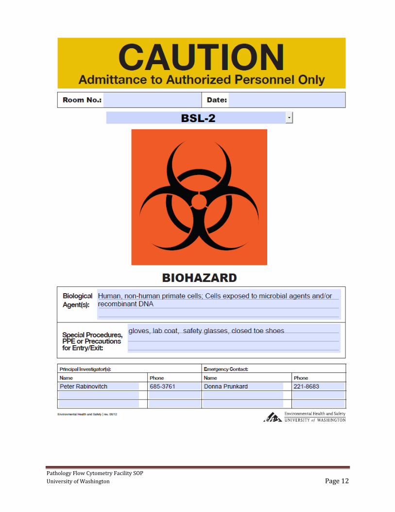
Exposure Control Plan Emergency Information
Eye / Mucous Membrane Exposure
Wounds or Needle sticks
1. Flush immediately at
nearest eyewash station for 15
min.
1. Wash the area immediately.
Use warm water and sudsing
soap to scrub the area for 15 min.
2. Notify your supervisor, they can help.
3. Seek care
Mon-Fri 9am-5pm:
Contact the Employee Health Center-UW (EHC-UW) at 685-1026
After Hours:
go to the UWMC Emergency Department (NE-207 UWMC)
-or-
nearest Emergency Department
4. Be prepared to give the following information to the healthcare providers:
1- Tell them you are a UW employee
2- Have information about the agent or cells involved in the accident, including information such
as agent, route of exposure, dose or concentration, unusual characteristics of the agent, animal
infection, cell line, and PI contact information.
5. Complete the On-line Accident Report
http://www.ehs.washington.edu/ohsoars/index.shtm
Pathology Flow Cytometry Facility SOP
University of Washington Page 14
Pathology Flow Cytometry Facility
Phone List
Flow Cytometry Facility 206-221-8683
Donna Prunkard, Facility Manager
Cell phone 206-616-8761
Jeanne Fredrickson, 206-616-8715
Dr. Peter Rabinovitch, 206-685-3761

Pathology Flow Cytometry Facility SOP
University of Washington Page 15
Path Flow Facility
Biological Sample Spill Log
Date:___________________ Time:_________________
Type of sample:_______________________________________
Personnel who cleaned the spill:__________________________
Manager notified:______________________________________
Any Exposure Reported? No Yes If yes:
Notify Your Supervisor
Complete on-line Accident Report:
http://www.ehs.washington.edu/ohsoars/index.shtm

Pathology Flow Cytometry Facility SOP
University of Washington Page 16
Pathology Flow Cytometry Facility
Orientation and Training
User's Name__________________________________
UW email address:_______________________________
Department___________________________ PI Name:______________________
General Laboratory Training
Have you received the following? Date
Registration and training for use of on-line scheduling calendar. Yes
UW Biosafety Training up to date. Yes
Facility orientation and training (will take place at time of first appointment).
Location of EH&S Manual and Path Flow SOP
H581c entry and exit procedures, door sign policy
Location of PPE
Decontamination of work area
Yes
Training for BSL2 cells
Date
I understand that a BUA must be on file with us before cells that require IBC approval can be brought into the facility. See chart on page 3. Yes
Training on the Aria cell sorter
Aerosol management system (AMS)
Procedures on completion of sorting.
Decontamination of work area
Yes
Emergencies – what to do for:.
Sorter clogs
Spills
exposures
Yes
User's Signature ________________________________________ Staff initials __________