MANUAL FOR THE DIAGNOSIS AND TREATMENT OF … · 1 Preface Leishmaniasis is a major health problem...
49
Republic of Sudan Federal Ministry of Health Neglected Tropical Disease Division (NTDs) MANUAL FOR THE DIAGNOSIS AND TREATMENT OF LEISHMANIASIS October 2014
Transcript of MANUAL FOR THE DIAGNOSIS AND TREATMENT OF … · 1 Preface Leishmaniasis is a major health problem...

Republic of SudanFederal Ministry of Health
Neglected Tropical Disease Division (NTDs)
MANUAL FOR THE DIAGNOSIS AND TREATMENTOF
LEISHMANIASIS
October 2014

1
TABLE OF CONTENTS
OBJECTIVES AND TARGETS OF THE MANUAL: ................................................................................................ 3
CHAPTER 1 .....................................................................................................................................4
INTRODUCTION ............................................................................................................................4
EPIDEMIOLOGY OF LEISHMANIASIS IN SUDAN ......................................................................................4PARASITES............................................................................................................................................6VECTORS ..............................................................................................................................................6RESERVOIR ............................................................................................. ERROR! BOOKMARK NOT DEFINED.TRANSMISSION CYCLES ........................................................................................................................7
CHAPTER 2 .....................................................................................................................................8
CLINICAL FEATURES OF VISCERAL LEISHMANIASIS AND DIAGNOSIS.........................8
VL CLINICAL FEATURES: MAIN SYMPTOMS AND SIGNS .........................................................................8THE MAIN SYMPTOMS AND SIGNS OF VL ARE: ......................................................................................8
DIAGNOSIS OF VISCERAL LEISHMANIASIS (VL): .................................................................9
CLINICAL DIAGNOSIS ............................................................................................................................9LABORATORY DIAGNOSIS ...................................................................................................................10
Parasitological diagnosis ..............................................................................................................10Serological diagnosis.....................................................................................................................11
FIRST-LINE TREATMENT FOR VISCERAL LEISHMANIASIS .....................................................................14OTHER ALTERNATIVE OPTIONS FOR FIRST LINE TREATMENT FOR VL .................................................14SECOND-LINE TREATMENT OF VISCERAL LEISHMANIASIS ...................................................................17
Follow-up after treatment ............................................................................................................21Criteria of cure .............................................................................................................................. 21Case definition according to follow-up .........................................................................................21
CUTANEOUS LEISHMANIASIS .................................................................................................23
MUCOSAL LEISHMANIASIS......................................................................................................24
CLINICAL FEATURES OF LEISHMANIA/HIV CO-INFECTION..................................................................27DIAGNOSIS OF VISCERAL LEISHMANIASIS IN HIV-POSITIVE PATIENTS ................................................27TREATMENT OF HIV/LEISHMANIA CO-INFECTION ..............................................................................28
Latex agglutination test (KAtex) ..................................................................................................32Leishmanin skin test ......................................................................................................................33
ANNEX 3: DEFINITIONS .............................................................................................................34
ANNEX 4: ASPIRATION OF LYMPH NODE, SPLEEN AND BONE MARROW ....................36
ANNEX 6: REFERENCES ............................................................................................................44

2
Acknowledgements
The National Leishmaniasis Control Programme (LCP), Federal Ministry of Health,
Sudan, would like to acknowledge all the efforts spent on studying, controlling and
reducing morbidity and mortality of leishmaniasis in Sudan, which culminated in the
formulation of this manual in April 2004, updated in 2012.
We would like to express our thanks to all institutions, organizations, research
groups and individuals for their support.

1
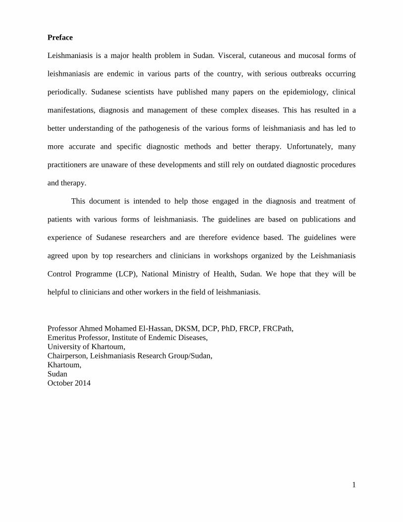
Preface
Leishmaniasis is a major health problem in Sudan. Visceral, cutaneous and mucosal forms of
leishmaniasis are endemic in various parts of the country, with serious outbreaks occurring
periodically. Sudanese scientists have published many papers on the epidemiology, clinical
manifestations, diagnosis and management of these complex diseases. This has resulted in a
better understanding of the pathogenesis of the various forms of leishmaniasis and has led to
more accurate and specific diagnostic methods and better therapy. Unfortunately, many
practitioners are unaware of these developments and still rely on outdated diagnostic procedures
and therapy.
This document is intended to help those engaged in the diagnosis and treatment of
patients with various forms of leishmaniasis. The guidelines are based on publications and
experience of Sudanese researchers and are therefore evidence based. The guidelines were
agreed upon by top researchers and clinicians in workshops organized by the Leishmaniasis
Control Programme (LCP), National Ministry of Health, Sudan. We hope that they will be
helpful to clinicians and other workers in the field of leishmaniasis.
Professor Ahmed Mohamed El-Hassan, DKSM, DCP, PhD, FRCP, FRCPath,Emeritus Professor, Institute of Endemic Diseases,University of Khartoum,Chairperson, Leishmaniasis Research Group/Sudan,Khartoum,SudanOctober 2014

2
AbbreviationsCL Cutaneous leishmaniasisDAT Direct agglutination testICT Immunochromatographic testIFAT Immunofluorescence antibody testIM IntramuscularIV IntravenousLCP Leishmaniasis Control ProgrammeLST Leishmanin skin testPCR Polymerase chain reactionPKDL Post Kala-azar dermal leishmaniasisPT Prothrombin timerK28 Recombinant 28 antigenrK39 Recombinant K39 antigenSGOT Aspartate aminotransferaseSSG Sodium stibogluconateTB tuberculosisTOC Test of cureVL Visceral leishmaniasisWHO World Health Organization

3
Objectives and Targets of the Manual:
to provide a standardized and simplified guide for diagnosis and management of
leishmaniasis;
to promote evidence-based, safe and rational use of anti leishmnanial drugs;
to serve as a training tool and reference material for health service providers,
programme managers and researchers.
Targets:
health care workers (physicians, medical assistants, nurses, pharmacy personnel,
laboratory technicians) providing care to people in endemic areas;
LCP managers, health planners, and researchers;
Organizations involved in leishmaniasis control.

4
Chapter 1
INTRODUCTION
History of the Leishmaniasis Control Programme
Visceral leishmaniasis (VL) is a vector-borne protozoal disease that is estimated to cause
200 000–400 000 cases annually. More than 90% of these cases are found in six countries:
Bangladesh, Brazil, Ethiopia, India, South Sudan and Sudan.
The Leishmaniasis Control Programme (LCP) was part of endemic disease
administration, together with schistosomiasis, guinea worm, sleeping sickness and zoonotic
diseases. In February 1998, the LCP was designated as a separate programme and a national
programme coordinator was nominated. Since September 2001, the programme has been
integrated with the National Malaria and Schistosomiasis Control Programmes within the Ministry
of Health. Recently, the LCP become under the Neglected Tropical disease directorate within the
MOH, with other control programs of neglected tropical disease.
Epidemiology of leishmaniasis in Sudan
Leishmaniasis is a group of diseases with a variety of clinical manifestations. There are four
main clinical forms of leishmaniasis in Sudan: VL (kala-azar); post-kala-azar dermal
leishmaniasis (PKDL); CL; and mucocutaneous leishmaniasis. VL is the most severe form, with
up to 100% fatality in untreated cases. In population-based studies, incidence rates of 38/1000
per year, and case fatality rates as high as 20.5% have been observed (Zijlstra et al., 1994).
The disease is reported to be more prevalent among poor people, individuals with
malnourishment, vagrants, farmers, labourers, water carriers and those who live in remote areas,
who have a limited capacity to meet the costs of the disease.

5
Disease burden
VL is among the most important health problems in Sudan, with >24 660 cases and 1193 deaths
being reported during 1996–2001, and from 2002 to 2011 the number of reported cases was
29 700 and 1120 deaths in seven states. The number of reported cases is mainly a reflection of
reporting rather than the actual disease transmission. Reports and published work from Sudan
show that the disease affects mainly children with few adult cases.
Cutaneous leishmaniasis (CL) is present in the different states of Sudan, with the main foci in the
northern state and North Darfur, and in 2011, 6000 cases of CL were reported, mainly .
Figure: 1 Map of VL (kala-azar) in Sudan based on cases reported in 2011 from 19 treatmentcentres. Cases are attributed to a population of 100 000.
> 15
10-15
5– 10
0 – 5
Uncertain

6
Parasites
Worldwide, over 20 pathogenic species of the Leishmania parasite are known. In Sudan, the
parasite isolated from humans and sand flies that causes VL belongs to the Leishmania donovani
sensu lato cluster. There are a few reports of isolation of Leishmania archbaldi and Leishmania
infantum from humans and dogs in Gedaref State, eastern Sudan. The causative parasite for CL
in Sudan is Leishmania major zymodeme LON-1 (El Hassan and Zijlstra, 2001).
Vectors
Phlebotomus orientalis is the primary vector for transmission of VL in Sudan. The termite hill
dweller Phlebotomus martini has also been reported. The sylvatic behaviour of adult P.
orientalis and lack of knowledge of its resting and breeding sites are the main reasons to prohibit
any plan to control this vector through spraying of insecticides. However, insecticide-
impregnated bed nets and insect repellents remains the only choice for protection against the
bites of P. orientalis. Observations on the bed time of people in VL-endemic areas imply that
impregnated bed nets could potentially give more protection to children than adults against P.
orientalis bites.
The vector of CL is Phlebotomus papatasi.
Figure 2 Phlebotomine sandfly

7
Transmission cycles
There are two different transmission cycles:
Anthroponotic transmission in which humans are the sole source of infection for the
vector;
Congenital transmission has been reported sporadically from some endemic areas. There are no
reports on other routes of transmission such as sexual or transdermal.

8
Chapter 2
CLINICAL FEATURES OF VISCERAL LEISHMANIASIS AND
DIAGNOSIS
VL clinical features: main symptoms and signs
The main symptoms and signs of VL are:
prolonged, irregular fever with or without rigors
enlarged spleen, which is soft at the start of the disease and later it can become hard
weight loss that progresses to wasting
enlarged lymph nodes
anaemia that is secondary to chronic illness, with iron-deficiency features
Cough.
About half of the Sudanese VL patients present with hepatomegaly, nasal bleeding, diarrhoea
and vomiting (see below table). Few patients show oedema or jaundice. Other signs and
symptoms are insomnia, arthralgia, ascites and uveitis. Patients become gradually ill over a
period of a few months and nearly always die if not treated.
Table 1
Occurrence of clinical features in patients with VL in Sudan
Symptoms/signs Percentage1- Fever2- Splenomegaly3- Uncomfortable spleen4- Weight loss (wasting)5- Anaemia6- Lymph node enlargement7- Loss of appetite8- Cough9- Hepatomegaly10- Epistaxis11- Diarrhoea12- Vomiting13- Jaundice14- Oedema
95%95%85%80%75%75%70%75%60%50%40%15%5%5%
Source: adapted by permission of the publisher, from WHO, 1996.
9
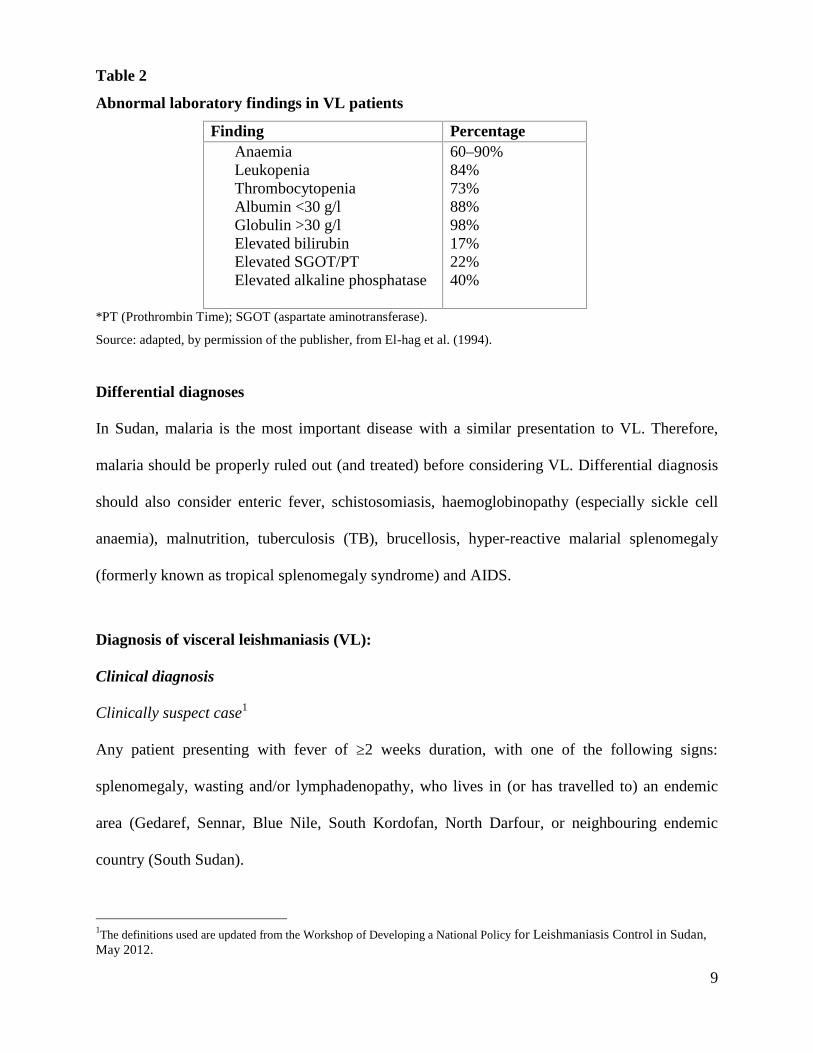
Table 2
Abnormal laboratory findings in VL patients
Finding PercentageAnaemiaLeukopeniaThrombocytopeniaAlbumin <30 g/lGlobulin >30 g/lElevated bilirubinElevated SGOT/PTElevated alkaline phosphatase
60–90%84%73%88%98%17%22%40%
*PT (Prothrombin Time); SGOT (aspartate aminotransferase).
Source: adapted, by permission of the publisher, from El-hag et al. (1994).
Differential diagnoses
In Sudan, malaria is the most important disease with a similar presentation to VL. Therefore,
malaria should be properly ruled out (and treated) before considering VL. Differential diagnosis
should also consider enteric fever, schistosomiasis, haemoglobinopathy (especially sickle cell
anaemia), malnutrition, tuberculosis (TB), brucellosis, hyper-reactive malarial splenomegaly
(formerly known as tropical splenomegaly syndrome) and AIDS.
Diagnosis of visceral leishmaniasis (VL):
Clinical diagnosis
Clinically suspect case1
Any patient presenting with fever of ≥2 weeks duration, with one of the following signs:
splenomegaly, wasting and/or lymphadenopathy, who lives in (or has travelled to) an endemic
area (Gedaref, Sennar, Blue Nile, South Kordofan, North Darfour, or neighbouring endemic
country (South Sudan).
1The definitions used are updated from the Workshop of Developing a National Policy for Leishmaniasis Control in Sudan,May 2012.

10
Confirmed case2
There are two accepted ways of confirming VL in a clinically suspected case: either by
parasitology or serology.
Laboratory diagnosis
After taking a proper history and performing a thorough clinical examination, parasitological or
serological tests are required to decide who should be treated (see the national recommended
diagnostic algorithm below).
Parasitological diagnosis
Microscopic examination of Giemsa-stained spleen, bone marrow or lymph node aspirates to
detect amastigotes remains the reference standard in VL diagnosis. However, these methods are
either invasive or insensitive. It takes <1 h from sample collection to obtain the result.
The sensitivity of lymph node aspiration is 52–65%, whereas the sensitivity of bone
marrow aspiration can reach up to 75% (Kager et al., 1983; Siddig et al., 1988; Zijlstra, 1998).
Splenic aspiration sensitivity is 90–95%. Negative slides do not prove absence of the parasite,
and low parasite density can be missed microscopically.
Lymph node aspiration is safe, easy and can be done by paramedical staff, compared with
bone marrow aspiration, which is painful and needs specific and sterile needles, and trained
personnel. Spleen aspiration is the most sensitive procedure, but it may be hazardous. It needs an
experienced physician and should be performed in hospital, where blood transfusion is available
because bleeding is a life-threatening complication.
Parasitological methods require highly trained laboratory personnel and the results are
dependent on the quality of microscopic examination and reagents.
2The definitions used are updated from the Workshop of Developing a National Policy for Leishmaniasis Control in Sudan,May 2012.

11
Serological diagnosis
Several immunological blood tests that identify specific antibodies against Leishmania are
available: immunofluorescence antibody test (IFAT), ELISA, direct agglutination test (DAT) and
recombinant K39 antigen (rK39) immunochromatographic test (ICT) (useful in field settings).
Leishmaniasis Control Program selected two serological tests (DAT and rK39 ICT) that can be
used for diagnosis of VL in Sudan.
Direct agglutination test
The DAT is simpler than many other tests but its drawbacks include:
prolonged incubation time (18 h)
requirement for microtitre plates and micropipettes
Requirement for well-trained laboratory personnel.
Moreover, the DAT (as for all serological tests for antibody detection) suffers from two major
limitations.
Antibodies can remain detectable up to several years after cure because of asymptomatic
infections, a significant proportion of healthy people living in endemic areas with no
history of VL are positive for Leishmania antibodies.
Therefore, the DAT cannot distinguish between active VL and subclinical and past infection, and
such tests cannot be used for diagnosis of relapse or for evaluation of cure.
DAT titres:
DAT Freeze driedPositive: ≥ 1:3200Borderline (BL): 1:1600; 1:800; 1:400Negative: ≤ 1:200

12
rK39-based immunochromatographic test:
rK39 is a cloned antigen of 39 amino acid repeats of a gene found in Leishmania chagasi. A
meta-analysis of 13 studies evaluating the rK39 ICT showed excellent diagnostic performance
(sensitivity and specificity estimates of 93.9% and 95.3%, respectively). The overall sensitivity
was lower in studies from East Africa than in those from South Asia. Several brands of the rK39
ICT have been evaluated. Higher values for both sensitivity (90%) and specificity (99%) have
been obtained with the rK39 ICT (IT-LEISH) manufactured by Bio-Rad (previously DiaMed)
the good diagnostic performance of IT-LEISH has been confirmed in a recent tropical disease
research multicenter study with 87.2% sensitivity and 96.4% specificity in East Africa. The test
is easy to use in the field, reproducible, rapid (10–20 min) and inexpensive, therefore, the
diagnosis can be established at the primary health care level. However, as an antibody detection
test, the rK39 ICT cannot be used for the diagnosis of relapse and as a test of cure.
Nucleic-acid-based assays
Polymerase chain reaction (PCR) is usually highly sensitive for detection of Leishmania infection, but
this does not imply it will be useful for the confirmation of acute VL disease in patients in an endemic
area, because many carriers of the infection in the area will be PCR positive without developing VL
disease.

13
National Diagnostic Algorithm of clinical suspect of VL
Clinical Suspect VL
Previously Treated for VL
Negative rK39Lymph node or Bone
marrow aspirate
DAT test if available orGo for parasitology
Look for alternativediagnosis and Treat
Yes NO
rK39 test
Positive rK39
PositiveTreat for VL
Positive DAT ≥ 1:3200
Negative
Negative DAT ≤ 1:200 Borderline DAT 1:1600;1:800; 1:400
Look for alternativediagnosis and treat
Treat for Relapse
Lymph node or Bonemarrow aspirate
Treat for VL
Treat for VL
Negative Positive
Look for alternativediagnosis and treat
14
Chapter 3
TREATMENT OF LEISHMANIASIS
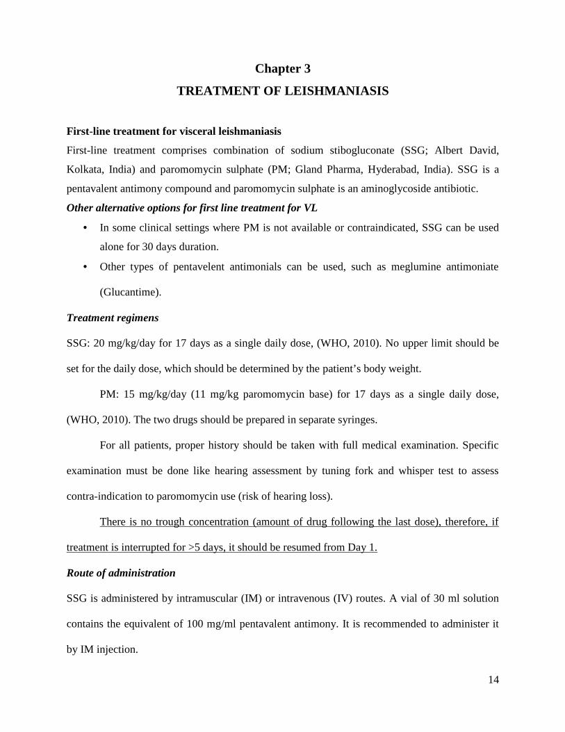
First-line treatment for visceral leishmaniasis
First-line treatment comprises combination of sodium stibogluconate (SSG; Albert David,
Kolkata, India) and paromomycin sulphate (PM; Gland Pharma, Hyderabad, India). SSG is a
pentavalent antimony compound and paromomycin sulphate is an aminoglycoside antibiotic.
Other alternative options for first line treatment for VL
In some clinical settings where PM is not available or contraindicated, SSG can be used
alone for 30 days duration.
Other types of pentavelent antimonials can be used, such as meglumine antimoniate
(Glucantime).
Treatment regimens
SSG: 20 mg/kg/day for 17 days as a single daily dose, (WHO, 2010). No upper limit should be
set for the daily dose, which should be determined by the patient’s body weight.
PM: 15 mg/kg/day (11 mg/kg paromomycin base) for 17 days as a single daily dose,
(WHO, 2010). The two drugs should be prepared in separate syringes.
For all patients, proper history should be taken with full medical examination. Specific
examination must be done like hearing assessment by tuning fork and whisper test to assess
contra-indication to paromomycin use (risk of hearing loss).
There is no trough concentration (amount of drug following the last dose), therefore, if
treatment is interrupted for >5 days, it should be resumed from Day 1.
Route of administration
SSG is administered by intramuscular (IM) or intravenous (IV) routes. A vial of 30 ml solution
contains the equivalent of 100 mg/ml pentavalent antimony. It is recommended to administer it
by IM injection.
15
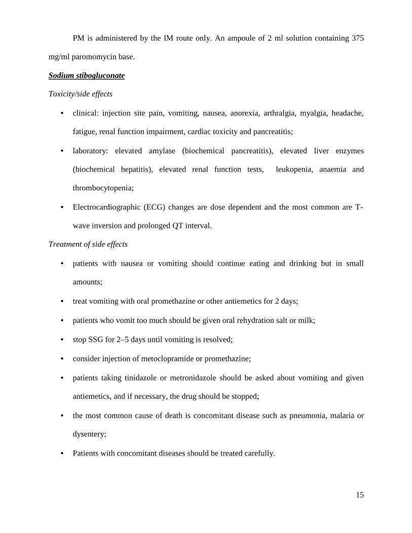
PM is administered by the IM route only. An ampoule of 2 ml solution containing 375
mg/ml paromomycin base.
Sodium stibogluconate
Toxicity/side effects
clinical: injection site pain, vomiting, nausea, anorexia, arthralgia, myalgia, headache,
fatigue, renal function impairment, cardiac toxicity and pancreatitis;
laboratory: elevated amylase (biochemical pancreatitis), elevated liver enzymes
(biochemical hepatitis), elevated renal function tests, leukopenia, anaemia and
thrombocytopenia;
Electrocardiographic (ECG) changes are dose dependent and the most common are T-
wave inversion and prolonged QT interval.
Treatment of side effects
patients with nausea or vomiting should continue eating and drinking but in small
amounts;
treat vomiting with oral promethazine or other antiemetics for 2 days;
patients who vomit too much should be given oral rehydration salt or milk;
stop SSG for 2–5 days until vomiting is resolved;
consider injection of metoclopramide or promethazine;
patients taking tinidazole or metronidazole should be asked about vomiting and given
antiemetics, and if necessary, the drug should be stopped;
the most common cause of death is concomitant disease such as pneumonia, malaria or
dysentery;
Patients with concomitant diseases should be treated carefully.

16
Contraindications
Contraindications to SSG include: Cardiac disease, liver disease (jaundice), renal failure, age
>45 years, HIV/AIDS, pregnancy, and infancy
Pregnant women and neonates
The data on the safety of pentavalent antimonials in pregnancy are confined to case reports,
which indicate that they are relatively safe. However, recent published work indicates some
reservations for using SSG in pregnant women and infants. There was some excretion of
antimony in breast milk following administration of SSG to one woman. More evaluation is
required before pronouncing on the safety of antimony in breast-feeding infants.
Paromomycin sulphate
The bioavailability of PMS is high (approaching 100%) following IM injection but it is
negligible following oral administration; thus the IM route is required to treat a systemic disease
such as VL. The half-life of PMS in humans is 2–3 h. It is not metabolized; it is excreted
unchanged in the urine, thus, accumulation can occur in patients with diminished renal function.
PMS should be stored below 30°C and protect from light. It should not be frozen.
Toxicity/side effects
The most common adverse effects are injection site pain, transient elevation of alanine
aminotransferase and aspartate aminotransferase, and increased blood alkaline phosphatase,
creatinine and bilirubin. Toxicological studies have been conducted in animals. At high doses,
paromomycin exhibits nephrotoxicity, ototoxicity and neuromuscular blockade. No cases of
overdose have occurred in clinical trials of IM PMS. General supportive care is indicated. The
effectiveness of renal dialysis as a treatment of overdose has not been formally studied.

17
Contraindications
Patients who have shown hypersensitivity to Paromomycin or other aminoglycosides.
Discontinue use if an allergic reaction occurs.
Relatively contraindicated in patients with impaired renal function.
Patients with pre-existing hearing impairment.
Pregnant women and neonates
Reproductive studies in rats and rabbits indicate that PMS is not teratogenic but does affect
fertility and implantation. Aminoglycosides can cross the placenta, thus, PM may cause foetal
injury when administered to pregnant women. It is therefore contraindicated during pregnancy.
PM can be recovered in breast milk. The absorption of PM is negligible after oral administration,
thus, it is expected that breast-fed infants would have no systemic exposure to PM.
Second-line treatment of visceral leishmaniasis
Liposomal amphotericin B (AmBisome)
AmBisome is administered at a dose of 3mg/kg/daily days for 10 to 14 days. 3-5 mg/kg per dose
for 6-10 days up to total of 30 mg/kg as initial dose please see annex
Given on alternate days: 1, 3, 5, 7, 9, 10, for special group for example: renal
problem or heart disease to avoid fluid overload and 11 see annex for calculation.
Indications
lack of response or relapse after SSG–PMS or SSG monotherapy
Contraindication to SSG or PM.
SSG-induced toxicity
age >45 or <2 years
HIV co-infection
Pregnancy
18
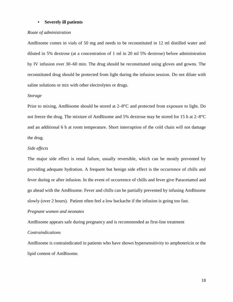
Severely ill patients
Route of administration
AmBisome comes in vials of 50 mg and needs to be reconstituted in 12 ml distilled water and
diluted in 5% dextrose (at a concentration of 1 ml in 20 ml 5% dextrose) before administration
by IV infusion over 30–60 min. The drug should be reconstituted using gloves and gowns. The
reconstituted drug should be protected from light during the infusion session. Do not dilute with
saline solutions or mix with other electrolytes or drugs.
Storage
Prior to mixing, AmBisome should be stored at 2–8°C and protected from exposure to light. Do
not freeze the drug. The mixture of AmBisome and 5% dextrose may be stored for 15 h at 2–8°C
and an additional 6 h at room temperature. Short interruption of the cold chain will not damage
the drug.
Side effects
The major side effect is renal failure, usually reversible, which can be mostly prevented by
providing adequate hydration. A frequent but benign side effect is the occurrence of chills and
fever during or after infusion. In the event of occurrence of chills and fever give Paracetamol and
go ahead with the AmBisome. Fever and chills can be partially prevented by infusing AmBisome
slowly (over 2 hours). Patient often feel a low backache if the infusion is going too fast.
Pregnant women and neonates
AmBisome appears safe during pregnancy and is recommended as first-line treatment
Contraindications
AmBisome is contraindicated in patients who have shown hypersensitivity to amphotericin or the
lipid content of AmBisome.

19
Other second-line drugs for treatment of leishmaniasis
Amphotericin B deoxycholate Amphotericin B deoxycholate is sometimes referred to as
conventional amphotericin B. Amphotericin B is a mixture of antifungal polyene antibiotics
produced by some strains of Streptomyces nodosus.
Dose
Amphotericin B is given at a dose of 1 mg/kg every other day for 30 days (total 15 mg/kg),
or 0.5 mg/kg daily for 30 days. It should be given initially at a test dose of 0.1 mg/kg.
Route of administration
IV infusion should be given as a colloidal complex with sodium deoxycholate, at a concentration
of 0.1 mg/ml in 5% glucose over 2–4 h. Amphotericin B is infused in 1 litter 5% dextrose over
12 h, to decrease infusion-related side effects (e.g., fever and chills).
Side effects
These side effects apply to the conventional form only: headache, nausea, vomiting, chills, fever,
malaise, muscle and joint pain, diarrhoea and gastrointestinal cramps, hypertension, hypotension,
cardiac arrhythmia (including ventricular fibrillation), cardiac arrest, skin rashes, anaphylactoid
reactions, blurred vision, tinnitus, hearing loss, vertigo, liver disorders, peripheral neuropathy,
convulsions, nephrotoxicity (which occurs in almost all patients receiving amphotericin B),
hypokalaemia, hypomagnesaemia, nephrocalcinosis, anaemia, and thrombophlebitis at the
injection site.
Pregnant women and neonates
There are case reports of successful treatment of fungal infections in pregnant women with
amphotericin B, without any adverse effects on the infant. However, care should still be
exercised.
20
Drugs under investigation
Miltefosine
Miltefosine was developed as an antineoplastic drug. It is the first oral drug with demonstrated
efficacy against VL but clinical trials are not yet complete. The drug has been extensively
studied in India, where it is registered. Miltefosine use in Sudan should be restricted to
compassionate use (e.g., treatment of multiple relapses).
Supportive treatment for patients with visceral leishmaniasis
Care under direct medical supervision and observation
Nutritional support.
Multivitamins.
Iron sulphate/gluconate and folic acid.
Tinidazole for parasitic infections.
Malaria prophylaxis in special circumstances.
Care for concurrent infection (e.g., malaria, pneumonia, TB and HIV/AIDS).
Blood transfusion is not generally needed; haemoglobin increases with successful
treatment. Blood transfusion should be undertaken if the setting is optimal for donor
screening and one has the ability to deal with any complications that may arise.
Nutrition protocol for visceral leishmaniasis patients
The overall objectives in nutritional care for patients with VL are to assist the recovery period
and improve the response to treatment. The specific objectives focus on:
supporting all patients with nutritional support during treatment
reducing the incidence of concomitant illness/infection
Treating severe and moderate acute malnutrition.

21
All patients with VL should receive adequate nutritional support. This can be done through
general food distribution, which must be appropriate in quality and quantity. The quality can be
improved through supplementation of food items.
Follow-up after treatment
Follow-up at the end of treatment, at 6 months post-treatment, and at any time whenever
symptoms return is important to detect treatment failure.
Criteria of cure
Primary VL: clinical cure with no fever, absence/reduction in the size of the spleen, weight gain,
good appetite, and increase in haemoglobin and albumin levels.
Case definition according to follow-up
Cases can be defined as follows:
Initial cure: patient who improves clinically and has negative test of cure (TOC).
Non-responder: patients with persistent clinical symptoms and signs and a positive
parasitology aspiration after completion of treatment (after 17 days of combination
therapy or 30 days of SSG treatment or full dose AmBisome) patients who do not show
any decrease in parasite load are also classified as non- responders.
Slow responder: Patients who show a slow clinical response and decrease but not a
disappearance of parasite load on lymph node or bone marrow aspirate examination after
completion of therapy. (After 17 days of combination therapy or 30 days of SSG
treatment or full dose Ambisome ).
Definitive cure: patients who have an initial cure and show no sign of relapse within 6
months.
Relapse: a patient with clinically and parasitologically confirmed VL within 6 months of
successful treatment of VL.

22
Test of cure
TOC is a lymph node or bone marrow aspiration performed at the end of treatment to assess the
parasitological response to therapy. If the TOC is performed too early (<28 days), parasites may
still be present and a diagnosis of non-response or slow response could be wrongly made. This is
particularly important now that shorter treatment regimens are administered (i.e. SSG/PM 17d,
Ambisome 10 day). Therefore, the interpretation of a positive TOC made earlier than day 28
must take into account the presence or absence of clinical and biological signs of non-response.
TOC is indicated in the following situations:
1. Primary Kala-azar patients who show no clinical improvement at the completion of first
line therapy (persistence of fever, Hb not increasing and/or no regression of the spleen)
2. Relapsed kala azar patients who received second line therapy.
3. In all HIV-VL co-infected patients.
4. Non-responders shifted to another anti-leishmanial treatment.
5. Slow responders during the extended course of treatment (weekly).
TOC is not necessary for primary VL if the patient improved clinically (No kala-azar symptom,
gain weight, Hb increase and decrease in spleen size).
Treatment of non-responders and relapses
Initial non-responders
Consider second-line drugs (e.g., AmBisome). Do not repeat combination therapy
(SSG+PM); instead switch to another antileishmanial drug. In case there are no others drugs are
available, one can stop PM and continue alone with SSG for first relapse up to 60 doses
First relapse: For any patients treated before by SSG alone should be receive SSG/PM and for
any patient treated by SSG/PM will be treated with AmBisome

23
Second and third relapse: AmBisome (dose as above) Patients should be examined for risk
factors for TB and HIV.
Chapter 4
OTHER TYPES OF LEISHMANIASIS
Cutaneous leishmaniasis
Cutaneous leishmaniasis (CL) in Sudan is caused by L. major. The disease is endemic in many
parts of the country.
The vector is P. papatasi and the animal reservoir is probably the Nile rat A. niloticus.
Clinically, patients usually present with papules, nodules, or noduloulcerative lesions,
mainly on the exposed parts of the skin. In 20% of cases, the parasite disseminates through the
lymphatics, producing sporotrichoid-like lesions.
Diagnosis is confirmed by the demonstration of parasites in slit smears in 50–70% of
cases and in histological sections in 70%. CL is a self-limiting disease; therefore, treatment is
confined to patients with severe disease (El-Hassan & Zijlstra, 2001).
Treatment of cutaneous leishmaniasis
Single lesions in the extremities or the trunk do not need treatment and can spontaneously heal
within 4–8 weeks. Two to three lesions that do not heal spontaneously and involve the
face/eyelids can safely be treated with a single oral dose of fluconazole (200-600 mg/day in
adults and 6-12 mg/kg/day for children) for 4–6 weeks. Liver function tests should be checked
initially and regularly. Multiple lesions and lesions in diabetic/immunocompromised patients can
be treated with systemic pentavalent antimony (SSG) at a dose of 10–20 mg/kg/day, and should
be given if the lesions are more severe, and continued until a few days after clinical and
parasitological cure is achieved.

24
Mucosal leishmaniasis
Mucosal leishmaniasis is a chronic infection of the upper respiratory tract and/or oral mucosa,
which is caused mainly by L. donovani. The disease occurs in areas of the country that are
endemic for VL, particularly among Masalit and other closely related tribes in western Sudan.
The condition may develop during or after an attack of VL, but in most cases it is a primary
mucosal disease.
The diagnosis is established by demonstration of parasites in smears or biopsies, by
culture or animal inoculation. Most patients yield positive results in the DAT and leishmanin
skin test (LST). Patients respond well to treatment with SSG (Pentostam) (El-Hassan & Zijlstra,
2001).
Treatment of mucosal leishmaniasis
Single doses of 10–20 mg/kg/day of SSG are given for a minimum of 4 weeks. Relapse should
be treated with the same drug given for at least twice as long as the original treatment. Only
when this fails should alternative treatment be given.
Post kala-azar dermal leishmaniasis
PKDL is a known complication of VL. In the majority of cases, dermal lesions develop several
months after the clinical cure of VL, but sometimes PKDL occurs during or even before
treatment. However, cases have been reported in the absence of a history of VL. About 55% of
successfully treated VL patients develop PKDL.
The lesions of PKDL start on the face as small scattered hypopigmented macules and papules.
The rash can become nodular and spread to the trunk and limbs. Lesions are symmetrical and not
itchy. A grading system is used to describe the spread of the skin lesions.

25
Grade 1: scattered macular, papular or nodular rash on the face with some lesions on the
upper chest and upper arms.
Grade 2: dense macular, papular or nodular rash covering most of the face and extending
on the chest, back, and upper arms and legs.
Grade 3: dense macular, papular rash, covering most of the body, including hands and
feet. In grade 3, crusting, ulcers, scaling and spreading to the mucosa of the lip and the
palate occur. It is referred to as PKDL with mucosal involvement (or combined PKDL and
post kala azar mucosal leishmaniasis). Lesions are often restricted to sun-exposed areas
(whole body in children, face and collar distribution for men, and face for women).
Parasites can be detected in skin smears, but not in all cases. Parasites have also been detected
in the normal skin in patients with VL. PKDL might persist for up to 10 years. It is speculated
that PKDL patients could form a reservoir of the parasite in the community. Bed nets may be
indicated to prevent transmission.
PKDL is distinct from CL, which consists of one or more ulcers caused by other species of
Leishmania. PKDL can easily be confused with diffuse leprosy.
Treatment of post kala-azar dermal leishmaniasis
Spontaneous healing occurs in mild cases (grade 1 or mild grade 2). The indications for
treatment are:
grade 3 and advanced grade 2 lesions
persistence of lesions for >12 months
Concomitant anterior uveitis or mucosal lesions.
Treat until the lesions are definitely improving and not until the lesions have disappeared. Once
healing begins during treatment, it usually continues off treatment. There are no parasitic criteria
for cure because of the difficulties of demonstrating the parasite in the lesions. Follow-up is
26
monthly until full resolution of lesions. It is advisable to take pictures of PKDL patients (after
obtaining consent) at admission to ensure proper follow-up of the clearing of lesions.
The following drugs can be used to treat PKDL:
AmBisome at a dose of 2.5 mg/kg/day for 20 days has been used successfully in some
patients (Hashim et al., 1995; Musa et al., 2005).
SSG at a dose of 20 mg/kg/day for 60–90 days is effective (El Hassan & Zijlstra, 2001).
It is advised to continue treatment until 4–7 days after clinical cure (lesions have
regressed and are no longer palpable, but discoloration is still visible). Approximately 4–
8 weeks of treatment are needed, but longer courses are common.
27
Chapter 5
LEISHMANIASIS AND HIV CO-INFECTION
HIV and leishmaniasis are both expanding due to the increasingly overlapping geographical
distribution of the two causative pathogens (in Asia, east Africa, South Africa and southern
Europe) the number of cases with co-infection is expected to rise. IV drug users represent the
main population at risk.
HIV infection and VL influence each other reciprocally. HIV has caused an increase in
VL cases. HIV patients with VL have a poor prognosis, and VL stimulates replication of HIV
(Davidson, 1997).
Clinical features of Leishmania/HIV co-infection
The diagnosis of VL in patients co-infected with HIV can be particularly difficult. The usual
clinical features of VL (splenomegaly, pancytopenia, fever and wasting) are not always present
or may be hidden by other opportunistic infections mimicking the same symptoms. Patients may
have atypical VL with slight or no splenomegaly and only non-specific symptoms of fever,
wasting and malaise.
VL may be rapidly progressive, resembling bacterial sepsis. Alternatively, VL may have
unusually slow progression with a few non-specific symptoms, and some patients with VL may
be entirely asymptomatic.
HIV/Leishmania co-infected patients may have predominantly gastrointestinal
involvement with symptoms of diarrhoea and wasting. Leishmania parasites can be detected in
biopsies of the oesophagus, duodenum and rectum. The larynx may also be involved, either
alone or with gastrointestinal involvement.
Diagnosis of visceral leishmaniasis in HIV-positive patients

28
In AIDS patients, the cells responsible for the immune response are destroyed, impairing the
capacity of the immune system to react to invasion by the leishmaniasis parasite. Consequently,
the serological test for detection of Leishmania antibodies (IFAT or ELISA) is negative in 20–40
% of patients with HIV and VL co-infection. It is expected that DAT titres in Africa will be
lower in such cases as well. The diagnosis is better by lymph node/bone marrow aspirate, and not
DAT alone. Parasites are abundant in lymph nodes/bone marrow, and microscopy of bone
marrow aspirates yields a diagnosis in 91–97% of cases.
In 50% of HIV/VL patients, the buffy coat (white cell concentrate) of peripheral blood is
positive. Sensitivity of culture of the buffy coat can reach 90%. About 80% of HIV/Leishmania
co-infected patients usually have another AIDS-defining opportunistic infection, and 90% have
CD4+ count <200/mm3 at the time of diagnosis. This may not be the case in Africa where the
causative species is L. donovani, which is probably more virulent than L. infantum.
Treatment of HIV/Leishmania co-infection
All VL patients should be counselled and tested for HIV because first-line antileishmanial
treatment in HIV-positive patients is different. The response to antimonials, AmBisome or other
anti-leishmaniasis drugs is both slow and less satisfactory in HIV/Leishmania co-infected
patients. The median survival among HIV/VL patients is about 12 months. These patients have a
huge parasite load and lack cell-mediated immunity to assist in parasite clearance.
Patients with VL/HIV co-infection should be treated with AmBisome.
Failure to clear parasites and treatment of relapse
A positive TOC in a patient with HIV/Leishmania co-infection should be followed by further
treatment. If patients do not have relief of the symptoms of VL by 30 days, they will not benefit
from a longer course of treatment. Thus, if HIV infection is suspected in patients with a positive
TOC, an HIV test should be preformed.

29
Relapse of VL in known HIV patients should only be treated if symptoms are present. If
no symptomatic relief occurs after 1 week, treatment should be stopped and palliative care
initiated. Thus, if HIV infection is suspected in patients who have relapsed, an HIV test should
be performed.
Prognosis of HIV/leishmaniasis
Thirty percent of HIV/VL patients will die during or within 1 month after treatment using drug
rather than AmBisome. The mean survival with optimal treatment is only 12 months. Only 16%
will survive for >3 years. Death is seldom due to VL alone. A sterile cure cannot be achieved by
any drug, and relapse is almost inevitable.
Public health implications of HIV/leishmaniasis
HIV/Leishmania co-infected patients must be considered as highly infectious reservoirs of
Leishmania in areas of active transmission because of the presence of amastigotes in the blood.
The reservoir of VL in Africa is probably humans, so drug-resistant transmission can occur.
There is a clear correlation between the severity of immunosuppression and the
percentage of sandflies becoming infected. Every VL patient in the should sleep under a
mosquito net, and every HIV-positive patient should be sleeping under an impregnated bed net,
whether or not they have VL.

30
ANNEX 1: OUTLINE OF THE NEW TREATMENT PROTOCOL OF
LEISHMANIASIS
1- Visceral leishmaniasis:
a- First-line treatment is SSG and Paromomycin sulphate injection
Drug GROUP Dose Duration Route of
administration
SSG
+
Paromomycin sulphate
All patients except
breast feeding infant
All patients except
breast feeding infant
20 mg/kg single dose
15 mg/kg single dose
17 days
17 days
IM/IV
IM only
b- Second-line treatment:
Drug GROUP Dose Duration Route of
administration
AmBisome®
(liposomal
amphotericin B)
All patients 3 mg/kg/day 10–14 days IV only
2- Treatment of cutaneous leishmaniasis
Drug GROUP Dose Duration Route of
administration
fluconazole Adults with 4 or more
lesions or not suitable
for local inj
200-600 mg single
dose
Daily 4–6 weeks Oral
fluconazole Children with 4 or
more lesions or not
suitable for local inj
6-12 mg/kg single
dose
Daily 4–6 weeks Oral
SSG Severe lesions
Diabetes
Immunocompromised
10–20 mg/kg single
dose
Until a few days after
clinical and
parasitological cure is
achieved
IM/IV

31
3- Mucosal leishmaniasis
Drug GROUP Dose/day Duration Route of
administration
SSG All new cases 20 mg/kg single dose Minimum of 4 weeks IM/IV
SSG Relapse 20 mg/kg twice Minimum of 4 weeks IM/IV
4- Post Kala-azar Dermal Leishmaniasis
Drug GROUP Dose Duration Route of
administration
AmBisome®
(liposomal
amphotericin B)
All patients 2.5 mg/kg/day Daily for 20 days IV only
SSG
All patients 20 mg/kg/day 60–90 days (4–7 days
after clinical cure);
resolution of
papules/nodules
IM/IV
5- Leishmaniasis/HIV co-infection
* SSG: sodium stibogluconate (pentostam) ®* Other types of pentavelent antimonials can be used include meglumine antimonite (Glucantime) © in the absence
of SSG.
Drug GROUP Dose Duration Route of
administration
AmBisome®
(liposomal
amphotericin B)
Co-infected patients 2.5 mg/kg/day 10–14 days Infusion
32
ANNEX 2: OTHER TESTS FOR VISCERAL LEISHMANIASIS
rK28-based
A new, synthetic, multiepitope polyprotein, rK28, was obtained by fusing L. infantum k9 gene
with single repeat units of k39 and k26 genes. This was followed by development and evaluation
of two new rK28-based RDT prototypes.
rK28-LF RDT was developed by EASE MedTrend (Shanghai, China) and evaluated in
Bangladesh. It had more favourable results (98.1% sensitivity, 92.5% specificity) compared to
Kalazar Detect (88.7% sensitivity, 100% specificity).
A second prototype test of rK28 (K28-DPP) was developed by Chembio Diagnostic
Systems (Medford, NY, USA) and evaluated in Sudan. K28-DPP RDT proved to be superior and
provided a sensitivity of 95.9% and specificity of 100% while the Kalazar Detect yielded a
sensitivity of 86.3% and specificity of 96.4%. Moreover, the results of a recent TDR multicentre
study proved that the new rK28-based RDTs have great potential to simplify VL disease
confirmation at the point-of-care (unpublished data).
Latex agglutination test (KAtex)
The KAtex latex agglutination test was developed to detect heat-stable, low-molecular-weight
carbohydrate antigen in urine (Attar et al., 2001). It is a simple, rapid and economical test, and is
most suitable for use in remote areas.
In a multicenter study in east Africa (Ethiopia, Kenya and Sudan), KAtex showed good
specificity but moderate to very low sensitivity (Boelaert et al., 2008). A field evaluation of the
33
test in Sudan indicated that the test had a sensitivity of 95.2% and good agreement with
microscopy but poor agreement with serological tests and a specificity of 100% (El-Safi et al.,
2003). In addition, the test was also positive for the two confirmed VL cases co-infected with
HIV. One of the advantages of the test is its ability to distinguish active from past infections.
Accordingly, it can be used as a TOC and for diagnosis of relapse. However, the need to boil the
urine before testing to avoid false-positive reactions is a limitation. Work to improve the format
of this urine antigen detection test is ongoing.
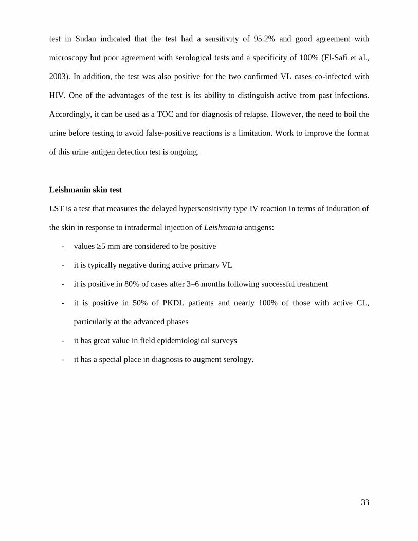
Leishmanin skin test
LST is a test that measures the delayed hypersensitivity type IV reaction in terms of induration of
the skin in response to intradermal injection of Leishmania antigens:
- values ≥5 mm are considered to be positive
- it is typically negative during active primary VL
- it is positive in 80% of cases after 3–6 months following successful treatment
- it is positive in 50% of PKDL patients and nearly 100% of those with active CL,
particularly at the advanced phases
- it has great value in field epidemiological surveys
- it has a special place in diagnosis to augment serology.
34
ANNEX 3: DEFINITIONS
Cutaneous leishmaniasis
Ulcers, macules, papules or nodules of the skin caused by leishmaniasis. Atypical forms are
frequent. No involvement of other organs.
Primary visceral leishmaniasis
A patient who is diagnosed with VL for the first time has primary VL. There is no previous
history of treatment of VL. Informal, incomplete treatments in the villages do occur and should
be asked for explicitly.
Post kala-azar dermal leishmaniasis
PKDL is a cutaneous manifestation caused by the same parasite that causes VL and usually
occurs several weeks after recovery from VL.
Relapse
A patient with a diagnosis of VL who has previously been treated successfully for VL.
Reinfection
A person who has VL more than once is considered to have a relapse rather than reinfection. The
term reinfection is not used with VL patients. We assume that reinfection occurs but does not
lead to clinical disease because of immunity after successful treatment.
Test of cure
The TOC is aspiration (usually of the spleen) to obtain proof of cure (to demonstrate the absence
of parasites). A smear is made on a slide and stained with Giemsa. A TOC is performed for
patients with primary VL between days 25 and 30. A positive TOC indicates that the patient has

35
not yet been fully cured. It is possible that in patients who improve clinically, the parasites in the
smear are reduced in number but are still detectable (slow responders).
Initial cure
Patients who improve clinically and have a negative TOC at discharge (after the first treatment).
Definite cure
Patients who have an initial cure and show no sign of relapse within 6 months.
36
ANNEX 4: ASPIRATION OF LYMPH NODE, SPLEEN AND BONE
MARROW
Lymph nodes
Lymph node aspiration is commonly taken from less hazardous clear lymph groups (e.g.,
inguinal or trochlear). The nodes are grasped between the thumb and finger after sterilization. A
21-gauge needle is attached to a 5-ml syringe and introduced, pushing the needle in and out to
force the cells into the needle. A few drops of the aspirated fluid are placed onto a slide, smeared
thinly, dried, fixed with methanol, stained with Giemsa, and examined.
Spleen
Spleen aspiration is the simplest, fastest and most reliable procedure for detection of Leishmania
parasites. However, it carries a risk of bleeding, visceral puncture and splenic rupture, therefore,
it requires expertise (physician) and should be performed in hospital with ready access to blood
and an operating theatre.
Contraindications for spleen aspiration are: impalpable spleen, bleeding tendency,
untrained staff, jaundice and late pregnancy.
Technique
Sterilization of the site and equipment.
A 21-gauge needle attached to a 5-ml syringe is inserted in the midline of the spleen. The
needle is inserted quickly to the full needle depth (3 cm). A vacuum is created by pulling
the plunger back and the needle is removed immediately. The speed ensures that no
tearing occurs. The content of the syringe is deposited on a slide; a thin smear is made;
and the slide is stained with Giemsa and examined.
Puncture is performed at expiration, especially in children.

37
No local anaesthesia is given.
Sedation is recommended in restless or crying child (lymph node or bone marrow
aspiration is preferred).
Patient must be observed for 24 h.
Bone marrow
Bone marrow aspirate is usually taken from the sternum or iliac crest under local anaesthesia,
with a sterile bone marrow aspirate needle attached to a 10-ml syringe. This smears are formed
immediately on at least three slides, fixed with 100% methanol, dried labelling, and stained with
Giemsa. Examined fewer than 100 magnifications (1000 field).
38
ANNEX 5: SOP for Preparation of AmBisome (reconstitution and dilution), Sudan

39
Procedure:1. Choose appropriate room for preparation of AmBisome and wash your hands with soap.2. Wear a pair of gloves.3. Take 12 ml of distilled water (in a 20 ml syringe, if available).4. Remove the rubber cap of the AmBisome vial and push the distilled water into the vial.5. Shake the vial immediately for 30 seconds until complete dispersion of the AmBisome.6. Allow bubbles to dissipate before use.7. Follow the same instruction for the next vial to be reconstituted using the same syringe;
calculate the total number of vials based on body weight according to the Table given below.8. Hypersensitivity test: take 0.25ml of reconstituted AmBisome in 1 ml disposable syringe
and dilute it up to 10 ml with 5% DA for test.9. AmBisome is NOT compatible with saline and must not be reconstituted with saline or
administered through an intravenous line that has previously been used for saline unless firstflushed with 5% dextrose solution. Do NOT mix AmBisome with other drugs or electrolytes
10. Calculate the volume of 5% DA to dilute the whole dose of AmBisome according to thepatient’s body weight using the tables.
11. Now hang the 500ml pack of 5% DA on an infusion stand.12. Fix an infusion set with the 5%DA bottle/pack. Keep the required volume of 5% DA in the
bottle/ pack and discard the excess amount of 5%DA in a bowl.13. Take another syringe to pull the reconstituted AmBisome from the vial, set a syringe filter on
it.14. Wash the pack/bottle surface with a ball of cotton soaked with chlorohexidine at the site of
injecting the reconstituted AmBisome.15. Inject the drug into that 5%DA solution.16. Use one syringe filter for one vial of AmBisome.17. Inject the total volume of reconstituted AmBisome in that 5% DA solution in the same
manner.18. Follow strict aseptic precaution from preparation site to infusion to the patient.Responsible person: Qualified and trained staff nurse is responsible for the task but the wholeprocedure should be closely observed by the duty physician.Glossary/ Definition:
1. 5% DA: 5% dextrose in aqua2. kg: kilogram3. ml: milliliter4. SOP: Standard Operating Procedure
Version - 2 Dated: 11/12/2012Objective of SOP: To prepare AmBisome for infusion accurately and with proper asepticprecautionScope and applicability 1. To administer AmBisome by infusion in a visceral leishmaniasis
patient2. Hypersensitivity test to AmBisome.Materials requiredA pair of disposable hand gloves 70% isopropyl alcohol/ spirit.Disposable syringe (1ml and 10 ml) withsterile needle
5% DA (500 ml) bottle/ packDistilled/ Sterile water AmBisome vialDisposable infusion set Syringe filter and Infusion stand

40
Reference: Leaflet provided with the AmBisome vial

41
AmBisome dosage in East Africa:5 mg / dose for 6 – 10 d
Total dose up to 30 mg / kg
Regimen:3 mg/kg/day for 10 days
Calculation:Dosage in ml = dosage in mg X 19 = ml of Dextrose 5%
4Rate for infusion: weight in Kg x 2,375
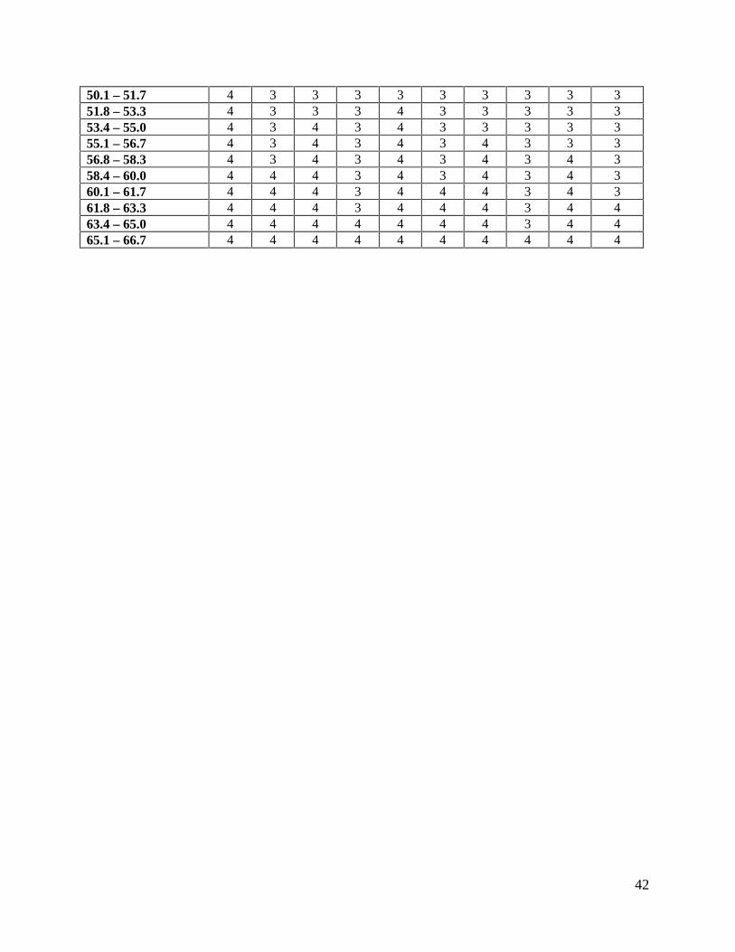
AmBisome doses (~30 mg/kg divided in 10 doses)
Weight (kg) D1 D2 D3 D4 D5 D6 D7 D8 D9 D103.5 – 5 1 - - - 1 - - - 1 -5.1 – 6.6 1 - 1 - 1 - - - 1 -6.7 – 8.3 1 - 1 - 1 - 1 - 1 -8.4 – 10 1 1 1 - 1 - 1 - 1 -10.1 – 11.7 1 1 1 - 1 1 1 - 1 -11.8 – 13.3 1 1 1 - 1 1 1 - 1 113.4 – 15.0 1 1 1 1 1 1 1 - 1 115.1 – 16.7 1 1 1 1 1 1 1 1 1 116.8 – 18.3 2 1 1 1 1 1 1 1 1 118.4 – 20.0 2 1 1 1 2 1 1 1 1 120.1 – 21.7 2 1 2 1 2 1 1 1 1 121.8 – 23.3 2 1 2 1 2 1 2 1 1 123.4 – 25.0 2 1 2 1 2 1 2 1 2 125.1 – 26.7 2 2 2 1 2 1 2 1 2 126.8 – 28.3 2 2 2 1 2 2 2 1 2 128.4 – 30.0 2 2 2 1 2 2 2 1 2 230.1 – 31.7 2 2 2 2 2 2 2 1 2 231.8 – 33.3 2 2 2 2 2 2 2 2 2 233.4 – 35.0 3 2 2 2 2 2 2 2 2 235.1 – 36.7 3 2 2 2 3 2 2 2 2 236.8 – 38.3 3 2 3 2 3 2 2 2 2 238.4 – 40.0 3 2 3 2 3 2 3 2 2 240.1 – 41.7 3 2 3 2 3 2 3 2 3 241.8 – 43.3 3 3 3 2 3 2 3 2 3 243.4 – 45.0 3 3 3 2 3 3 3 2 3 245.1 – 46.7 3 3 3 2 3 3 3 2 3 346.8 – 48.3 3 3 3 3 3 3 3 2 3 348.4 – 50.0 3 3 3 3 3 3 3 3 3 3
42
50.1 – 51.7 4 3 3 3 3 3 3 3 3 351.8 – 53.3 4 3 3 3 4 3 3 3 3 353.4 – 55.0 4 3 4 3 4 3 3 3 3 355.1 – 56.7 4 3 4 3 4 3 4 3 3 356.8 – 58.3 4 3 4 3 4 3 4 3 4 358.4 – 60.0 4 4 4 3 4 3 4 3 4 360.1 – 61.7 4 4 4 3 4 4 4 3 4 361.8 – 63.3 4 4 4 3 4 4 4 3 4 463.4 – 65.0 4 4 4 4 4 4 4 3 4 465.1 – 66.7 4 4 4 4 4 4 4 4 4 4

43

44
ANNEX 6: REFERENCES
Davidson R. AIDS and leishmaniasis. Genitourinary Medicine, 1997, 73:237–239.
El Hassan A et al. Post Kala-azar dermal leishmaniasis in the Sudan: clinical features pathology
and treatment. Transactions of the Royal Society of Tropical Medicine and Hygiene, 1992,
86:245–248.
El Hassan AM, Zijlstra EE. Leishmaniasis in Sudan. 1. Cutaneous leishmaniasis. Transactions of
the Royal Society of Tropical Medicine and Hygiene, 2001, 95(Suppl. 1):1–17.
El Harith A et al. A simple and economical DAT test for sero diagnosis and sero epidemiological
studies of visceral leishmaniasis. Transactions of the Royal Society of Tropical Medicine and
Hygiene, 1986, 80:583–586.
El Harith A et al. Improvement of a direct agglutination test for field studies of visceral
leishmaniasis. Journal Clinical Microbiology, 1988, 26:1321–1325.
Kager ,P. and Rees, P. Splenic aspiration: review of the literature. Tropical and Geographical
Medicine. 1983; 35:111-124.
Kager P et al. Splenic aspiration; experience in Kenya. Tropical and Geographical Medicine,
1983, 53:125–131.
Kirk, R. (1956). African leishmaniasis. Central African Journal of Medicine, 2, 199-203.
Neave, S. H. M. (1904). Leishmania donovani in the Sudan. British Medicinal Journal. i, 1252.
Sati, M. H. (1958) Early Phases of an outbreak of Kala-azar in the Southern Fung. Sudan
Medical Journal. 1, 98-111.
Siddig M et al. Visceral leishmaniasis in the Sudan: comparative parasitological methods of
diagnosis. Transactions of the Royal Society of Tropical Medicine and Hygiene, 1988, 82:66–68.
45
Stephenson, R. W. (1940). An epidemic of Kala-azar in the upper Nile Province of the Anglo-
Egyptian Sudan. Annals of Tropical Medicine and Parasitology. 34, 175-179.
Sundar, S. et al. Rapid accurate field diagnosis of Indian visceral leishmaniasis. Lancet.
1998;351:563-5.
Thomson, M. et al. Environmental determinates of the distribution of P.orientalis in Sudan ()
the development of risking mapping using soil and mean maxim daily temperature.
LiverpoolSchool Tropical Medicine 1997, UK.
Thomson,M .and Ashford R.Consultancy report on entomological and parasitological
investigations in Leer and Duar,West Upper Nile, South Sudan, July 1990.
Liverpool School of Tropical |Medicine, Pembroke Place, Liverpool, L3 5QA.England.
WHO (1990). Control of the leishmaniasis. Report of a WHO Expert Committee. Geneva:
World Health Organization, Technical Report Series, no.793.
WHO. Manual on Visceral Leishmaniasis Control. Geneva, WHO, 1996 (WHO/LEISH/96.40).
WHO. Technical report series, no 793, Geneva1990.
Zijlstra EE et al. Endemic Kala-azar in eastern Sudan: a longitudinal study on the incidence of
clinical and subclinical disease and post-Kala-azar dermal leishmaniasis. American Journal of
Tropical Medicine and Hygiene, 1994, 51:826–836.
Zijlstra EE. rK39 Enzyme-linked immunosorbent assay for diagnosis of Leishmania donovani
infection. Clinical and Diagnostic Laboratory Immunology, 1998, 5:717–720.
Zijlstra EE et al. The treatment of kala-azar in the Sudan with sodium stibogluconate: a
randomized trial of three dosage regimens. Transactions of the Royal Society of Tropical
Medicine and Hygiene, 1993, 87:307–309.