Manual: AdEasy Adenoviral Vector System - …„¢ Adenoviral Vector System Instruction Manual...
43
AdEasy™ Adenoviral Vector System Instruction Manual Catalog #240009 (AdEasy™ Adenoviral Vector System) #240005 (pAdEasy-1 vector) #240006 (pShuttle vector) #240007 (pShuttle-CMV vector) #240008 (pShuttle-CMV-lacZ vector) #240081 (pShuttle-IRES-hrGFP-1 vector) #240082 (pShuttle-IRES-hrGFP-2 vector) Revision C Research Use Only. Not for Use in Diagnostic Procedures. XXXXXX-12
Transcript of Manual: AdEasy Adenoviral Vector System - …„¢ Adenoviral Vector System Instruction Manual...

AdEasy™ Adenoviral Vector System
Instruction Manual
Catalog #240009 (AdEasy™ Adenoviral Vector System) #240005 (pAdEasy-1 vector) #240006 (pShuttle vector) #240007 (pShuttle-CMV vector) #240008 (pShuttle-CMV-lacZ vector) #240081 (pShuttle-IRES-hrGFP-1 vector) #240082 (pShuttle-IRES-hrGFP-2 vector)
Revision C
Research Use Only. Not for Use in Diagnostic Procedures. XXXXXX-12

LIMITED PRODUCT WARRANTY This warranty limits our liability to replacement of this product. No other warranties of any kind, express or implied, including without limitation, implied warranties of merchantability or fitness for a particular purpose, are provided by Agilent. Agilent shall have no liability for any direct, indirect, consequential, or incidental damages arising out of the use, the results of use, or the inability to use this product.
ORDERING INFORMATION AND TECHNICAL SERVICES
United States and Canada Agilent Technologies Stratagene Products Division 11011 North Torrey Pines Road La Jolla, CA 92037 Telephone (858) 373-6300 Order Toll Free (800) 424-5444 Technical Services (800) 894-1304 email [email protected] World Wide Web www.genomics.agilent.com
Europe Location Telephone
Austria 01 25125 6800
Benelux 02 404 92 22
Denmark 45 70 13 00 30
Finland 010 802 220
France 0810 446 446
Germany 0800 603 1000
Italy 800 012575
Netherlands 020 547 2600
Spain 901 11 68 90
Sweden 08 506 4 8960
Switzerland 0848 8035 60
UK/Ireland 0845 712 5292
All Other Countries Please contact your local distributor. A complete list of distributors is available at www.genomics.agilent.com.
AdEasy™ Adenoviral Vector System
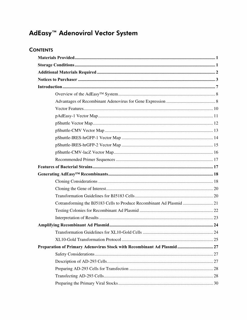
CONTENTS Materials Provided.............................................................................................................................. 1 Storage Conditions.............................................................................................................................. 1 Additional Materials Required .......................................................................................................... 2 Notices to Purchaser ........................................................................................................................... 3 Introduction......................................................................................................................................... 7
Overview of the AdEasy™ System....................................................................................... 8 Advantages of Recombinant Adenovirus for Gene Expression ............................................ 8 Vector Features.................................................................................................................... 10 pAdEasy-1 Vector Map....................................................................................................... 11 pShuttle Vector Map............................................................................................................ 12 pShuttle-CMV Vector Map ................................................................................................. 13 pShuttle-IRES-hrGFP-1 Vector Map .................................................................................. 14 pShuttle-IRES-hrGFP-2 Vector Map .................................................................................. 15 pShuttle-CMV-lacZ Vector Map......................................................................................... 16 Recommended Primer Sequences ....................................................................................... 17
Features of Bacterial Strains............................................................................................................ 17 Generating AdEasy™ Recombinants.............................................................................................. 18
Cloning Considerations ....................................................................................................... 18 Cloning the Gene of Interest................................................................................................ 20 Transformation Guidelines for BJ5183 Cells ...................................................................... 20 Cotransforming the BJ5183 Cells to Produce Recombinant Ad Plasmid ........................... 21 Testing Colonies for Recombinant Ad Plasmid .................................................................. 22 Interpretation of Results ...................................................................................................... 23
Amplifying Recombinant Ad Plasmid............................................................................................. 24 Transformation Guidelines for XL10-Gold Cells ............................................................... 24 XL10-Gold Transformation Protocol .................................................................................. 25
Preparation of Primary Adenovirus Stock with Recombinant Ad Plasmid ................................ 27 Safety Considerations.......................................................................................................... 27 Description of AD-293 Cells............................................................................................... 27 Preparing AD-293 Cells for Transfection ........................................................................... 28 Transfecting AD-293 Cells.................................................................................................. 28 Preparing the Primary Viral Stocks ..................................................................................... 30

Guidelines for Infection Conditions and Amplification of the Primary Viral Stock .................. 31 Titer Considerations ............................................................................................................ 31 Amplification Guidelines .................................................................................................... 31 Infection Procedure Guidelines ........................................................................................... 31 Optimizing Infection Conditions......................................................................................... 32 Monitoring the Infection...................................................................................................... 32
Detection of hrGFP ........................................................................................................................... 32 Monitoring Transfection and Infection of Cells .................................................................. 32 Specifications for hrGFP and EGFP Excitation and Emission Spectra............................... 33
LacZ Control: Detection and Applications..................................................................................... 33 Transfection Control............................................................................................................ 33 Virus Control ....................................................................................................................... 33
Appendix: Plaque Assay using Agarose Overlay ........................................................................... 34 Preparing Viral Stock Dilutions .......................................................................................... 34 Overlaying the Infected Cells with Agarose........................................................................ 34 Plaque Isolation ................................................................................................................... 35
Troubleshooting ................................................................................................................................ 36 Preparation of Media and Reagents ................................................................................................ 37 References .......................................................................................................................................... 39 Endnotes............................................................................................................................................. 39 MSDS Information............................................................................................................................ 39
AdEasy™ Adenoviral Vector System 1
AdEasy™ Adenoviral Vector System
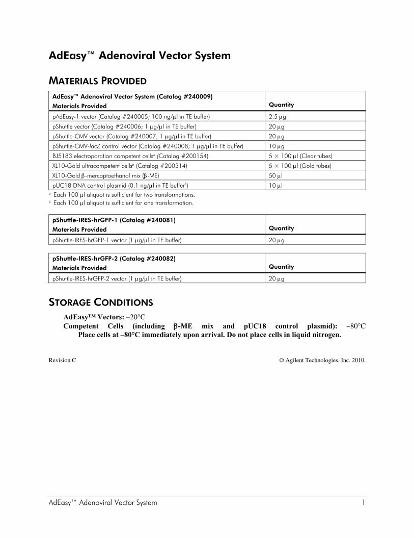
MATERIALS PROVIDED AdEasy™ Adenoviral Vector System (Catalog #240009)
Materials Provided
Quantity
pAdEasy-1 vector (Catalog #240005; 100 ng/μl in TE buffer) 2.5 μg
pShuttle vector (Catalog #240006; 1 μg/μl in TE buffer) 20 μg
pShuttle-CMV vector (Catalog #240007; 1 μg/μl in TE buffer) 20 μg
pShuttle-CMV-lacZ control vector (Catalog #240008; 1 μg/μl in TE buffer) 10 μg
BJ5183 electroporation competent cellsa (Catalog #200154) 5 × 100 μl (Clear tubes)
XL10-Gold ultracompetent cellsb (Catalog #200314) 5 × 100 μl (Gold tubes)
XL10-Gold β-mercaptoethanol mix (β-ME) 50 μl
pUC18 DNA control plasmid (0.1 ng/μl in TE bufferb) 10 μl a Each 100 μl aliquot is sufficient for two transformations. b Each 100 μl aliquot is sufficient for one transformation. pShuttle-IRES-hrGFP-1 (Catalog #240081)
Materials Provided
Quantity
pShuttle-IRES-hrGFP-1 vector (1 μg/μl in TE buffer) 20 μg
pShuttle-IRES-hrGFP-2 (Catalog #240082)
Materials Provided
Quantity
pShuttle-IRES-hrGFP-2 vector (1 μg/μl in TE buffer) 20 μg
STORAGE CONDITIONS AdEasy™ Vectors: –20°C Competent Cells (including β-ME mix and pUC18 control plasmid): –80°C
Place cells at –80°C immediately upon arrival. Do not place cells in liquid nitrogen.
Revision C © Agilent Technologies, Inc. 2010.

2 AdEasy™ Adenoviral Vector System
ADDITIONAL MATERIALS REQUIRED Pac I restriction enzyme Pme I restriction enzyme Alkaline phosphatase, molecular biology grade Chloroquine§ ViraPack Transfection Kit [Stratagene Catalog #200488] In Situ β-Galactosidase Staining Kit [Stratagene Catalog #200384] StrataPrep PCR Purification Kit [Stratagene Catalog #400771] Electroporation cuvettes, 0.2 cm gap Electroporator 14-ml BD Falcon polypropylene round-bottom tubes (BD Biosciences Catalog #352059) 5-ml BD Falcon polystyrene round bottom tubes (BD Biosciences Catalog #352054) AD–293 cells (recommended) [Stratagene Catalog #240085] or HEK293 cells Growth medium for AD-293 cells§ [Invitrogen Life Technologies (Gibco) Catalog #11995] SeaPlaque® agarose [FMC Corporation]
§ See Preparation of Media and Reagents.

AdEasy™ Adenoviral Vector System 3
NOTICES TO PURCHASER
Limited License Agreement: AdEasy™ Vectors Agilent hereby licenses Customer to use Stratagene AdEasy™ Vectors provided herewith (referred to hereinafter as the “Products”) on the following terms and conditions. (For purposes of this Notice, “Customer” shall include any person or entity which ordered the Products or at any time uses the Products.) Customer’s acceptance of delivery and/or use of the Products shall constitute Customer’s binding agreement to the following terms and conditions. If Customer is unwilling to accept such terms and conditions, Agilent is willing to accept return of the Products prior to any use of the Products, for a full refund.
1. The Products shall be used solely for research purposes, solely on premises under the control of Customer, solely by employees or agents of Customer, and solely in compliance with all laws, regulations, rules and guidelines applicable to the Products and their use, testing, handling, or other disposition thereof, or otherwise applicable to Customer’s activities hereunder.
2. Customer acknowledges and agrees that Customer is experienced and/or informed regarding the characteristics and use of adenoviral-based products such as the Products, for what purposes the Products are to be used, and how the Products should be used.
3. THE PRODUCTS ARE EXPERIMENTAL IN NATURE AND ARE PROVIDED WITHOUT WARRANTIES OF ANY KIND, EXPRESS OR IMPLIED, INCLUDING, WITHOUT LIMITATION, WARRANTIES OF MERCHANTABILITY OR FITNESS FOR A PARTICULAR PURPOSE. Customer hereby waives, releases and renounces any and all warranties, guarantees, obligations, liabilities, rights and remedies, express or implied, arising by law or otherwise, with respect to the usefulness or freedom from defects of the products, including, but not limited to, (a) any implied warranty or merchantability or fitness for a particular purpose, (b) any implied warranty arising from course of performance, course of dealing or usage in the trade, and (c) any obligation, right, liability, claim or remedy for (1) loss of use, revenue or profit, or any other damages, (2) infringement of third party intangible property rights, and (3) incidental or consequential damages.
4. Customer shall bear all risks associated with the Products and their use, testing, handling or other disposition thereof, and all risks associated with Customer's activities under this Agreement. Customer hereby assumes all risks of damage or injury to Customer's facilities, employees or agents and to any third party arising from possession or use of the Products. Agilent shall have no liability to Customer, its employees or agents or to any third party, regardless of the form or theory of action (whether contract, tort or otherwise, including but not limited to, negligence and strict liability), for any direct, indirect, consequential, incidental or other damages arising out of or relating to the Products or this Agreement.
5. Customer may at any time properly dispose of the Products in a manner which ensures their prompt destruction and is consistent with all applicable laws, regulations, rules and guidelines.
6. No modification or waiver of any terms or conditions of this Notice shall be effective unless in a writing signed by Customer and an authorized representative of Stratagene Products Division, Agilent Technologies Inc.
Limited License Agreement: CMV Promoter The use of the CMV Promoter is covered under U.S. Patent Nos. 5,168,062 and 5,385,839 owned by the University of Iowa Research Foundation and licensed FOR RESEARCH USE ONLY. For further information, please contact UIRF at 319-335-4546.

4 AdEasy™ Adenoviral Vector System
Limited License Agreement: AdEasy™ Products AdEasy™ products are sold under license from Johns Hopkins University. Rights to use these products are limited to non-commercial research only. No other rights are conveyed. Inquiry into the availability of a license to broader rights or the use of these products for commercial purposes should be directed to Johns Hopkins University School of Medicine Office of Technology Licensing, 111 Market Street, Suite 906, Baltimore, MD 21202. Purchase of these products does not grant rights to:(1) offer the vectors or any derivatives thereof for resale; or (2) to distribute or transfer the vector or any derivative thereof to third parties.
FLAG® License Agreement The enclosed DNA expression vector and/or antibody are specifically adapted for a method of producing selected protein molecules covered by one or more of the following patents owned by Sigma-Aldrich Co.: U.S. Patent Nos. (5,011912, 4,703,004, 4,782,137 and 4,851,341;EP Patent No. 150,126 (Austria, Belgium, Switzerland, France, United Kingdom, Italy, Netherlands and Sweden); EP Patent No. 335,899 (Belgium, Switzerland, Germany, France, United Kingdom, Italy, Luxembourg and Sweden); German Patent No. P3584260.1; Canadian Patent No. 1,307,752; and Japanese Patent Nos. 1,983,150 and 2,665,359. Your payment includes a limited license under these patents to make only the following uses of these products:
A. Vector License: You may use the enclosed vector to transform cells to produce proteins containing the amino acid sequence DYKDDDDK for research purposes provided, however, such research purposes do not include binding an unlicensed antibody to any portion of this amino acid sequence nor using such proteins for the preparation of antibodies having an affinity for any portion of this amino acid sequence. B. Antibody License: You may only use the enclosed antibody for research purposes to perform a method of producing a protein in which the protein is expressed in a host cell and purified by use of the antibody in accordance with a claim in one of the above patents in force in a country where the use actually occurs so long as: (1) you perform such method with a DNA expression vector licensed from Sigma-Aldrich Co.; and (2) you do not bind (or allow others to bind) an unlicensed antibody to any DYKDDDDK epitope of any fusion protein that is produced by use of the method.
This license does not include any rights under any other patents. You are not licensed to use the vector and/or antibody in any manner or for any purposed not recited above. As used above, the term “unlicensed antibody” means any antibody which Sigma-Aldrich Co. has not expressly licensed pursuant to Paragraph B, above. Sigma-Aldrich Co. hereby expressly retains all rights in the above listed patents not expressly licensed hereunder. If the terms and conditions of this License Agreement are acceptable to you, then you may open the vessel(s) containing the vector and/or antibody and, through such act of opening a vessel, will have shown your acceptance to these terms and conditions. If the terms and conditions of this License Agreement are not acceptable to you, then please return the vessel(s) unopened to Stratagene Products Division for a complete refund of your payment. For additional licensing information or to receive a copy of any of the above patents, please contact the Sigma-Aldrich Co. licensing department at telephone number 314-771-5765.

AdEasy™ Adenoviral Vector System 5
Non-Commercial Research Use License For Nonprofit Entities: pShuttle-IRES-hrGFP-1 and pShuttle-IRES-hrGFP-2 Agilent agrees to sell, and Licensee agrees to purchase, Stratagene Vitality fluorescent protein products provided herewith (referred to as the “Products”) on the following terms and conditions. (For purposes of this License, “Licensee” shall include any person or entity which ordered the Products or at any time uses the Products.) LICENSEE’S ACCEPTANCE OF DELIVERY AND/OR USE OF THE PRODUCTS SHALL CONSTITUTE LICENSEE’S BINDING AGREEMENT TO THE FOLLOWING TERMS AND CONDITIONS. IF LICENSEE IS UNWILLING TO ACCEPT SUCH TERMS AND CONDITIONS, AGILENT IS WILLING TO ACCEPT RETURN OF THE PRODUCTS PRIOR TO ANY USE OF THE PRODUCTS, FOR A FULL REFUND. 1. The Products, containing DNA sequences encoding for fluorescent protein or variants thereof, are proprietary or exclusively licensed to Agilent and licensed hereunder for non-commercial research purposes only. Licensee may modify only the non-coding region outside of the nucleic acid encoding the fluorescent protein of the Products to facilitate non-commercial research. Licensee shall not have any rights to (i) modify the coding region of the nucleic acid encoding the fluorescent protein of the Products, (ii) offer the Products, or any component, derivative or modification thereof, for resale, or (iii) distribute, transfer, or otherwise provide access to, the Products, or any component, derivative or modification thereof, to any third party for any purpose or use. 2. Except as set forth above, no other rights, express or implied, are conveyed to Licensee. No rights are granted to Licensee to use the Products for (i) the provision of services to any for-profit third party (e.g., screening and profiling), (ii) diagnostic applications, (iii) methods employed in screens to evaluate compounds (e.g., high throughput screening (“HTS”), (iv) profiling chemicals for selectivity, bioavailability, drug metabolism or toxicity, (v) use in vivo in multicellular organisms (and methods therein) for gene therapy, (vi) quality control or quality assurance processes, including food and environmental testing, or (vii) use in manufacturing. 3. The Products shall be used solely on premises under the control of Licensee, and in compliance with all laws, regulations, rules and guidelines applicable to the Products and their use, testing, handling, or other disposition thereof, or otherwise applicable to Licensee’s activities hereunder. 4. Title to the Products shall not transfer to Licensee. 5. Agilent warrants that, at the time of shipment, the Products will conform to the specifications which accompany the Products. This warranty limits Agilent’s liability to replacement of the Products. AGILENT MAKES NO OTHER WARRANTIES, EXPRESS OR IMPLIED, WITH RESPECT TO THE PRODUCTS, INCLUDING ANY WARRANTIES OF MERCHANTABILITY OR FITNESS FOR ANY PARTICULAR PURPOSE OR THAT THE PRODUCTS DO NOT INFRINGE ANY PROPRIETARY RIGHTS OF ANY THIRD PARTY. Licensee hereby waives, releases and renounces any and all warranties, guarantees, obligations, liabilities, rights and remedies, express or implied, arising by law or otherwise, with respect to the usefulness or freedom from defects of the Products, including, but not limited to, (a) any implied warranty or merchantability or fitness for a particular purpose, (b) any implied warranty arising from course of performance, course of dealing or usage in the trade, and (c) any obligation, right, liability, claim or remedy for (1) loss of use, revenue or profit, or any other damages, (2) infringement of third party intellectual property rights, and (3) incidental or consequential damages. 6. Licensee agrees to bear all risks associated with the Products and their use, testing, handling or other disposition thereof, and all risks associated with Licensee's use of the Products purchased hereunder. Licensee hereby assumes all risks of damage or injury to Licensee's facilities, employees

6 AdEasy™ Adenoviral Vector System
or agents and to any third party arising from possession or use of the Products. Agilent shall have no liability to Licensee, its employees or agents or to any third party, regardless of the form or theory of action (whether contract, tort or otherwise, including, but not limited to, negligence and strict liability), for any direct, indirect, consequential, incidental or other damages arising out of or relating to the Products or this License. 7. Licensee shall indemnify, defend and hold Agilent, its affiliates, distributors, suppliers, directors, officers, employees and agents, harmless from and against any and all claims, actions, demands, liabilities, damages and expenses (including attorneys’ fees) relating to or arising out of any damage or injury, including, but not limited to, product liability and intellectual property infringement claims of any nature, alleged to have been caused by the Products or the use, testing, handling or other disposition thereof or Licensee’s activities hereunder. 8. Licensee may at any time properly dispose of the Products in a manner which ensures their prompt destruction and is consistent with all applicable laws, regulations, rules and guidelines. 9. No modification or waiver of any terms or conditions of this License shall be effective unless in writing signed by Licensee and an authorized representative of Stratagene Products Division, Agilent Technologies Inc. For information on acquiring a license to use the Products for commercial purposes, including commercial research purposes, please contact Stratagene Products Division, Business Development, 11011 North Torrey Pines Road, La Jolla, California 92037, telephone number (858) 373-6300, facsimile number 1-866-725-7207.
IRES Sequence Use of the translation enhancer of the pShuttle-IRES-hrGFP-1 and p-Shuttle-IRES-hrGFP-2 vectors is covered by U.S. Patent No. 4,937,190 and is limited to use solely for research purposes. Any other use of the translation enhancer of the pIRES-hrGFP-1a and pIRES-hrGFP-2a vectors requires a license from WARF. WARF can be reached at P.O. Box 7365 Madison, WI 53707-7365.

AdEasy™ Adenoviral Vector System 7
INTRODUCTION Recombinant adenoviruses are a versatile tool for gene delivery and expression. Several features of adenovirus biology have made such viruses the vectors of choice for certain applications. For example, adenoviruses are capable of infecting a broad range of cell types and infection is not dependent on active host cell division. Additionally, high virus titers and high-level gene expression can be obtained, which are important considerations for protein production techniques in mammalian cells. The most commonly used adenoviral vector is human adenovirus serotype 5, which is rendered replication defective by the deletion of the E1 and E3 genes. The E1 gene is essential for the assembly of infectious virus particles and is complemented in vivo by an adenovirus packaging cell line (e.g. AD-293). The E3 gene encodes proteins involved in evading host immunity and is dispensable. Not only do these deletions render the virus incapable of replicating itself, but they also create space for up to 7.5 kb of foreign DNA.1 Two methods have traditionally been used to generate recombinant adenoviruses. The first involves direct ligation of the gene of interest into the adenoviral genome. Because the adenovirus genome is large (36 kb) and contains few useful restriction sites, this method is technically very challenging. The second, and more commonly used method, involves cloning the gene of interest into a shuttle vector and transferring the gene into the adenovirus genome by means of homologous recombination in an adenovirus packaging cell line.2 The isolation of recombinant adenovirus by this method involves performing multiple plaque isolations and is extremely laborious and time consuming. More recently, a different approach has been developed by T.C. He and colleagues.3 Employing the efficient homologous recombination machinery in E. coli, a recombinant adenovirus is produced by a double-recombination event between cotransformed adenoviral backbone plasmid vector, pAdEasy-1, and a shuttle vector carrying the gene of interest. This eliminates the need to manipulate the large adenovirus DNA molecule in vitro (in restriction and ligation reactions). There is no need to carry out laborious plaque purification rounds. The time needed to generate a recombinant adenovirus is reduced by several weeks.
Note The AdEasy™ XL Adenoviral Vector System is also available (Stratagene Catalog #240010). The XL kit includes BJ5183 cells pre-transformed with the pAdEasy-1 plasmid (BJ5183-AD-1 cells), providing a streamlined recombinant Ad DNA production method. This kit also includes the AD-293 cells recommended for use with the AdEasy vector system.

8 AdEasy™ Adenoviral Vector System
Overview of the AdEasy™ System A schematic overview of the production of recombinant adenovirus is shown in Figure 1. In the AdEasy system, the DNA of interest is cloned into any of the four shuttle vectors (pShuttle-CMV, pShuttle, pShuttle-IRES-hrGFP-1 (Catalog #240081) or pShuttle-IRES-hrGFP-2 (Catalog #240082). Once constructed, the shuttle vector is linearized with Pme I and cotransformed into BJ5183 together with pAdEasy-1, the supercoiled viral DNA plasmid. Transformants are selected for kanamycin resistance, and recombinants are subsequently identified by restriction digestion. Once a recombinant is identified, it is produced in bulk using the recombination-deficient XL10-Gold strain. Purified recombinant Ad plasmid DNA is digested with Pac I to expose its inverted terminal repeats (ITR), and then used to transfect AD–293 (or HEK293) cells where deleted viral assembly genes are complemented in vivo.
Advantages of Recombinant Adenovirus for Gene Expression Broad range of infectivity and high titer Adenoviruses can infect a broad range of mammalian cells and have been used successfully to express human and non-human proteins. Recombinant adenoviruses can produce high titers (107 to 108 pfu/ml following transient transfection). Infection does not require an actively dividing host cell Recombinant adenovirus can infect both dividing and non-dividing cells. Expressed human proteins are properly folded and modified Because the AdEasy vector system employs human adenovirus and human host cell lines, human proteins expressed using this system are abundant and have the correct posttranslational modification and folding. Large insert size The AdEasy vector system allows for the insertion of up to 7.5 kb of foreign DNA. AdEasy vector is non-insertional Because the recombinant adenovirus remains epichromosomal in the human host cell, there is only a remote possibility of activation or inactivation of host cell genes resulting from interruption by the transfected gene(s).

AdEasy™ Adenoviral Vector System 9
FIGURE 1 Production of recombinant adenovirus
gene of interest
Ori
Ori
Kan
Pac I
Pac I
Pac I
RecombinantAd
Plasmid
Transfect AD-293 (or HEK293) cells
Digest with IPac
LITR
Encapsidation signal
RITR
LITREncapsidation
signal
Promotergene of interest
poly AAdenoviral DNA
Pac IPac I
cotransformB 5183 cells, select for Kan
JR
linearize with IPme
MCSShuttle Vector
R ight A
rm
Ori
Kan
gene of interest
Pac I
Pac I
LITR
Encapsidation signal
RITR
poly APme ILeft ArmCloning Geneof Interest
HomologousRecombination
in Bacteriain vivo
Virus Productionin AD-293 Cells
Regions ofHomologous
Recombination
Left Arm
pAdEasy-1Vector
Ori Right Arm
Pac I
Amp
RITR
RITR
RITR

10 AdEasy™ Adenoviral Vector System
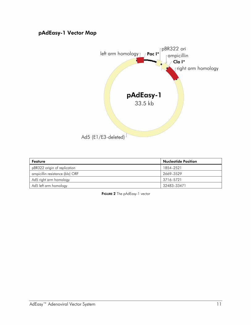
Vector Features pAdEasy-1 and the set of shuttle vectors contain different resistance cassettes: ampicillin and kanamycin, respectively. All plasmids have the pBR322 origin of replication. The circular map of the pAdEasy-1 vector is shown in Figure 2, and circular maps of the shuttle vectors that may be used in the AdEasy adenoviral vector system are shown in Figures 3–7.
pAdEasy-1 The plasmid pAdEasy-1, containing most of the human adenovirus serotype 5 (Ad5) genome, is deleted for the genes E1 and E3. The removal of these two viral genes creates space for foreign DNA and eliminates self-replication capabilities. The E1 deletion renders the viruses defective for replication and incapable of producing infectious viral particles in target cells (provided there is no complementation by the host cell); the E3 region encodes proteins involved in evading host immunity and is dispensable. The deletion of both genes creates room for up to 7.5 kb of foreign DNA that can be inserted into the Ad5 genome. The E1 gene, which is necessary for production of viral particles, is provided in trans by AD-293 cells. pAdEasy-1 carries the ampicillin resistance gene, which is lost after recombination with a shuttle vector.
pShuttle and pShuttle-CMV The vector pShuttle-CMV contains a multiple cloning site sandwiched between the CMV promoter and the SV40 polyadenylation signal and is suitable for insertion of a large cDNA (up to 6.6 kb). pShuttle contains only a multiple cloning site. This allows for the insertion of an entire expression cassette, including specialized promoters and termination signals (up to 7.5 kb). The regions indicated as arms are the stretches of sequence homology with pAdEasy-1 where the homologous recombination occurs. The R-ITR and L-ITR regions are short inverted terminal repeats (Left and Right) which have a role in replication of the viral DNA.2
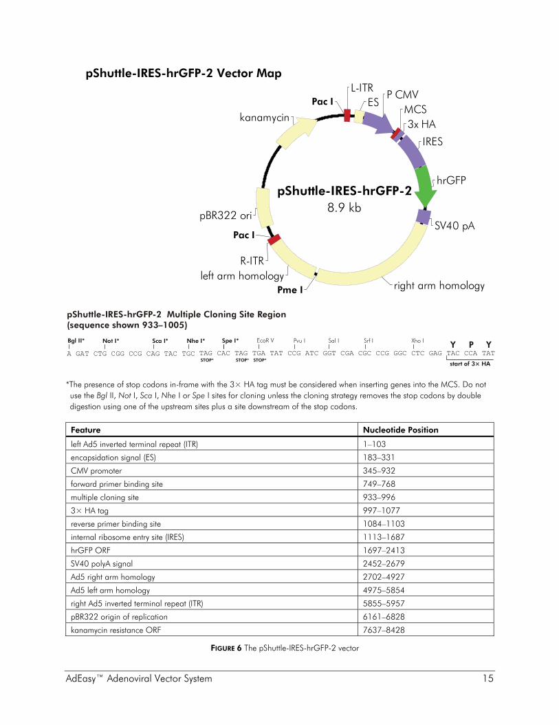
pShuttle-IRES-hrGFP-1 and pShuttle-IRES-hrGFP-2 Two additional shuttle vectors, pShuttle-IRES-hrGFP-1 (Catalog #240081) and pShuttle-IRES-hrGFP-2 (Catalog #240082) are available separately. Both of these vectors contain the CMV promoter and a dicistronic expression cassette in which the multiple cloning site (MCS) is followed by the EMCV-IRES, which directs translation of a humanized recombinant green fluorescent protein (hrGFP) from a novel marine organism as a second open reading frame. This design allows the expression of the gene of interest (up to 5.2 kb) to be monitored at the single-cell level due to expression of the hrGFP on the same transcript. The gene of interest may be fused to three contiguous copies of either the FLAG® epitope (pShuttle-IRES-hrGFP-1) or the HA epitope (pShuttle-IRES-hrGFP-2).
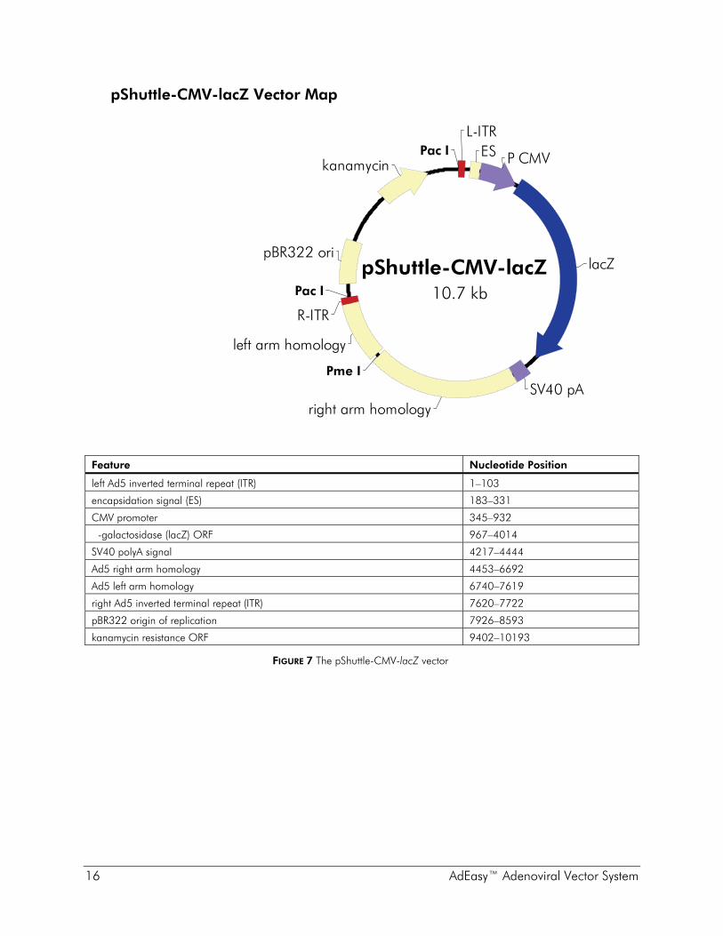
pShuttle-CMV-lacZ The lacZ gene was inserted in the MCS site of the pShuttle-CMV to produce pShuttle-CMV-lacZ. This construct is provided as a control for the production of recombinant adenovirus.
AdEasy™ Adenoviral Vector System 11
pAdEasy-1 Vector Map
Feature Nucleotide Position
pBR322 origin of replication 1854–2521
ampicillin resistance (bla) ORF 2669–3529
Ad5 right arm homology 3716–5721
Ad5 left arm homology 32483–33471
FIGURE 2 The pAdEasy-1 vector
pBR322 oriampicillin
right arm homology
left arm homology
Cla I*
Pac I*
Ad5 (E1/E3-deleted)
pAdEasy-133.5 kb

12 AdEasy™ Adenoviral Vector System
pShuttle Vector Map
Feature Nucleotide Position
left Ad5 inverted terminal repeat (ITR) 1–103
encapsidation signal (ES) 183–331
forward primer binding site 299–323
multiple cloning site 345–404
Ad5 right arm homology 414–2652
reverse primer binding site 438–459
Ad5 left arm homology 2701–3580
right Ad5 inverted terminal repeat (ITR) 3581–3683
pBR322 origin of replication 3887–4554
kanamycin resistance ORF 5363–6154
FIGURE 3 The pShuttle vector
pShuttle Multiple Cloning Site Region(sequence shown 299–459)
GAAGTGAAATCTGAATAATTTTGTGTTACTCATAGCGCGTAATACT...Forward primer binding site
...GGGCGTGGTTAAGGGTGGGAAAGAATATATAAGGTGGGGGTCTTATGTAGTTTTGReverse primer binding site
...GGTACCGCGGCCGCCTCGAGTCTAGAGATATCGAATTCAAGCTTGTCGACTCGAAGATCT...
Kpn I Not I Xho I Xba I EcoR V Hind III Sal I Bgl II
kanamycin
right arm homology
left arm homology
pBR322 ori
ESL-ITR
R-ITR
MCS
Pac I
Pac I
Pme I
pShuttle6.6 kb

AdEasy™ Adenoviral Vector System 13
pShuttle-CMV Vector Map
Feature Nucleotide Position
left Ad5 inverted terminal repeat (ITR) 1–103
encapsidation signal (ES) 183–331
CMV promoter 341–933
forward primer binding site 888–907
multiple cloning site 940–987
reverse primer binding site 1009–1031
SV40 polyA signal 1011–1238
Ad5 right arm homology 1243–3497
Ad5 left arm homology 3545–4428
right Ad5 inverted terminal repeat (ITR) 4429–4531
pBR322 origin of replication 4735–5402
kanamycin resistance ORF 6211–7002
FIGURE 4 The pShuttle-CMV vector
L-ITR
MCS
R-ITR
pBR322 ori
Pac I
Pac I ES
kanamycinSV40 pA
P CMV
right arm homology
Pme Ileft arm homology
pShuttle-CMV7.5 kb
pShuttle-CMV Multiple Cloning Site Region(sequence shown 888–1031)Forward primer binding site
GGTCTATATAAGCAGAGCTGGTTTAGTGAACCGTCAGATCCGCTAG...
Kpn I Not I Xho I EcoR VHind IIISal I
...AGATCTGGTACCGTCGACGCGGCCGCTCGAGCCTAAGCTTCTAGATAAGATATC...
Reverse primer binding site...CGATCCACCGGATCTAGATAACTGATCATAATCAGCCATACCAC

14 AdEasy™ Adenoviral Vector System
pShuttle-IRES-hrGFP-1 Vector Map
*The presence of stop codons in-frame with the 3× FLAG tag must be considered when inserting genes into the MCS. Do not use the Bgl II, Not I, Sca I, Nhe I or Spe I sites for cloning unless the cloning strategy removes the stop codons by double digestion using one of the upstream sites plus a site downstream of the stop codons.
Feature Nucleotide Position
left Ad5 inverted terminal repeat (ITR) 1–103
encapsidation signal (ES) 183–331
CMV promoter 345–932
forward primer binding site 749–768
multiple cloning site 933–996
3× FLAG tag 997–1068
reverse primer binding site 1075–1094
internal ribosome entry site (IRES) 1104–1678
hrGFP ORF 1688–2404
SV40 polyA signal 2443–2670
Ad5 right arm homology 2693–4918
Ad5 left arm homology 4966–5845
right Ad5 inverted terminal repeat (ITR) 5846–5948
pBR322 origin of replication 6152–6819
kanamycin resistance ORF 7628–8419
FIGURE 5 The pShuttle-IRES-hrGFP-1 vector
pShuttle-IRES-hrGFP-1 Multiple Cloning Site Region(sequence shown 933–1005)
TAG CAC TAG TGA TAT CCG ATC GGT CGA CGC CCG GGC CTC GAG GAC TAC AAG
Bgl II* Sca I*Not I* Nhe I*
A GAT CTG CGG CCG CAG TAC TGCSTOP*STOP*STOP*
EcoR V Pvu ISpe I* Sal I Xho ISrf I
start of 3× FLAG
D Y K
hrGFP
pBR322 ori
kanamycin
Pac I
Pac I
Pme I
P CMVES
L-ITR
MCS3x FLAG
IRES
SV40 pA
right arm homologyleft arm homology
R-ITR
pShuttle-IRES-hrGFP-18.9 kb
AdEasy™ Adenoviral Vector System 15
pShuttle-IRES-hrGFP-2 Vector Map
*The presence of stop codons in-frame with the 3× HA tag must be considered when inserting genes into the MCS. Do not use the Bgl II, Not I, Sca I, Nhe I or Spe I sites for cloning unless the cloning strategy removes the stop codons by double digestion using one of the upstream sites plus a site downstream of the stop codons.
Feature Nucleotide Position
left Ad5 inverted terminal repeat (ITR) 1–103
encapsidation signal (ES) 183–331
CMV promoter 345–932
forward primer binding site 749–768
multiple cloning site 933–996
3× HA tag 997–1077
reverse primer binding site 1084–1103
internal ribosome entry site (IRES) 1113–1687
hrGFP ORF 1697–2413
SV40 polyA signal 2452–2679
Ad5 right arm homology 2702–4927
Ad5 left arm homology 4975–5854
right Ad5 inverted terminal repeat (ITR) 5855–5957
pBR322 origin of replication 6161–6828
kanamycin resistance ORF 7637–8428
FIGURE 6 The pShuttle-IRES-hrGFP-2 vector
pShuttle-IRES-hrGFP-2 Multiple Cloning Site Region(sequence shown 933–1005)
Y P YTAG CAC TAG TGA TAT CCG ATC GGT CGA CGC CCG GGC CTC GAG TAC CCA TAT
Bgl II* Sca I*Not I* Nhe I*
A GAT CTG CGG CCG CAG TAC TGCSTOP*STOP*STOP*
EcoR V Pvu ISpe I* Sal I Xho ISrf I
start of 3× HA
Pac I
Pac I
Pme I
L-ITRES
P CMVMCS3x HA
IRES
hrGFP
SV40 pA
right arm homologyleft arm homology
R-ITR
pBR322 ori
kanamycin
pShuttle-IRES-hrGFP-28.9 kb
16 AdEasy™ Adenoviral Vector System
pShuttle-CMV-lacZ Vector Map
Feature Nucleotide Position
left Ad5 inverted terminal repeat (ITR) 1–103
encapsidation signal (ES) 183–331
CMV promoter 345–932
�-galactosidase (lacZ) ORF 967–4014
SV40 polyA signal 4217–4444
Ad5 right arm homology 4453–6692
Ad5 left arm homology 6740–7619
right Ad5 inverted terminal repeat (ITR) 7620–7722
pBR322 origin of replication 7926–8593
kanamycin resistance ORF 9402–10193
FIGURE 7 The pShuttle-CMV-lacZ vector
Pac I
Pac I
Pme I
kanamycin
L-ITR
R-ITR
right arm homology
P CMV
lacZ
left arm homology
pBR322 ori
SV40 pA
ES
pShuttle-CMV-lacZ10.7 kb

AdEasy™ Adenoviral Vector System 17
Recommended Primer Sequences Recommended sequences of primers flanking the MCS suitable for PCR amplification and/or sequencing applications are given in Table I.
TABLE I Vector Direction Sequence Position in Vector
Forward 5´ GAAGTGAAATCTGAATAATTTTGTG 3´ 299–323 pShuttle
Reverse 5´ CAAAACTACATAAGACCCCCAC 3´ 438–459
Forward 5´ GGTCTATATAAGCAGAGCTG 3´ 888–907 pShuttle-CMV
Reverse 5´ GTGGTATGGCTGATTATGATCAG 3´ 1009–1031
Forward 5´ CTCACGGGGATTTCCAAGTC 3´ 749–768 pShuttle-IRES-hrGFP-1 Reverse 5´ ATGCAGTCGTCGAGGAATTG 3´ 1075–1094
Forward 5´ CTCACGGGGATTTCCAAGTC 3´ 749–768 pShuttle-IRES-hrGFP-2 Reverse 5´ ATGCAGTCGTCGAGGAATTG 3´ 1084–1103
FEATURES OF BACTERIAL STRAINS In order to produce and amplify recombinant adenovirus with the AdEasy adenoviral system, two different prokaryotic host strains are required. The first, BJ5183, is recA proficient and supplies the machinery necessary to execute the recombination event between the shuttle vector and the pAdEasy vector. The second strain, provided as XL10-Gold* ultracompetent cells, is used to amplify the recombined adenovirus plasmid. This strain is both endonuclease deficient (endA1) and recombination deficient (recA). The endA1 mutation greatly improves the quality of plasmid miniprep DNA, and the recA mutation helps ensure insert stability. In the following table, the genes indicated in italics signify that the bacterium carries a mutant allele. The genes present on the F´ episome represent the wild-type bacterial alleles.
Host strain References Genotype
BJ5183 4 endA1 sbcBC recBC galK met thi-1 bioT hsdR (Strr)
XL10-Gold ultracompetent cells
5, 6 TetR Δ(mcrA)183 Δ(mcrCB-hsdSMR-mrr)173 endA1 supE44 thi-1 recA1 gyrA96 relA1 lac Hte [F´ proAB lacIqZΔM15 Tn10 (TetR) Amy CamR]
* U.S. Patent Nos.5,512,468 and 5,707,841 and equivalent foreign patents.

18 AdEasy™ Adenoviral Vector System
GENERATING ADEASY™ RECOMBINANTS
Cloning Considerations Refer to Figures 3–6 for circular maps and corresponding MCS sequences for the AdEasy shuttle vectors and to www.genomics.agilent.com for the complete nucleotide sequence and restriction maps for the pShuttle and pShuttle-CMV vectors. Each vector sequence for vectors in the AdEasy system has been verified for accuracy at the cloning junctions. The remainder of the sequence of each of the AdEasy vectors has been compiled from existing data.
Absence of Pme I and Pac I Sites in the Insert DNA The shuttle vector must be linearized using Pme I before cotransformation of BJ5183 bacteria and the recombinant Ad plasmid must be digested with Pac I before transfection of the packaging cell line. Ensure that the gene of interest does not contain either of these restriction sites. If these sites exist in the gene of interest, it will be necessary to perform site-directed mutagenesis before proceeding to the cloning steps.
Inclusion of Transcriptional and Translational Control Sequences in the Insert DNA The choice of shuttle vector (pShuttle, pShuttle-CMV, pShuttle-IRES-hrGFP-1 or pShuttle-IRES-hrGFP-2) depends on the user's desired application. The pShuttle vector allows insertion of an entire expression cassette, such that gene expression is controlled by insert-provided transcriptional promoter and terminator sequences as well as translation initiation and stop codons. If transcription via the CMV promoter is desired, choose from among the pShuttle-CMV, pShuttle-IRES-hrGFP-1 or pShuttle-IRES-hrGFP-2 vectors. If the gene of interest is cloned into one of these three CMV-containing vectors, the insert must include an initiation codon. We recommend the use of the Kozak initiation sequence. A complete Kozak sequence includes CCACCATGG, although CCATGG, or the core ATG, is sufficient. If the pShuttle-CMV vector is used, the insert must also contain a stop codon. Conversely, the pShuttle-IRES-hrGFP-1 and pShuttle-IRES-hrGFP-2 vectors already contain in-frame stop codons at the C-terminus of the fusion tags. Proteins may also be expressed from either of the pShuttle-IRES-hrGFP-1 or pShuttle-IRES-hrGFP-2 vectors in the absence of a fusion tag if an in-frame stop codon is included in the insert DNA that is cloned into the MCS upstream of the tag.

AdEasy™ Adenoviral Vector System 19
Cloning Capacity of the Shuttle Vectors It is important to adhere to the upper size limit of insert DNA (cloning capacity) for any of the shuttle vectors. Inserting larger fragments results in considerable decreases in efficiency of the AdEasy system. See the table below for shuttle vector cloning capacities and for an outline of the general features for the AdEasy shuttle vectors.
Shuttle Vector Features
Vector
Cloning capacity
Promoter
Poly A
MCS restriction sites
Description
pShuttle 7.5 kb — — Kpn I, Not I, Xho I, Xba I, EcoR V, Hind III, Sal I, Bgl II
Ligate an entire expression cassette into MCS
pShuttle-CMV 6.6 kb CMV + Kpn I Sal I, Not I, Xho I, Hind III, EcoR V
Ligate gene of interest into MCS between the CMV promoter and poly A
pShuttle-IRES-hrGFP-1 or -2
5.2 kb CMV + Bgl II*, Not I*, Sca I*, Nhe I* Spe I*, EcoR V, Pvu I, Sal I, Srf I, Xho I
Ligate gene of interest into MCS, in-frame with the FLAG or HA tag. Dicistronic transcript encoding hrGFP allows monitoring of the expression of the gene of interest by GFP fluorescence
*These restriction sites are upstream of in-frame stop codons. Do not use these sites for cloning unless the cloning strategy removes the stop codons by double digestion using one of the upstream sites plus a site downstream of the stop codons.

20 AdEasy™ Adenoviral Vector System
Cloning the Gene of Interest
1. Clone the gene of interest into the shuttle vector of choice using appropriate site(s) in the MCS.
2. Confirm the presence of the insert by restriction digestion or sequence analysis.
3. Purify DNA (shuttle vector plus gene of interest) in sufficient quantity for the subsequent cotransformation steps (2 μg of linearized shuttle vector DNA is required for each cotransformation reaction; 1 μg for each test and control transformation reactions).
4. Linearize shuttle plasmid DNA using Pme I and confirm complete digestion by agarose gel electrophoresis. It is necessary to prepare linearized samples of both the test plasmid DNA (shuttle vector plus gene of interest) and the control shuttle vector, pShuttle-CMV-lacZ.
5. Once complete digestion with Pme I is confirmed, remove the enzyme and buffer by method of choice. The Stratagene StrataPrep PCR Purification Kit is suitable for this purpose.
6. Treat the purified DNA with alkaline phosphatase for 30 minutes at 37°C.
7. Separate the linearized, dephosphorylated shuttle vector using agarose gel electrophoresis. Put samples of uncut shuttle vector in adjacent lanes for comparison. Ensure that the gel is run long enough to visualize good separation between uncut and cut DNA.
8. Gel purify the linearized shuttle vector by method of choice.
Note Ensure that the method chosen to purify the shuttle vector results in a DNA sample that does not contain salts, which will severely damage cells during electroporation.
9. Resuspend the purified DNAs in sterile dH2O to a final concentration of 1 μg/μl.
Transformation Guidelines for BJ5183 Cells
Storage Conditions Electroporation competent cells are sensitive to even small variations in temperature and must be stored at the bottom of a –80°C freezer. Transferring tubes from one freezer to another may result in a loss of efficiency. Electroporation competent cells should be placed at –80°C directly from the dry ice shipping container. When aliquoting, keep electroporation competent cells on ice at all times.

AdEasy™ Adenoviral Vector System 21
Cotransforming the BJ5183 Cells to Produce Recombinant Ad Plasmid
Note In this portion of the protocol, the BJ5183 cells are cotransformed with the linearized shuttle vector (containing the gene of interest or containing the control gene, lacZ) and the pAdEasy-1 vector. A recombination event that takes place in the bacterial cells results in the production of recombinant AdEasy plasmid DNA. It is very important that this cotransformation takes place in BJ5183 cells (supplied in this kit) and not in the XL10-Gold cells (also supplied in this kit). Only the BJ5183 cells have the cellular components necessary to carry out recombination.
1. Prechill four DNase-free microcentrifuge tubes and four electroporation cuvettes (0.2 cm gap) on ice.
2. Remove two aliquots of BJ5183 electroporation competent cells from –80°C storage and thaw on ice.
3. Gently pipet 40 μl of the competent cells into each of the chilled microcentrifuge tubes.
4. Into one of the tubes, pipet 1 μl (1 μg) of linearized, dephosphorylated shuttle vector and 1 μl of pAdEasy-1 supercoiled vector (100 ng/μl). Mix by tapping the tube gently and keep on ice.
5. Into the second tube, pipet 1 μl (1 μg) of linearized, dephosphorylated pShuttle-CMV-lacZ vector and 1 μl of pAdEasy-1 supercoiled vector (100 ng/μl). Mix by tapping the tube gently and keep on ice.
6. Into the third tube, pipet (1 μl) 1 μg of linearized, dephosphorylated shuttle vector. Mix by tapping the tube gently and keep on ice. This controls for background contributed by the shuttle vector.
7. Into the fourth tube, pipet, 1 μl of pUC18 DNA control plasmid (diluted 1:10 in sterile dH2O).
8. Set the electroporator to the following settings by referring to the instructions provided with the instrument: 200 Ω, 2.5 kV, 25 μF.
9. Transfer the contents of one microcentrifuge tube from step 4 into one of the chilled electroporation cuvettes and tap the cuvette gently to settle the mixture to the bottom.
10. Slide the cuvette into the electroporation chamber until the cuvette connects with the electrical contacts.
11. Pulse the sample once, then quickly remove the cuvette. Immediately add 1 ml of sterile LB broth (see Preparation of Media and Reagents) and pipet up and down to resuspend the cells.

22 AdEasy™ Adenoviral Vector System
12. Transfer the cell suspension to a sterile 14-ml BD Falcon polypropylene round-bottom tube.
13. Repeat the electroporation for the other three transformation reactions. Incubate all of the transformations at 37°C for 1 hour while shaking at 225–250 rpm.
14. For the three recombination reactions and associated controls, plate the entire volume of recovered cells onto 3–5 LB-kanamycin plates§ (e.g. four plates containing 50 μl, 100 μl, 250 μl, and 600 μl respectively).
15. For the pUC18 transformation, first place a 100-μl pool of LB broth on an LB–ampicillin agar plate.§ Add 5 μl of the transformed cells to the pool of LB broth. Use a sterile spreader to spread the mixture. The pUC18 control transformation should yield an efficiency of > 108 cfu/μg.
16. Incubate the plates overnight at 37°C.
Testing Colonies for Recombinant Ad Plasmid
1. Examine the transformation plates. The plates containing the transformation of linearized shuttle vector alone have uniform-sized colonies arising from uncut/recircularized vector. When compared to the control shuttle vector plates, the transformants on the plates containing the pAdEasy-1 recombinants will appear as two populations: normal size and tiny at an approximate ratio of 3:1. The tiny colonies are the potential recombinants and the normal-sized colonies are background from the shuttle vector.
2. Examine the pUC18 transformation to determine transformation efficiency, if desired.
3. Pick 10 or more of the smallest, well isolated colonies each from the test recombination plate (shuttle vector plus gene of interest) and the control recombination plate (pShuttle-CMV-lacZ) into 3–5 ml cultures of LB-kanamycin broth. §
Note The recombinants will be low copy number plasmids approximately 40 kb in size. As implied by the small colony size on the test plates, cultures will grow slowly and plasmid yields will be low. Miniprep procedures should be adapted accordingly. Procedures suitable for purification of cosmids or large plasmids are recommended.
4. Incubate at 37°C overnight while shaking at 225–250 rpm. § See Preparation of Media and Reagents.

AdEasy™ Adenoviral Vector System 23
5. Prepare miniprep DNA from 2 ml (or more) of overnight culture using a procedure suitable to purify large plasmids or cosmids. The final volume of miniprep DNA should be 50 μl and the DNA should be resuspended in sterile dH2O or TE buffer.
6. Cut 10 μl of the miniprep DNA with Pac I and run the entire digest on a 0.8% agarose TAE§ gel next to 10 μl of uncut miniprep DNA. As a control, also cut a small amount (~0.2μg) unrecombined shuttle vector (prepared in step 3 of Cloning the Gene of Interest) and run in an adjacent lane.
Interpretation of Results
Restriction of recombinant Ad plasmid DNA with Pac I should yield a large fragment of ~30 kb*, and a smaller fragment of either 3.0 kb (if recombination took place between the left arms) or 4.5 kb (if recombination took place at the origins of replication). Uncut recombinants will give a large smear at the top of the gel very close to the wells (and often have a smaller band that runs just below 23 kb). Potential recombinants may be difficult to identify (in some instances you can only visualize the 30 kb band) if the quality or yield of the miniprep is low. If this is the case, prepare DNA from a greater volume of culture with a procedure adapted for purification of large plasmids or cosmids. More than 20% of the colonies should contain a recombinant plasmid close to 40 kb. There are often faint background bands in BJ5183 minipreps. If the PREDOMINANT bands are the expected sizes, redigest potential recombinants prepared from XL10-Gold cells. If after that second preparation there are still bands that are unaccounted for, discard the clone. For this reason it is recommended that more than one potential recombinant be amplified in XL10-Gold cells.
Note Remember to reserve a small amount of each recombinant Ad plasmid DNA sample for transforming in the subsequent protocol.
* Due to limitations in the resolution of large DNA fragments on 0.8% agarose gels, the
30 kb band can be observed to migrate next to the 23 kb marker of the λ Hind III DNA size ladder.

24 AdEasy™ Adenoviral Vector System
AMPLIFYING RECOMBINANT AD PLASMID In this part of the protocol, individual positive recombinant Ad plasmids, identified by restriction digest, are used to transform XL10-Gold ultracompetent cells so that the recombinant adenovirus plasmid DNA can be amplified. It is possible that some clones identified as positive by restriction digest will produce low titers of packaged virus or will express the gene of interest poorly. It is recommended that more than one positive clone be used to transform XL10-Gold cells so that a few individuals can be tested in human cell transfection.
Transformation Guidelines for XL10-Gold Cells
Storage Conditions Ultracompetent cells are sensitive to even small variations in temperature and must be stored at the bottom of a –80°C freezer. Transferring tubes from one freezer to another may result in a loss of efficiency. Ultracompetent cells should be placed at –80°C directly from the dry ice shipping container.
Aliquoting Cells When aliquoting, keep ultracompetent cells on ice at all times. It is essential that the 14-ml BD Falcon polypropylene tubes are placed on ice before the cells are thawed and that the cells are aliquoted directly into the prechilled tubes. It is also important to use at least 100 μl of ultracompetent cells/transformation. Using a smaller volume will result in lower efficiencies.
Use of 14-ml BD Falcon Polypropylene Round-Bottom Tubes It is important that 14-ml BD Falcon polypropylene round-bottom tubes are used for the transformation protocol, since other tubes may be degraded by the β-mercaptoethanol used in step 3 of the Transformation Protocol. In addition, the incubation period during the heat-pulse step is critical and has been optimized specifically for the thickness and shape of 14-ml BD Falcon polypropylene tubes.
Use of β-Mercaptoethanol β-Mercaptoethanol (β-ME) has been shown to increase transformation efficiency. The XL10-Gold β-mercaptoethanol mix provided in this kit is diluted and ready to use. For optimum efficiency, use 4 μl of the β-ME mix. (We cannot guarantee highest efficiencies with β-ME from other sources.)
Length and Temperature of the Heat Pulse There is a defined window of highest efficiency resulting from the heat pulse during transformation. Optimal efficiencies are observed when cells are heat-pulsed for 30 seconds. Do not exceed 42°C.
AdEasy™ Adenoviral Vector System 25
XL10-Gold Transformation Protocol
Note Each 100 μl aliquot of XL10-Gold ultracompetent cells is sufficient for one transformation. Thaw as many aliquots as required to transform the test recombinant adenovirus plasmid(s) (shuttle vector plus gene of interest recombined with pAdEasy-1) and the control recombinant adenovirus plasmid (pShuttle-CMV-lacZ recombined with pAdEasy-1). It is recommended that more than one test recombinant adenovirus plasmid be transformed in this step so that a few individuals can be tested in human cell transfection experiments.
1. Prepare a 42°C water bath
2. Prechill 14-ml BD Falcon polypropylene round-bottom tubes.
3. Thaw the XL10-Gold ultracompetent cells on ice.
4. Gently mix the cells by hand. Aliquot 100 μl of the cells into prechilled 15-ml tubes.
5. Add 4 μl of the β-ME mix provided with the kit to the 100 μl of cells. (We cannot guarantee highest efficiencies with β-ME from other sources.)
6. Swirl the contents of the tube gently. Incubate the cells on ice for 10 minutes, swirling gently every 2 minutes.
7. For each plasmid to be transformed, add 0.1–50 ng of DNA to one of the aliquots of cells and swirl gently. To a separate tube of cells add 1 μl of the pUC18 control plasmid (diluted 1:10 in sterile dH2O) and swirl gently.
8. Incubate the tubes on ice for 30 minutes.
9. Prewarm NZY+ broth (see Preparation of Media and Reagents) in a 42°C water bath for use in step 12.
Note Transformation of XL10-Gold ultracompetent cells has been optimized with NZY+ broth.
10. Heat-pulse the tubes in a 42°C water bath for 30 seconds. The duration of the heat pulse is critical for obtaining the highest efficiencies. Do not exceed 42°C.
11. Incubate the tubes on ice for 2 minutes.
12. Add 0.9 ml of prewarmed (42°C) NZY+ broth to each tube and incubate the tubes at 37°C for 1 hour with shaking at 225-250 rpm.

26 AdEasy™ Adenoviral Vector System
13. Plate 5 μl, 25 μl, and 100 μl of each transformation reaction on LB-kanamycin agar plates using a sterile spreader.
To plate the cells transformed with the pUC18 control plasmid, first place a 195-μl pool of NZY+ broth on an LB–ampicillin agar plate. Add 5 μl of the control transformation reaction to the pool of NZY+ broth. Use a sterile spreader to spread the mixture. This pUC18 control transformation should yield an efficiency of > 109 cfu/μg.
14. Incubate the plates overnight at 37°C.
15. The next afternoon, pick one colony from each transformation and transfer into 10 ml LB-kanamycin broth and grow overnight at 37°C while shaking at 225-250 rpm.
16. The next morning transfer 5 ml of the overnight culture into a clean tube and store at 4°C.
17. (OPTIONAL) Prepare miniprep DNA from some or all of the remaining 5 ml using any standard miniprep procedure. Digest 5 μl of the miniprep DNA with Pac I and analyze by agarose gel electrophoresis (using a 0.8% agarose TAE gel). Confirm the desired restriction pattern (one band ~30 kb and a second band of either 3.0 kb or 4.5 kb) prior to inoculating a 500-ml flask in step 18.
18. Use the 5 ml of overnight culture stored at 4°C to inoculate a flask containing 500 ml LB-kanamycin broth. Grow the culture overnight at 37° while shaking at 225–250 rpm.
19. The following morning, prepare maxiprep DNA from the liquid culture. This DNA will be used to transfect human cells and must be of suitable quality and purity (e.g. prepared using standard cesium chloride density gradient centrifugation or affinity column purification that produces DNA of equivalent quality).
20. Digest a sufficient amount of each purified recombinant adenovirus plasmid with Pac I. (5 μg of DNA is needed for each transfection.)
21. Run 0.2 μg of each cut DNA on a 0.8% agarose TAE gel and confirm the desired restriction pattern (one band ~30 kb and a second band of either 3.0 kb or 4.5 kb).
22. Remove buffer and enzyme from the remainder of the restriction reactions by phenol extraction/ethanol precipitation or using a similar DNA purification kit, such as the Stratagene StrataPrep PCR Purification Kit.
23. Under sterile conditions, resuspend the DNA in 50 μl of sterile 0.1 × TE buffer or dH2O. Store the resuspended DNA at –20°C.

AdEasy™ Adenoviral Vector System 27
PREPARATION OF PRIMARY ADENOVIRUS STOCK WITH RECOMBINANT AD PLASMID
Safety Considerations
Note The safety guidelines presented in this section are not intended to replace the BSL 2+ safety procedures already in place at your facility. The information set forth below is intended as an additional resource and to supplement existing protocols in your laboratory.
Prior to use of the AdEasy vectors, the user should become thoroughly familiar with the safety considerations concerning the production and handling of adenovirus. For a description of laboratory biosafety level criteria, consult the Centers for Disease Control Office of Health and Safety Web site http://www.cdc.gov/od/ohs/biosfty/bmbl4/bmbl4s3.htm. Production of adenovirus and use of adenoviral vectors fall within NIH Biosafety Level 2 criteria. For more information regarding BSL-2+ practices, consult the UCSD Environmental Health and Safety Web site http://www-ehs.ucsd.edu/ADENO.HTM.
Note The steps performed in this section, Preparation of Primary Adenovirus Stock with Recombinant Ad Plasmid, need to be carried out under sterile conditions in a laminar flow hood which is designated for use with virus. For handling adenovirus-containing solutions, use disposable pipets or pipettors with filter tips to prevent the transfer of contaminated aerosols.
Description of AD-293 Cells Prepare and titer AdEasy recombinant virus stocks using the AD-293 cell line [provided with the AdEasy XL adenoviral vector system (Catalog #240010) and available separately (Catalog #240085)]. AD-293 cells are derived from the commonly used HEK293 cell line, but have improved cell adherence and plaque formation properties. HEK293 cells are human embryonic kidney cells transformed by sheared adenovirus type 5 DNA.7 AD-293 cells, like HEK293 cells, produce the adenovirus E1 gene in trans, allowing the production of infectious virus particles when cells are transfected with E1-deleted adenovirus vectors such as the pAdEasy-1 vector. Standard HEK293 cells do not adhere well to tissue culture dishes, hindering adherent cell culture and plaque assay procedures. AD-293 cells demonstrate improved adherence to tissue culture dishes, making AD-293 cell monolayers less susceptible to disruption.
Note Despite the improved adherence of AD-293 cells, it is important to minimize monolayer disruption during passaging and plaque assays by gently pipeting liquids down the side of the culture dish instead of pipetting directly onto the cells.

28 AdEasy™ Adenoviral Vector System
Preparing AD-293 Cells for Transfection Plate AD-293 cells at 7–8 × 10⁵ cells per 60-mm tissue culture dish in growth medium§ 24 hours prior to the transfection.
Notes To achieve optimal titers, it is important that the AD-293 cells are healthy and plated at optimal density. Cells should be passaged at ≤50% confluence, and ideally passaged no more than 30 times. It is thus prudent to initially prepare a large number of frozen vials of the cells while they are at a low passage and healthy. Care should be taken to avoid clumping of the cells during passaging and plating for transfection.
HEK293 cells may also be used for these procedures, but these cells display decreased adherence compared to AD-293 cells and additional care must be taken during the following manipulations.
Transfecting AD-293 Cells A variety of transfection protocols may be successfully used with these vectors and the AD-293 cell line, including the protocol below. This protocol is a modification of the Stratagene ViraPack Transfection Kit protocol. As a general guideline, the following protocol is expected to produce a viral titer of approximately 107 plaque forming units (pfu)/ml when transducing AD-293 cells. Titers of 1011–1013 can be achieved by concentrating the virus by CsCl gradient banding.8
Note The procedure in this section, Transfecting AD-293 Cells, will take a minimum of 10 hours to complete.
Adding the MBS-Containing Medium to the Cells
1. Inspect the host cells that were split the day before; they should be approximately 70% confluent.
2. Prepare the MBS-containing medium. § This must be done immediately prior to the transfection. For each 60-mm tissue culture plate, 4 ml of MBS-containing medium must be prepared.
3. Aspirate growth medium, wash the cells twice with phosphate-buffered saline (PBS), then replace with 4 ml of MBS-containing medium in each 60-mm plate. Return the plates to the 37°C incubator. This must be done 20–30 minutes before the addition of the DNA suspension.
§ See Preparation of Media and Reagents.

AdEasy™ Adenoviral Vector System 29
Adding the DNA Suspension to the Cells
Note Begin the preparation of the transfection DNA mixtures, as described in this section, approximately 10 minutes prior to the end of the 20–30 minute incubation from the previous section.
1. Remove the resuspended, Pac I digested recombinant Ad plasmid DNA samples from storage at –20°C and transfer them to the laminar flow hood.
2. For each transfection, pipet 5 μg of Pac I digested, recombinant Ad plasmid DNA in a 5-ml BD Falcon polystyrene tube containing sterile dH2O such that the final volume of dH2O plus DNA is 225 μl.
Note The volumes above are for a single transfection. If duplicates are desired, the volumes of DNA and dH2O may be scaled up proportionally.
3. Add 25 μl Solution I and 250 μl Solution II from the ViraPack Transfection Kit to the tubes containing the DNA. Immediately following the addition of Solutions I and II, gently mix the contents of the tube by tapping the tube.
4. Incubate the DNA mixture at room temperature for 10 minutes.
5. Remove the plates containing AD-293 cells in MBS-containing medium from the incubator. Gently mix the DNA suspension by pipetting up and down to resuspended any DNA precipitate, then add the DNA suspensions to the plates in a dropwise fashion. Swirl the plate gently while adding the DNA suspension to prevent lifting of cells from the plate and to distribute the DNA suspension evenly.
Note From this point on, it should be assumed that adenovirus is present in plates containing the transfected cells. Gloves and disposable lab coats should be worn while working with the virus. When pipetting solutions and transferring plates to and from the laminar flow hood, contamination with aerosols should be avoided. In case of spills, follow the procedures recommended at your facility. Additionally, consult the Web sites described in Safety Considerations.
6. Return the tissue culture plates to the 37°C incubator.
7. After incubating for 3 hours, remove the medium from the plates and replace it with 4 ml of growth medium supplemented with 25 μM chloroquine (see Preparation of Media and Reagents). Return the plates to the 37°C incubator.
30 AdEasy™ Adenoviral Vector System
8. After incubating for an additional 6–7 hours, remove the growth medium containing 25 μM chloroquine and replace with 4 ml growth medium—no chloroquine.
9. Incubate the culture plates at 37°C for 7–10 days, replenishing the growth medium when needed (based on media color). If the cells appear to be well attached to the plate, replace the medium with 4 ml of fresh medium, taking care not to dislodge the cells. Alternatively, if detached cells are observed in the growth medium, add an equal volume of fresh medium to the existing medium. These cells will be used to prepare the primary viral stocks. If using one of the hrGFP-containing vectors, the progress of the transfection may be monitored by fluorescence microscopy (see Detection of hrGFP).
Preparing the Primary Viral Stocks
1. Prepare a small dry ice-methanol bath and a small 37°C water bath and place them in the laminar flow hood.
2. Carefully remove growth medium from adenovirus-producing AD-293 plates and wash the cells once with PBS. Take care not to lose any clusters of floating and partially attached cells during this process.
Note If the cells are already mostly detached, pipet up and down gently in the growth medium until cells become completely resuspended. Transfer cell suspension to a screw cap centrifuge tube and pellet the cells by low speed centrifugation. Aspirate medium, and wash the cells once with 0.5 ml of sterile PBS. Resuspend the cell pellet in a fresh 0.5 ml sterile PBS (per 60-mM dish) and proceed to Step 5.
3. Add 0.5 ml of PBS to each plate of cells to be harvested. Collect the cells by holding the plate at an angle and scraping the cells into the pool of PBS with a cell lifter.
4. Transfer the cell suspension to a 1.7-ml screw-cap microcentrifuge tube. If duplicate DNA samples were transfected, the cells from duplicate samples may be combined in the microcentrifuge tube at this stage.
5. Subject the cell suspension to four rounds of freeze/thaw by alternating the tubes between the dry ice-methanol bath and the 37°C water bath, vortexing briefly after each thaw.
Note Each freeze and each thaw will require approximately 5 minutes’ incubation time.
6. Collect cellular debris by microcentrifugation at 12,000 × g for 10 minutes at room temperature.
7. Transfer the supernatant (primary virus stock) to a fresh screw-cap microcentrifuge tube. Viral stocks can be stored for more than one year at –80°C.

AdEasy™ Adenoviral Vector System 31
GUIDELINES FOR INFECTION CONDITIONS AND AMPLIFICATION OF THE PRIMARY VIRAL STOCK
Titer Considerations Primary viral stocks produced with the above protocol are generally expected to be in the 107–108 pfu/ml range. However, there is significant variation in titer achieved based on differences in constructs and recombinant clones, as well as user methodology and fluctuations in transfection efficiency. The titer of the virus stock can be determined by plaque assay (see Appendix). In practice, it generally saves time to proceed with the virus stock amplification, monitor the infection visually (see Monitoring the Infection), and then titer the amplified virus stock for use in subsequent applications.
Amplification Guidelines Amplification of a virus stock is achieved by infection of AD-293 cultures with a low passage virus stock. One round of amplification generally produces a 10-fold increase in titer.
Minimizing the Production of Replication-Competent Adenovirus (RCA) Since AD-293 cells possess integrated human Ad5 DNA, there is a low frequency of homologous recombination between the E1-deleted vector and the host DNA resulting in the production of some replication competent adenovirus (RCA). The frequency of occurrence is very low, but the percentage of RCA in a given virus stock goes up with each amplification of that stock. The primary viral stock contains the lowest numbers of RCA, and it is recommended that all amplifications be initiated with virus stock at the lowest possible passage number.
Infection Procedure Guidelines Infection of AD-293 cells may be achieved simply by adding a solution of viral particles to adherent cells in tissue culture dishes. To amplify a virus stock, prepare cultures of AD-293 cells that are 50–70% confluent (see Optimizing Infection Conditions). Dilute the primary virus stock into a minimal volume of growth medium (just enough to cover cells) and add the virus suspension to the cell culture dishes. Incubate the infection reactions for 2 hours, preferably on a rocking platform to disperse the solution evenly. After two hours, supply additional growth medium to the culture. After the desired number of days of incubation,* harvest the cells in a minimal volume of PBS. Prepare the amplified virus stock by 4 rounds of freeze/thaw as described in Preparing the Primary Viral Stock. * The number of days infection/amplification is allowed to proceed will depend on the
confluency of the cells at the time of infection and on the initial ratio of virus particles to cells (multiplicity of infection). See Monitoring the Infection for additional information.

32 AdEasy™ Adenoviral Vector System
Optimizing Infection Conditions The multiplicity of infection (MOI) is the number of virus per cell used to infect a culture. At high MOIs (10–20), cells should be plated at high densities (near confluence) as once the virus takes over the cell machinery, the cell will cease to divide. The opposite is optimal for low MOI infections; cells should be infected near 50% confluence as only a fraction of the cells will become infected initially and the uninfected cells can continue to grow until they become infected.
Confluence (%) Cells needed for a 60-mm plate
Cells needed for a 100-mm plate
50% 1.5 ×10⁶ 3.5 ×10⁶
75% 2.5 ×10⁶ 5.5 ×10⁶
100% 3.5 ×10⁶ 7.5 ×10⁶
Monitoring the Infection To monitor the progress of an adenoviral infection, it is necessary to observe phenotypic changes to infected cells. The cells will show evidence of a cytopathic effect (CPE): cells will round up and detach from the plate, and the nucleus will occupy a major part of the cell due to the high level of virus production. High MOI infections will show complete CPE and can be harvested as soon as three days post-infection, whereas low MOI infections will need to incubate for longer periods until CPE is observed (up to 10 days). If pShuttle-IRES-hrGFP-1 or pShuttle-IRES-hrGFP-2 was used as the shuttle vector, infection may also be monitored by hrGFP fluorescence in infected cells. The time of appearance of hrGFP fluorescence depends on the MOI of the infection, but hrGFP fluorescence should precede the appearance of CPE in the cellular phenotype. hrGFP fluorescence of cells in culture plates may be observed one day post-infection for high MOI infections, and may require incubation up to five days post-infection for detection for low MOI infections.
DETECTION OF HRGFP
Monitoring Transfection and Infection of Cells AD-293 (or HEK293) cells transfected with recombinant Ad DNA derived from pShuttle-IRES-hrGFP-1 or pShuttle-IRES-hrGFP-2 should show expression of hrGFP 24–72 hours after transfection. The time of observation of hrGFP fluorescence in cultured cells after infection depends on the multiplicity of infection (MOI). hrGFP fluorescence may be observed as early as one day post-infection for high MOI infections and may become detectable as late as five days post-infection for low MOI infections.

AdEasy™ Adenoviral Vector System 33
Fluorescing cells growing in tissue culture dishes can be observed using an inverted fluorescence microscope. Fluorescence of populations of harvested cells can also be measured using FACS analysis or fluorometer assays. The table below lists excitation and emission spectra peaks for the Stratagene hrGFP as compared to the widely used EGFP.
Specifications for hrGFP and EGFP Excitation and Emission Spectra
GFP Forma Excitation/Emission Spectra Maxima (nm)
hrGFP 500/506
EGFP 488/509b a Both forms of GFP compared in this table have been codon-optimized for maximum
expression in human cells. b The emission spectrum for EGFP also shows a shoulder at 540 nm.
Note Filter sets compatible with the detection of hrGFP and EGFP are sold by Omega Optical, Inc. (Phone: 802 254 2690, or see www.omegafilters.com):
Exciter filter: XF1073 Emitter filter: XF3084 Beam splitter: XF2010 Microscope cube set with the exciter filter, emitter filter and
beam splitter: XF100-2
LACZ CONTROL: DETECTION AND APPLICATIONS The control vector pShuttle-CMV-lacZ can be used to monitor your success at various points during the AdEasy procedure. To detect the presence of LacZ the cells are stained with X-gal. Any in situ X-gal staining procedure will work, but we recommend the Stratagene In Situ β-galactosidase staining kit. The β-galactosidase activity is easily detected, making this a useful control for the recombinant Ad plasmid transfection.
Transfection Control A minimum of two days post-transfection with Pac I digested LacZ recombinant Ad plasmid, cells can be stained with X-gal to evaluate the success of the transfection. Keep in mind that if transfected into an E1-complementing cell line such as AD-293, adenovirus will be present and the staining procedure should be performed under BSL-2+ guidelines.
Virus Control An X-gal stain can be performed on adenovirus infected cells to (1) confirm that the transfection was successful and that infectious virus particles were produced (2) estimate titer of stocks produced from transfection or amplification and (3) test the ability of adenovirus vectors to infect a potential target cell. Three days of incubation post-infection is adequate to detect the presence of LacZ.

34 AdEasy™ Adenoviral Vector System
APPENDIX: PLAQUE ASSAY USING AGAROSE OVERLAY The following protocol may be used to determine the titer (pfu/ml) of a viral stock. In addition, although the AdEasy system essentially obviates the need to plaque purify clones for a viral stock, if desired, the following protocol can also be used to isolate a single virus clone.
Preparing Viral Stock Dilutions
1. Plate AD-293 cells at a density of 5 × 10⁵ per well of 6-well tissue culture plates.
2. Incubate overnight at 37°C.
3. Dilute viral stocks in 1-ml volumes over a 10-fold series from 10–5 to 10–9 in growth medium. Carry dilutions in duplicate.
4. Add 1 ml of each dilution to a separate well of the 6-well plate. Leave one well “medium only” (no virus added) as a control.
5. Incubate at 37°C for 2 hours. Gentle rocking during the incubation is beneficial but not required.
6. Proceed to Overlaying the Infected Cells with Agarose.
Overlaying the Infected Cells with Agarose
Note The agarose overlay should be applied so that it spreads fast enough to cover the plate before solidifying but not so fast that the cells are disrupted. It may be necessary to practice the overlaying technique on uninfected cells prior to performing the plaque assay. Prior to the addition of the agarose overlay, inspect the plates containing the cells to ensure adequate adherence.
1. Prepare a solution of 5% SeaPlaque® agarose in sterile PBS, autoclave, and store in 10-ml aliquots at 4°C in 50-ml sterile conical tubes.
2. Prior to use, melt the agarose by placing the tube in a beaker of boiling water. Do not microwave the tube of agarose.
3. Once melted, cool the agarose to 45°C.
4. Add 30 ml of growth medium previously equilibrated to 37°C and mix. This makes the final agarose concentration 1.25%. Proceed immediately to the next step.
5. Completely remove the growth medium from the wells that will receive the overlay.
6. Gently pipet 3 ml of agarose/growth medium mix very gently along the side of the well and allow it to completely cover the bottom of the well.

AdEasy™ Adenoviral Vector System 35
7. Incubate the plate at 37°C. Plaques, having the appearance of small white spots, should be visible to the naked eye within 12–21 days. During that time, if the agarose/growth medium overlay becomes yellow, pour additional overlays at a volume of 1.5 ml per addition.
8. To determine titer, count plaques from wells where isolated plaques are clearly visible and countable. Average the counts from duplicate wells and multiply that number by the dilution factor to estimate pfu/ml.
Plaque Isolation
1. Under sterile conditions, core-out well isolated plaques (as agarose plugs) with a sterile Pasteur pipette and transfer to 250 μl of growth medium in a sterile microcentrifuge tube.
2. Elute virus for 24 hours at 37°C.
3. Use this virus stock to perform an additional round of plaque purification if desired, or amplify for later use.

36 AdEasy™ Adenoviral Vector System
TROUBLESHOOTING Observation Suggestion
No colonies following cotransformation of BJ5183 cells with shuttle vector and pAdEasy-1 vectors
Perform a control transformation of the electroporation competent BJ5183 cells using the pUC18 control DNA to ensure that the cells are highly competent (≥ 108 cfu/μg). Be sure to plate pUC transformations on ampicillin plates.
Ensure that the Pme I cut shuttle vector (containing the gene of interest) is gel purified and dephosphorylated, and that the DNA is resuspended in sterile dH2O prior to cotransformation.
All of the colonies following cotransformation of BJ5183 cells are normal size
This results from poor Pme I digestion or too much shuttle vector used in the cotransformation. Ensure that the shuttle vector is completely digested with Pme I and dephosphorylated, and that no more than 1 μg is used in the cotransformation.
Restriction digestion to confirm correct recombination produces bands of sizes other than 3.0, 4.5, and 30 kb following agarose gel electrophoresis
Provided the quality and yield of the miniprep DNA is adequate to make an evaluation, plasmids not producing bands of expected sizes following restriction digestion should be discarded. Choose more isolated small colonies from the original plates and test by restriction digestion There are often faint background bands in BJ5183 minipreps. If the PREDOMINANT bands are the expected sizes, redigest potential recombinants prepared from XL10-Gold cells. If after that second preparation there are still bands that are unaccounted for, discard the clone. For this reason it is recommended that more than one potential recombinant be amplified in XL10-Gold cells.
No colonies following transformation of XL10-Gold ultracompetent cells using recombinant Ad plasmid DNA
Perform a control transformation of the XL10-Gold cells using the pUC18 control DNA to ensure that the cells are competent (≥ 5 × 10⁹ cfu/μg).
No growth in liquid LB-kanamycin miniprep cultures
LB-kanamycin agar plates weak or bad. Small colonies will grow alone or as satellites if the kanamycin plates are not at full strength. Prepare fresh LB-kanamycin agar plates, and do not add the kanamycin until the molten agar solution is ≤ 42°C.
Insert is cloned out of frame Sequence to ensure correct reading frame. Reclone if insert is out of reading frame.
Western analysis does not detect the FLAG- or HA-fusion protein
Assay is not sufficiently sensitive or is being performed incorrectly. Use a positive control. Perform western analysis after viral amplification to ensure sufficient fusion protein is present for detection.

AdEasy™ Adenoviral Vector System 37
PREPARATION OF MEDIA AND REAGENTS
LB Broth (per Liter) 10 g of NaCl 10 g of tryptone 5 g of yeast extract Add deionized H2O to a final volume of
1 liter Adjust to pH 7.0 with 5 N NaOH Autoclave
LB-Kanamycin Broth (per Liter) Make 1 liter of LB Broth. Autoclave Cool to 48°C Add 5 ml of 10-mg/ml-filter-sterilized
kanamycin
LB-Kanamycin Agar (per Liter) 10 g of NaCl 10 g of tryptone 5 g of yeast extract 20 g of agar Adjust pH to 7.0 with 5 N NaOH Add deionized H2O to a final volume of
1 liter Autoclave Cool to 48°C Add 5 ml of 10-mg/ml-filter-sterilized
kanamycin Pour into petri dishes (~25 ml/100-mm dish)
NZY+ Broth (per Liter) 10 g of NZ amine (casein hydrolysate) 5 g of yeast extract 5 g of NaCl Add deionized H2O to a final volume
of 1 liter Adjust to pH 7.5 using NaOH Autoclave Add the following filer-sterilized
supplements prior to use: 12.5 ml of 1 M MgCl2 12.5 ml of 1 M MgSO4 20 ml of 20% (w/v) glucose (or 10 ml
of 2 M glucose)
LB-Ampicillin Agar (per Liter) 10 g of NaCl 10 g of tryptone 5 g of yeast extract 20 g of agar Adjust pH to 7.0 with 5 N NaOH Add deionized H2O to a final volume of
1 liter Autoclave Cool to 48°C Add 10 ml of 10-mg/ml-filter-sterilized
ampicillin Pour into petri dishes (~25 ml/100-mm dish)
TE Buffer (1×) 10 mM Tris-HCl (pH 7.5) 1 mM EDTA Autoclave
TE Buffer (0.1×) 100 μl TE Buffer (1×) 900 μl sterile dH20
1× TAE Buffer 40 mM Tris-acetate 1 mM EDTA
PBS 137 mM NaCl 2.6 mM KCl 10 mM Na2HPO4 1.8 mM KH2PO4 Adjust the pH to 7.4 with HCl
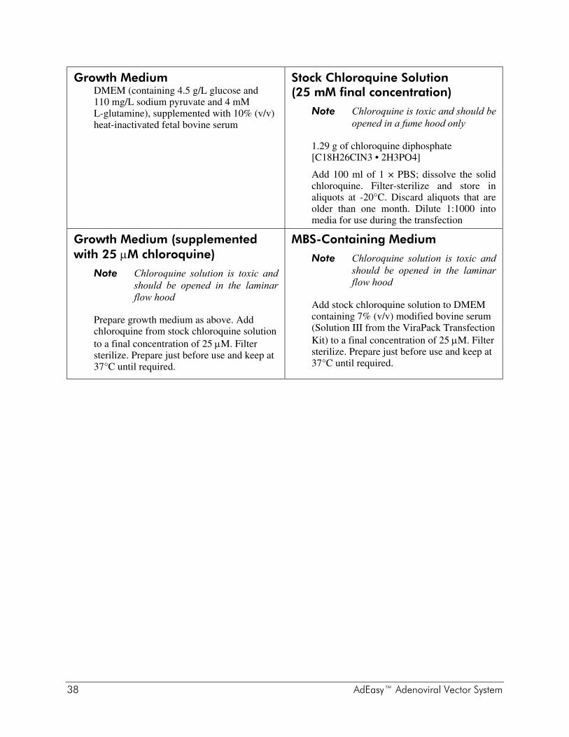
38 AdEasy™ Adenoviral Vector System
Growth Medium DMEM (containing 4.5 g/L glucose and 110 mg/L sodium pyruvate and 4 mM L-glutamine), supplemented with 10% (v/v) heat-inactivated fetal bovine serum
Stock Chloroquine Solution (25 mM final concentration)
Note Chloroquine is toxic and should be opened in a fume hood only
1.29 g of chloroquine diphosphate [C18H26CIN3 • 2H3PO4]
Add 100 ml of 1 × PBS; dissolve the solid chloroquine. Filter-sterilize and store in aliquots at -20°C. Discard aliquots that are older than one month. Dilute 1:1000 into media for use during the transfection
Growth Medium (supplemented with 25 μM chloroquine)
Note Chloroquine solution is toxic and should be opened in the laminar flow hood
Prepare growth medium as above. Add chloroquine from stock chloroquine solution to a final concentration of 25 μM. Filter sterilize. Prepare just before use and keep at 37°C until required.
MBS-Containing Medium
Note Chloroquine solution is toxic and should be opened in the laminar flow hood
Add stock chloroquine solution to DMEM containing 7% (v/v) modified bovine serum (Solution III from the ViraPack Transfection Kit) to a final concentration of 25 μM. Filter sterilize. Prepare just before use and keep at 37°C until required.

AdEasy™ Adenoviral Vector System 39
REFERENCES 1. Benihoud, K., Yeh, P. and Perricaudet, M. (1999) Curr Opin Biotechnol 10(5):440-7. 2. Berkner, K. L. (1988) Biotechniques 6(7):616-29. 3. He, T. C., Zhou, S., da Costa, L. T., Yu, J., Kinzler, K. W. et al. (1998) Proc Natl Acad
Sci U S A 95(5):2509-14. 4. Hanahan, D. (1983) J Mol Biol 166(4):557-80. 5. Jerpseth, B., Callahan, M. and Greener, A. (1997) Strategies 10(2):37–38. 6. Bullock, W. O., Fernandez, J. M. and Short, J. M. (1987) Biotechniques 5(4):376–378. 7. Graham, F. L., Smiley, J., Russell, W. C. and Nairn, R. (1977) J Gen Virol 36(1):59-74. 8. Tollefson, A. E., Hermiston, T. W. and Wold, W. S. M. (1998) Methods in Molecular
Medicine 21:1–9.
ENDNOTES SeaPlaque® is a registered trademark of FMC Corporation. FLAG® is a registered trademark of Sigma-Aldrich Co. AdEasy is a trademark of Johns Hopkins University.
MSDS INFORMATION The Material Safety Data Sheet (MSDS) information for Stratagene products is provided on the web at http://www.genomics.agilent.com. MSDS documents are not included with product shipments.