Manifestations of gastrointestinal diseases in the oral cavity · · 2014-05-28Crohn’s disease...
18
Manifestations of gastrointestinal diseases in the oral cavity Nabil El-Lababidi
Transcript of Manifestations of gastrointestinal diseases in the oral cavity · · 2014-05-28Crohn’s disease...
Manifestations of gastrointestinal diseases in the oral cavity Nabil El-Lababidi

Types of mouth affections in conjunction with GIT diseases I.
Glossitis:
Crohn’s disease
Coeliac disease
Kwashiorkhor
Malabsorption syndromes
Gastro-duodenal ulcers
Teeth disorders:
Coeliac disease
Gardner syndrome
Ulcers, erosions:
Crohn’s disease
Ulcerative colitis
Coeliac disease
Malabsorption syndromes
Candidiasis:
Steroid treatment
Lip affections:
Crohn’s disease
Types of mouth affections in conjunction with GIT diseases II.
Gingivitis:
Crohn’s disease
Coeliac disease
Tingling sensations of the mouth:
Coeliac disease
Malabsorption syndromes
Cheilitis:
Crohn’s disease
Coeliac disease
Malabsorption syndromes
Pigmentations:
Peutz-Jeghers syndrome
Coeliac disease I.
Malabsorption syndrome
Permanent gluten (gliadin) intolerance
Gliadin is mainly in oats, rye, wheat and barley
Etiology is unclear. A coincidence of genetic predisposition and autoimmune mechanisms is suspected
Heavily underdiagnosed. Estimates are that 1% of North American population is affected. 90% of patients are still undiagnosed!
Increased risk of developing coeliac disease in patients with:
Diabetes Mellitus type 1
Autoimmune thyroiditis
Down’s syndrome
Coeliac disease II.
Clinical manifestations:
Typical:
Abdominal pain
Diarrhea
Weight loss, failure to thrive, growth delay
Other:
Anemia
Significant weakness
Osteoporosis
Menstrual cycle disorders/infertility
Delayed puberty
Dermatitis herpetiformis Dühring

Coeliac disease III.
Clinical manifestation in the oral cavity:
Enamel defects
Delayed teeth eruptions
Recurring mouth ulcers
Cheilosis
Oral lichen ruber planus
Atrophic glossitis
Coeliac disaease IV.
Laboratory diagnosis:
Whole IgA levels (selective IgA deficiency incidence = 1:600!)
Anti tissue transglutaminase antibodies
Anti endomysial antibodies
Endoscopic enterobiopsy
Treatment:
Life long, strict, gluten-free diet
Gastro esophageal reflux I.
Dental enamel destruction caused by gastric acids in patients with chronic gastro esophageal reflux in:
Gastro esophageal reflux disease
Hiatal hernia
Bulimia nervosa
Loss of dental enamel in the surfaces exposed to gastric acids, so called erosions
The maximum teeth damage in bulimic patients is in the oral surfaces of the upper frontal teeth
Eroded dental enamel is smooth, shiny and hard
In cases of long term damage the tooth dentin can be seen as a brown-greenish streaming. Teeth are sensitive to thermal stimuli
Jaundice I.
Excessive bilirubin accumulates in tissues including the oral mucosa thus leading to their yellowish discoloration
The degree of the yellowish discoloration depends on the bilirubin levels and on the duration of hyperbilirubinemia
Bilirubin has affinity to elastin » increased accumulation in the tongue frenulum and the soft palate
Cave! Similar discolorations can be seen in patients with excessive vitamin A intake!
In childhood, biliverdin forms teeth depositions » yellowish to greenish discoloration of the teeth, for instance in children with biliary atresia

Peutz-Jeghers syndrome I.
Mutation in the LKB1 gene
Autosomal dominant pattern of inheritance or sporadic mutations
Associated with hamartomas affecting mainly the thin intestine and perioral and oral pigmentations
Flat, painless, brown spots in the oral cavity, mainly on the buccal mucosa, tongue and lips
Microscopically, acanthosis with increased melanocytes and near-by keratinocytes pigmentation is present
Treatment is not necessary, only for social or cosmetic reasons
Zaheri et al. have proven good results of ablation with potassium-titanyl-phosphate laser

Gardner syndrome I.
Autosomal dominant pattern of inheritance, rarely, spontaneous mutation in a gene on the 5th chromosome
Clinical manifestations include:
Intestinal polyposis with high risk of malignant transformation
Skin manifestations:
Epidermoid cysts
Fibromas
Sebaceous cysts
Bone manifestations:
Osteomas of the skull
Tumors of the thyroid gland

Gardner syndrome II.
Affections of the head and neck usually appear during childhood or adolescence:
Multiple enostoses of the jaws, usually affection the teeth alveoli, asymptomatic
Supernumerary and/or missing teeth:
Usually affecting the canine teeth and sparing molars
Supernumerary teeth usually wedge-shaped
Increased risk of odontomas, in the same distribution like in supernumerary teeth
Osteomas of the jaws and paranasal cavitis
Epidermoid cysts of the head and the neck

Gardner syndrome III.
A dentist can alert the gatroenterologist in regards to the possibility of Gardner syndrome via oral manifestations
According to Ide et al. :
Patients with 3 – 6 jaw osteomas are suspicious of Gardner syndrome
Patients with more than 6 osteomas are regarded as diagnosed with Gardner syndrome until proven different
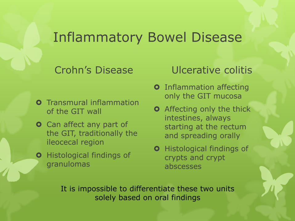
Inflammatory Bowel Disease
Crohn’s Disease
Transmural inflammation of the GIT wall
Can affect any part of the GIT, traditionally the ileocecal region
Histological findings of granulomas
Ulcerative colitis
Inflammation affecting only the GIT mucosa
Affecting only the thick intestines, always starting at the rectum and spreading orally
Histological findings of crypts and crypt abscesses
It is impossible to differentiate these two units solely based on oral findings
Oral manifestations of Crohn’s disease I.
According to Dupuy et al. only in 0.5% of patients with Crohn’s disease
Patients with oral manifestations are more likely to have affections of the esophagus and the anus
Male predominance, usually manifests in early age
Rarely, oral manifestations can be the first presentation of Crohn’s disease
Usually multifocal, linear, nodular, polypoid or diffuse affections of the oral mucosa
Predilection of affecting the labial and buccal mucosa
Usually hard, pink and painless
Painful on touch or due to ingestion of acidic, spicy or hot food only when ulcers are present
Ulcers can be persistent, linear and deep » diff. dg. blisters

Oral manifestations of Crohn’s disease II.
Microscopically:
Subepithelial, non-caseating granulomas. Characteristic epitheloid histiocytosis, large-cell and lymphocytic infiltrate
The changes are identical to those seen in the intestines
Oral manifestations are typically persistent, remitting and relapsing
Response to systemic treatment is individual, variable and unpredictable
Oral manifestations don’t always correspond with the degree of GIT inflammation activity
Some oral ulcers respond to topical or infiltrative administration of steroids
Oral manifestations of ulcerative colitis I.
Affections of the oral cavity are called pyostomatitis vegetans
Very rare, much rarer than oral manifestations in Crohn’s disease
Male predominance
Oral manifestations can develop at any age
They can precede GIT manifestations but usually they appear at a similar time
They are pustules on a red basis, affecting any part of the oral cavity with the exception of the dorsum of the tongue
Long lasting lesions can granulate or appear as polypoid shape or rippled
Some patients have ulcers of the oral cavity
10% of patients with oral manifestations also have arthritis of the temporomandibular joint
Oral manifestations of ulcerative colitis II.
Microscopically:
Crypt abscesses with lack of granulomas
Similar to changes of the thick intestine
Inflammatory infiltration with neutrophile, eosinophile and lymphocyte predominance is usually present
Oral manifestations usually respond well to systemic steroid treatment
Oral manifestations usually correspond with the degree of thick intestine inflammatory activity