Mandava IJPSR, 2012; Vol. 3(4): 967and Thavva, -980 ISSN ...
14
Mandava and Thavva, IJPSR, 2012; Vol. 3(4): 967-980 ISSN: 0975-8232 Available online on www.ijpsr.com 967 IJPSR (2012), Vol. 3, Issue 04 (Review Article) Received on 29 November, 2011; received in revised form 08 January, 2012; accepted 25 March, 2012 NOVEL APPROACH: MICROSPONGE DRUG DELIVERY SYSTEM Shyam Sunder Mandava* and Vedavathi Thavva Department of Pharmaceutics, CMR College of Pharmacy, Kandlakoya (V), Medchal Road, Hyderabad-501401, Andhra Pradesh, India ABSTRACT Transdermal drug delivery system (TDS) is not practically for delivery of materials whose final target is skin itself. Application topical agents generally offer many problems such as rashes, skin irritancy and burning sensation etc due to higher percutaneous absorption of drugs on the skin. Some conventional dosage e.g., gels and ointments which are often aesthetically unappealing, greasiness and stickiness etc. that often result into lack of patient compliance. For reduce this side effects, microsponge technology offers many advantage over the conventional drug delivery. The microsponge based drug delivery system is a unique technology for controlled release and enhanced drug deposition in the skin while minimizing transdermal penetration of topically active agents. Drug loaded microsponge consist of microporous beads, typically 10-25 μm in diameter. Microsponge delivery system (MDS) can provide increased efficacy for topically active agents with enhanced safety, extended product stability, enhanced formulation flexibility, reduced side effects and improved aesthetic properties in an efficient and novel manner. In addition these are non-irritating, non- allergenic, non-mutagenic, and non-toxic. MDS technology is being used currently in cosmetics, over-the-counter skin care, sunscreen and prescription products. INTRODUCTION: Several predictable and reliable systems have been developed for systemic drugs under the heading of trans-dermal delivery system (TDS) using the skin as portal of entry. It has improved the efficacy and safety of many drugs that may be better administered through skin. But TDS is not practical for delivery of materials whose final target is skin itself 1 . Controlled release of drugs onto the epidermis with assurance that the drug remains primarily localized and does not enter the systemic circulation in significant amounts, is an area of research that has only recently been addressed with success. No efficient vehicles have been developed for controlled and localized delivery of drugs into the stratum-corneum and underlying skin layers and not beyond the epidermis 2 . Moreover, the application of topical drugs suffers many problems, such as, ointments, which are often aesthetically unappealing, greasiness, stickiness, and so on, that often results into lack of patient compliance. These kinds of formulations require high concentrations of active ingredients for effective therapy because of their low efficiency of delivery system, resulting into irritation and allergic reactions in significant users. Other drawbacks of topical formulations are uncontrolled evaporation of active ingredient, unpleasant odor and potential incompatibility of drugs with the vehicles. Conventional formulations of topical drugs are intended to work on the outer layer of the skin. Keywords: Microsponge, Controlled diffusion, Topical delivery, Oral delivery, Biopharmaceutical delivery Correspondence to Author: Shyam Sunder Mandava Department of Pharmaceutics, CMR College of Pharmacy, Kandlakoya (V), Medchal Road, Hyderabad-501401, Andhra Pradesh, India
Transcript of Mandava IJPSR, 2012; Vol. 3(4): 967and Thavva, -980 ISSN ...

Mandava and Thavva, IJPSR, 2012; Vol. 3(4): 967-980 ISSN: 0975-8232
Available online on www.ijpsr.com 967
IJPSR (2012), Vol. 3, Issue 04 (Review Article)
Received on 29 November, 2011; received in revised form 08 January, 2012; accepted 25 March, 2012
NOVEL APPROACH: MICROSPONGE DRUG DELIVERY SYSTEM
Shyam Sunder Mandava* and Vedavathi Thavva
Department of Pharmaceutics, CMR College of Pharmacy, Kandlakoya (V), Medchal Road, Hyderabad-501401, Andhra Pradesh, India
ABSTRACT
Transdermal drug delivery system (TDS) is not practically for delivery of materials whose final target is skin itself. Application topical agents generally offer many problems such as rashes, skin irritancy and burning sensation etc due to higher percutaneous absorption of drugs on the skin. Some conventional dosage e.g., gels and ointments which are often aesthetically unappealing, greasiness and stickiness etc. that often result into lack of patient compliance. For reduce this side effects, microsponge technology offers many advantage over the conventional drug delivery. The microsponge based drug delivery system is a unique technology for controlled release and enhanced drug deposition in the skin while minimizing transdermal penetration of topically active agents. Drug loaded microsponge consist of microporous beads, typically 10-25 μm in diameter. Microsponge delivery system (MDS) can provide increased efficacy for topically active agents with enhanced safety, extended product stability, enhanced formulation flexibility, reduced side effects and improved aesthetic properties in an efficient and novel manner. In addition these are non-irritating, non-allergenic, non-mutagenic, and non-toxic. MDS technology is being used currently in cosmetics, over-the-counter skin care, sunscreen and prescription products.
INTRODUCTION: Several predictable and reliable systems have been developed for systemic drugs under the heading of trans-dermal delivery system (TDS) using the skin as portal of entry. It has improved the efficacy and safety of many drugs that may be better administered through skin. But TDS is not practical for delivery of materials whose final target is skin itself 1.
Controlled release of drugs onto the epidermis with assurance that the drug remains primarily localized and does not enter the systemic circulation in significant amounts, is an area of research that has only recently been addressed with success. No efficient vehicles have been developed for controlled and localized delivery of drugs into the stratum-corneum and underlying skin layers and not beyond the
epidermis 2. Moreover, the application of topical drugs suffers many problems, such as, ointments, which are often aesthetically unappealing, greasiness, stickiness, and so on, that often results into lack of patient compliance. These kinds of formulations require high concentrations of active ingredients for effective therapy because of their low efficiency of delivery system, resulting into irritation and allergic reactions in significant users.
Other drawbacks of topical formulations are uncontrolled evaporation of active ingredient, unpleasant odor and potential incompatibility of drugs with the vehicles. Conventional formulations of topical drugs are intended to work on the outer layer of the skin.
Keywords:
Microsponge,
Controlled diffusion,
Topical delivery,
Oral delivery,
Biopharmaceutical delivery
Correspondence to Author:
Shyam Sunder Mandava
Department of Pharmaceutics, CMR College of Pharmacy, Kandlakoya (V), Medchal Road, Hyderabad-501401, Andhra Pradesh, India

Mandava and Thavva, IJPSR, 2012; Vol. 3(4): 967-980 ISSN: 0975-8232
Available online on www.ijpsr.com 968
Typically, such products release their active ingredients upon application, producing a highly concentrated layer of active ingredient that is rapidly absorbed. Actual requirement to maximize the amount of time to release of an active ingredient is present either on skin surface or within the epidermis, while minimizing its transdermal penetration into the body.
In recent years, there has been considerable emphasis given to the development of microsponge base novel drug delivery systems, in order to modify and control the release behavior of the drugs. By incorporation into a carrier system, it is possible to alter the therapeutic index and duration of the activity of drugs. Microsponges are porous microspheres, biologically inert particles that are made of synthetic polymers and the particles serve to protect the entrapped drug compound from physical and environmental degradation. Their high degree of cross-linking results in particles that are insoluble, inert and sufficient strength to stand up to the high shear commonly used in manufacturing of creams, lotions, and powders. Their characteristic feature is the capacity to adsorb or “load” a high degree of active materials into the particle and on to its surface and it is delivered to skin via controlled diffusion.
Microsponges are microscopic spheres capable of absorbing skin secretions, therefore reducing oiliness and shine from the skin. Spherical particles composed of clusters of even tinier spheres are capable of holding four times their weight in skin secretions. Microsponge particles are extremely small, inert, indestructible spheres that do not pass through the skin. Rather, they collect in the tiny nooks and crannies of the skin and slowly release the entrapped drug, as the skin needs it. The size of the microsponges can be varied, usually from 5-300 µm in diameter, depending upon the degree of smoothness or after-feel required for the end formula.
Although the microsponge size may vary, a typical 25 µm sphere can have up to 250000 pores and an internal pore structure equivalent to 10 ft in length. The microsponge system can prevent excessive accumulation of ingredients within the epidermis and the dermis. Potentially, the microsponge system can significantly reduce the irritation of effective drugs without reducing their efficacy.
The microsponges are then washed away with the next cleansing. The MDS fulfills these requirements and has resulted in a new generation of very well-tolerated and highly efficacious, novel products (Microsponge shown in fig. 1).
FIG. 1: MICROSPONGE
Advantages 3:
Oil control: it can absorb oil up to 6 times its weight without drying.
Extended release up to 12 hours
Reduced irritation and better tolerance hence improved patient compliance
Improvement of product aesthetics
Improved thermal, physical, and chemical stability
Incorporation of immiscible products
Flexibility to develop novel product forms
Improves material processing e.g. Liquid can be converted into powders
Improves efficacy in treatment
Increased bioavailability
Characterstics of Microsponges 4:
Stable over a pH range of 1 to 11
Stable up to 130oC temperature
Compatible with the many of the vehicles and active ingredients

Mandava and Thavva, IJPSR, 2012; Vol. 3(4): 967-980 ISSN: 0975-8232
Available online on www.ijpsr.com 969
Self sterilizing as their average pore size is 0.25μm where bacteria cannot penetrate
Higher pay load (50 to 60%)
Free flowing and cost effective
Characterstics of Active Ingredients: Active ingredients that can be entrapped in microsponges must meet following requirements 5.
It should be miscible in monomer
Miscible by addition of small amount of a water immiscible solvent.
It should be water immiscible or at most only slightly soluble.
It should be inert to monomers.
It should be stable in contact with polymerization catalyst and conditions of polymerization.
Formulation: The MDS contain drug, polymer, vehicle and other additives like plasticizers that help stabilize the structure.
Various drugs used in MDS are
Benzoyl peroxide,
Retinol,
Tretinoin,
Flucinolone acetonide,
Ketoprofen,
Ibuprofen,
Flurbiprofen,
Paracetamol,
Dicyclomine,
Fluconazole etc.
Various polymers can form a microsponge cage. These include Ethyl cellulose, Eudragit RS 100, Polystyrene, acrylic polymers and PHEMA etc 6, 7.
Preparation of Microsponges: Drug entrapped in microsponges can take place in two ways, based on physicochemical properties of drug. One-step process and two-step process with respective liquid-liquid suspension polymerization and quasi emulsion solvent diffusion techniques. If the drug is typically an inert non-polar material, will create the porous structure it is called porogen.
Liquid-Liquid Suspension Polymerization: It is also referred to as Bottom-up approach (starting with monomer). In general, a solution is made comprising of monomers and the active ingredients (non polar). This phase is then suspended with agitation in an aqueous phase containing additives such as surfactants and dispersing agents. Once the suspension is established with discrete droplets of desired size, polymerization is effected by activating the monomers either by catalysis, increased temperature or irradiation 8.
The various steps summarized 9;
1. Selection of monomer or combination of
monomers
2. Formation of chain monomers as
polymerization begins
3. Formation of ladders as a result of crosses
linking between chain monomers
4. Folding of monomer ladder to form spherical
particles
5. Agglomeration of microspheres, which give rise
to formation, bunches of microspheres
6. Binding of bunches to form microsponges.
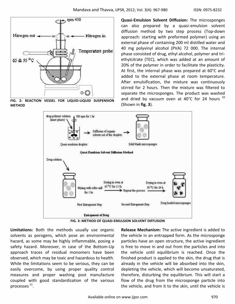
The polymerization process leads to the formation of a reservoir type of system, which opens at the surface through pores. Impregnating them within preformed microsponges then incorporates the functional substances. Some times solvent may be used for faster and efficient incorporation of the active substances. Once the polymerization is complete the solid that result from the process are recovered from the suspension. The particles are then washed and processed until they are substantially ready for use. The microsponge product can be made using styrene and divinyl benzene or methyl methacrylate and ethylene glycol dimethacrylate as starting materials (Reaction vessel is shown in fig. 2).
Mandava and Thavva, IJPSR, 2012; Vol. 3(4): 967-980 ISSN: 0975-8232
Available online on www.ijpsr.com 970
FIG. 2: REACTION VESSEL FOR LIQUID-LIQUID SUSPENSION METHOD
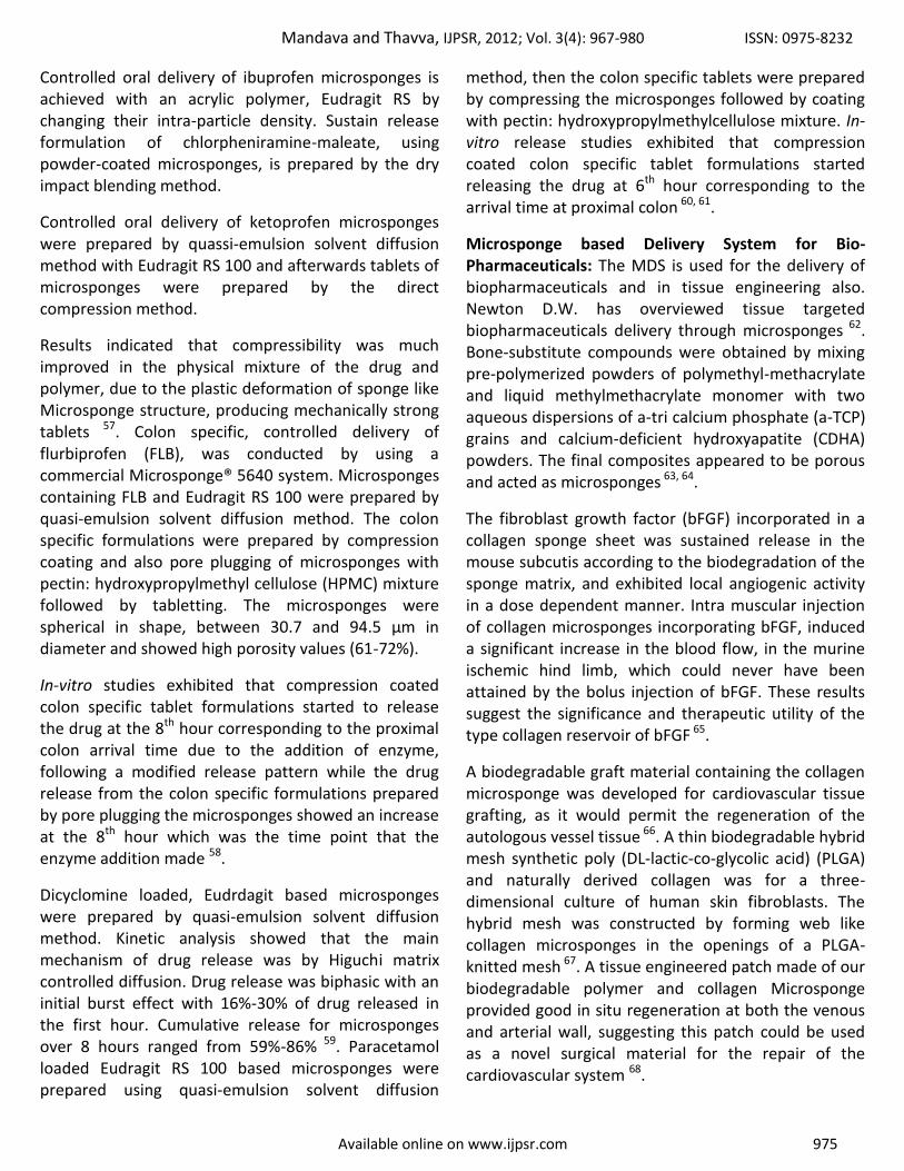
Quasi-Emulsion Solvent Diffusion: The microsponges can also prepared by a quasi-emulsion solvent diffusion method by two step process (Top-down approach: starting with preformed polymer) using an external phase of containing 200 ml distilled water and 40 mg polyvinyl alcohol (PVA) 72 000. The internal phase consisted of drug, ethyl alcohol, polymer and tri-ethylcitrate (TEC), which was added at an amount of 20% of the polymer in order to facilitate the plasticity. At first, the internal phase was prepared at 60°C and added to the external phase at room temperature. After emulsification, the mixture was continuously stirred for 2 hours. Then the mixture was filtered to separate the microsponges. The product was washed and dried by vacuum oven at 40°C for 24 hours 10 (Shown in fig. 3).
FIG. 3: METHOD OF QUASI-EMULSION SOLVENT DIFFUSION
Limitations: Both the methods usually use organic solvents as porogens, which pose an environmental hazard, as some may be highly inflammable, posing a safety hazard. Moreover, in case of the Bottom-Up approach traces of residual monomers have been observed, which may be toxic and hazardous to health. While the limitations seem to be serious, they can be easily overcome, by using proper quality control measures and proper washing post manufacture coupled with good standardization of the various processes 11.
Release Mechanism: The active ingredient is added to the vehicle in an entrapped form. As the microsponge particles have an open structure, the active ingredient is free to move in and out from the particles and into the vehicle until equilibrium is reached. Once the finished product is applied to the skin, the drug that is already in the vehicle will be absorbed into the skin, depleting the vehicle, which will become unsaturated, therefore, disturbing the equilibrium. This will start a flow of the drug from the microsponge particle into the vehicle, and from it to the skin, until the vehicle is

Mandava and Thavva, IJPSR, 2012; Vol. 3(4): 967-980 ISSN: 0975-8232
Available online on www.ijpsr.com 971
either dried or absorbed. Even after that the microsponge particles retained on the surface of the stratum-corneum will continue to gradually release the drug to the skin, providing prolonged release over time.
Release can also be controlled through diffusion or other external triggers such as pressure, temperature and solubility 12, 13, 14.
Pressure: Pressure/ Rubbing applied can release active ingredient from microsponge onto skin.
Temperature change: At room temperature, few entrapped active ingredients can be too viscous to flow suddenly from microsponges onto the skin. With increase in skin temperature, flow rate is also increased and therefore release is also enhanced,
Solubility: The microsponge loaded with water-soluble ingredients like anti-perspirants and antiseptics will release the ingredient in the presence of water. Thus release may be achieved based on the ability of external medium to dissolve the active ingredient, the concentration gradient varies or the ability to swell the microsponge network.
Evaluation Parameters of Microsponges: Various factors are affecting the drug release from microsponges. So it can be evaluated by following factors.
Particle Size Determination: Particle size analysis of loaded and unload microsponges can be performed by laser light diffractometry or any other suitable method. The values (d50) can be expressed for all formulations as mean size range. Cumulative percentage drug release from microsponges of different particle size will be plotted against time to study effect of particle size on drug release. Particles larger than 30 μm can impart gritty feeling and hence particles of sizes between 10 and 25 μm are preferred to use in final topical formulation 15.
Morphology and Surface Topography of Microsponges: For morphology and surface topography, prepared microsponges can be coated with gold–palladium under an argon atmosphere at
room temperature and then the surface morphology of the microsponges can be studied by scanning electron microscopy (SEM).SEM of a fractured microsponge particle can also be taken to illustrate its ultra structure 16.
Determination of Loading Efficiency and Production Yield: The loading efficiency (%) of the microsponges can be calculated according to the following equation
17.
The production yield of the microsponges can be determined by calculating accurately the initial weight of the raw materials and the last weight of the microsponge obtained.
Determination of True Density: The true density of microsponges were measured using an ultra-pycnometer under helium gas and was calculated from a mean of repeated determinations 18.
Characterization of Pore Structure: Pore volume and diameter are vital in controlling the intensity as well as duration of effectiveness of the active ingredient. Pore diameter also affects the migration of active ingredients from microsponges into the vehicle in which the material is dispersed. Mercury intrusion porosimetry can be employed to study effect of pore diameter and volume on rate of drug release from microsponges.
Porosity parameters of microsponges such as intrusion-extrusion isotherms pore size distribution, total pore surface area, average pore diameters, shape and morphology of the pores, bulk and apparent density can be determined by using mercury intrusion porosimetry 19.
The pore diameter of microsponges can be calculated by using Washburn equation 20,

Mandava and Thavva, IJPSR, 2012; Vol. 3(4): 967-980 ISSN: 0975-8232
Available online on www.ijpsr.com 972
Where, D is the pore diameter (μm); γ the surface tension of mercury (485 dyn cm−1); θ the contact angle (130o); and P is the pressure (psia).
Total pore area (Atot) was calculated by using equation,
Where P is the pressure (psia); V the intrusion volume (mL g−1); Vtot is the total specific intrusion volume (mL g−1).
The average pore diameter (Dm) was calculated by using equation,
Envelope (bulk) density (ρse) of the microsponges was calculated by using equation,
Where Ws is the weight of the microsponge sample (g); Vp the empty penetrometer (mL); VHg is the volume of mercury (mL).
Absolute (skeletal) density (ρsa) of microsponges was calculated by using equation,
Where Vse = the volume of the penetrometer -the volume of the mercury (mL).
Finally, the percent porosity of the sample was found from equation,
Pore morphology can be characterized from the intrusion–extrusion profiles of mercury in the microsponges 21.
Compatibility studies: Compatibility of drug with reaction adjuncts can be studied by thin layer chromatography (TLC) and Fourier Transform Infra-red spectroscopy (FT-IR). Effect of polymerization on crystallinity of the drug can be studied by powder X-ray diffraction (XRD) and Differential Scanning Colorimetry (DSC). For DSC approximately 5 mg samples can be accurately weighed into aluminum pans and sealed and can be run at a heating rate of 15oC/min over a temperature range 25–430oC in atmosphere of nitrogen 22, 23, 24.
Polymer / Monomer Composition: Polymer composition of the MDS can affect partition coefficient of the entrapped drug between the vehicle and the microsponge system and have direct influence on the release rate of entrapped drug. Release of drug from microsponge systems of different polymer compositions can be studied by plotting cumulative % drug release against time. The choice of monomer is dictated both by the vehicle into which it will be dispersed and characteristics of active ingredient to be entrapped. Various monomer combinations will be screened for their suitability with the drugs by studying their drug release profile. Release rate and total amount of drug released from the system composed of methyl methacrylate/ ethylene glycol dimethacrylate is slower than styrene/ divinyl benzene system. Polymers with varying electrical charges or degrees of hydrophobicity or lipophilicity may be prepared to provide flexibility in the release of active ingredients 25.
Resiliency: Resiliency (visco-elastic properties) of microsponges can be modified to produce bead lets that is softer or firmer according to the needs of the final formulation. Increased cross-linking tends to slow down the rate of release. Hence resiliency of microsponges will be studied and optimized as per the requirement by considering release as a function of cross-linking with time 26.
Dissolution Tests: Dissolution profile of microsponges can be studied by use of dissolution apparatus USP XXIII with a modified basket consisted of 5μm stainless steel mesh at 37oC under 150 rpm. The dissolution medium is selected while considering solubility of drug to ensure sink conditions. Samples from the dissolution medium can be analyzed by suitable analytical method at various intervals 27.

Mandava and Thavva, IJPSR, 2012; Vol. 3(4): 967-980 ISSN: 0975-8232
Available online on www.ijpsr.com 973
Drug Release from the Semi Solid Dosage Forms and Drug Deposition Studies: Drug release from the semi solid dosage forms are performed by the Franz- type static diffusion cells. In this epidermal side of the skin was exposed to ambient condition. While dermal side was kept facing the receptor solution. Receptor compartment containing 20mL phosphate buffer pH 5.8 was thermo stated at 32±0.5°C and stirred at 600 rpm. Skin was saturated with diffusion medium for 1 h before the application of sample. A 200-mg of sample was applied on the donor compartment. For determination of drug deposited in the skin, the diffusion cell was dismantled after a period of 4, 8, 16, and 24 h. The skin was carefully removed, and drug present on the skin surface was cleaned with distilled water 28.
In-vitro Diffusion Studies: The in-vitro diffusion studies of prepared microsponge gel were carried out in Keshary-Chien diffusion cell using through a cellophane membrane. 100 ml of phosphate buffer was used as receptor compartment, and then 500 mg of gel containing 10 mg of drug was spread uniformly on the membrane. The donor compartment was kept in contact with a receptor compartment and the temperature was maintained at 37±0.50. The solution
on the receptor side were stirred by externally driven Teflon coated magnetic bars at predetermined time intervals, pipette out 5ml of solution from the receptor compartment and immediately replaced with the fresh 5ml phosphate buffer. The drug concentration on the receptor fluid was determined spectro-photometrically against appropriate blank. The experiment was carried out in triplicate 29.
Safety Considerations 30, 31:
Skin and eye irritation studies in rabbits
Oral toxicity studies in rats
Mutagenicity in bacteria
Allergenicity in guinea pigs
Applications: Microsponges are used mostly for topical and recently for oral administration as well as biopharmaceutical delivery. It offers the formulator a range of alternatives to develop drug and cosmetic products. These are developed to deliver an active ingredient efficiently at the low dose and also to enhance stability, reduce side effects and modify drug release 32 (Various applications are shown in table 1).
TABLE 1: APPLICATION OF MICROSPONGES Active agents Applications
Sunscreens Long lasting product efficacy, with improved protection against sunburns and sun related
injuries even at elevated concentration and with reduced irritancy and sensitization.
Anti-acne e.g. Benzoyl peroxide Maintained efficacy with decreased skin irritation and sensitization. Anti-inflammatory e.g. hydrocortisone Long lasting activity with reduction of skin allergic response and dermatoses.
Anti-fungals Sustained release of actives. Anti-dandruffs e.g. zinc pyrithione, sele-nium sulfide Reduced unpleasant odour with lowered irritation with extended safety and efficacy.
Antipruritics Extended and improved activity.
Skin depigmenting agents e.g. hydroquinone Improved stabilization against oxidation with improved efficacy and aesthetic appeal.
Rubefacients Prolonged activity with reduced irritancy greasi-ness and odour.
Topical Drug Delivery 33: Genetically engineered melanin is incorporated in microsponges (sunscreens), melanosponge-α to spread it evenly hence give protection against UV-A and UV-B radiation 34.
Fluocinolone acetonide (FA) is a corticosteroid primarily used in dermatology to reduce skin inflammation and relieve itching 35. Benzyl peroxide (BPO) is used for the treatment of acne and athletes foot.
Skin irritation is a common side effect, and it has been shown that controlled release of BPO from the
microsponge on to the skin could reduce the side effect while reducing percutaneous absorption 36, 37, 38,
39.
Hydroquinone (HQ) bleaching creams are considered the gold standard for treating hyper pigmentation. A new formulation of HQ 4% with retinol 0.15% entrapped in microsponge reservoirs was developed to release HQ gradually to prolong exposure to treatment and to minimize skin irritation 40. Hydroquinone prevents the overproduction of melanin, while lightening the brown spots on the skin 41, 42.

Mandava and Thavva, IJPSR, 2012; Vol. 3(4): 967-980 ISSN: 0975-8232
Available online on www.ijpsr.com 974
0.1% and 0.04% tretinoin is entrapped in MDS for topical treatment of acne vulgaris. This formulation uses patented methyl methacrylate/glycol dimethacrylate cross polymer porous microspheres to enable inclusion of the active ingredient, tretinoin, in an aqueous gel. It has been marketed as Retin-A-Micro by Ortho-McNeil pharmaceutical 43. In a 21 day cumulative irritancy study and a split face study, microsponge formulation showed a reduced level of systemic absorption of tretinoin ranging from 25% - 40% reduction helping to explain the improved tolerability. Numerous studies have been conducted and published comparing the tolerability of various retinoids: tazarotene, Adapalene 44, 45, 46, 47, 48.
Tretinoin is used for the treatment of photo-damage and Acne, Which contributes to the premature aging of skin and has been implicated in skin cancer.
5-Fluorouracil (5-FU) is an effective chemotherapeutic agent for treating actinic keratosis, a pre-cancerous, hardened-skin condition caused by excessive exposure to sunlight 49.
Mupirocine containing microsponges were prepared by emulsion solvent diffusion method. It is used for the treatment of primary and secondary skin infections such as impetigo, eczema and atopic dermatitis 50.
The entrapped dimethicone of microsponge 5700. Dimethicone is a blend of 78% 350 cst(centistokes) polydimethylsiloxane and 22% 1000 cst polydimethylsiloxane 51.
Fluconazole is an active agent against yeasts, yeasts like fungi and dimorphic fungi, with possible drawback of itching in topical therapy. Microsponges were prepared by liquid-liquid suspension polymerization of styrene and methyl methacrylate 52.
Aceclofenac is a NSAIDs having excellent anti-inflammatory and analgesic activity but NSAID produces GIT ulceration, liver and kidney trouble especially in case of oral administration. In view, of adverse drug reaction associated with oral formulations, aceclofenac is increasingly administered by topical route. Aceclofenac loaded microsponge are prepared by using quasi-emulsion solvent diffusion method. It is incorporated in gel base and various parameters are studied.
Gel formulation is subjected to rats and studied anti-inflammatory activity by Carrageen an induced paw edema method 53.
Hydroxyzine HCl loaded with microsponges was prepared solvent diffusion method using Eudragit RS-100 polymer. In this preparation, acetone as dispersing solvent and liquid paraffin as the continuous medium. Magnesium stearate was added to the dispersed phase to prevent flocculation of Eudragit RS-100 microsponges. Pore inducers such as sucrose and pre-gelatinized starch were used to enhance the rate of drug release. Microsponges of nearly 98% encapsulation efficiency and 60-70% porosity were produced. The pharmacodynamic effect of the chosen preparation was tested on the shaved back of histamine-sensitized rabbits 54.
Itraconazole loaded microsponges were prepared using Quasi-emulsion solvent diffusion technique. Itraconazole is a triazole antifungal agent used to treat both superficial and systemic fungal infections 55.
Oral Drug Delivery: A Microsponge system offers the potential to hold active ingredients in a protected environment and provide controlled delivery of oral medication to the lower gastrointestinal (GI) tract, where it will be released upon exposure to specific enzymes in the colon. This approach if successful should open up entirely new opportunities for MDS. In oral applications, the Microsponge system has been shown to increase the rate of solubilization of poorly water-soluble drugs by entrapping such drugs in the Microsponge system pores. Because these pores are very small, the drug is in effect reduced to microscopic particles and the significantly increased surface area thus greatly increases the rate of solubilization.
Additionally, the time it takes the microsponge system to pass through the small and large intestine is considerably increased as a result maximizing the amount of drug that is absorbed.
Controlled release of drug which is considered as micro-balloons were prepared by the dissolving the drug in ethanol, on addition of water, the ethanol diffused from the emulsion droplets to leave a highly porous particle by changing the inter particle friction sensing capability 56.
Mandava and Thavva, IJPSR, 2012; Vol. 3(4): 967-980 ISSN: 0975-8232
Available online on www.ijpsr.com 975
Controlled oral delivery of ibuprofen microsponges is achieved with an acrylic polymer, Eudragit RS by changing their intra-particle density. Sustain release formulation of chlorpheniramine-maleate, using powder-coated microsponges, is prepared by the dry impact blending method.
Controlled oral delivery of ketoprofen microsponges were prepared by quassi-emulsion solvent diffusion method with Eudragit RS 100 and afterwards tablets of microsponges were prepared by the direct compression method.
Results indicated that compressibility was much improved in the physical mixture of the drug and polymer, due to the plastic deformation of sponge like Microsponge structure, producing mechanically strong tablets 57. Colon specific, controlled delivery of flurbiprofen (FLB), was conducted by using a commercial Microsponge® 5640 system. Microsponges containing FLB and Eudragit RS 100 were prepared by quasi-emulsion solvent diffusion method. The colon specific formulations were prepared by compression coating and also pore plugging of microsponges with pectin: hydroxypropylmethyl cellulose (HPMC) mixture followed by tabletting. The microsponges were spherical in shape, between 30.7 and 94.5 μm in diameter and showed high porosity values (61-72%).
In-vitro studies exhibited that compression coated colon specific tablet formulations started to release the drug at the 8th hour corresponding to the proximal colon arrival time due to the addition of enzyme, following a modified release pattern while the drug release from the colon specific formulations prepared by pore plugging the microsponges showed an increase at the 8th hour which was the time point that the enzyme addition made 58.
Dicyclomine loaded, Eudrdagit based microsponges were prepared by quasi-emulsion solvent diffusion method. Kinetic analysis showed that the main mechanism of drug release was by Higuchi matrix controlled diffusion. Drug release was biphasic with an initial burst effect with 16%-30% of drug released in the first hour. Cumulative release for microsponges over 8 hours ranged from 59%-86% 59. Paracetamol loaded Eudragit RS 100 based microsponges were prepared using quasi-emulsion solvent diffusion
method, then the colon specific tablets were prepared by compressing the microsponges followed by coating with pectin: hydroxypropylmethylcellulose mixture. In-vitro release studies exhibited that compression coated colon specific tablet formulations started releasing the drug at 6th hour corresponding to the arrival time at proximal colon 60, 61.
Microsponge based Delivery System for Bio-Pharmaceuticals: The MDS is used for the delivery of biopharmaceuticals and in tissue engineering also. Newton D.W. has overviewed tissue targeted biopharmaceuticals delivery through microsponges 62. Bone-substitute compounds were obtained by mixing pre-polymerized powders of polymethyl-methacrylate and liquid methylmethacrylate monomer with two aqueous dispersions of a-tri calcium phosphate (a-TCP) grains and calcium-deficient hydroxyapatite (CDHA) powders. The final composites appeared to be porous and acted as microsponges 63, 64.
The fibroblast growth factor (bFGF) incorporated in a collagen sponge sheet was sustained release in the mouse subcutis according to the biodegradation of the sponge matrix, and exhibited local angiogenic activity in a dose dependent manner. Intra muscular injection of collagen microsponges incorporating bFGF, induced a significant increase in the blood flow, in the murine ischemic hind limb, which could never have been attained by the bolus injection of bFGF. These results suggest the significance and therapeutic utility of the type collagen reservoir of bFGF 65.
A biodegradable graft material containing the collagen microsponge was developed for cardiovascular tissue grafting, as it would permit the regeneration of the autologous vessel tissue 66. A thin biodegradable hybrid mesh synthetic poly (DL-lactic-co-glycolic acid) (PLGA) and naturally derived collagen was for a three-dimensional culture of human skin fibroblasts. The hybrid mesh was constructed by forming web like collagen microsponges in the openings of a PLGA-knitted mesh 67. A tissue engineered patch made of our biodegradable polymer and collagen Microsponge provided good in situ regeneration at both the venous and arterial wall, suggesting this patch could be used as a novel surgical material for the repair of the cardiovascular system 68.
Mandava and Thavva, IJPSR, 2012; Vol. 3(4): 967-980 ISSN: 0975-8232
Available online on www.ijpsr.com 976
Marketed Formulations: Microsponge delivery systems are used for topical prescription, over-the-counter (OTC) and personal care products. Products under development or in the marketplace utilize the topical microsponge system in three primary ways 69
(The marketed products are shown in table 2).
As reservoirs releasing active ingredients over an extended period of time
As receptacles for adsorbing undesirable substances, such as excess skin oils
As closed containers holding ingredients away from the skin for superficial action
TABLE 2: MARKETED FORMULATIONS OF MICROSPONGES
Product name Advantages Company
Retin-A-Micro 0.1 And 0.04% tretinoin entrapped in MDS, for topical treatment of acne vulgaris. This formulation uses patented methyl methacrylate / glycol+8 dimethacrylate cross-polymer porous microspheres.
Ortho-McNeil Pharmaceutical,Inc.
Carac cream, 0.5% Carac cream contains 0.5% fluorouracil, with 0.35% being incorporated into a patented porous microsphere (Microsponge) composed of methyl methacrylate / glycol dimethacrylate cross-polymer and dimethicone.
Dermik Laboratories, Inc.Berwyn,PA19312
USA
Line eliminator dual retinol facial treatment
Lightweight cream with a retinol (Vitamin A) in MDS, delivers both immediate and time-released wrinkle-fighting action.
Avon
Retinol cream
The retinol molecule is kept in the microsponge system to protect the potency of vitamin A. This helps to maximize the retinol dosage, while reducing the possibility of irritation. Retinol is a topical vitamin A derivative, which helps maintain healthy skin, hair, and mucous membranes.
Biomedic
Retinol 15 nightcream A nighttime treatment cream with Microsponge system. The formula contain of pure retinol. Continuous use of Retinol 15 will result in the visible diminishment of fine lines and wrinkles, and improve in skin discolorations.
Biomedic,sothys
EpiQuin micro The Microsponge® system entrap hydroquinone and retinol. The microsponges release these ingredients into the skin gradually throughout the day, which may minimize skin irritation
Skin Medica Inc
Sports cream RS and XS Topical analgesic-anti-inflammatory and counterirritant actives in a Microsponge® Delivery System (MDS) for the management of musculoskeletal conditions
Embil Pharmaceutical.
Salicylic peel 20 and 30 Deep BHA peeling agent: Salicylic acid 20% and 30%, Microsponge Technology, Excellent exfoliation and stimulation of the skin for more resistant skin types or for faster results. Will dramatically improve fine lines, pigmentation, and acne concerns.
Biophora
Micro peel plus
The MicroPeel® Plus,stimulates cell turnover through the application of salicylic acid in the form of microcrystals using Microsponge® technology.The MicroPeel® Plus aggressively outperforms other superficial chemical peels by freeing the skin of all dead cells, while doing no damage to the skin.
Biomedic
Oil free matte block spf-20
This sunscreen provides a shield for the skin from damaging UV rays and controls oil production. Microsponge technology absorbs the oil, maintaining an all-day matte finish. Oil-free formula contains soothing Green Tea to help calm inflammation caused by breakouts. Cornstarch and Vinyl Dimethicone / Methicone Silsesquioxane Cross-polymer act as microsponges to absorb excess surface oils on skin.
Dermalogica
Oil control lotion
Feature-light lotion microsponges that absorb oil on the skin's surface during the day, for a matte finish.Eliminate shine for hours with this feature-weight lotion. The natural-antibiotic Skin Response Complex soothes inflammation and tightness to promote healing, Acne-Prone, oily skin conditions
Fountain Cosmetics
Lactrex™ 12% moisturizing cream
It contains 12% lactic acid as the neutral ammonium salt and ammonium lactate. Lactrex™ also contains water and glycerin, a natural humectant, to soften and help moisturize dry, flaky, cracked skin.
SDRPharmaceuticals, Inc., Andover, NJ, .S.A.
07821
Dermalogica oil control lotion
It is a feather-light lotion, formulated with oil absorbing Microsponge® technology and hydrating botanicals. The naturally antiseptic skin response complex helps soothe and purify the skin.
John and Ginger Dermalogica skin care
products
Aramis fragrances
24-Hour high performance antiperspirant spray sustained release of fragrance in the microsponge.The microsponge comes in the form of an ultra light powder, and because it is micro in size, it can absorb fragrant oil easily, while maintaining a free-flowing powder characteristic where release is controlled due to moisture and temperature.
Aramis Inc
Ultra guard Microsponge system that contains dimethicone to help protect a baby's skin from diaper rash Scott Paper

Mandava and Thavva, IJPSR, 2012; Vol. 3(4): 967-980 ISSN: 0975-8232
Available online on www.ijpsr.com 977
Patent information of Microsponge Products:
In September 1, 1987, Won; Richard (Palo Alto, CA) of Advanced Polymer Systems, Inc. (Redwood City, CA) received US patent for developing Method for delivering an active ingredient by controlled time release utilizing a novel delivery vehicle which can be prepared by a process utilizing the active ingredient as a porogen 70 (United States Patent 4, 690, 825).
September 8, 1992, won; Richard (Palo Alto, CA) of Advanced Polymer Systems, Inc. (Redwood City, CA) received US patent for developing Two-step method for preparation of controlled release formulations 71 (United States Patent 5, 145, 675).
Dean, Jr. et al., received US patent no. 4863856 for the development of weighted collagen microsponges having a highly cross-linked collagen matrix are described suitable for use in culturing organisms in motive reactor systems. The microsponges have an open to the surface pore structure, pore sizes and volumes suitable for immobilizing a variety of bioactive materials
72.
Advanced Polymer Systems Inc. has been granted a patent for the use of its Microsponge technology in tretinoin formulations. The patent will provide coverage through September 21, 2016 73.
EpiQuin® Micro Hydroquinone USP 4%, under license from Amcol Intl. Corp. U.S. Patent Numbers 5, 851, 538 and 6, 896, 890 74.
Future Perspective:
Nanosponges: Today, as we realize the immense advantages offered by the nano-size, the micro sized products are likely to be outdated. The nanosized particles have a very high surface area to size ratio and a greater potential to modulate the release of actives compared to micro-sized particles. While inorganic nanosponges have many applications in electronics, the first pharmaceutical nanosponges based on cross linked cyclodextrins have been reported 75, 76. These are nano sized, highly porous materials composed of
beta-cyclodextrins cross linked with carbonate bonds. Econazole nitrate nanosponges loaded carbapol hydrogel were recently developed. These are prepared using ethyl cellulose and poly vinyl alcohol by emulsion solvent evaporation method 77.
Going the natural way using a Functional Active: Although natural actives are important consumer attractants, now the focus has shifted on using multifunctional natural ingredients. For example, Marinosomes®, liposomes made from natural anti-inflammatory lipid extracts, have set a new paradigm in using such functional 'active excipients'. The possibility of using such substances for constructing a microsponge structure appears to be cost effective and innovative.
Microsponges in Oral Care Cosmetics: An interesting application of the microsponge technology could be in oral cosmetics, such as to sustain the release of volatile ingredients, thus increasing the duration of the 'fresh feel'. Microsponges of such volatile ingredients may be easily incorporated in tooth pastes or mouth washes.
Long lasting Coloured Cosmetics; a new application for Microsponges: Colours entrapped in microsponges may be used in a variety of coloured cosmetic products such as rouge or lipsticks to make them long lasting. As stated above, microsponges help in uniform spreading and improving covering power. Thus, coloured cosmetics formulated with microsponges would be highly elegant 78.
CONCLUSION: A Microsponge Delivery System consist of microporous beads, have entrap wide range of active ingredients and then controlled release of actives onto the skin over a time and in response to other triggers such as pressure, ambient skin temperature and moisture. MDS is originally developed for topical delivery. Now a days it can also used for tissue engineering and controlled oral delivery using bio-erodible polymers, especially for colon specific delivery. MDS holds a promising future in various pharmaceutical applications in the coming years as they have unique properties like extended release, reduced irritancy, small size, self sterilize and compatible with most of vehicles and ingredients, so flexible to develop novel product forms. Thus, MDS is a very emerging field which is needed to be explored.

Mandava and Thavva, IJPSR, 2012; Vol. 3(4): 967-980 ISSN: 0975-8232
Available online on www.ijpsr.com 978
REFERENCES:
1. Kydonieus A. F., Berner B: Transdermal Delivery of Drugs, CRC Press, Boca Raton, 1987.
2. Chowdary K. P. R., Rao Y. S: Mucoadhesive Micro-spheres for Controlled Drug Delivery, Biol. Pharm. Bull., 2004, 27(11), 1717-1724.
3. Vyas SP, Khar RK: Targeted and controlled Drug Delivery, 1
st Ed, CBS Publication, 2002.
4. Aritomi H, Yamasaki Y, Yamada K, Honda H and Koshi M: Development of sustained release formulation of chlorpheniramine maleate using powder coated microsponges prepared by dry impact blending method. Journal of Pharmaceutical Sciences and Technology. 1996, 56(1): 49-56.
5. Kawashima Y., Niwa T., Takeuchi H., Hino T., Itoh Y: Control of Prolonged Drug Release and Compression Properties of Ibuprofen Microsponges with Acrylic Polymer, Eudragit RS, by changing their Intraparticle Density., Chem. Pharm. Bull., 1992; 40 (1); 196-201.
6. Ruckenstein E, Hong L: Concentrated emulsion polymerization pathway to hydrophobic and hydrophilic microsponge molecular reservoirs. Chem. Mater.1992; 4: 1032-1037.
7. Chadawar V, Shaji J: Microsponge delivery system. Current drug delivery, 2007; 4: 123-129.
8. Tansel C¸ Omoglu, Nurs¸In Gonu, Tamer Baykara: The effects of pressure and direct compression on tablet-ting of microsponges, International Journal of Phar-maceutics, 2002; 242; 191–195.
9. Anderson D.L. Cheng C.H. Nacht S; Flow Characteristics of Loosely Compacted Macroporous Microsponge(R) polymeric systems. Powder Technology, 1994; 78:15-18.
10. Tansel C¸ omoglu, Nurs¸ in Gonu l, Tamer Baykara: Preparation and in vitro evaluation of modified release ketoprofen Microsponges, Il Farmaco, 2003; 58; 101-106.
11. Kirti deshmukh :Solid Porous Microsphere: Emerging Trend in Pharmaceutical Technology., International Journal of Pharma and Bio Sciences, Jan-.March 2011; Vol 2, Issue 1.
12. Embil K., Nacht S: The microsponge delivery system (MDS) a Topical Delivery System with reduced irritancy incorporating multiple triggering Mechanisms for the release of actives.J. Microencapsule. 1996; 13: 575–88.
13. Khopade AJ, Jain S, Jain NK: The microsponge, Eastern pharmacist, 1996, 49-53.
14. Shah V.P.:Determination of In-vitro Release from Hydrocortisone Creams. International Journal of Pharmaceutics. 1989; 53: 53-59.
15. Martin A., Swarbrick J. & Cammarrata A., In: Physical Pharmacy- Physical Chemical Principles in Pharma-ceutical Sciences. 3rd Ed., 1991.
16. Emanuele AD, Dinarvand R: Preparation, Characterization and Drug Release from Thermo responsive Microspheres. International Journal of Pharmaceu-tics.1995, 118:237-242.
17. Kilicarslan, M., Baykara, T: The effect of the drug/polymer ratio on the properties of Verapamil HCl loaded microspheres. Int. J. Pharm. 2003: 252,; 99–109.
18. Barkai A, Pathak V, Benita S: Polyacrylate (Eudragit retard) microspheres for oral controlled release of nifedipine. I. Formulation design and process optimization. Drug Dev. Ind. Pharm. 1990; 16:2057-2075.
19. Poresizer Model No. 9310, Micromeritics Instrument Corp., Nor-cross, Georgia.
20. Washburn, E.W.: Note on a method of determining the distribution of pore sizes in a porous material. Proc. Natl. Acad. Sci. U.S.A. 1921; 7; 115–116.
21. Orr Jr C; Application of mercury penetration to material analysis. Powder Technol 1969; 3:117-23.
22. Kawashima Y, Niwa T, Takeuchi H, Hino T, Itoh Y, Furuyama S: Characterization of polymorphs of tranilast anhydrate and tranilast monohydrate when crystallized by two solvent change spherical crystallization techniques. J. Pharm. Sci. 1991; 81:472-478.
23. Bodmeier R, Chen H: Preparation and characterization of microspheres containing the anti-inflammatory agents, indomethacin, ibu-profen, and ketoprofen. J. Control. Release. 1989; 10:167-175.
24. Jones DS, Pearce KJ: Investigation of the effects of some process variables on, microencapsulation of propranolol HCl by solvent evaporation method. Int. J. Pharm. 1995; 118: 99-205.
25. Wakiyama N, Juni K, Nakano M: Preparation and evaluation in vitro of polylactic acid microspheres containing local anesthetic. Chem. Pharm. Bull. (Tokyo). 1981; 29:3363-3368.
26. D’souza JI: The Microsponge Drug Delivery System: For Delivering an Active Ingredient by Controlled Time Release. Pharma. info.net, 2008, 6 (3): 62.
27. D’souza JI: In-vitro Antibacterial and Skin Irritation Stu-dies of Microsponges of Benzoyl Peroxide. Indian Drugs. 2001, 38(7): 23.
28. Franz T. J: Percutaneous absorption, On the relevance of invitro date. J. Invest. Dermatol. 1975; 45; 498-503.
29. Shobha rani R Hiremath, Text book of industrial pharmacy, published by universities press private limited, pg.no.44-45.
30. Sato T, Kanke M, Schroeder G, Deluca: Porous biodegradable microspheres for controlled drug delivery. I: Assessment of processing conditions and solvent removal techniques. Pharm Res 1988; 5: 21-30.
31. Draize JH, Woodard G, Calvery HO: Methods for the study of irritation and toxicity of substances applied topically to the skin and Mucous Membranes, J Pharmacol Exp ther, 1994,; 82; 377-389.
32. Khopade AJ, Jain sanjay, Jain NK. “The Microsponge”. Eastern Pharmacist, 1996, 49-53.
33. Geeta Patel, Patel JK: Use of a Microsponge in Drug Delivery Systems, Pharmaceutical processing, 2008, 158.
34. V.B. Patravale, S.D. Mandawgade: Novel cosmetic delivery systems: an application update. Int.J.Cosmetic Sci., 2008; 30:19-33.
35. D’souza JI, Harinath NM :Topical anti-inflammatory gels of flucinolone acetonide entrapped in eudragit based microsponge delivery system, Research J Pharm and Tech, 2008 ;1; 502-506.
36. Jelvehgari M, Siahi-Shadbad MR, Azarmi S: The microsponge delivery system of benzoyl peroxide: Preparation, characterization and release studies. Int J Pharm 2006; 308:124-132.
37. D’soza JI, Jagadish K, Saboji, Suresh G, Killedar, Harinath N: Design and evaluation of benzoyl peroxide microsponges to enhance therapeutic efficacy in acne treatement, accepted for presentation in 20
th FAPA congress, Bangkok,
2004. 38. Nokhodchi A, Jelvehgari M, Mozafari M: Factors affecting
the morphology of benzoyl peroxide microsponges, Micron, 2007; 38; 834-840.

Mandava and Thavva, IJPSR, 2012; Vol. 3(4): 967-980 ISSN: 0975-8232
Available online on www.ijpsr.com 979
39. Wester R., Patel R., Natch S., Leyden J., Melendres J., Maibach H. :Controlled release of benzoyl peroxide from a porous microsphere polymeric system can reduce topical irritancy, J. Am. Acad. Derm., 1991, 24, 720-726.
40. Grimes PE: A microsponge formulation of hydroquinone 4% and retinol 0.15% in the treatement of melasma and post-inflammatory hyperpigmentation, Cutis, 2004; 74; 362-368.
41. Palumbo A, d'Ischia M, Misuraca G, Prota G: Mechanism of inhibition of melanogenesis by hydroquinone. Biochim Biophys Acta. 1991; 1073(1):85-90.
42. Nishimura T, Kometani T, Okada S, Ueno N, Yamamoto T: Inhibitory effects of hydroquinone-alpha-glucoside on melanin synthesis. Yakugaku Zasshi. 1995; 115(8):626-632.
43. Summary Basis of Approval Retin-A Micro 0.1%. Available at: www.accessdata.FDA.com.Accessed April 5, 2006.
44. Leyden JJ, Tanghetti EA, Miller B, Ung M, Berson D, Lee J: Once-daily tazarotene 0.1% gel versus once-daily tretinoin 0.1% microsponge gel for the treatment of facial acne vulgaris: a double-blind randomized trial. Cutis, 2002; 69(supply):12-19.
45. Cunliffe WJ, Danby FW, Dunlap F, Gold MH, Gratton D, Greenspan A: Randomised, controlled trial of the efficacy and safety of adapalene gel 0.1% and tretinoin cream 0.05% in patients with acne vulgaris. Eur J Dermatol. 2002 12:350-354.
46. Egan N, Loesche MC, Baker MM.: Randomized, controlled, bilateral (split-face) comparison trial of the tolerability and patient preference of adapalene gel 0.1% and tretinoin microsphere gel 0.1% for the treatment of acne vulgaris. Cutis. 2001: 68(suppl):20-24.
47. Thiboutot D, Gold MH, Jarratt MT, et al: Randomized controlled trial of the tolerability, safety, and efficacy of adapalene gel 0.1% and tretinoin microsphere gel 0.1% for the treatment of acne vulgaris. Cutis. 2001; 68 (suppl):10-19.
48. Kakita L: Tazarotene versus tretinoin or adapalene in the treatment of acne vulgaris. J Am Acad Dermatol. 2000; 43:S51-54.
49. Talisuna AO, Bloland P, D’Alessandro U: History, Dynamics, and public haelth importance of malaria parasite resistance. Clinical microbiology reviews. 2004; 17; 235-254.
50. Amrutiya N, Bajaj A, Madan M: Development of microsponges for topical delivery of mupirocin, AAPS Pharm Sci Tech, 2009; 10; 402-408.
51. icis.advancedpolymers-new-entrapment-system. Articles/1998/04/27/87201.
52. James J. Leyden, Alan Shalita, Diane Thiboutot, Kenneth Washenik, and Guy Webster: Topical Retinoids in Inflammatory Acne: A Retrospective, Investigator-Blinded, Vehicle-Controlled, Photographic Assessment, Clin. Therapeutics, 2005; 27:216-224.
53. P. M. Dandagi, M. R. Upadhyay, A. P. Gadad, V. S. Mastiholimath : Design and Evaluation of Aceclofenac Loaded Microsponge for Topical Delivery, Journal of Pharmaceutical Research & Clinical Practice, Apr-June 2011; 1(2):90-101.
54. Zaki Rizkalla CM, Latif Aziz R, Soliman II: In Vitro and In Vivo Evaluation of Hydroxyzine Hydrochloride Microsponges for Topical Delivery., AAPS Pharm.Sci.Tech, DOI: 10.1208/s12249-011-9663-5.
55. Ramadevi Bhimavarapu, Karuna Priya Chitra, P. Karunkiran, G. Raviteja, Y. Meharagavendra,
S.Sundaramma, D. Chaitanya: Itraconazole Loaded Microsponges- A Novel Carrier System, The Pharma Professionals, May - August 2011; Volume 1 – Issue 2.
56. Kawashima Y, Niawa T, Takeuchi H, Hino T, Itoh Y: Hollow macrospheres for use as a Floating controlled drug effect system in the stomach, J Pharm. Sci 82(2)135-140,1992.
57. Tansel C, Omoglu CT, Gonul N, Baykara, T: Preparation and in vitro evaluation of modified release ketoprofen microsponges. Il Farmaco 2003; 58:101–106.
58. Orlu M, Cevher E, Araman A: Design and evaluation of colon specific drug delivery system containing flurbiprofen microsponges. Int J Pharm 2006; 318: 103-117.
59. Jain V, Sigh R: Dicyclomine loaded eudragit based microsponge with potential for colonic delivery: preparation and characterization, Trop J Pharm Res, 2010; 9; 67-72.
60. Jain V, Sigh R: Development and characterization of eudragit RS 100 loaded microsponges and its colonic delivery using natural polysacharides, Acta poloniae pharmaceutica-Drug Research, 2010; 67; 407-415.
61. Manoj Kumar Mishra, Mukesh Shikhri, Rishikesh Sharma and Mahesh Prasad Goojar: Optimization, formulation development and characterization of Eudragit RS 100 loaded microsponges and subsequent colonic delivery, International Journal of Drug Discovery And Herbal Res Earch (IJDDHR) 1(1): Jan-Mar: (2011), 8-13.
62. Newton D.W., Biotechnology Frontier: Targeted Drug Delivery, US Pharmacist, 1991, 16(Jun), 38-39, 43-44, 46-48, 50-51.
63. Dario T. Berutoa, Rodolfo Bottera, Milena Fini: The effect of water in inorganic microsponges of calcium phosphates on the porosity and permeability of composites made with polymethylmethacrylate, Biomaterials, 2002; 23; 2509–2517.
64. Cheng, Ushida T, Tateishi T: A Biodegradable Hybrid Sponge Nested with collagen microsponges, J Biomed Mater Res, 2000; 51:273-9.
65. Kanematsu A, Marui A, Yamamoto S, Ozeki K,Hirano Y, Yamamoto M, Ogawa O, Komeda M, Tabata Y: Type 1 collagen can function as a reservoir of basic fibroblast growth factor, J control Release, 2004, 99, 281-292.
66. Iwai S, Sawa Y, Ichikawa H, Taketani S, Uchimura E, Chen G, Hara M, Miyake J, Matsuda H: Biodegradable polymer with collagen microsponge serves as a new bioengineered cardiovascular prosthesis, J Thorac Cardiovasc surg, 2004; 128; 472-479.
67. Chen G, Sato T, Ohgusi H, Ushida T, Tateishi T, Tanaka J: Culturing of skin fibroblasts in a thin PLGA_collagen hybrid mesh, Biomaterials, 2005; 26; 2559-2566.
68. Tateishi T, Chen G, Ushida T: Biodegradable porous scaffolds for tissue engineering, J Artif Organs, 2002 5; 77-83.
69. Embil VP : OTC external analgesic cream/topical analgesic anti-inflammatory, counter irritant utilizing the microsponge delivery system for controlled release of actives, UK Patent 01010586; 2000.
70. Won, Richard: Method for delivering an active ingredient by controlled time release utilizing a novel delivery vehicle which can be prepared by a process utilizing the active ingredient as a porogen, United States Patent 4690825; 1987.
71. Won, Richard (Palo Alto, CA): Two step method for preparation of con-trolled release formulations, United States Patent 5145675; 1992.

Mandava and Thavva, IJPSR, 2012; Vol. 3(4): 967-980 ISSN: 0975-8232
Available online on www.ijpsr.com 980
72. Dean, Jr., Robert C, Silver, Frederick H. Berg, Richard A. Phillips, Philip G. Runstadler, Jr.Peter W. Maffia, Gennaro J :weighted collagen microsponge for immobilizing bioactive materials, United States Patent 4863856; 1989.
73. http://www.icis.com/Articles/1999/10/18/97738/news-capsule.html.
74. http://www.epiquinmicro.com/microsponge. 75. R. Cavalli, W. Tumiatii et al. J.Inclusion Phenomena and
Macrocyclic Chemistry, 2006; 56:209-213.
76. S. Swaminathan, S. Torne et al.J.Inclusion Phenomena and Macrocyclic Chemistry, 2007; 57: 89-94.
77. Renuka Sharma, Roderick B, Walker and Kamla Pathak: Evaluation of the Kinetics and Mechanism of Drug Release from Econazole nitrate Nanosponge Loaded Carbapol Hydrogel. Ind J Pharm Edu Res, Jan-Mar, 2011/ Vol 45/ Issue 1.
78. Aditya pattani, Sulbha A. Phadnis, Vandana B. Patravale: Microsponges: a path-breaking cosmetic innovation. Household and Personal Care Today 4/2008.
***********************