Management of Venous Thromboembolisms: Part I. … · Management of Venous Thromboembolisms: Part...
22
Management of Venous Thromboembolisms: Part I. The Consensus for Deep Vein Thrombosis Kang-Ling Wang, 1 Pao-Hsien Chu, 2 * Cheng-Han Lee, 3 Pei-Ying Pai, 4 Pao-Yen Lin, 5 Kou-Gi Shyu, 6 Wei-Tien Chang, 7 Kuan-Ming Chiu, 8 Chien-Lung Huang, 9 Chung-Yi Lee, 10 Yen-Hung Lin, 11 Chun-Chieh Wang, 12 Hsueh-Wei Yen, 13 Wei-Hsian Yin, 9 Hung-I Yeh, 14 Chern-En Chiang, 1 Shing-Jong Lin 15 and San-Jou Yeh 2 * Deep vein thrombosis (DVT) is a potentially catastrophic condition because thrombosis, left untreated, can result in detrimental pulmonary embolism. Yet in the absence of thrombosis, anticoagulation increases the risk of bleeding. In the existing literature, knowledge about the epidemiology of DVT is primarily based on investigations among Caucasian populations. There has been little information available about the epidemiology of DVT in Taiwan, and it is generally believed that DVT is less common in Asian patients than in Caucasian patients. However, DVT is a multifactorial disease that represents the interaction between genetic and environmental factors, and the majority of patients with incident DVT have either inherited thrombophilia or acquired risk factors. Furthermore, DVT is often overlooked. Although symptomatic DVT commonly presents with lower extremity pain, swelling and tenderness, diagnosing DVT is a clinical challenge for physicians. Such a diagnosis of DVT requires a timely systematic assessment, including the use of the Wells score and a D-dimer test to exclude low-risk patients, and imaging modalities to confirm DVT. Compression ultrasound with high sensitivity and specificity is the front-line imaging modality in the diagnostic process for patients with suspected DVT in addition to conventional invasive contrast venography. Most patients require anticoagulation therapy, which typically consists of parenteral heparin bridged to a vitamin K antagonist, with variable duration. The development of non-vitamin K oral anticoagulants has revolutionized the landscape of venous thromboembolism treatment, with 4 agents available,including rivaroxaban, dabigatran, apixaban, and edoxaban. Presently, all 4 drugs have finished their large phase III clinical trial programs and come to the clinical uses in North America and Europe. It is encouraging to note that the published data to date regarding Asian patients indicates that such new therapies are safe and efficacious. Ultimately, our efforts to improve outcomes in patients with DVT rely on the awareness in the scientific and medical community regarding the importance of DVT. Key Words: Anticoagulants · Deep vein thrombosis · Diagnosis · Treatment 1 Acta Cardiol Sin 2016;32:1-22 Consensus doi: 10.6515/ACS20151228A Acta Cardiol Sin 2016;32:1-22 Received: September 8, 2015 Accepted: December 28, 2015 1 General Clinical Research Center, Taipei Veterans General Hospital; School of Medicine, National Yang-Ming University; 2 Division of Cardiology, Department of Internal Medicine, Heart Failure Center, Healthcare Center, Chang Gung Memorial Hospital; College of Medicine, Chang Gung University; 3 Department of Internal Medicine, National Cheng Kung University Hospital; College of Medicine, National Cheng Kung University, ; 4 Division of Cardiology, Department of Internal Medicine, China Medical University Hospital; School of Medicine, China Medical University; 5 Division of Cardiovascular Surgery, Department of Surgery, National Cheng Kung University Hospital; 6 Division of Cardiology, Shin Kong Wu Ho-Su Memorial Hospital; 7 Department of Emergency Medicine, National Taiwan University Hospital; 8 Division of Cardiovascular Surgery, Cardiovascular Center, Far Eastern Memorial Hospital; 9 Division of Cardiology, Department of Internal Medicine, Cheng Hsin General Hospital; 10 Department of Cardiovascular Surgery, Department of Surgery, Tri-Service General Hospital; 11 Department of Internal Medicine, National Taiwan University Hospital; 12 Department of Cardiology, Chang Gung Memorial Hospital; College of Medicine, Chang Gung University; 13 Division of Cardiology, Department of Internal Medicine; Kaohsiung Medical University Hospital; 14 Division of Cardiology, Department of Internal Medicine, Mackay Memorial Hospital; Mackay Medical College; 15 Department of Medical Research, Taipei Veterans General Hospital; School of Medicine, National Yang-Ming University. Address correspondence and reprint requests to: Dr. Pao-Hsien Chu, Division of Cardiology, Department of Internal Medicine, Heart Failure Center, Healthcare Center, Chang Gung Memorial Hospital, Taoyuan, Taiwan. Tel: 886-3-328-1200 ext. 8162; Fax: 886-3-327-1192; E-mail: [email protected] *Pao-Hsien Chu and San-Jou Yeh share equal contribution as corresponding authors.
Transcript of Management of Venous Thromboembolisms: Part I. … · Management of Venous Thromboembolisms: Part...
Management of Venous Thromboembolisms:
Part I. The Consensus for Deep Vein Thrombosis
Kang-Ling Wang,1
Pao-Hsien Chu,2* Cheng-Han Lee,
3Pei-Ying Pai,
4Pao-Yen Lin,
5Kou-Gi Shyu,
6Wei-Tien Chang,
7
Kuan-Ming Chiu,8
Chien-Lung Huang,9
Chung-Yi Lee,10
Yen-Hung Lin,11
Chun-Chieh Wang,12
Hsueh-Wei Yen,13
Wei-Hsian Yin,9
Hung-I Yeh,14
Chern-En Chiang,1
Shing-Jong Lin15
and San-Jou Yeh2*
Deep vein thrombosis (DVT) is a potentially catastrophic condition because thrombosis, left untreated, can result
in detrimental pulmonary embolism. Yet in the absence of thrombosis, anticoagulation increases the risk of
bleeding. In the existing literature, knowledge about the epidemiology of DVT is primarily based on investigations
among Caucasian populations. There has been little information available about the epidemiology of DVT in
Taiwan, and it is generally believed that DVT is less common in Asian patients than in Caucasian patients. However,
DVT is a multifactorial disease that represents the interaction between genetic and environmental factors, and the
majority of patients with incident DVT have either inherited thrombophilia or acquired risk factors. Furthermore,
DVT is often overlooked. Although symptomatic DVT commonly presents with lower extremity pain, swelling and
tenderness, diagnosing DVT is a clinical challenge for physicians. Such a diagnosis of DVT requires a timely
systematic assessment, including the use of the Wells score and a D-dimer test to exclude low-risk patients, and
imaging modalities to confirm DVT. Compression ultrasound with high sensitivity and specificity is the front-line
imaging modality in the diagnostic process for patients with suspected DVT in addition to conventional invasive
contrast venography. Most patients require anticoagulation therapy, which typically consists of parenteral heparin
bridged to a vitamin K antagonist, with variable duration. The development of non-vitamin K oral anticoagulants
has revolutionized the landscape of venous thromboembolism treatment, with 4 agents available,including
rivaroxaban, dabigatran, apixaban, and edoxaban. Presently, all 4 drugs have finished their large phase III clinical
trial programs and come to the clinical uses in North America and Europe. It is encouraging to note that the
published data to date regarding Asian patients indicates that such new therapies are safe and efficacious.
Ultimately, our efforts to improve outcomes in patients with DVT rely on the awareness in the scientific and
medical community regarding the importance of DVT.
Key Words: Anticoagulants � Deep vein thrombosis � Diagnosis � Treatment
1 Acta Cardiol Sin 2016;32:1�22
Consensus doi: 10.6515/ACS20151228A
Acta Cardiol Sin 2016;32:1�22
Received: September 8, 2015 Accepted: December 28, 20151General Clinical Research Center, Taipei Veterans General Hospital; School of Medicine, National Yang-Ming University;
2Division of
Cardiology, Department of Internal Medicine, Heart Failure Center, Healthcare Center, Chang Gung Memorial Hospital; College of Medicine,
Chang Gung University;3Department of Internal Medicine, National Cheng Kung University Hospital; College of Medicine, National Cheng
Kung University, ;4Division of Cardiology, Department of Internal Medicine, China Medical University Hospital; School of Medicine, China
Medical University;5Division of Cardiovascular Surgery, Department of Surgery, National Cheng Kung University Hospital;
6Division of
Cardiology, Shin Kong Wu Ho-Su Memorial Hospital;7Department of Emergency Medicine, National Taiwan University Hospital;
8Division
of Cardiovascular Surgery, Cardiovascular Center, Far Eastern Memorial Hospital;9Division of Cardiology, Department of Internal Medicine,
Cheng Hsin General Hospital;10
Department of Cardiovascular Surgery, Department of Surgery, Tri-Service General Hospital;11
Department of
Internal Medicine, National Taiwan University Hospital;12
Department of Cardiology, Chang Gung Memorial Hospital; College of Medicine,
Chang Gung University;13
Division of Cardiology, Department of Internal Medicine; Kaohsiung Medical University Hospital;14
Division of
Cardiology, Department of Internal Medicine, Mackay Memorial Hospital; Mackay Medical College;15
Department of Medical Research,
Taipei Veterans General Hospital; School of Medicine, National Yang-Ming University.
Address correspondence and reprint requests to: Dr. Pao-Hsien Chu, Division of Cardiology, Department of Internal Medicine, Heart Failure
Center, Healthcare Center, Chang Gung Memorial Hospital, Taoyuan, Taiwan. Tel: 886-3-328-1200 ext. 8162; Fax: 886-3-327-1192; E-mail:
*Pao-Hsien Chu and San-Jou Yeh share equal contribution as corresponding authors.

INTRODUCTION
Venous thromboembolism (VTE), including deep
vein thrombosis (DVT) and pulmonary embolism (PE),
represents a significant healthcare burden worldwide.
For the past 150 years, DVT and PE have been catego-
rized as two separate entities but share a common
pathogenesis centered on Virchow’s triad of blood flow
stasis, vessel wall damage and increased blood viscosity.
Actually, both conditions are related aspects of the
same dynamic disease process, VTE, that not only share
risk factors and treatment but also require a coordi-
nated approach to make the timely diagnosis.1
In partic-
ular, VTE may be lethal as PE may result in acute right
ventricular failure.2-4
In Europe, it has been estimated
that more than 500,000 deaths per annum are attribut-
able to VTE or its associated complications.5
Besides the
high fatality rate, VTE also has features of high recur-
rence rate,6,7
and intractable thrombogenesis that may
cause severe chronic sequela,8,9
as well as poor patient
quality of life.10
The serious and chronic nature of VTE
requires considerable healthcare resources to be pro-
perly managed.11,12
Furthermore, VTE is predominantly
a disease that affects the elderly.13
In fact, the incidence
rates of VTE increase exponentially as the patient popu-
lation increases in age.14,15
In addition, hospitalized pa-
tients undergoing major surgery and patients with acute
exacerbations of a variety of medical conditions are at
an increased risk for VTE.16-18
Since a greater proportion
of frail elderly patients have either complex medical or
surgical conditions, it is expected that an ever-increasing
number of patients would be diagnosed with VTE in
their coming years.19,20
Clinically, it has been long postulated that PE is a se-
quel of DVT as clots, forming in the lower extremity or
pelvic veins, break off and travel into the pulmonary cir-
culation.21
Indeed, more than 40% of patients with DVT
have asymptomatic PE identified by imaging stduies,1,22
and the majority of patients with fatal PE have demon-
strable DVT on autopsy, mostly asymptomatic before
death.23
Even though DVT and PE are part of the same
disease, VTE, early mortality rates are considerably dif-
ferent,14,24,25
particularly a small proportion of patients
with PE have substantially worse outcomes within the
first 3 months.2,26
Therefore, the clinical assessment, the
diagnostic and therapeutic approaches for PE and DVT
are manifestly different.4
In this document, experts have reviewed the up-
dated information of DVT regarding epidemiology, diag-
nostic approaches, and pharmacological management.
Ultimately, this document has made consensus recom-
mendations for clinical practices. With these renewed
efforts, we hope to remind the medical community of
VTE as a mitigable disease that has been long overlooked.
EPIDEMIOLOGY
VTE is a common clinical condition in Western
countries, with an incidence of 100 cases or greater in
100,000 person-years.5,14,25,27
In general, incident symp-
tomatic DVT occurs twice as frequently as incident
symptomatic PE.28,29
Among patients with lower extrem-
ity DVT, regardless of symptoms, isolated distal DVT
without concomitant proximal extension represents the
majority of all diagnoses with this illness.30-32
However,
most of the patients with symptomatic DVT also have
extensive and proximal thrombosis.33
Studies conducted in various communities have re-
ported that the incidence of DVT ranged from 93-124
cases per 100,000 person-years in West Europe,25,34
116
cases per 100,000 person-years in North America,6
and
52-55 cases per 100,000 person-years in Australia and
New Zealand.35,36
It has been generally perceived that
VTE, including DVT, is less common in Asian patients
than in Caucasian patients.36,37
The studies conducted in
Asia, primarily in ethnic Chinese populations, consis-
tently reported lower incidences of VTE in Asian pa-
tients with rates on average four-fold or more lower
than in Caucasian patients.38
The average incidences
were 17, 12, and 6 cases per 100,000 person-years for
VTE, DVT, and PE, respectively.38
However, the informa-
tion was comparable to data obtained in patients of
Asian ethnicity who lived in Western countries.36,37
The
reported incidence of VTE among the general popula-
tion in Taiwan was 16-17 cases per 100,000 person-
years.15,39
However, comparisons between Asian and
Western data should be interpreted cautiously. Owing to
the frequently stealthy nature of VTE, the diagnosis re-
quires an elevated level of awareness both from pa-
tients and physicians, in conjunction with the compre-
hensive approaches and the sensitivity and specificity of
Acta Cardiol Sin 2016;32:1�22 2
Kang-Ling Wang et al.

diagnostic modalities. The long-time belief that VTE is
less common in Asian populations may have provided
false reassurance and leads to a lack of awareness.40
It
remains unknown to what extent that the observed dif-
ferences between Asian and Caucasian patients are
genuine or reflect discrepancies between study de-
signs, patient demographics, and physician practices.
In addition, population-base epidemiology data has de-
monstrated yearly increases in incident VTE in Asian
patients,20
particularly in high-risk and elderly pa-
tients.39,41
Risk factors
DVT, as part of the continuum of VTE, is a multi-
factorial disease that represents the interaction of ge-
netic and environmental factors. The majority of pa-
tients with incident or recurrent VTE have at least one
recognized risk factor.42,43
Common risk factors are listed
in Table 1. Acquired risk factors are far more common
than inherited thrombophilia. Additionally, advanced
age, cancer, immobilization, recent trauma or surgery,
and hospitalization are all important risk factors.44,45
Orthopedic surgery
Major orthopedic surgeries such as hip fracture re-
pair and total hip/knee arthroplasty, including replace-
ment surgery, represent a substantially high risk for
VTE.19
Prior to the 1980s, while no thromboprophylaxis
was conducted, the rates of symptomatic VTE in pa-
tients after hip fracture surgery without prophylaxis
ranged from 15 to 30%.46
In the modern era, with th-
romboprophylaxis in Western populations, symptomatic
VTE in patients undergoing major orthopedic surgery
was 2.7%, of which 1.5% had DVT.47
However, it should
be noted that the asymptomatic DVT confirmed by
venograms was 3 times higher than symptomatic DVT.48
Recent meta-analysis also suggested that asymptomatic
DVT accounted for most of the events in patients after
knee arthroplasty.49
Customarily, it has been perceived that Asian pa-
tients are at a lower risk for VTE following orthopedic
procedures.50
However, evidence supporting an under-
estimated incidence of VTE in Asia stems from the re-
view of postoperative VTE reported in Asian coun-
tries.40,51,52
Overall, the adjusted incidences of total DVT
were 13%, 16%, 50%, and 18% for general surgery, total
hip replacement, total knee replacement, and hip frac-
ture surgery, respectively.40
Regardless of the methodol-
ogies used for diagnosing DVT, the reported incidences
of post-operative DVT were notably high across Asian
countries.51
The SMART study was a prospective investi-
gation of the incidence of symptomatic VTE up to 1
month postoperative in Asia.53
Patients undergoing ma-
jor orthopedic surgery without thromboprophylaxis
were included, and patients from Taiwan were jointly
enrolled in the study.54
Among 2,420 Asian patients,
postoperative symptomatic DVT occurred in 0.9% of pa-
tients; whereas in 326 evaluable venograms, 35.6% of
patients had asymptomatic DVT.54
The AIDA program
also showed a similar result to the SMART venography
study. Asian patients undergoing total knee replacement
had the highest risk for incident DVT.55
Findings of the
SMART and AIDA studies were consistent with Western
data.40
Some studies based upon claims data inferred
that the incidence of symptomatic VTE after knee or hip
arthroplasty was low in Taiwan.56,57
However, a single
hospital report based on routine postoperative veno-
grams indicated that 63.6% of patients with total knee
arthroplasty had DVT, of which the majority were symp-
tomatic distal DVT.32
The risk of late DVT and thrombo-
sis propagation was similar in patients in Taiwan with
isolated distal DVT, or a more complicated DVT.58
Appar-
ently, further research is needed to elucidate those dis-
crepancies.
3 Acta Cardiol Sin 2016;32:1�22
Diagnosis and Treatment for DVT
Table 1. Risk factors for venous thromboembolism
Advancing age
Obesity
Recent surgery (including hip or knee replacement)
Major trauma (including fracture)
Active cancer
Acute medical illnesses (e.g. heart failure, respiratory failure)
Paralytic stroke or immobilization
Antiphospholipid syndrome
Inherited thrombophilia
Previous venous thromboembolism
Congenital venous malformation
Varicose veins
Central venous catheter or vena cava filter
Long-distance travel
Pregnancy/antepartum
Oral contraceptives or hormone replacement therapy
References19,21,45

Medical illness
VTE is also a common complication in patients who
suffer critical illness.59
In Europe and North America,
VTE occurred in 14.9% of patients hospitalized for acute
illness without thromboprophylaxis, of which 4.9% had
proximal DVT on venograms.60
Symptomatic DVT repre-
sented only 10% of the total number of asymptomatic
events in patients with acute medical illness.61
The risk
of asymptomatic DVT, including both proximal and distal
DVT, proven by venograms was as high as 9.0%.62
Never-
theless, prospective observations in Asian countries in-
dicated that the rate of symptomatic VTE without th-
romboprophylaxis in hospitalized non-surgical patients
was 1.1%,63
which was comparable to rates in Western
populations.64
In addition, most of Asian patients were not as-
signed with thromboprophylaxis measures in the setting
of acute illness or surgical conditions. The low reported
incidence of symptomatic VTE may only reflect a re-
duced awareness by patients and physicians. Finally, au-
topsy data suggested a similar incidence of PE in Asia
and in the West.65-67
Some of the apparent differences
in the incidence of symptomatic VTE between Western
and Asian populations probably arose from variations in
access to healthcare.68
Inherited thrombophilia
The common inheritable factors for thrombophilia
include factor V Leiden, prothrombin G20210A, and de-
ficiencies of natural inhibitors.21,44,69
Factor V Leiden and
prothrombin G20210A are more prevalent in Cauca-
sians,70,71
whereas protein S deficiency is the most com-
mon defect, followed by protein C and antithrombin de-
ficiency in Asians.40
In Taiwan, factor V Leiden and pro-
thrombin genetic mutations are rare conditions; the
most common inherited thrombophilia is protein S defi-
ciency regardless of the presence of VTE.72-75
The ethnic
differences in the distribution of genetic predisposition
to VTE are shown in Table 2.
It is important to realize the limitations of throm-
bophilia testing. Diagnosing a heritable thrombophilia
has rarely changed the course of patient management.
The presence of a heritable thrombophilia increases
the risk of first thrombosis in varying degrees,21
and it
does not strongly predict the risk of recurrence after
discontinuation of anticoagulation in unselected VTE
patients.19,76,77
It is generally recommended not to of-
fer heritable thrombophilia testing to patients with
provoked VTE.78
In NICE recommendations, the only
clear indications to perform heritable thrombophilia
testing are when consideration is given to discontinu-
ing anticoagulation following an unprovoked VTE, and
for patients with a history of a first-degree relative with
VTE.79
Recommendations
� Asian patients might have a lower risk for symptomatic
VTE compared with Caucasian patients.
� Thromboprophylaxis should be incorporated into the
daily care regimen for Asian patients complicated with
high-risk medical or surgical conditions.
� Factor V Leiden and prothrombin G20210A mutation
are rare in Asian patients with VTE, and therefore
should not be routinely tested.
Acta Cardiol Sin 2016;32:1�22 4
Kang-Ling Wang et al.
Table 2. Ethnic differences in the distribution of inherited thrombophilias
Factor V
Leiden
Prothrombin
G20210A
mutation
Protein S deficiency Protein C deficiency Antithrombin deficiency
General populations, %
Caucasians 4.871
2.0,71
2.740
0.03-0.1340
0.2-0.471
0.02-0.1671
Asians 0-0.240
0-0.240
0.06-3.740
0.3-1.140
0-2.340
Taiwanese NA NA 6.474
4.074
6.474
Patients with VTE, %
Caucasians 18.871
7.171
2.371
3.771
1.971
Asians 040
040
10.7-17.840
8.9-10.740
4.7-8.140
Taiwanese 072,73,75
073
3.6,75
8.0,73
32.9,72
33.674
4.0,73
10.7,75
17.2,74
18.872
3.5,72
4.0,73
5.2,74
7.175
NA, not applicable; VTE, venous thromboembolism.
Diagnosis
Venous thrombosis in the lower extremity can in-
volve the superficial leg veins, the deep veins of the calf,
and proximal veins, that include popliteal, superficial
femoral, common femoral, and iliac veins. DVT at the
calf is generally asymptomatic as complete dissolution
of small thrombi occurs quite frequently.30
These th-
rombi can extend proximally and become dangerous,
whereas more proximal DVT results in symptoms associ-
ated with venous outflow obstruction, venous or peri-
vascular inflammation, or PE.80,81
Diagnosing DVT is an ongoing clinical challenge
for physicians. The accurate and timely diagnosis of
this disease is mandatory in patients with suspected
DVT because thrombi left untreated can result in det-
rimental PE, whereas anticoagulation in the absence
of thrombosis is medically inappropriate. Common
symptoms such as pain, swelling and tenderness make
it sometimes difficult to distinguish DVT from other
medical conditions such as heart failure, local infec-
tion, or the popliteal cyst.82,83
As a result, the clinical
diagnosis of DVT has been proven to be surprisingly
unreliable.84
Contrast venography
Contrast venography is the benchmark for diagnos-
ing DVT.80
The diagnosis is established based on the
presence of a constant intraluminal filling defect on at
least two projections. Treatment with anticoagulants
could be safely withheld in patients with a technically
adequate normal venogram, and only 1.3% of patients
developed symptomatic DVT within 3 months.80
How-
ever, venography is invasive, not uniformly available,
and has limitations for patients with renal insufficiency
and allergic reactions to contrast medium. To illustrate
the entire venous drainage of the lower extremity, a
dorsal foot vein cannulation is necessary. The technical
difficulties frequently encountered have been cannu-
lation failure,85
and inadequate visualization of a venous
segment.86
A certain and reliable diagnosis of DVT can only be
established by means of imaging. On the other hand,
ruling out acute DVT can be safely achieved by using a
standardized clinical probability assessment and a D-
dimer blood test. The main advantage of such an ap-
proach is the reduction in the required number of imag-
ing tests, which are in general time consuming, costly
and associated with radiation exposure and other com-
plications. Therefore, venography is currently seldom
used, but still serves as a reference standard and should
be used if other tests are unable to definitely confirm or
exclude the diagnosis of DVT.87
Clinical assessment
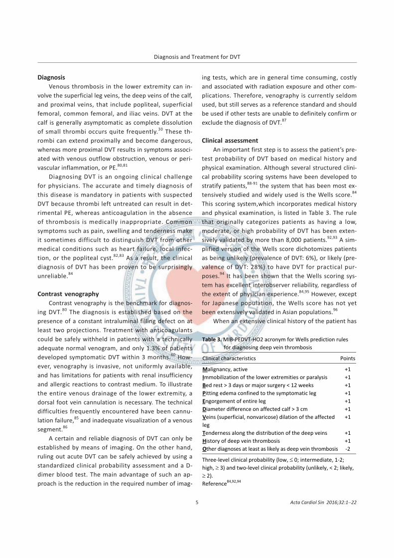
An important first step is to assess the patient’s pre-
test probability of DVT based on medical history and
physical examination. Although several structured clini-
cal probability scoring systems have been developed to
stratify patients,88-91
the system that has been most ex-
tensively studied and widely used is the Wells score.84
This scoring system,which incorporates medical history
and physical examination, is listed in Table 3. The rule
that originally categorizes patients as having a low,
moderate, or high probability of DVT has been exten-
sively validated by more than 8,000 patients.92,93
A sim-
plified version of the Wells score dichotomizes patients
as being unlikely (prevalence of DVT: 6%), or likely (pre-
valence of DVT: 28%) to have DVT for practical pur-
poses.94
It has been shown that the Wells scoring sys-
tem has excellent interobserver reliability, regardless of
the extent of physician experience.84,95
However, except
for Japanese population, the Wells score has not yet
been extensively validated in Asian populations.96
When an extensive clinical history of the patient has
5 Acta Cardiol Sin 2016;32:1�22
Diagnosis and Treatment for DVT
Table 3. MIB-PEDVT-HO2 acronym for Wells prediction rules
for diagnosing deep vein thrombosis
Clinical characteristics Points
Malignancy, active +1
Immobilization of the lower extremities or paralysis +1
Bed rest > 3 days or major surgery < 12 weeks +1
Pitting edema confined to the symptomatic leg +1
Engorgement of entire leg +1
Diameter difference on affected calf > 3 cm +1
Veins (superficial, nonvaricose) dilation of the affected
leg
+1
Tenderness along the distribution of the deep veins +1
History of deep vein thrombosis +1
Other diagnoses at least as likely as deep vein thrombosis -2
Three-level clinical probability (low, � 0; intermediate, 1-2;
high, � 3) and two-level clinical probability (unlikely, < 2; likely,
� 2).
Reference84,92,94

been obtained, the clinician can get confirmation of
whether any instances of active cancer (Malignancy),
paraplysis that leads to immobilization (Immobilization
of the lower extremities or paralysis), and prolonged
bed rest or a recent major surgery (Bed rest > 3 days or
major surgery < 12 weeks) are major risk factors for DVT
and thus related to the patient’s course of disease. The
presentation indicative of DVT includes leg swelling
(Pitting edema confined to the symptomatic leg and/or
Engorgement of the entire leg, and a Diameter differ-
ence on the affected calf > 3 cm), visible collateral circu-
lation (Veins, nonvaricose superficial dilation of the af-
fected leg), and leg pain (Tenderness along the distribu-
tion of the deep veins). A prior history of DVT (History of
deep vein thrombosis) is also an independent risk factor.
Finally, if there is an alternative tentative diagnosis
other than DVT (Other diagnoses at least as likely as
deep vein thrombosis), the likelihood of DVT is de-
creased. In this instance, the acronym MIB-PEDVT-HO2
is a convenient tool.
D-dimer
The major criticism in using the Wells score to as-
sess DVT probability is the subjective element of con-
sidering an alternative diagnosis. This clinical probability
can be subsequently supplemented by the combination
with a determination of the level of D-dimer for better
risk stratification.94
Fibrin D-dimer is the degradation product of cross-
linked fibrin. D-dimer levels are typically elevated in pa-
tients with acute thrombosis because of simultaneous ac-
tivation of coagulation and fibrinolysis. The sensitivity of
an elevated level of D-dimer is high, but the specificity is
rather moderate.97
A high D-dimer level is also observed in
conditions such as malignancy, infection, pregnancy, post
surgery or trauma, and increasing age. Consequently, a
negative result can facilitate the exclusion of VTE.98
A number of D-dimer assays are available. The
meta-analysis of 217 D-dimer tests evaluated for DVT in-
dicated that the sensitivities of the D-dimer enzyme-
linked immunofluorescence assay, microplate enzyme-
linked immunosorbent assay and latex quantitative assay
are 96%, 94%, and 93%, respectively; they are superior to
those of the whole-blood D-dimer assay, latex semi-
quantitative assay and latex qualitative assay as the sensi-
tivities are 83%, 85%, and 69%, respectively.99
The higher
sensitivity yields a higher negative predictive value and
introduces fewer concerns regarding false-negative re-
sults. In addition, the age-dependent decrease in specific-
ity of D-dimer with suspected PE was observed.100
Recent
investigations suggested that age-adjusted cut-offs (age �
10 ug/L above 50 years) improved specificity of D-dimer
testing in the elderly with suspected PE.101-103
The Euro-
pean guidelines have adopted the age-adjusted D-dimer
cut-offs for the diagnosis of acute PE.4
The clinical appli-
cation of the aforementioned aspect regarding D-dimer
testing has not been prospectively validated in patients
with suspected DVT and not yet endorsed by American
guidelines for the diagnosis of DVT.
Compression ultrasound
Compression ultrasound (CUS) is the front-line im-
aging modality in the diagnostic process for patients
with suspected DVT. The sensitivity and specificity of
the CUS in diagnosing proximal DVT is 94% and 98%, re-
spectively.104
However, the sensitivity decreases in de-
tecting asymptomatic proximal DVT with similar speci-
ficity.105
The sensitivity of CUS in diagnosing distal DVT is
relatively low, at 57%,104
and is only 48% for detecting
asymptomatic calf vein thrombosis.105
For proximal DVT,
ultrasound studies have a positive predictive value of
100% and 71% for symptomatic and asymptomatic
events, respectively, whereas the negative predictive
value for symptomatic and asymptomatic events are
100% and 94%, respectively.106
Conventionally, 3 protocols are presently available.
The proximal CUS (so-called 2-point CUS) assesses com-
pressibility of the femoral and popliteal veins. Loss of
compressibility of either segment is diagnostic for inci-
dent proximal DVT. The second approach is to conduct a
complete CUS along the proximal till distal deep veins of
the leg. The prior study suggested that only 0.5% of pa-
tients withholding anticoagulation after a single, nega-
tive, complete CUS had distal DVT within 3 months and
no proximal DVT occurred.82
Although it has good accu-
racy, complete CUS is time consuming and a modest
proportion of imaging results are inadequate, particu-
larly in the calf veins.107
As an alternative to the afore-
mentioned measures, repeated proximal CUS after 5-7
days is recommended in patients with an initial negative
proximal CUS finding. This approach detects if any distal
DVT propagates into the proximal vein that may increase
Acta Cardiol Sin 2016;32:1�22 6
Kang-Ling Wang et al.

the risk of PE. The repeated proximal CUS strategy in the
intermediate to high-risk patients is safe with regard to
withholding anticoagulation.108,109
Computed tomography and magnetic resonance
imaging
Both computed tomography venography (CTV) and
magnetic resonance venography (MRV) are alternatives
to CUS as a means to diagnose DVT.110
In particular, CTV
provides direct imaging of the inferior vena cava, pelvic
and lower extremity veins immediately after computed
tomography pulmonary angiography. Since DVT is the
most important risk for PE, it seems ideal that a single
examination simplifies and shortens VTE work-up. How-
ever, this single comprehensive imaging modality for
VTE requires a larger amount of iodinated contrast me-
dium to produce adequate opacification of the pulmo-
nary arteries, and pelvic and lower extremity veins,111,112
and exposure to ionizing radiation may be greater than
for either test alone.113,114
The pooled analysis showed
that for the diagnosis of DVT, in particular proximal DVT,
CTV had a sensitivity and specificity of 96% and 95%, re-
spectively.115
In addition, to facilitate the diagnosis of
DVT, MRV has a sensitivity and specificity of 92% and
95%, respectively, and the sensitivity for the diagnosis
of proximal DVT increases to 94%.116
The sensitivity and specificity of both CTV and MRV
for the diagnosis of DVT are comparable to CUS. How-
ever, the safety of withholding anticoagulation on the
basis of a normal CTV or MTV result has yet to be eva-
luated prospectively. Furthermore, CTV has generally
been tested along with computed tomography pulmo-
nary angiogram in patients with suspected PE. CTV,
however, has yet to be more extensively assessed as a
standalone diagnostic modality in patients with sus-
pected DVT. Therefore, both imaging modalities should
not be routinely used as front-line approaches.87
It is
reasonable to utilize CTV or MRV in conditions that CUS
cannot be performed or is less reliable, such as for pa-
tients with morbid obesity or casts, and patients with
suspected DVT in the iliac or inferior cava vein or sus-
pected venous anomaly.87,98,117
Recommendations
� To diagnose DVT requires systematic assessment (Fig-
ure 1).
� An acronym of MIB-PEDVT-HO2 for the Wells score is
the comprehensive initial to stratify patients.
� The combination of the Wells score and a D-dimer test
is validated in deciding whether to initially withhold
anticoagulation.
� CUS, either proximal, whole leg or repeated measure-
7 Acta Cardiol Sin 2016;32:1�22
Diagnosis and Treatment for DVT
Figure 1. Diagnosis flowchart. * Moderately or highly sensitive D-dimer test.
ments, should constitute the first-line imaging for most
patients with the increased likelihood of DVT.
� CTV and MRV have similar sensitivities and speci-
ficities to CUS in diagnosing DVT. However, those
measures should be reserved for conditions where
patients cannot be evaluated properly by CUS or
thrombosis in pelvic veins, or inferior vena cava is
suspected.
� Contrast venography is the reference modality for di-
agnosing DVT and is reserved for occasions when
other tests are unable to definitely establish or ex-
clude DVT.
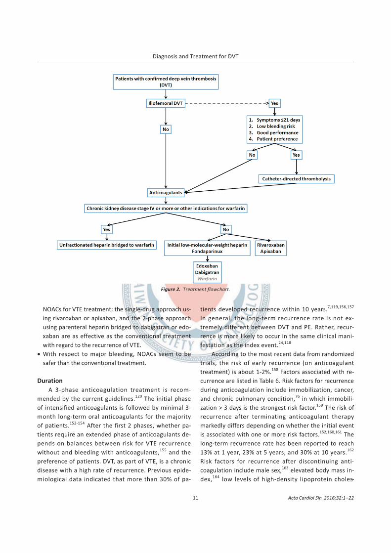
Treatment
DVT can become complicated with PE, and has a
high recurrence at the early stage without treatment.118
The risk of recurrence and post-thrombotic syndrome
(PTS) develop steadily after the first episode of DVT.7,119
The most effective manner in which to prevent throm-
bus extension, recurrence and late complications pri-
marily depends on effective pharmacological and/or
mechanical treatment.120
Thrombolysis
Despite effective anticoagulation, only 20%, 39%
and 58% of the patients had normal CUS at 3 months, 6
months and 1 year, respectively.121
Other studies re-
ported a similar rate of the presence of residual throm-
bus at 3 months and beyond.122,123
The presence of re-
sidual thrombus has been associated with the risk of re-
currence and the occurrence of PTS.121,124,125
The possi-
bility to reduce thrombus burden and to restore venous
drainage in the early stage of DVT in patients with ex-
treme high risk for thromboembolism complications is
an appealing one. The evidence suggested that systemic
thrombolysis had the potential to reduce PTS at the ex-
pense of an increase in major bleeding. As a result, sys-
temic thrombolysis is not generally recommended by
the current guidelines.120,126
A variety of endovascular
interventions have been tested in patients with ilio-
femoral DVT, including catheter-directed thrombolysis
(CDT) and/or thrombectomy. CDT reduces PTS at 24
months with a number needed to treat of 7 in a ran-
domized trial that involved 209 patients with iliofemoral
DVT.127
There was no significant reduction in recurrence
by CDT in this small-sized trial. However, bleeding is a
primary concern of a broad application of thrombo-
lysis.128
Currently, CDT is preferred for patients with
iliofemoral DVT and/or limb-threatening circulatory
compromise, acute or subacute symptoms, and a low
risk of bleeding.120,126
Endovascular thrombectomy and stenting have re-
duced recurrence and PTS at 30 months compared with
anticoagulation only in a randomized trial involving 169
patients with proximal DVT.129
Whether pharmaco-
mechanical CDT truly reduces recurrence and PTS in pa-
tients with proximal DVT is being evaluated currently in
a larger trial with a target of more than 600 patients and
a primary endpoint of PTS occurrence.130
Conventional anticoagulation
The initial and standard pharmacological approach in
patients with DVT started with parenteral anticoagulants,
either unfractionated or low-molecular-weight heparin,
followed by long-term vitamin K antagonists (VKAs). The
importance of effective initial parenteral anticoagulants
has been emphasized by the results in early trials.131,132
It
has been suggested to start parenteral anticoagulation
within a short period of time while waiting for the results
of diagnostic tests in patients with intermediate to high
clinical suspicion of acute DVT.120
In general, twice-daily
low-molecular-weight heparin (LMWH) administration is
preferred over once-daily LMWH administration or un-
fractionated heparin for a lower risk of inducing major
bleeding and heparin-induced thrombocytopenia.133,134
An oral VKA is preferably initiated on the same day
as the start of parenteral anticoagulants, which should
be continued for at least 5 days until the international
normalized ratio (INR) is 2.0 or above for 2 consecutive
days.120,135
The targeted INR for VKA treatment is 2.0-
3.0. The subtherapeutic dose is associated with higher
recurrence,136
and the occurrence of PTS.137
To maintain
adequate anticoagulation without an excessive risk of
bleeding in the use of VKAs, primarily warfarin, is a les-
son that every clinician has learned for the past 50 years
due to the limitations of new effective treatment.138
Non-vitamin K oral anticoagulants (NOACs)
NOACs have been developed to optimize VTE man-
agement and overcome the limitations of traditional
treatments. Five NOACs have been extensively tested in
patients with VTE in clinical trials. Except for ximela-
Acta Cardiol Sin 2016;32:1�22 8
Kang-Ling Wang et al.

gatran, which has been withdrawn, rivaroxaban, dabi-
gatran, apixaban, and edoxaban have been approved in
North America and Europe for VTE treatment; edoxaban
has completed the largest clinical trial ever in VTE treat-
ment. Except for acute treatment, rivaroxaban, dabi-
gatran, and apixaban have been compared with placebo
or warfarin in the setting of extended treatment for
VTE.
Rivaroxaban
Patients with acute DVT or PE were enrolled in the
EINSTEIN program. In the EINSTEIN DVT, 3,449 patients
with acute symptomatic DVT were randomized to re-
ceive oral rivaroxaban alone (15 mg twice daily for 3
weeks, followed by 20 mg once daily) or subcutaneous
enoxaparin followed by warfarin for 3, 6, or 12 months.139
In the EINSTEIN PE, 4,832 patients who had acute symp-
tomatic PE with or without DVT, were randomized ac-
cording to the same protocol.140
In both the EINSTEIN
DVT and EINSTEIN PE, a single-drug approach by rivar-
oxaban was non-inferior to the standard treatment with
regard to VTE recurrence and had no excessive major or
clinically relevant nonmajor bleeding.139,140
In particular,
there was no signal of excessive VTE recurrence by day
21, at the end of the twice-daily treatment. The pool
analysis of 8,282 patients showed very similar results of
separate trials but there was significantly less major
bleeding in patients with rivaroxaban compared to the
standard therapy (hazard ratio: 0.54; 95% confidence in-
terval: 0.37-0.79, p = 0.002).141
Dabigatran
Dabigatran has been tested in 2 trials for acute VTE
treatment (RE-COVER and RE-COVER II).142,143
The RE-
COVER series consisted of 2 trials because of low rate of
recurrent VTE observed in the first trial with a small
population. Dabigatran 150 mg twice daily was com-
pared with warfarin for 6 months after the initial par-
enteral heparin. There were 2,564 and 2,589 patients
with acute VTE enrolled in the RE-COVER and the RE-
COVER II, respectively. In both trials, dabigatran was
non-inferior to warfarin with regard to recurrent VTE
but had significantly less major or clinically relevant
nonmajor bleeding (hazard ratio: 0.63; 95% confidence
interval: 0.47-0.84, in the RECOVER; hazard ratio: 0.62;
95% confidence interval: 0.45-0.84, in the RECOVER II).
The extent of major bleeding was similar in patients
with dabigatran or warfarin. The conclusions were con-
sistent in the pooled analysis.142
Apixaban
In the AMPLIFY study, 5,400 patients with acute
VTE were randomized to receive apixaban (10 mg
twice daily for 7 days, followed by 5 mg twice daily) or
subcutaneous enoxaparin followed by warfarin for 6
months.144
Apixaban, as a single-drug approach, was
non-inferior to the standard treatment with respect
to recurrent VTE. Both major bleeding or clinically re-
levant nonmajor bleeding were significantly less in
patients with apixaban (relative risk: 0.44, 95% confi-
dence interval: 0.36-0.55, p < 0.001; relative risk:
0.31, 95% confidence interval: 0.17-0.55, p < 0.001,
respectively).
Edoxaban
Edoxaban has been compared with warfarin in the
largest phase III clinical trial in VTE treatment. In the
HOKUSAI VTE, 8,240 patients with acute VTE were ran-
domly assigned to edoxaban (60 mg once daily or 30 mg
once daily in patients with renal impairment or low
body weight) or warfarin for 3 to 12 months after the
initial parenteral heparin.145
Edoxaban was non-inferior
to warfarin with respect to recurrent VTE. Furthermore,
edoxaban had significantly less major or clinically rele-
vant nonmajor bleeding (hazard ratio: 0.81; 95% confi-
dence interval: 0.71-0.94, p = 0.004), and had no exces-
sive risk for major bleeding.
The risk of recurrent VTE and bleeding are funda-
mental considerations for recommendation of VTE man-
agement. Although clinical trials of NOACs in VTE treat-
ment have differences with regard to the study designs,
the conclusions are rather similar. The design and re-
sults of pivotal NOAC trials for VTE treatment are listed
in Table 4 and 5. The availability of NOACs that do not
require monitoring will create a treatment shift for pa-
tients with VTE as an effective, safer, and more conve-
nient treatment for patients, physicians, and healthcare
systems.146
The additional advantage of NOACs against
VKAs is that Asians are prone to bleeding when treated
with VKAs, and the optimal INR use has yet to be deter-
mined in Asian patients.147,148
In trials evaluating NOACs
for stroke prevention in atrial fibrillation, NOACs have
9 Acta Cardiol Sin 2016;32:1�22
Diagnosis and Treatment for DVT

less critical bleeding than VKAs in Asian patients than in
patients with other ethnicities.147,149,150
In trials evaluat-
ing NOACs for VTE treatment, both rivaroxaban and edo-
xaban have published data of Asian patients.150,151
Both
analyses reconfirmed the similar efficacy and the pro-
bable safety advantage of rivaroxaban and edoxaban
against dose-adjusted VKAs.
Recommendations
� Timely administration of anticoagulation is essential in
the treatment of DVT (Figure 2).
� Conventional treatment involves parenteral heparin
bridging to VKAs with a maintenance target INR of
2.0-3.0.
� Two strategies have been tested in 6 pivotal trials of
Acta Cardiol Sin 2016;32:1�22 10
Kang-Ling Wang et al.
Table 5. Hazard ratio (95% confident interval) of selected outcomes for non-vitamin K oral anticoagulants in venous
thromboembolism treatment
Rivaroxaban vs. VKAs
EINSTEIN pooled analysis
Dabigatran vs. VKAs
RE-COVER pooled analysis
Apixaban vs. VKAs
AMPLIFY*
Edoxaban vs. VKAs
HOKUSAI-VTE
Recurrent VTE 0.89 (0.66-1.19) 1.09 (0.76-1.57) 0.84 (0.60-1.18)#0.82 (0.60-1.14)
#
Major bleeding 0.54 (0.37-0.79) 0.73 (0.48-1.11) 0.31 (0.17-0.55) 0.84 (0.59-1.21)
Major or CRNM bleeding 0.93 (0.81-1.06) 0.62 (0.50-0.76) 0.44 (0.36-0.55) 0.81 (0.71-0.94)
Death 0.89 (0.67-1.18) 1.00 (0.67-1.51) 0.79 (0.53-1.19) 1.05 (0.82-1.35)
* Relative risks are reported for the AMPLIFY study.#
Recurrent VTE during on-treatment period.
CRNM, clinically relevant nonmajor; VKA, vitamin K antagonist; VTE, venous thromboembolism.
Table 4. Patient characteristics in clinical trials of non-vitamin K oral anticoagulants for venous thromboembolism treatment
EINSTEIN-DVT EINSTEIN-PE RE-COVER RE-COVER II AMPLIFY HOKUSAI-VTE
Rivaroxaban VKA Rivaroxaban VKA Dabigatran VKA Dabigatran VKA Apixaban VKA Edoxaban VKA
Number of
patients
1,731 1,718 2,419 2,413 1,273 1,266 1,280 1,288 2,691 2,704 4,118 4,122
Design Open-label Open-label Double-blinded Double-blinded Double-blinded Double-blinded
Treatment
duration
3, 6 or 12 months* 3, 6 or 12 months* 6 months 6 months 6 months 3, 6 or 12 months*
Initial
treatment
Rivaroxaban
15 mg twice
daily for 3
weeks
LMWH
� 5
days
Rivaroxaban
15 mg twice
daily for 3
weeks
LMWH
� 5 days
LMWH or UFH
� 5 days
LMWH or UFH
� 5 days
Apixaban 10 mg
twice daily for 7
days
LMWH
or UFH
� 5 days
LMWH or UFH
� 5 days
Long-term
treatment
Rivaroxaban
20 mg once
daily
INR:
2-3
Rivaroxaban
20 mg once
daily
INR:
2-3
Dabigatran
150 mg
twice daily
INR:
2-3
Dabigatran
150mg
twice daily
INR: 2-3 Apixaban 5mg
twice daily
INR: 2-3 Edoxaban
60 mg once
daily#
INR:
2-3
Age, years 56 56 58 58 55 54 55 55 57 57 56 56
Female, % 43 44 46 48 42 41 39 40 42 41 43 43
Index event, %
DVT 99 99 0 0 69 69 69 68 65 66 60 60
PE � DVT 1 1 100 100 31 31 31 32 35 34 40 40
Risk factor, %
Unprovoked 61 63 65 64 NR NR NR NR 90 90 66 65
Prior VTE 19 19 19 20 26 25 19 16 17 15 19 18
Cancer 7 5 5 5 5 5 4 4 2 3 9 10
TTR, % NA 58 NA 63 NA 60 NA 57 NA 60 NA 64
* Treatment duration was left to the discretion of the treating physician.#
An edoxaban 30 mg once daily was administered in certain conditions.
DVT, deep vein thrombosis; INR, international normalized ratio; LMWH, low-molecular-weight heparin; NA, not applicable; NR, not reported; PE,
pulmonary embolism; TTR, time in therapeutic range; UFH, unfractionated heparin; VKA, vitamin K antagonist; VTE, venous thromboembolism.
NOACs for VTE treatment; the single-drug approach us-
ing rivaroxban or apixaban, and the 2-phase approach
using parenteral heparin bridged to dabigatran or edo-
xaban are as effective as the conventional treatment
with regard to the recurrence of VTE.
� With respect to major bleeding, NOACs seem to be
safer than the conventional treatment.
Duration
A 3-phase anticoagulation treatment is recom-
mended by the current guidelines.120
The initial phase
of intensified anticoagulants is followed by minimal 3-
month long-term oral anticoagulants for the majority
of patients.152-154
After the first 2 phases, whether pa-
tients require an extended phase of anticoagulants de-
pends on balances between risk for VTE recurrence
without and bleeding with anticoagulants,155
and the
preference of patients. DVT, as part of VTE, is a chronic
disease with a high rate of recurrence. Previous epide-
miological data indicated that more than 30% of pa-
tients developed recurrence within 10 years.7,119,156,157
In general, the long-term recurrence rate is not ex-
tremely different between DVT and PE. Rather, recur-
rence is more likely to occur in the same clinical mani-
festation as the index event.24,118
According to the most recent data from randomized
trials, the risk of early recurrence (on anticoagulant
treatment) is about 1-2%.158
Factors associated with re-
currence are listed in Table 6. Risk factors for recurrence
during anticoagulation include immobilization, cancer,
and chronic pulmonary condition,76
in which immobili-
zation > 3 days is the strongest risk factor.159
The risk of
recurrence after terminating anticoagulant therapy
markedly differs depending on whether the initial event
is associated with one or more risk factors.152,160,161
The
long-term recurrence rate has been reported to reach
13% at 1 year, 23% at 5 years, and 30% at 10 years.162
Risk factors for recurrence after discontinuing anti-
coagulation include male sex,163
elevated body mass in-
dex,164
low levels of high-density lipoprotein choles-
11 Acta Cardiol Sin 2016;32:1�22
Diagnosis and Treatment for DVT
Figure 2. Treatment flowchart.
terol,165
idiopathic presentation, thrombophilia, increas-
ing age, shorter duration of anticoagulation, cancer, and
residual thrombus.7,121,166
In fact, the provoked nature of an index event is a
major determinant for the risk of VTE recurrence.15,167
In
patients with proximal DVT and PE, the estimated cumu-
lative risk of recurrence after stopping anticoagulants is
as follows: VTE provoked by surgery, 1% after 1 year and
3% after 5 years; VTE provoked by a nonsurgical revers-
ible risk factor, 5% after 1 year and 15% after 5 years;
and unprovoked VTE, 10% recurrence after 1 year and
30% after 5 years, and 15% annually for patients with
cancer.120
Previous trials have compared different antithrom-
botic strategies in the reduction of VTE recurrence in pa-
tients with an unprovoked event.152,160,168
These trials in-
dicated that extending anticoagulation for 1 year has yet
to eliminate long-term recurrence in patients with un-
provoked VTE. Two trials have intended to extend the
anticoagulation treatment beyond 1 year.169,170
In the
ELATE study, low-intensity warfarin (target INR of 1.5-
1.9) has been compared with standard-intensity warfa-
rin (target INR of 2.0-3.0), and patients have been fol-
lowed for an average of 2.4 years. The recurrence rate
was 1.9 per 100 patient-years in the low-intensity group
compared with 0.7 per 100 patient-years in the stan-
dard-intensity group (p = 0.03).170
In the PREVENT study,
508 patients with unprovoked VTE were randomized to
low-intensity warfarin (target INR of 1.5-2.0) or placebo.
The trial was terminated after 2 years of therapy due to
extreme beneficial effect with warfarin. The recurrent
VTE was 2.6 per 100 patient-years with warfarin com-
pared with 7.2 per 100 patient-years with placebo (p <
0.001).169
Incidentially, the major bleeding was remark-
able low in both trials (around 1 per 100 patient-years in
all treatment groups).
Besides warfarin, extended treatment trials for VTE
involving rivaroxaban, dabigatran and apixaban have
been completed, with variable treatment duration and
comparators.139,171,172
All 3 NOACs have largely reduced
recurrent or fatal VTE and had no excessive risk for ma-
jor bleeding compared with placebo. The recurrent or
fatal VTE and major bleeding was similar with dabi-
gatran and standard-intensity warfarin, but the risk of
major or clinically relevant bleeding event was lower
with dabigatran compared with standard-intensity war-
farin.171
The information associated with the NOACs tri-
als involving extended treatment for VTE is listed in
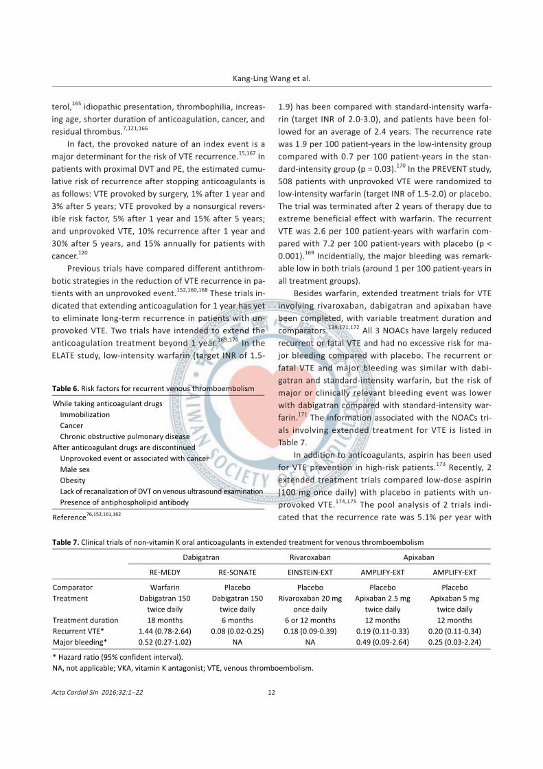
Table 7.
In addition to anticoagulants, aspirin has been used
for VTE prevention in high-risk patients.173
Recently, 2
extended treatment trials compared low-dose aspirin
(100 mg once daily) with placebo in patients with un-
provoked VTE.174,175
The pool analysis of 2 trials indi-
cated that the recurrence rate was 5.1% per year with
Acta Cardiol Sin 2016;32:1�22 12
Kang-Ling Wang et al.
Table 7. Clinical trials of non-vitamin K oral anticoagulants in extended treatment for venous thromboembolism
Dabigatran Rivaroxaban Apixaban
RE-MEDY RE-SONATE EINSTEIN-EXT AMPLIFY-EXT AMPLIFY-EXT
Comparator Warfarin Placebo Placebo Placebo Placebo
Treatment Dabigatran 150
twice daily
Dabigatran 150
twice daily
Rivaroxaban 20 mg
once daily
Apixaban 2.5 mg
twice daily
Apixaban 5 mg
twice daily
Treatment duration 18 months 6 months 6 or 12 months 12 months 12 months
Recurrent VTE* 1.44 (0.78-2.64) 0.08 (0.02-0.25) 0.18 (0.09-0.39) 0.19 (0.11-0.33) 0.20 (0.11-0.34)
Major bleeding* 0.52 (0.27-1.02) NA NA 0.49 (0.09-2.64) 0.25 (0.03-2.24)
* Hazard ratio (95% confident interval).
NA, not applicable; VKA, vitamin K antagonist; VTE, venous thromboembolism.
Table 6. Risk factors for recurrent venous thromboembolism
While taking anticoagulant drugs
Immobilization
Cancer
Chronic obstructive pulmonary disease
After anticoagulant drugs are discontinued
Unprovoked event or associated with cancer
Male sex
Obesity
Lack of recanalization of DVT on venous ultrasound examination
Presence of antiphospholipid antibody
Reference76,152,161,162
low-dose aspirin compared with 7.5% per year with pla-
cebo (p = 0.008), whereas there was no excessive risk
for major bleeding.176
Despite the probability that extended treatment is
effective and generally safe in reducing VTE recurrence
in patients with higher risk for recurrence, anticoagu-
lation treatment of indefinite duration, or even probably
for a lifetime, is a major undertaking both for patients
and healthcare professionals.177
The decision of the
treatment duration for DVT requires a complex evalua-
tion that balances the risks of recurrence in the absence
of anticoagulation against the risks of bleeding compli-
cations with continued pharmacological therapy.152,178
There is not yet a universally accepted method for pre-
dicting the risk of VTE recurrence in the absence of
anticoagulation. The DASH scoring system and Vienna
prediction model are newly developed assessment for
patients with unprovoked VTE.179,180
To address the risk
of major bleeding with anticoagulation in the first 3
months of treatment, the practice guidelines recom-
mend a complex matrix whereas the RIETE scoring is a
simple assessment.181
Recommendations
� The long-term treatment comprises minimally a 3-
month duration of oral anticoagulants.
� Patients without a transient reversible risk factor for
VTE are more likely to develop recurrences after cessa-
tion of anticoagulant treatment.
� For extended treatment, normal or low intensity warfa-
rin, dabigatran, rivaroxaban, apixaban, and aspirin are
effective on the reduction of VTE recurrence in patients
with unprovoked VTE.
� Whether patients require an extended phase of antico-
agulants depends on balances between the risk of VTE
recurrence without anticoagulants and bleeding with
anticoagulants, and the preference of patients.
� There is not yet a widely accepted scoring system for
prediction of VTE recurrence without anticoagulation
and bleeding with anticoagulation.
Pregnancy-related VTE
Pregnancy is associated with a hypercoagulable
state characterized by increased concentrations of
procoagulant factors and by a simultaneous decrease
of anticoagulant factors.182
It is important to note that
the likelihood of VTE is increased at least 4-fold dur-
ing pregnancy compared with a non-pregnant status,
and is a major cause of maternal mortality in the de-
veloped world.183,184
The overall incidence of VTE in
pregnancy is about 1-2 cases per 1,000 deliveries in
the West, and around 1 case per 10,000 individuals in
Asia.20,185
The risk of VTE remains high until the first 6
weeks after delivery have passed.186
Furthermore,
VTE occurs at a rate of 5.4, 7.2, and 4.3 cases per
10,000 pregnancies for antepartum, peripartum, and
postpartum, respectively.185
Isolated DVT, particularly
left iliac and/or femoral vein, is more common in
pregnant patients due to the enlarged uterus’ com-
pressive effects on veins.187
The use of diagnostic algorithms for DVT, the combi-
nation of structured clinical prediction rules and the use
of D-dimer testing, in non-pregnant patients is neither
adequate nor validated when applied in pregnancy. Leg
swelling, the cardinal sign and symptom of DVT, may be
associated with a normal pregnancy and levels of D-
dimer increase with the progression of a normal preg-
nancy.188
The cornerstone of diagnosing DVT in preg-
nancy is the demonstration of presence of a clot by CUS.
Compression maneuvers should be performed along the
entire venous system from the femoral to the popliteal
vein. The sensitivity and negative predictive value are
90.9% and 98.9%, respectively,189
with a reduced accu-
racy for isolated calf- and iliac-vein thrombosis.190
CUS
could be repeated within 7 days if the initial study is
negative,191,192
or MRV could be used if the initial CUS
study is negative or for detecting iliac-vein thrombo-
sis.193
It is safe to withhold anticoagulation in pregnant pa-
tients with suspected DVT following negative serial CUS
and iliac vein imaging.194
Maternal complications from
anticoagulant therapy are similar to those seen in
non-pregnant patients.195
However, additional consider-
ations regarding the health and life of the fetus will af-
fect patients’ choices on anticoagulation therapy. For
the treatment of acute DVT, particularly proximal DVT
with or without PE, inpatient treatment with parenteral
heparin is recommended and VKAs should only be consid-
ered in exceptional circumstances.192,196
Pregnant women
were excluded from participating in clinical trials evalu-
ating NOACs, and NOACs are not approved for such con-
ditions and should not be used in breast-feeding pa-
13 Acta Cardiol Sin 2016;32:1�22
Diagnosis and Treatment for DVT

tients. In general, LMWH is recommended over
unfractionated heparin for use in pregnant patients. It is
reasonable to use the twice-daily regimen of LMWH in
pregnancy, especially for the first month when the risk
of recurrence is the greatest. This practice also stems
from the altered renal elimination of LMWH and the im-
pact of weight gain during pregnancy.
No study has assessed the optimal duration of anti-
coagulant therapy for treatment of pregnancy-related
VTE. In non-pregnant patients with VTE, evidence sup-
ports a minimum duration of 3 months of treatment.
Major guidelines recommend anticoagulants, which
should be continued for at least 6 weeks postpartum
(for a minimum total therapy duration of 3
months).196,197
And for pregnant women with a prior
history of VTE associated with transient risk factors,
withholding antepartum anticoagulation therapy is
generally safe.198,199
Cancer-related VTE
Patients with cancer are at the increased risk for
VTE,200
and carry a substantial risk for VTE recurrence.15,201
Meanwhile, VTE can be attributed to major morbidity
and mortality in patients with cancer,202
and has a signif-
icant negative impact on quality of life in patients with
cancer.203
Although long-term VKAs are warranted and
prevent recurrence in the majority of patients, VKA
treatment therapy is problematic in patients with cancer
because drug interactions, malnutrition, vomiting, and
liver dysfunction can lead to unpredictable levels of
anticoagulation in addition to the potential need for in-
vasive procedures requiring interruption of VKAs.204
These limitations may contribute to the higher risk of re-
current thromboembolism and bleeding in patients with
cancer than in patients without cancer.
Weight-adjusted LMWH as a parenteral agent is ef-
fective and appealing for treatment in patients with
cancer. The remote and recent clinical trials have dem-
onstrated the advantage of treatment with LMWH over
VKAs for 6 months.205,206
NOACs had at least a similar
clinical benefit as VKAs in the subgroup analyses,141,207,208
and might be even comparable to LMWH in the pooled
analyses.209,210
However, additional studies are needed
to confirm this concept and to compare NOACs with
LMWH directly in patients with cancer.211
Currently, 3
protocols that will randomize cancer patients with VTE
to either NOACs or LMWH with a variable treatment
duration are approved (NCT02583191, NCT02585713,
NCT02073682). Despite practicing guidelines recom-
mending LMWH for at least 3-6 months and possibly
indefinitely for patients with active cancer, the optimal
duration of LMWH treatment beyond 6 months is yet
to be determined by the evidence from the random-
ized controlled trials.212,213
In the Hokusai VTE-cancer
study, patients will be treated for an extended period
up to 12 months on the basis of the risk-benefit assess-
ment by the treating physician and/or on patient pre-
ference.214
Areas with uncertainty
Although a large number of cohort studies have pro-
vided convincing data, and the clinical assessment path-
way has been long constructed, these studies describe
and have been validated mainly in Western populations.
The therapeutic trials had a very low proportion of both
young and elderly patients, and patients with minimal
comorbidity. In addition, a small proportion of Asian pa-
tients participated in these trials. The NOACs have been
tested in extended treatment with limited follow-up pe-
riods. It will be necessary to elaborate on (i) whether
Asian patients indeed have a low risk for the develop-
ment of DVT and PE, (ii) whether the diagnostic algo-
rithm is feasible if Asian patients truly have a lower
prevalence of DVT and PE, (iii) long-term recurrence
without and bleeding risk with anticoagulation treat-
ment in Asian patients, and (iv) whether treatment for
VTE in patients with pregnancy or cancer is practical in
Asian patients who might have different thrombosis and
bleeding profiles. Nevertheless, the accumulation of
clinical experience with new therapeutics in the real-
world will have to proceed at a prudent pace. For those
trials that enrolled but have yet to publish data regard-
ing Asian patients, we welcome public dissemination of
the research results, so that physicians, patients, and
other health care professionals can benefit from the
results.215,216
SUMMARY
The heavy burden of DVT, as part of VTE continuum,
is seriously overlooked in Taiwan. Routine risk assess-
Acta Cardiol Sin 2016;32:1�22 14
Kang-Ling Wang et al.

ment and thromboprophylaxis for high-risk patient have
not been established, and the guidelines are not yet de-
veloped. On the other hand, the diagnosis of DVT re-
quires a sophisticated evaluation from pre-test risk as-
sessment until imaging confirmation. The implantation
of the comprehensive diagnostic algorithm facilitates
the timely administration of therapeutics for the appro-
priate patients. For patients with DVT, anticoagulation is
essential to prevent thrombus extension and recur-
rence. The conventional strategy of initial parenteral
heparin followed by warfarin is effective, but somehow
inconvenient and has its limitations. Quickly-adopted
NOACs as an alternative strategy for acute VTE treat-
ment have their unique features with respect to each
other. Some of the NOACs are taken once daily, others
twice daily, some require the use of a parenteral hepa-
rin lead in, and others use the single-drug approach. The
single-drug approach without preceding parenteral
treatments is appealing to low risk outpatients whereas
the conventional parenteral lead in strategy is feasible
for intermediate/high risk inpatients. Nevertheless,
NOACs have been proven effectively safe in the large-
scale clinical trials, particularly with respect to the im-
portant outcome of bleeding. Finally, DVT is a chronic
disease and unprovoked DVT carries a higher risk for re-
currence. Extended treatment should be considered in
such patients in part based upon the weight of preven-
tion of recurrence and risk of bleeding. Growing studies
have paid attention to the idea of constructing a more
user-friendly scoring system to predict VTE recurrence
without and bleeding events with anticoagulation. Fi-
nally, this consensus document hopefully can upsurge
the awareness of DVT in the medical community, and
improve patient outcomes during the acute phase and
thereafter in the long run.
DISCLOSURE
Dr. Kang-Ling Wang received some benefit from
Bayer, Boehringer Ingelheim, and Daiichi Sankyo. Dr.
Kou-Gi Shyu has been on the speaker’s bureau for
AstraZeneca, Bayer, Daiichi Sankyo, MSD, Pfizer, Sanofi,
and Takeda. Dr. Chern-En Chiang has been on the
speaker’s bureau for Bayer, Boehringer Ingelheim, Daii-
chi Sankyo, and Pfizer.
FUNDING SOURCES
This work was supported, in part, by grants from the
Ministry of Health and Welfare (MOHW104-TDU-B-
211-113-003), the Ministry of Science and Technology
(102-2314-B-182A-060-MY2 and 104-2314-B-182A-131),
and Chang Gung Memorial Hospital (CIRPG3E0011).
REFERENCES
1. Moser KM, Fedullo PF, LitteJohn JK, Crawford R. Frequent
asymptomatic pulmonary embolism in patients with deep ve-
nous thrombosis. JAMA 1994;271(3):223-5.
2. Goldhaber SZ, Visani L, De Rosa M. Acute pulmonary embolism:
clinical outcomes in the International Cooperative Pulmonary
Embolism Registry (ICOPER). Lancet 1999;353(9162):1386-9.
3. Kucher N, Rossi E, De Rosa M, Goldhaber SZ. Prognostic role of
echocardiography among patients with acute pulmonary em-
bolism and a systolic arterial pressure of 90 mm Hg or higher.
Arch Intern Med 2005;165(15):1777-81.
4. Konstantinides SV, Torbicki A, Agnelli G, et al. 2014 ESC guide-
lines on the diagnosis and management of acute pulmonary
embolism. Eur Heart J 2014;35(43):3033-69.
5. Cohen AT, Agnelli G, Anderson FA, et al. Venous thromboem-
bolism (VTE) in Europe. The number of VTE events and associ-
ated morbidity and mortality. Thromb Haemost 2007;98(4):
756-64.
6. Spencer FA, Gore JM, Lessard D, et al. Patient outcomes after
deep vein thrombosis and pulmonary embolism: the Worcester
Venous Thromboembolism Study. Arch Intern Med 2008;
168(4):425-30.
7. Prandoni P, Noventa F, Ghirarduzzi A, et al. The risk of recurrent
venous thromboembolism after discontinuing anticoagulation
in patients with acute proximal deep vein thrombosis or pulmo-
nary embolism. A prospective cohort study in 1,626 patients.
Haematologica 2007;92(2):199-205.
8. Pengo V, Lensing AW, Prins MH, et al. Incidence of chronic
thromboembolic pulmonary hypertension after pulmonary
embolism. N Engl J Med 2004;350(22):2257-64.
9. Kahn SR. The post-thrombotic syndrome: the forgotten morbid-
ity of deep venous thrombosis. J Thromb Thrombolysis 2006;
21(1):41-8.
10. Roberts LN, Patel RK, Donaldson N, et al. Post-thrombotic syn-
drome is an independent determinant of health-related quality
of life following both first proximal and distal deep vein throm-
bosis. Haematologica 2014;99(3):e41-3.
11. Lefebvre P, Laliberte F, Nutescu EA, et al. All-cause and dis-
ease-related health care costs associated with recurrent ve-
nous thromboembolism. Thromb Haemost 2013;110(6):1288-
97.
15 Acta Cardiol Sin 2016;32:1�22
Diagnosis and Treatment for DVT

12. Sorensen HT, Horvath-Puho E, Pedersen L, et al. Venous throm-
boembolism and subsequent hospitalisation due to acute ar-
terial cardiovascular events: a 20-year cohort study. Lancet
2007;370(9601):1773-9.
13. Silverstein MD, Heit JA, Mohr DN, et al. Trends in the incidence
of deep vein thrombosis and pulmonary embolism: a 25-year
population-based study. Arch Intern Med 1998;158(6):585-93.
14. White RH. The epidemiology of venous thromboembolism. Cir-
culation 2003;107(23 Suppl 1): I4-8.
15. Lee CH, Lin LJ, Cheng CL, et al. Incidence and cumulative recur-
rence rates of venous thromboembolism in the Taiwanese
population. J Thromb Haemost 2010;8(7):1515-23.
16. Alikhan R, Cohen AT, Combe S, et al. Risk factors for venous
thromboembolism in hospitalized patients with acute medical
illness: analysis of the MEDENOX Study. Arch Intern Med 2004;
164(9):963-8.
17. Samama MM. An epidemiologic study of risk factors for deep
vein thrombosis in medical outpatients: the Sirius study. Arch
Intern Med 2000;160(22):3415-20.
18. Cohen AT, Alikhan R, Arcelus JI, et al. Assessment of venous
thromboembolism risk and the benefits of thromboprophylaxis
in medical patients. Thromb Haemost 2005;94(4):750-9.
19. Anderson FA Jr, Spencer FA. Risk factors for venous throm-
boembolism. Circulation 2003;107(23 Suppl 1):I9-16.
20. Jang MJ, Bang SM, Oh D. Incidence of venous thromboem-
bolism in Korea: from the Health Insurance Review and Assess-
ment Service database. J Thromb Haemost 2011;9(1):85-91.
21. Kyrle PA, Eichinger S. Deep vein thrombosis. Lancet 2005;
365(9465):1163-74.
22. Meignan M, Rosso J, Gauthier H, et al. Systematic lung scans re-
veal a high frequency of silent pulmonary embolism in patients
with proximal deep venous thrombosis. Arch Intern Med 2000;
160(2):159-64.
23. Sandler DA, Martin JF. Autopsy proven pulmonary embolism in
hospital patients: are we detecting enough deep vein thrombo-
sis? J R Soc Med 1989;82(4):203-5.
24. Murin S, Romano PS, White RH. Comparison of outcomes after
hospitalization for deep venous thrombosis or pulmonary em-
bolism. Thromb Haemost 2002;88(3):407-14.
25. Naess IA, Christiansen SC, Romundstad P, et al. Incidence and
mortality of venous thrombosis: a population-based study. J
Thromb Haemost 2007;5(4):692-9.
26. Kucher N, Rossi E, De Rosa M, Goldhaber SZ. Massive pulmo-
nary embolism. Circulation 2006;113(4):577-82.
27. Heit JA. The epidemiology of venous thromboembolism in the
community. Arterioscler Thromb Vasc Biol 2008;28(3):370-2.
28. Guijarro R, Montes J, Sanroman C, et al. Venous thromboem-
bolism in Spain. Comparison between an administrative data-
base and the RIETE registry. Eur J Intern Med 2008;19(6):443-6.
29. Spencer FA, Emery C, Joffe SW, et al. Incidence rates, clinical
profile, and outcomes of patients with venous thromboem-
bolism. The Worcester VTE study. J Thromb Thrombolysis 2009;
28(4):401-9.
30. Kakkar VV, Howe CT, Flanc C, Clarke MB. Natural history of post-
operative deep-vein thrombosis. Lancet 1969;2(7614):230-2.
31. Galanaud JP, Sevestre MA, Genty C, et al. Incidence and predic-
tors of venous thromboembolism recurrence after a first iso-
lated distal deep vein thrombosis. J Thromb Haemost 2014;
12(4):436-43.
32. Wang CJ, Wang JW, Chen LM, et al. Deep vein thrombosis after
total knee arthroplasty. J Formos Med Assoc 2000;99(11):
848-53.
33. Cogo A, Lensing AW, Prandoni P, Hirsh J. Distribution of throm-
bosis in patients with symptomatic deep vein thrombosis. Im-
plications for simplifying the diagnostic process with compres-
sion ultrasound. Arch Intern Med 1993;153(24):2777-80.
34. Oger E. Incidence of venous thromboembolism: a community-
based study in Western France. EPI-GETBP Study Group.
Groupe d’Etude de la Thrombose de Bretagne Occidentale.
Thromb Haemost 2000;83(5):657-60.
35. Ho WK, Hankey GJ, Eikelboom JW. The incidence of venous
thromboembolism: a prospective, community-based study in
Perth, Western Australia. Med J Aust 2008;189(3):144-7.
36. Liao S, Woulfe T, Hyder S, et al. Incidence of venous throm-
boembolism in different ethnic groups: a regional direct com-
parison study. J Thromb Haemost 2014;12(2):214-9.
37. White RH, Zhou H, Murin S, Harvey D. Effect of ethnicity and
gender on the incidence of venous thromboembolism in a di-
verse population in California in 1996. Thromb Haemost 2005;
93(2):298-305.
38. Cohen A, Chiu KM, Park K, et al. Managing venous throm-
boembolism in Asia: winds of change in the era of new oral anti-
coagulants. Thromb Res 2012;130(3):291-301.
39. Lee CH, Cheng CL, Lin LJ, et al. Epidemiology and predictors of
short-term mortality in symptomatic venous thromboembo-
lism. Circ J 2011;75(8):1998-2004.
40. Angchaisuksiri P. Venous thromboembolism in Asia--an unre-
cognised and under-treated problem? Thromb Haemost 2011;
106(4):585-90.
41. Yu YB, Gau JP, Liu CY, et al. A nation-wide analysis of venous
thromboembolism in 497,180 cancer patients with the devel-
opment and validation of a risk-stratification scoring system.
Thromb Haemost 2012;108(2):225-35.
42. Anderson FA Jr, Wheeler HB. Physician practices in the manage-
ment of venous thromboembolism: a community-wide survey.
J Vasc Surg 1992;16(5):707-14.
43. Kyrle PA, Rosendaal FR, Eichinger S. Risk assessment for recur-
rent venous thrombosis. Lancet 2010;376(9757):2032-9.
44. Goldhaber SZ. Venous thromboembolism: epidemiology and
magnitude of the problem. Best Pract Res Clin Haematol 2012;
25(3):235-42.
45. Goldhaber SZ, Bounameaux H. Pulmonary embolism and deep
vein thrombosis. Lancet 2012;379(9828):1835-46.
46. Falck-Ytter Y, Francis CW, Johanson NA, et al. Prevention of VTE
in orthopedic surgery patients: Antithrombotic Therapy and
Prevention of Thrombosis, 9th ed: American College of Chest
Acta Cardiol Sin 2016;32:1�22 16
Kang-Ling Wang et al.
Physicians Evidence-Based Clinical Practice Guidelines. Chest
2012;141(2 Suppl):e278S-325S.
47. Bjornara BT, Gudmundsen TE, Dahl OE. Frequency and timing of
clinical venous thromboembolism after major joint surgery. J
Bone Joint Surg Br 2006;88(3):386-91.
48. Bergqvist D, Benoni G, Bjorgell O, et al. Low-molecular-weight
heparin (enoxaparin) as prophylaxis against venous throm-
boembolism after total hip replacement. N Engl J Med 1996;
335(10):696-700.
49. Lee WS, Kim KI, Lee HJ, et al. The incidence of pulmonary embo-
lism and deep vein thrombosis after knee arthroplasty in Asians
remains low: a meta-analysis. Clin Orthop Relat Res 2013;
471(5):1523-32.
50. Chung LH, Chen WM, Chen CF, et al. Deep vein thrombosis after
total knee arthroplasty in asian patients without prophylactic
anticoagulation. Orthopedics 2011;34(1):15.
51. Cohen AT, Asia-Pacific Thrombosis Advisory B. Asia-Pacific
Thrombosis Advisory Board consensus paper on prevention of
venous thromboembolism after major orthopaedic surgery.
Thromb Haemost 2010;104(5):919-30.
52. Liew NC, Chang YH, Choi G, et al. Asian venous thromboem-
bolism guidelines: prevention of venous thromboembolism. Int
Angiol 2012;31(6):501-16.
53. Leizorovicz A, Turpie AG, Cohen AT, et al. Epidemiology of ve-
nous thromboembolism in Asian patients undergoing major or-
thopedic surgery without thromboprophylaxis. The SMART
study. J Thromb Haemost 2005;3(1):28-34.
54. Leizorovicz A, Committee SVSS. Epidemiology of post-operative
venous thromboembolism in Asian patients. Results of the
SMART venography study. Haematologica 2007;92(9):1194-
200.
55. Piovella F, Wang CJ, Lu H, et al. Deep-vein thrombosis rates after
major orthopedic surgery in Asia. An epidemiological study
based on postoperative screening with centrally adjudicated bi-
lateral venography. J Thromb Haemost 2005;3(12):2664-70.
56. Lee CH, Cheng CL, Chang CH, et al. Universal pharmacological
thromboprophylaxis for total knee arthroplasty may not be
necessary in low-risk populations: a nationwide study in Tai-
wan. J Thromb Haemost 2012;10(1):56-63.
57. Wu PK, Chen CF, Chung LH, et al. Population-based epidemio-
logy of postoperative venous thromboembolism in Taiwanese
patients receiving hip or knee arthroplasty without pharmaco-
logical thromboprophylaxis. Thromb Res 2014;133(5):719-24.
58. Wang CJ, Wang JW, Weng LH, et al. Clinical significance of mus-
cular deep-vein thrombosis after total knee arthroplasty. Chang
Gung Med J 2007;30(1):41-6.
59. Attia J, Ray JG, Cook DJ, et al. Deep vein thrombosis and its pre-
vention in critically ill adults. Thromb Res 2001;161(10):1268-
79.
60. Turpie AG. Thrombosis prophylaxis in the acutely ill medical pa-
tient: insights from the prophylaxis in MEDical patients with
ENOXaparin (MEDENOX) trial. Am J Cardiol 2000;86(12B):
48M-52M.
61. Leizorovicz A, Cohen AT, Turpie AG, et al. Randomized, placebo-
controlled trial of dalteparin for the prevention of venous
thromboembolism in acutely ill medical patients. Circulation
2004;110(7):874-9.
62. Cohen AT, Davidson BL, Gallus AS, et al. Efficacy and safety of
fondaparinux for the prevention of venous thromboembolism
in older acute medical patients: randomised placebo controlled
trial. BMJ 2006;332(7537):325-9.
63. Leung V, Leung V, Lui W, et al. Incidence of deep vein thrombo-
sis in hospitalized Chinese medical patients is similar to that in
western populations. Thromb Res 2006;118(6):763-4.
64. Dunn AS, Brenner A, Halm EA. The magnitude of an iatrogenic
disorder: a systematic review of the incidence of venous throm-
boembolism for general medical inpatients. Thromb Haemost
2006;95(5):758-62.
65. Nakamura M, Fujioka H, Yamada N, et al. Clinical characteristics
of acute pulmonary thromboembolism in Japan: results of a
multicenter registry in the Japanese Society of Pulmonary Em-
bolism Research. Clin Cardiol 2001;24(2):132-8.
66. Chau KY, Yuen ST, Wong MP. Clinicopathological pattern of pul-
monary thromboembolism in Chinese autopsy patients: com-
parison with Caucasian series. Pathology 1997;29(3):263-6.
67. Dickens P, Knight BH, Ip P, Fung WS. Fatal pulmonary embolism:
a comparative study of autopsy incidence in Hong Kong and
Cardiff, Wales. Forensic Sci Int 1997;90(3):171-4.
68. Zakai NA, McClure LA. Racial differences in venous throm-
boembolism. J Thromb Haemost 2011;9(10):1877-82.
69. Seligsohn U, Lubetsky A. Genetic susceptibility to venous
thrombosis. N Engl J Med 2001;344(16):1222-31.
70. Margaglione M, Grandone E. Population genetics of venous
thromboembolism. A narrative review. Thromb Haemost 2011;
105(2):221-31.
71. De Stefano V, Rossi E, Paciaroni K, Leone G. Screening for inher-
ited thrombophilia: indications and therapeutic implications.
Haematologica 2002;87(10):1095-108.
72. Shen MC, Lin JS, Tsay W. High prevalence of antithrombin III,
protein C and protein S deficiency, but no factor V Leiden
mutation in venous thrombophilic Chinese patients in Taiwan.
Thromb Res 1997;87(4):377-85.
73. Ho CH, Chau WK, Hsu HC, et al. Causes of venous thrombosis in
fifty Chinese patients. Am J Hematol 2000;63(2):74-8.
74. Shen MC, Lin JS, Tsay W. Protein C and protein S deficiencies are
the most important risk factors associated with thrombosis in
Chinese venous thrombophilic patients in Taiwan. Thromb Res
2000;99(5):447-52.
75. Chen TY, Su WC, Tsao CJ. Incidence of thrombophilia detected in
southern Taiwanese patients with venous thrombosis. Ann
Hematol 2003;82(2):114-7.
76. Goldhaber SZ. Risk factors for venous thromboembolism. J Am
Coll Cardiol 2010;56(1):1-7.
77. De Stefano V, Rossi E. Testing for inherited thrombophilia and
consequences for antithrombotic prophylaxis in patients with
venous thromboembolism and their relatives. A review of the
17 Acta Cardiol Sin 2016;32:1�22
Diagnosis and Treatment for DVT

Guidelines from Scientific Societies and Working Groups.
Thromb Haemost 2013;110(4):697-705.
78. Chong LY, Fenu E, Stansby G, Hodgkinson S, Guideline Develop-
ment G. Management of venous thromboembolic diseases and
the role of thrombophilia testing: summary of NICE guidance.
BMJ 2012;344:e3979.
79. Howard LS, Hughes RJ. NICE guideline: management of venous
thromboembolic diseases and role of thrombophilia testing.
Thorax 2013;68(4):391-3.
80. Hull R, Hirsh J, Sackett DL, et al. Clinical validity of a negative
venogram in patients with clinically suspected venous throm-
bosis. Circulation 1981;64(3):622-5.
81. Hirsh J, Hoak J. Management of deep vein thrombosis and pul-
monary embolism. A statement for healthcare professionals.
Council on Thrombosis (in consultation with the Council on Car-
diovascular Radiology), American Heart Association. Circula-
tion 1996;93(12):2212-45.
82. Elias A, Mallard L, Elias M, et al. A single complete ultrasound in-
vestigation of the venous network for the diagnostic manage-
ment of patients with a clinically suspected first episode of
deep venous thrombosis of the lower limbs. Thromb Haemost
2003;89(2):221-7.
83. Grune S, Orlik J, Von Korn H, et al. Clinical signs in the diagnosis
of deep vein thrombosis. Int Angiol 2011;30(1):64-70.
84. Wells PS, Hirsh J, Anderson DR, et al. Accuracy of clinical assess-
ment of deep-vein thrombosis. Lancet 1995;345(8961):1326-
30.
85. Heijboer H, Buller HR, Lensing AW, et al. A comparison of
real-time compression ultrasonography with impedance ple-
thysmography for the diagnosis of deep-vein thrombosis in
symptomatic outpatients. N Engl J Med 1993;329(19):1365-9.
86. Lensing AW, Buller HR, Prandoni P, et al. Contrast venography,
the gold standard for the diagnosis of deep-vein thrombosis:
improvement in observer agreement. Thromb Haemost 1992;
67(1):8-12.
87. Bates SM, Jaeschke R, Stevens SM, et al. Diagnosis of DVT:
Antithrombotic Therapy and Prevention of Thrombosis, 9th ed:
American College of Chest Physicians Evidence-Based Clinical
Practice Guidelines. Chest 2012;141(2 Suppl):e351S-418S.
88. Constans J, Nelzy ML, Salmi LR, et al. Clinical prediction of lower
limb deep vein thrombosis in symptomatic hospitalized pa-
tients. Thromb Haemost 2001;86(4):985-90.
89. Kahn SR, Joseph L, Abenhaim L, Leclerc JR. Clinical prediction of
deep vein thrombosis in patients with leg symptoms. Thromb
Haemost 1999;81(3):353-7.
90. Buller HR, Ten Cate-Hoek AJ, Hoes AW, et al. Safely ruling out
deep venous thrombosis in primary care. Ann Intern Med
2009;150(4):229-35.
91. Goodacre S, Sutton AJ, Sampson FC. Meta-analysis: the value of
clinical assessment in the diagnosis of deep venous thrombosis.
Ann Intern Med 2005;143(2):129-39.
92. Wells PS, Anderson DR, Bormanis J, et al. Value of assessment of
pretest probability of deep-vein thrombosis in clinical manage-
ment. Lancet 1997;350(9094):1795-8.
93. Wells PS, Owen C, Doucette S, et al. Does this patient have deep
vein thrombosis? JAMA 2006;295(2):199-207.
94. Wells PS, Anderson DR, Rodger M, et al. Evaluation of D-dimer
in the diagnosis of suspected deep-vein thrombosis. N Engl J
Med 2003;349(13):1227-35.
95. Cornuz J, Ghali WA, Hayoz D, et al. Clinical prediction of deep ve-
nous thrombosis using two risk assessment methods in combi-
nation with rapid quantitative D-dimer testing. Am J Med 2002;
112(3):198-203.
96. Yamaki T, Nozaki M, Sakurai H, et al. Combined use of pretest
clinical probability score and latex agglutination D-dimer test-
ing for excluding acute deep vein thrombosis. J Vasc Surg 2009;
50(5):1099-105.
97. Righini M, Perrier A, De Moerloose P, Bounameaux H. D-Dimer
for venous thromboembolism diagnosis: 20 years later. J
Thromb Haemost 2008;6(7):1059-71.
98. Huisman MV, Klok FA. Diagnostic management of acute deep
vein thrombosis and pulmonary embolism. J Thromb Haemost
2013;11(3):412-22.
99. Di Nisio M, Squizzato A, Rutjes AW, et al. Diagnostic accuracy of
D-dimer test for exclusion of venous thromboembolism: a sys-
tematic review. J Thromb Haemost 2007;5(2):296-304.
100. Righini M, Goehring C, Bounameaux H, Perrier A. Effects of age
on the performance of common diagnostic tests for pulmonary
embolism. Am J Med 2000;109(5):357-61.
101. Douma RA, le Gal G, Sohne M, et al. Potential of an age adjusted
D-dimer cut-off value to improve the exclusion of pulmonary
embolism in older patients: a retrospective analysis of three
large cohorts. BMJ 2010;340:c1475.
102. Schouten HJ, Geersing GJ, Koek HL, et al. Diagnostic accuracy of
conventional or age adjusted D-dimer cut-off values in older pa-
tients with suspected venous thromboembolism: systematic
review and meta-analysis. BMJ 2013;346:f2492.
103. Righini M, Van Es J, Den Exter PL, et al. Age-adjusted D-dimer
cutoff levels to rule out pulmonary embolism: the ADJUST-PE
study. JAMA 2014;311(11):1117-24.
104. Goodacre S, Sampson F, Thomas S, et al. Systematic review and
meta-analysis of the diagnostic accuracy of ultrasonography for
deep vein thrombosis. BMC Med Imaging 2005;5:6.
105. Wells PS, Lensing AW, Davidson BL, et al. Accuracy of ultrasound
for the diagnosis of deep venous thrombosis in asymptomatic
patients after orthopedic surgery. A meta-analysis. Ann Intern
Med 1995;122(1):47-53.
106. Kearon C, Julian JA, Newman TE, Ginsberg JS. Noninvasive diag-
nosis of deep venous thrombosis. McMaster Diagnostic Imag-
ing Practice Guidelines Initiative. Ann Intern Med 1998;128(8):
663-77.
107. Gottlieb RH, Widjaja J, Tian L, et al. Calf sonography for detect-
ing deep venous thrombosis in symptomatic patients: experi-
ence and review of the literature. J Clin Ultrasound 1999;
27(8):415-20.
108. Cogo A, Lensing AW, Koopman MM, et al. Compression
Acta Cardiol Sin 2016;32:1�22 18
Kang-Ling Wang et al.

ultrasonography for diagnostic management of patients with
clinically suspected deep vein thrombosis: prospective cohort
study. BMJ 1998;316(7124):17-20.
109. Ten Wolde M, Hagen PJ, Macgillavry MR, et al. Non-invasive di-
agnostic work-up of patients with clinically suspected pulmo-
nary embolism; results of a management study. J Thromb
Haemost 2004;2(7):1110-7.
110. Kanne JP, Lalani TA. Role of computed tomography and mag-
netic resonance imaging for deep venous thrombosis and pul-
monary embolism. Circulation 2004;109(12 Suppl 1):I15-21.
111. Bruce D, Loud PA, Klippenstein DL, et al. Combined CT veno-
graphy and pulmonary angiography: how much venous en-
hancement is routinely obtained? AJR Am J Roentgenol 2001;
176(5):1281-5.
112. Garg K, Kemp JL, Wojcik D, et al. Thromboembolic disease: com-
parison of combined CT pulmonary angiography and veno-
graphy with bilateral leg sonography in 70 patients. AJR Am J
Roentgenol 2000;175(4):997-1001.
113. Rademaker J, Griesshaber V, Hidajat N, et al. Combined CT pul-
monary angiography and venography for diagnosis of pulmo-
nary embolism and deep vein thrombosis: radiation dose. J
Thorac Imaging 2001;16(4):297-9.
114. Reichert M, Henzler T, Krissak R, et al. Venous thromboem-
bolism: additional diagnostic value and radiation dose of pelvic
CT venography in patients with suspected pulmonary embo-
lism. Eur J Radiol 2011;80(1):50-3.
115. Thomas SM, Goodacre SW, Sampson FC, van Beek EJ. Diagnostic
value of CT for deep vein thrombosis: results of a systematic re-
view and meta-analysis. Clin Radiol 2008;63(3):299-304.
116. Sampson FC, Goodacre SW, Thomas SM, van Beek EJ. The accu-
racy of MRI in diagnosis of suspected deep vein thrombosis:
systematic review and meta-analysis. Eur Radiol 2007;17(1):
175-81.
117. Dupas B, el Kouri D, Curtet C, et al. Angiomagnetic resonance
imaging of iliofemorocaval venous thrombosis. Lancet 1995;
346(8966):17-9.
118. Kearon C. Natural history of venous thromboembolism. Circula-
tion 2003;107(23 Suppl 1):I22-30.
119. Prandoni P, Lensing AW, Cogo A, et al. The long-term clinical
course of acute deep venous thrombosis. Ann Intern Med 1996;
125(1):1-7.
120. Kearon C, Akl EA, Comerota AJ, et al. Antithrombotic therapy for
VTE disease: Antithrombotic Therapy and Prevention of Throm-
bosis, 9th ed: American College of Chest Physicians Evidence-
Based Clinical Practice Guidelines. Chest 2012;141(2 Suppl):
e419S-94S.
121. Prandoni P, Lensing AW, Prins MH, et al. Residual venous throm-
bosis as a predictive factor of recurrent venous thromboem-
bolism. Ann Intern Med 2002;137(12):955-60.
122. Prandoni P, Prins MH, Lensing AW, et al. Residual thrombosis on
ultrasonography to guide the duration of anticoagulation in pa-
tients with deep venous thrombosis: a randomized trial. Ann In-
tern Med 2009;150(9):577-85.
123. Enden T, Klow NE, Sandvik L, et al. Catheter-directed throm-
bolysis vs. anticoagulant therapy alone in deep vein thrombo-
sis: results of an open randomized, controlled trial reporting on
short-term patency. J Thromb Haemost 2009;7(8):1268-75.
124. Hull RD, Marder VJ, Mah AF, et al. Quantitative assessment of
thrombus burden predicts the outcome of treatment for ve-
nous thrombosis: a systematic review. Am J Med 2005;118(5):
456-64.
125. de Wolf MA, Wittens CH, Kahn SR. Incidence and risk factors of
the post-thrombotic syndrome. Phlebology 2012;27(Suppl 1):
85-94.
126. Jaff MR, McMurtry MS, Archer SL, et al. Management of mas-
sive and submassive pulmonary embolism, iliofemoral deep
vein thrombosis, and chronic thromboembolic pulmonary hy-
pertension: a scientific statement from the American Heart As-
sociation. Circulation 2011;123(16):1788-830.
127. Enden T, Haig Y, Klow NE, et al. Long-term outcome after addi-
tional catheter-directed thrombolysis versus standard treat-
ment for acute iliofemoral deep vein thrombosis (the CaVenT
study): a randomised controlled trial. Lancet 2012;379(9810):
31-8.
128. Watson L, Broderick C, Armon MP. Thrombolysis for acute deep
vein thrombosis. Cochrane Database Syst Rev 2014;1:CD002783.
129. Sharifi M, Bay C, Mehdipour M, et al. Thrombus Obliteration by
Rapid Percutaneous Endovenous Intervention in Deep Venous
Occlusion (TORPEDO) trial: midterm results. J Endovasc Ther
2012;19(2):273-80.
130. Vedantham S, Goldhaber SZ, Kahn SR, et al. Rationale and de-
sign of the ATTRACT Study: a multicenter randomized trial to
evaluate pharmacomechanical catheter-directed thrombolysis
for the prevention of postthrombotic syndrome in patients
with proximal deep vein thrombosis. Am Heart J 2013;165(4):
523-30.e3.
131. Brandjes DP, Heijboer H, Buller HR, et al. Acenocoumarol and
heparin compared with acenocoumarol alone in the initial
treatment of proximal-vein thrombosis. N Engl J Med 1992;
327(21):1485-9.
132. Raschke RA, Reilly BM, Guidry JR, et al. The weight-based hepa-
rin dosing nomogram compared with a “standard care” nomo-
gram. A randomized controlled trial. Ann Intern Med 1993;
119(9):874-81.
133. Cossette B, Pelletier ME, Carrier N, et al. Evaluation of bleeding
risk in patients exposed to therapeutic unfractionated or low-
molecular-weight heparin: a cohort study in the context of a
quality improvement initiative. Ann Pharmacother 2010;
44(6):994-1002.
134. Prandoni P, Siragusa S, Girolami B, et al. The incidence of hepa-
rin-induced thrombocytopenia in medical patients treated with
low-molecular-weight heparin: a prospective cohort study.
Blood 2005;106(9):3049-54.
135. Optimum duration of anticoagulation for deep-vein thrombosis
and pulmonary embolism. Research Committee of the British
Thoracic Society. Lancet 1992;340(8824):873-6.
19 Acta Cardiol Sin 2016;32:1�22
Diagnosis and Treatment for DVT

136. Palareti G, Legnani C, Cosmi B, et al. Poor anticoagulation qual-
ity in the first 3 months after unprovoked venous throm-
boembolism is a risk factor for long-term recurrence. J Thromb
Haemost 2005;3(5):955-61.
137. van Dongen CJ, Prandoni P, Frulla M, et al. Relation between
quality of anticoagulant treatment and the development of the
postthrombotic syndrome. J Thromb Haemost 2005;3(5):939-
42.
138. Haas S. New oral Xa and IIa inhibitors: updates on clinical trial
results. J Thromb Thrombolysis 2008;25(1):52-60.
139. Investigators E, Bauersachs R, Berkowitz SD, et al. Oral rivaro-
xaban for symptomatic venous thromboembolism. N Engl J
Med 2010;363(26):2499-510.
140. Investigators E-P, Buller HR, Prins MH, et al. Oral rivaroxaban for
the treatment of symptomatic pulmonary embolism. N Engl J
Med 2012;366(14):1287-97.
141. Prins MH, Lensing AW, Bauersachs R, et al. Oral rivaroxaban ver-
sus standard therapy for the treatment of symptomatic venous
thromboembolism: a pooled analysis of the EINSTEIN-DVT and
PE randomized studies. Thromb J 2013;11(1):21.
142. Schulman S, Kakkar AK, Goldhaber SZ, et al. Treatment of acute
venous thromboembolism with dabigatran or warfarin and
pooled analysis. Circulation 2014;129(7):764-72.
143. Schulman S, Kearon C, Kakkar AK, et al. Dabigatran versus war-
farin in the treatment of acute venous thromboembolism. N
Engl J Med 2009;361(24):2342-52.
144. Agnelli G, Buller HR, Cohen A, et al. Oral apixaban for the treat-
ment of acute venous thromboembolism. N Engl J Med 2013;
369(9):799-808.
145. Hokusai VTEI, Buller HR, Decousus H, et al. Edoxaban versus
warfarin for the treatment of symptomatic venous throm-
boembolism. N Engl J Med 2013;369(15):1406-15.
146. Verhamme P, Bounameaux H. Direct oral anticoagulants for
acute venous thromboembolism: closing the circle? Circulation
2014;129(7):725-7.
147. Chiang CE, Wang KL, Lip GY. Stroke prevention in atrial fibrilla-
tion: an Asian perspective. Thromb Haemost 2014;111(5):789-
97.
148. Wang KL, Chiang CE. Optimal international normalized ratio for
atrial fibrillation in Asians and Japanese: do we really know?
Circ J 2013;77(9):2242-3.
149. Lip GY, Wang KL, Chiang CE. Non-vitamin K antagonist oral anti-
coagulants (NOACs) for stroke prevention in Asian patients with
atrial fibrillation: time for a reappraisal. Int J Cardiol 2015;
180:246-54.
150. Nakamura M, Wang YQ, Wang C, et al. Efficacy and safety of
edoxaban for treatment of venous thromboembolism: a sub-
analysis of East Asian patients in the Hokusai-VTE trial. J Thromb
Haemost 2015;13(9):1606-14.
151. Wang Y, Wang C, Chen Z, et al. Rivaroxaban for the treatment of
symptomatic deep-vein thrombosis and pulmonary embolism
in Chinese patients: a subgroup analysis of the EINSTEIN DVT
and PE studies. Thromb J 2013;11(1):25.
152. Kearon C, Gent M, Hirsh J, et al. A comparison of three months
of anticoagulation with extended anticoagulation for a first epi-
sode of idiopathic venous thromboembolism. N Engl J Med
1999;340(12):901-7.
153. Hull R, Delmore T, Genton E, et al. Warfarin sodium versus low-
dose heparin in the long-term treatment of venous thrombosis.
N Engl J Med 1979;301(16):855-8.
154. Schulman S, Rhedin AS, Lindmarker P, et al. A comparison of six
weeks with six months of oral anticoagulant therapy after a first
episode of venous thromboembolism. Duration of Anticoagu-
lation Trial Study Group. N Engl J Med 1995;332(25):1661-5.
155. Kaatz S, Fu AC, AbuDagga A, et al. Association between antico-
agulant treatment duration and risk of venous thromboem-
bolism recurrence and bleeding in clinical practice. Thromb Res
2014;134(4):807-13.
156. Heit JA, Mohr DN, Silverstein MD, et al. Predictors of recurrence
after deep vein thrombosis and pulmonary embolism: a popu-
lation-based cohort study. Arch Intern Med 2000;160(6):761-8.
157. Schulman S, Lindmarker P, Holmstrom M, et al. Post-thrombotic
syndrome, recurrence, and death 10 years after the first epi-
sode of venous thromboembolism treated with warfarin for 6
weeks or 6 months. J Thromb Haemost 2006;4(4):734-42.
158. Agnelli G, Becattini C. Treatment of DVT: how long is enough
and how do you predict recurrence. J Thromb Thrombolysis
2008;25(1):37-44.
159. Nijkeuter M, Sohne M, Tick LW, et al. The natural course of
hemodynamically stable pulmonary embolism: Clinical out-
come and risk factors in a large prospective cohort study. Chest
2007;131(2):517-23.
160. Agnelli G, Prandoni P, Santamaria MG, et al. Three months ver-
sus one year of oral anticoagulant therapy for idiopathic deep
venous thrombosis. Warfarin Optimal Duration Italian Trial In-
vestigators. N Engl J Med 2001;345(3):165-9.
161. Pinede L, Ninet J, Duhaut P, et al. Comparison of 3 and 6 months
of oral anticoagulant therapy after a first episode of proximal
deep vein thrombosis or pulmonary embolism and comparison
of 6 and 12 weeks of therapy after isolated calf deep vein
thrombosis. Circulation 2001;103(20):2453-60.
162. Heit JA. Predicting the risk of venous thromboembolism recur-
rence. Am J Hematol 2012;87(Suppl 1):S63-7.
163. Kyrle PA, Minar E, Bialonczyk C, et al. The risk of recurrent ve-
nous thromboembolism in men and women. N Engl J Med
2004;350(25):2558-63.
164. Eichinger S, Hron G, Bialonczyk C, et al. Overweight, obesity,
and the risk of recurrent venous thromboembolism. Arch Intern
Med 2008;168(15):1678-83.
165. Eichinger S, Pecheniuk NM, Hron G, et al. High-density lipopro-
tein and the risk of recurrent venous thromboembolism. Circu-
lation 2007;115(12):1609-14.
166. Hansson PO, Sorbo J, Eriksson H. Recurrent venous throm-
boembolism after deep vein thrombosis: incidence and risk fac-
tors. Arch Intern Med 2000;160(6):769-74.
167. Iorio A, Kearon C, Filippucci E, et al. Risk of recurrence after a
Acta Cardiol Sin 2016;32:1�22 20
Kang-Ling Wang et al.

first episode of symptomatic venous thromboembolism pro-
voked by a transient risk factor: a systematic review. Arch Intern
Med 2010;170(19):1710-6.
168. Agnelli G, Prandoni P, Becattini C, et al. Extended oral anticoag-
ulant therapy after a first episode of pulmonary embolism. Ann
Intern Med 2003;139(1):19-25.
169. Ridker PM, Goldhaber SZ, Danielson E, et al. Long-term, low-in-
tensity warfarin therapy for the prevention of recurrent venous
thromboembolism. N Engl J Med 2003;348(15):1425-34.
170. Kearon C, Ginsberg JS, Kovacs MJ, et al. Comparison of low-in-
tensity warfarin therapy with conventional-intensity warfarin
therapy for long-term prevention of recurrent venous throm-
boembolism. N Engl J Med 2003;349(7):631-9.
171. Schulman S, Kearon C, Kakkar AK, et al. Extended use of dabi-
gatran, warfarin, or placebo in venous thromboembolism. N
Engl J Med 2013;368(8):709-18.
172. Agnelli G, Buller HR, Cohen A, et al. Apixaban for extended
treatment of venous thromboembolism. N Engl J Med 2013;
368(8):699-708.
173. Hovens MM, Snoep JD, Tamsma JT, Huisman MV. Aspirin in the
prevention and treatment of venous thromboembolism. J
Thromb Haemost 2006;4(7):1470-5.
174. Brighton TA, Eikelboom JW, Mann K, et al. Low-dose aspirin for
preventing recurrent venous thromboembolism. N Engl J Med
2012;367(21):1979-87.
175. Becattini C, Agnelli G, Schenone A, et al. Aspirin for preventing
the recurrence of venous thromboembolism. N Engl J Med
2012;366(21):1959-67.
176. Simes J, Becattini C, Agnelli G, et al. Aspirin for the Prevention of
Recurrent Venous Thromboembolism: The INSPIRE Collabora-
tion. Circulation 2014;130(13):1062-71.
177. Goldhaber SZ. Prevention of recurrent idiopathic venous
thromboembolism. Circulation 2004;110(24 Suppl 1):IV20-4.
178. Zhu T, Martinez I, Emmerich J. Venous thromboembolism: risk
factors for recurrence. Arterioscler Thromb Vasc Biol 2009;
29(3):298-310.
179. Eichinger S, Heinze G, Jandeck LM, Kyrle PA. Risk assessment of
recurrence in patients with unprovoked deep vein thrombosis
or pulmonary embolism: the Vienna prediction model. Circula-
tion 2010;121(14):1630-6.
180. Tosetto A, Iorio A, Marcucci M, et al. Predicting disease recur-
rence in patients with previous unprovoked venous throm-
boembolism: a proposed prediction score (DASH). J Thromb
Haemost 2012;10(6):1019-25.
181. Ruiz-Gimenez N, Suarez C, Gonzalez R, et al. Predictive variables
for major bleeding events in patients presenting with docu-
mented acute venous thromboembolism. Findings from the
RIETE Registry. Thromb Haemost 2008;100(1):26-31.
182. Eichinger S, Weltermann A, Philipp K, et al. Prospective evalua-
tion of hemostatic system activation and thrombin potential in
healthy pregnant women with and without factor V Leiden.
Thromb Haemost 1999;82(4):1232-6.
183. Heit JA, Kobbervig CE, James AH, et al. Trends in the incidence
of venous thromboembolism during pregnancy or postpartum:
a 30-year population-based study. Ann Intern Med 2005;
143(10):697-706.
184. Sultan AA, Tata LJ, West J, et al. Risk factors for first venous
thromboembolism around pregnancy: a population-based co-
hort study from the United Kingdom. Blood 2013;121(19):
3953-61.
185. Liu S, Rouleau J, Joseph KS, et al. Epidemiology of pregnancy-as-
sociated venous thromboembolism: a population-based study
in Canada. J Obstet Gynaecol Can 2009;31(7):611-20.
186. Pomp ER, Lenselink AM, Rosendaal FR, Doggen CJ. Pregnancy,
the postpartum period and prothrombotic defects: risk of ve-
nous thrombosis in the MEGA study. J Thromb Haemost 2008;
6(4):632-7.
187. Chan WS, Spencer FA, Ginsberg JS. Anatomic distribution of
deep vein thrombosis in pregnancy. CMAJ 2010;182(7):657-60.
188. Kawaguchi S, Yamada T, Takeda M, et al. Changes in d-dimer
levels in pregnant women according to gestational week.
Pregnancy Hypertens 2013;3(3):172-7.
189. Le Gal G, Kercret G, Ben Yahmed K, et al. Diagnostic value of sin-
gle complete compression ultrasonography in pregnant and
postpartum women with suspected deep vein thrombosis: pro-
spective study. BMJ 2012;344:e2635.
190. Eskandari MK, Sugimoto H, Richardson T, et al. Is color-flow du-
plex a good diagnostic test for detection of isolated calf vein
thrombosis in high-risk patients? Angiology 2000;51(9):705-10.
191. Ralli E, Zezza L, Caserta D. Pregnancy and venous thromboem-
bolism. Curr Opin Obstet Gynecol 2014;26(6):469-75.
192. Chan WS, Rey E, Kent NE, et al. Venous thromboembolism and
antithrombotic therapy in pregnancy. J Obstet Gynaecol Can
2014;36(6):527-53.
193. Marik PE, Plante LA. Venous thromboembolic disease and preg-
nancy. N Engl J Med 2008;359(19):2025-33.
194. Chan WS, Spencer FA, Lee AY, et al. Safety of withholding
anticoagulation in pregnant women with suspected deep vein
thrombosis following negative serial compression ultrasound
and iliac vein imaging. CMAJ 2013;185(4):E194-200.
195. Romualdi E, Dentali F, Rancan E, et al. Anticoagulant therapy for
venous thromboembolism during pregnancy: a systematic re-
view and a meta-analysis of the literature. J Thromb Haemost
2013;11(2):270-81.
196. Bates SM, Greer IA, Middeldorp S, et al. VTE, thrombophilia,
antithrombotic therapy, and pregnancy: Antithrombotic Ther-
apy and Prevention of Thrombosis, 9th ed: American College of
Chest Physicians Evidence-Based Clinical Practice Guidelines.
Chest 2012;141(2 Suppl):e691S-736S.
197. European Society of G, Association for European Paediatric C,
German Society for Gender M, et al. ESC Guidelines on the man-
agement of cardiovascular diseases during pregnancy: the Task
Force on the Management of Cardiovascular Diseases during
Pregnancy of the European Society of Cardiology (ESC). Eur
Heart J 2011;32(24):3147-97.
198. Brill-Edwards P, Ginsberg JS, Gent M, et al. Safety of withholding
21 Acta Cardiol Sin 2016;32:1�22
Diagnosis and Treatment for DVT

heparin in pregnant women with a history of venous throm-
boembolism. Recurrence of Clot in This Pregnancy Study
Group. N Engl J Med 2000;343(20):1439-44.
199. Bauersachs RM, Dudenhausen J, Faridi A, et al. Risk stratifica-
tion and heparin prophylaxis to prevent venous thromboem-
bolism in pregnant women. Thromb Haemost 2007;98(6):
1237-45.
200. Lee AY, Levine MN. Venous thromboembolism and cancer: risks
and outcomes. Circulation 2003;107(23 Suppl 1):I17-21.
201. Schulman S, Zondag M, Linkins L, et al. Recurrent venous
thromboembolism in anticoagulated patients with cancer:
management and short-term prognosis. J Thromb Haemost
2015;13(6):1010-8.
202. Sorensen HT, Mellemkjaer L, Olsen JH, Baron JA. Prognosis of
cancers associated with venous thromboembolism. N Engl J
Med 2000;343(25):1846-50.
203. Bura A, Cailleux N, Bienvenu B, et al. Incidence and prognosis of
cancer associated with bilateral venous thrombosis: a prospec-
tive study of 103 patients. J Thromb Haemost 2004;2(3):441-4.
204. Prandoni P, Lensing AW, Piccioli A, et al. Recurrent venous
thromboembolism and bleeding complications during antico-
agulant treatment in patients with cancer and venous throm-
bosis. Blood 2002;100(10):3484-8.
205. Lee AY, Levine MN, Baker RI, et al. Low-molecular-weight hepa-
rin versus a coumarin for the prevention of recurrent venous
thromboembolism in patients with cancer. N Engl J Med 2003;
349(2):146-53.
206. Lee AY, Kamphuisen PW, Meyer G, et al. Tinzaparin vs Warfarin
for Treatment of Acute Venous Thromboembolism in Patients
With Active Cancer: A Randomized Clinical Trial. JAMA 2015;
314(7):677-86.
207. Schulman S, Goldhaber SZ, Kearon C, et al. Treatment with
dabigatran or warfarin in patients with venous thromboem-
bolism and cancer. Thromb Haemost 2015;114(1):150-7.
208. Agnelli G, Buller HR, Cohen A, et al. Oral apixaban for the treat-
ment of venous thromboembolism in cancer patients: results
from the AMPLIFY trial. J Thromb Haemost 2015;13(12):2187-
91.
209. Larsen TB, Nielsen PB, Skjoth F, et al. Non-vitamin K antagonist
oral anticoagulants and the treatment of venous thromboem-
bolism in cancer patients: a semi systematic review and meta-
analysis of safety and efficacy outcomes. PLoS One 2014;9(12):
e114445.
210. Posch F, Konigsbrugge O, Zielinski C, et al. Treatment of venous
thromboembolism in patients with cancer: a network meta-
analysis comparing efficacy and safety of anticoagulants.
Thromb Res 2015;136(3):582-9.
211. Carrier M, Cameron C, Delluc A, et al. Efficacy and safety of anti-
coagulant therapy for the treatment of acute cancer-associated
thrombosis: a systematic review and meta-analysis. Thromb
Res 2014;134(6):1214-9.
212. Akl EA, Kahale L, Barba M, et al. Anticoagulation for the
long-term treatment of venous thromboembolism in patients
with cancer. Cochrane Database Syst Rev 2014;7:CD006650.
213. Francis CW, Kessler CM, Goldhaber SZ, et al. Treatment of ve-
nous thromboembolism in cancer patients with dalteparin for
up to 12 months: the DALTECAN Study. J Thromb Haemost
2015;13(6):1028-35.
214. van Es N, Di Nisio M, Bleker SM, et al. Edoxaban for treatment of
venous thromboembolism in patients with cancer. Rationale
and design of the Hokusai VTE-cancer study. Thromb Haemost
2015;114(6):1268-76.
215. Hudson KL, Collins FS. Sharing and reporting the results of clini-
cal trials. JAMA 2015;313(4):355-6.
216. Jones CW, Handler L, Crowell KE, et al. Non-publication of large
randomized clinical trials: cross sectional analysis. BMJ 2013;
347:f6104.
Acta Cardiol Sin 2016;32:1�22 22
Kang-Ling Wang et al.