Management of the Trauma Patient - Stritch School of … · Management of the Trauma Patient Hieu...
27
Management of the Trauma Patient Hieu Ton-That, MD, FACS Loyola University Medical Center Division of Burns, Trauma and Surgical Critical Care
Transcript of Management of the Trauma Patient - Stritch School of … · Management of the Trauma Patient Hieu...

Management of the Trauma Patient
Hieu Ton-That, MD, FACSLoyola University Medical Center
Division of Burns, Trauma and Surgical Critical Care

Trauma in the United States
• 2.7 million hospital admissions per year• Leading cause of death for ages 1-44
years• 100,000 deaths per year from traumatic
injuries– Half die before they reach medical care
• Hemorrhage is second-leading cause of death in trauma

Figure 6A: Number of Incidents by Age
Number of Incidents by Age
0
5,000
10,000
15,000
20,000
25,000
30,000
35,000
40,000
1 8 15 22 29 36 43 50 57 64 71 78 85 92 99 106
Age (years)
Num
ber
of In
cide
nts

Figure 7A: Number of Incidents by Age and Gender
Number of Incidents by Age and Gender
0
5,000
10,000
15,000
20,000
25,000
30,0000 6 12 18 24 30 36 42 48 54 60 66 72 78 84 90 96 102
Age (years)
Num
ber
of In
cide
nts
MalesFemales

Figure 8A: Case Fatality Rate by Age
Case Fatality Rate by Age
0.0
1.0
2.0
3.0
4.0
5.0
6.0
7.0
8.0
9.0
10.0
0 10 20 30 40 50 60 70 80Age (years)
Cas
e Fa
talit
y R
ate

Figure 10A: Number of Incidents by Mechanism of Injury
Number of Incidents by Mechanism of Injury
0
100,000
200,000
300,000
400,000
500,000
600,000
Motor v
ehicle
traffic Fall
Struck
by, a
gains
tTrans
port, o
ther
Firearm
Cut/pier
ce
Other s
pecif
ied an
d clas
sifiab
lePed
al cy
clist, o
ther
Fire/bu
rnMac
hinery
Mechanism of Injury
Num
ber o
f Inc
iden
ts

Primary Survey
• Advanced Trauma Life Support• Assess and address life threatening
injuries in order• “ABCDE of trauma”
– Airway– Breathing– Circulation– Neurologic “deficit”– Exposure of patient

Airway
– Identify airway obstruction– Maintain cervical spine immobilization– May require definitive airway
• Orotracheal intubation• Blind nasotracheal intubation• Cricothyroidotomy• Tracheotomy

Breathing
– Identify life threatening deficits in breathing mechanism
• Simple pneumothorax• Tension pneumothorax• Massive hemothorax• Open pneumothorax (“sucking chest wound”)• Flail chest

Circulation
• Or, identification of shockDefinition of shock – inadequate organ
perfusion• Causes of shock
– Hemorrhage/hypovolemia– Compressive– Cardiogenic– Neurogenic– Sepsis

Crystalloid and blood
Crystalloid and blood
CrystalloidCrystalloidFluid(3:1 rule)
Confused, lethargic
Anxious, confused
Mildly anxious
Slightly anxious
Mental status
Negligible5-1520-30>30Urine output
>3530-4020-3014-20Respiratory rate
DecreasedDecreasedDecreasedNormalPulse pressure
DecreasedDecreasedNormalNormalSystolic blood pressure
>140>120>100<100Pulse rate
>40%30-40%15-30%Up to 15%Blood Loss%
>20001500-2000750-1500Up to 750Blood Loss mL
Class IVClass IIIClass IIClass I
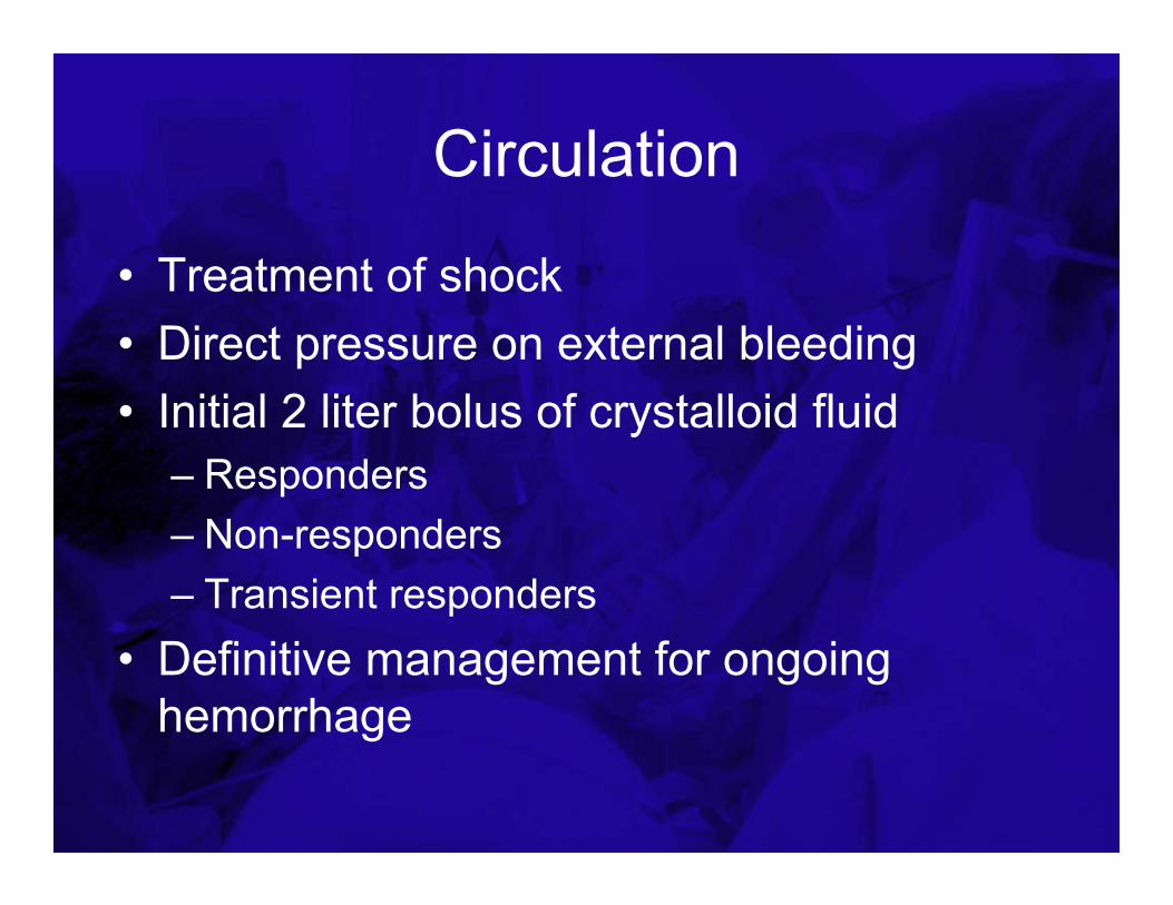
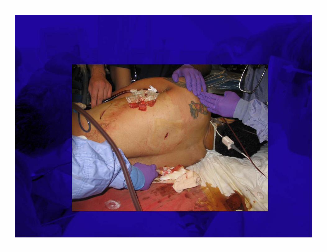
Circulation
• Treatment of shock• Direct pressure on external bleeding• Initial 2 liter bolus of crystalloid fluid
– Responders– Non-responders– Transient responders
• Definitive management for ongoing hemorrhage

Neurologic “deficit”
• Rapid assessment of neurologic status to identify life-threatening injury– Pupil size and response– Mental status (Glascow coma scale)– Motor and sensory exam

Glascow Coma Scale
• 3 – 15 point scale to assess mental status only
• Best observed response• Modified scale for children• GCS ≤ 8 is a “coma” and requires
intubation for airway protesction
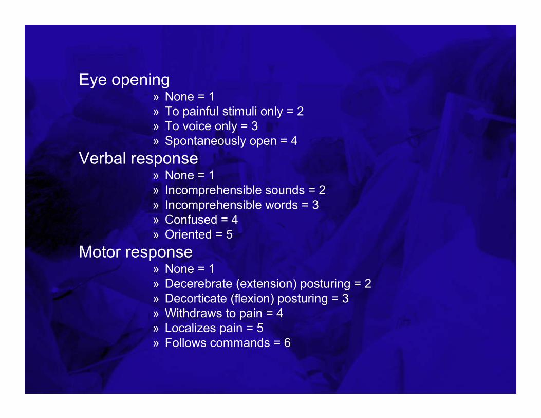
Eye opening» None = 1» To painful stimuli only = 2» To voice only = 3» Spontaneously open = 4
Verbal response» None = 1» Incomprehensible sounds = 2» Incomprehensible words = 3» Confused = 4» Oriented = 5
Motor response» None = 1» Decerebrate (extension) posturing = 2» Decorticate (flexion) posturing = 3» Withdraws to pain = 4» Localizes pain = 5» Follows commands = 6
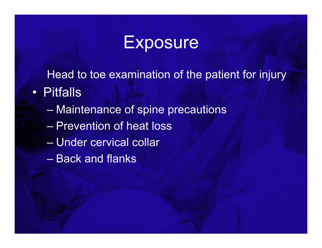
Exposure
Head to toe examination of the patient for injury• Pitfalls
– Maintenance of spine precautions– Prevention of heat loss– Under cervical collar– Back and flanks
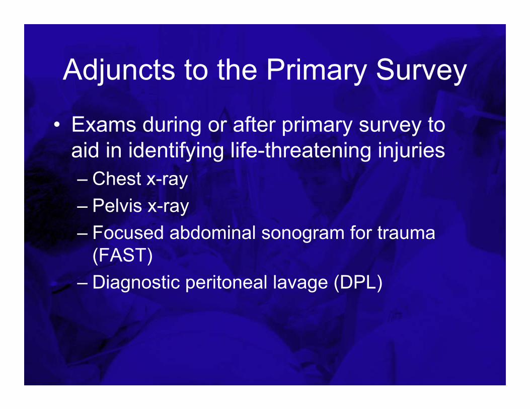
Adjuncts to the Primary Survey
• Exams during or after primary survey to aid in identifying life-threatening injuries– Chest x-ray– Pelvis x-ray– Focused abdominal sonogram for trauma
(FAST)– Diagnostic peritoneal lavage (DPL)


Secondary Surveyand Definitive Treatment
• The secondary survey is a complete head to toe evaluation of the patient
• Adjuncts to the secondary survey include CT’s, plain radiographs, blood tests
• Treatment plans, especially for multiple injuries, based on clinical status and specific injuries
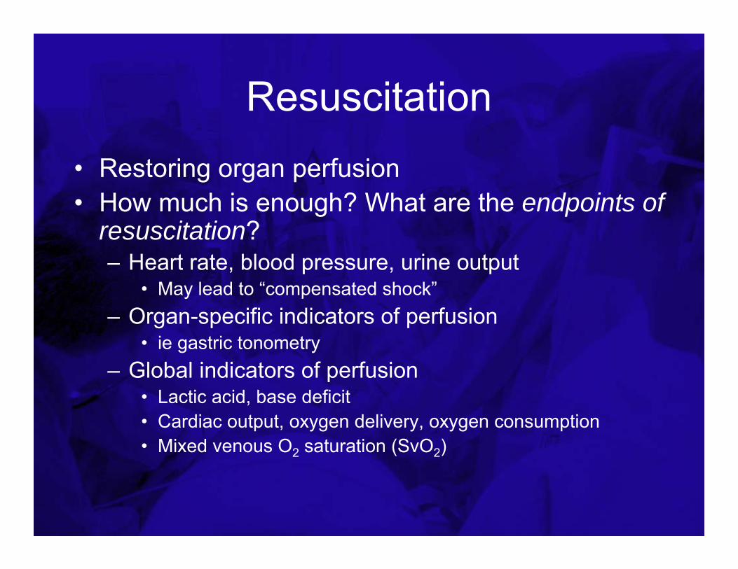
Resuscitation• Restoring organ perfusion• How much is enough? What are the endpoints of
resuscitation?– Heart rate, blood pressure, urine output
• May lead to “compensated shock”– Organ-specific indicators of perfusion
• ie gastric tonometry– Global indicators of perfusion
• Lactic acid, base deficit• Cardiac output, oxygen delivery, oxygen consumption• Mixed venous O2 saturation (SvO2)
Lactic acid and base deficit
• Initial BD and serum LA are reliable indicators of the need for ongoing resuscitation
• Time to normalization of LA and BD are predictive of MSOF and mortality

Damage-control laparotomy
• A shift from definitive management of abdominal injuries to stabilizing the patient for resuscitation
• Goals– Stop bleeding– Control contamination– Temporary abdominal closure


Critical care and rehabilitation

Questions?