Management of Splenic injury following blunt abdominal trauma · Discussion under the following...
101
Management of Splenic injury following blunt abdominal trauma Dr.D.Sirish Bharadwaj General Surgery
Transcript of Management of Splenic injury following blunt abdominal trauma · Discussion under the following...

Management of Splenic injury following blunt
abdominal traumaDr.D.Sirish Bharadwaj
General Surgery

Discussion under the following headingsIntroduction to traumaBlunt injury abdomen ‐ IntroductionMost commonly involved organsPatho‐physiology of injuryInitial management General physical examinationClassification based on hamodynamic statusMajor organ injuries

Splenic injuries:IntroductionTypesGeneral managementClinical presentationDiagnosisTreatment

INTRODUCTIONWHAT IS TRAUMA?Trauma is the study of medical problems associated with physical injury, which is an adverse effect of physical force upon a person.
Varieties:Mechanical: 1)Blunt 2)PenetratingThermalChemicalIonizing radiation etc..

“Golden Hour”ACS concept that deaths & complications are
reduced when trauma victims receive definitive treatment within the 1st hour after injury

BLUNT INJURY ABDOMENBlunt abdominal injuries carry a greater risk of morbidity and mortality than peneterating abdominal injuries.

Most commonly involved organs:
Spleen (40‐55%) Liver (35‐45%)Small bowel (5‐10%)

Pathophysiology of injury

Crushing effect: Compression from crush between solid objects such as the steering wheel/seat belt & the vertebrae
Seat belt injuries ‐ “seat belt sign” = highly correlated with intraperitoneal injury
Acceleration and deceleration forces → shear injury

10
Initial management of blunt injury abdomen

Primary survey and resuscitation:
A – AirwayB – BreathingC – CirculationD – DisabilityE – Exposure

Secondary SurveyA – AllergyM – MedicationP – Past medical historyL – Last mealE – Events of the incident

Events of the incident
Details about accidentDamage to carVelocitySteering wheel damageType of seatbelts usedAir bags deployed All patients involved in deceleration injuries and bicycle injuries should be suspected of having intraabdominal injury

Secondary survey (cont)Subsequent physical examRe‐evaluationAnalgesiaDocumentation & legal considerationsDefinitive care & transfer
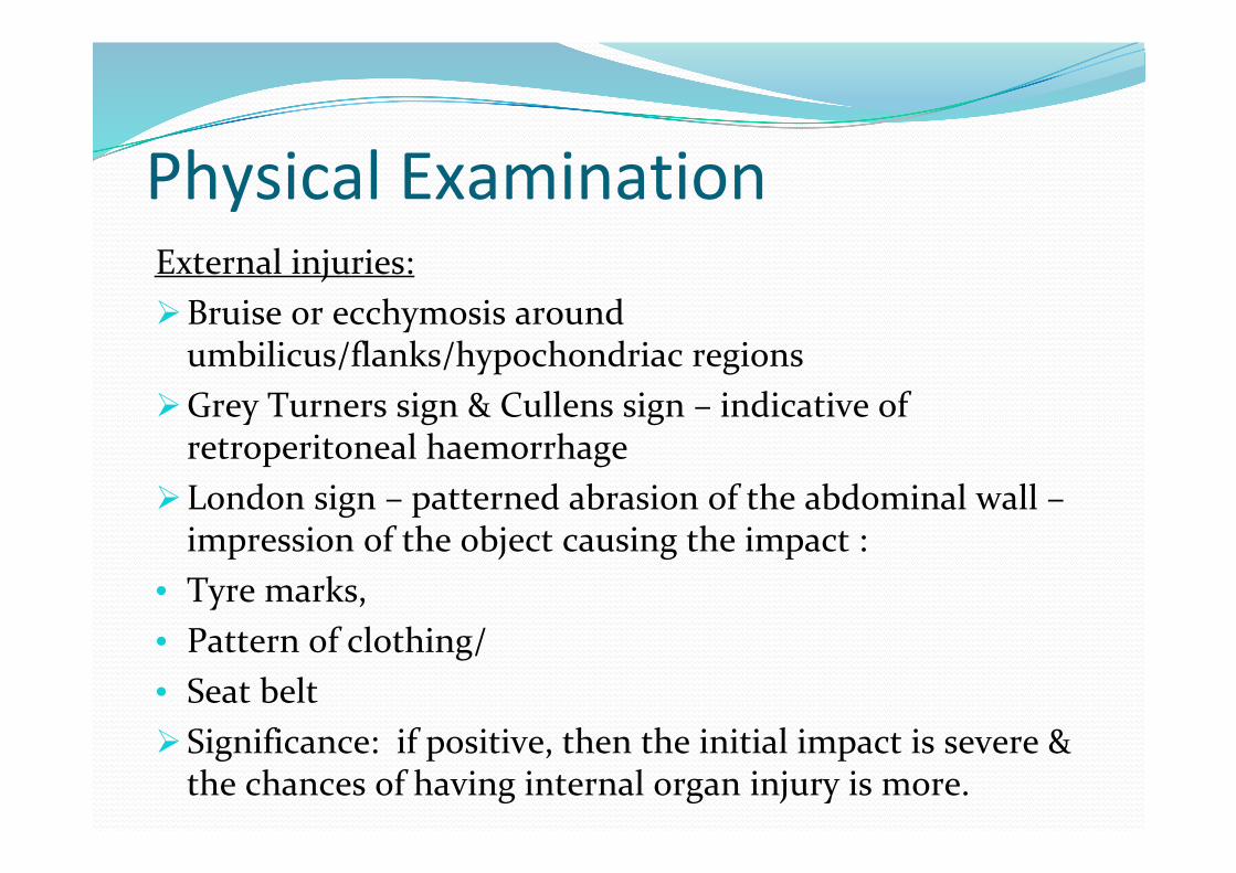
Physical ExaminationExternal injuries:Bruise or ecchymosis around umbilicus/flanks/hypochondriac regions
Grey Turners sign & Cullens sign – indicative of retroperitoneal haemorrhage
London sign – patterned abrasion of the abdominal wall – impression of the object causing the impact :
• Tyre marks,• Pattern of clothing/• Seat beltSignificance: if positive, then the initial impact is severe & the chances of having internal organ injury is more.
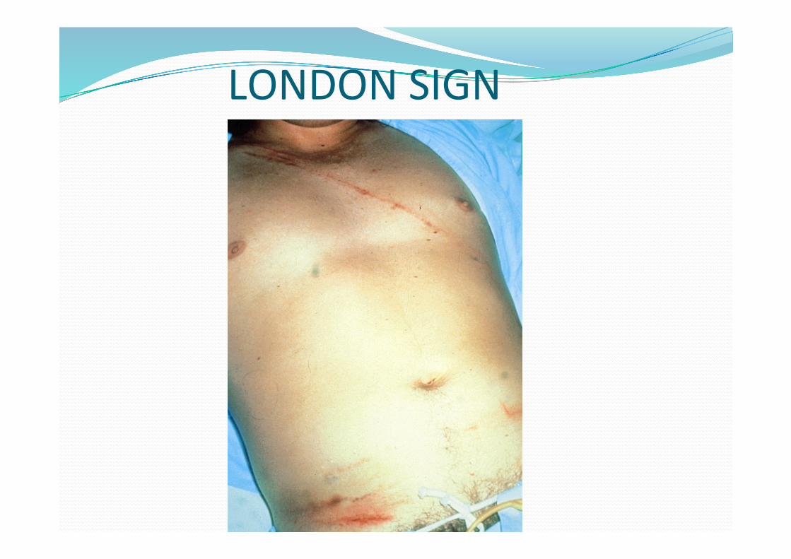
LONDON SIGN

CULLENS SIGN

GREY‐TURNERS SIGN

Abdominal findings:Distension of abdomenTenderness – suggests inflammation/haemoperitoneumRebound tendernessGuarding, rigidityFluid thrillObliteration of liver dullness – suggests perforation of hollow viscera
Increased hepatic/splenic dullnessFree fluid in abdomen detected by shifting dullnessAbsent or decreased bowel sounds

Unstable vitalsHypotension in the acute stage results from hemorrhage that is most often from a solid visceral or vascular injury.
Unexplained Hypotension associated with significant blunt abdominal trauma, one should exclude intraperitoneal hemorrhage with solid organ injury or vascular injury.

Abdominal injuries categorised into:Haemodynamically normal: Full investigations & planned treatment
Haemodynamically stable: Limited investigationsNon‐operative : Angio embolizationOperative : Which cavity?
Haemodynamically unstable: Immediate surgical correctionTrauma laparotomy

MAJOR ORGAN INJURIES

Hepatic Injuries2nd most commonly injured intraabdominal organAssociated with R 8‐12 rib fracturesSubcapsular hematomasLacerationsVascular injuries
TreatmentDamage Control ‐ Pack the liver to control bleeding & close at a later timeOperative repair
Hollow Organ InjuriesSmall or large bowel, gastric injuriesPerforation with spillage of contents into peritoneal cavitySigns & symptoms of peritonitis
TreatmentSurgical repairDiversion of the injured bowel with re‐anastamosis at a later time
TPN

SPLENIC INJURIES

Introduction to splenic injuriesAssociated with left rib 10‐12 fxs, falls, contact sports, assaults
Most commonly injured organ from blunt trauma 40% have no symptomsBleeding may be contained by capsule

Types of splenic injurySubcapsular haematomaLaceration of spleenPolar tearAvulsion of splenic pedicle

General Management of Abdominal TraumaScene survey for MOIRapid evaluation of the patientSecure airway with spinal precautionsProvide ventilatory supportHigh concentration O2Stabilize – Direct pressure, compression mattress external sheet or pelvic binder for hemorrhage
Wound managementVitals monitoringManage shockCardiac monitorRapid and safe Transport

Clinical presentationClassical features of splenic injuries: seen in severe injuries and polar tear with symptoms and signs of intraperitoneal haemorrhage.
Silent: There may be slight pain abdomen with no symptoms & signs.

Delayed rupture : seen in subcapsular haematomaMild left hypochondriac pain with no symptoms or signs at initial presentation and the injury is often missedAfter 7‐14 days the haematoma grows and ruptures and patient presents with symptoms & signs of intraperitoneal haemorrhage.

Classical features of major splenic traumaGeneral symptoms and signs of internal haemorrhage: Pallor Tachycardia Hypotension Restlessness Sweating Deep, sighing respiration Cold, clammy extremities Collapsed veins

There may be cutaneous bruising in the left upper quadrant of abdomen.
Increasing left upper abdominal pain & tendernessIncreasing left upper abdominal guarding & rigidityIncreasing abdominal distension
Symptoms & Signs of splenic injury

(cont)Diminished or absent bowel sounds.Kehr’s sign ‐ Referred pain in the left shoulder on elevation of foot end due to irritation of the adjacent diaphragm
Ballance’s sign – fixed dullness in the left flank due to clotted blood around the spleen and shifting dullness in the right flank due to unclotted blood in the peritoneal cavity.

DIAGNOSIS 1. Laboratory tests2. Radiological investigations

Lab tests:
Complete haemogram – May have ↓ Hct, ↑ WBCinitially Hb% may be normal for 6 hrs,
May have, ↑ lactate, LFTs, lipase, toxicology screen.
36
Radiological investigationsTo determine if there is hemoperitoneum or organ injury requiring surgical repair
X‐ray abdomen erectFAST DPL CT
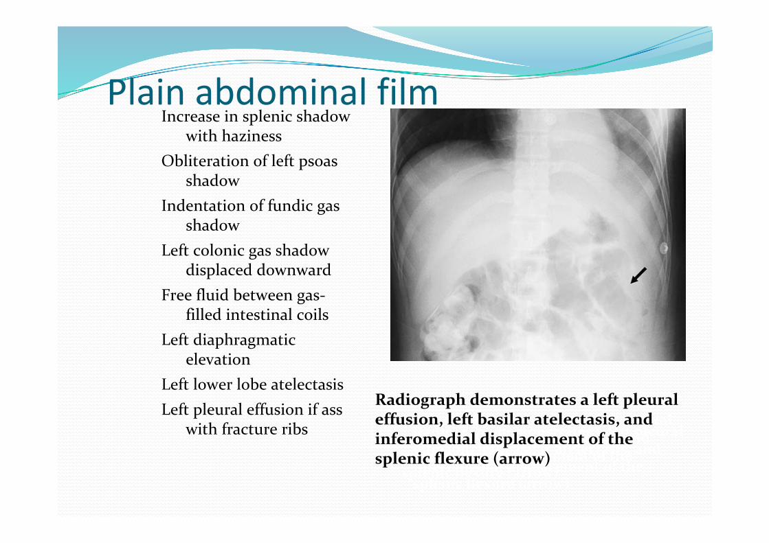
Plain abdominal filmIncrease in splenic shadow
with hazinessObliteration of left psoas
shadowIndentation of fundic gas
shadowLeft colonic gas shadow
displaced downwardFree fluid between gas‐
filled intestinal coilsLeft diaphragmatic
elevationLeft lower lobe atelectasisLeft pleural effusion if ass
with fracture ribs Radiograph demonstrates a left pleural effusion, left basilar atelectasis, and inferomedial displacement of thesplenic flexure (arrow)
Radiograph demonstrates a left pleural effusion, left basilar atelectasis, and inferomedial displacement of thesplenic flexure (arrow)
Radiograph demonstrates a left pleural effusion, left basilar atelectasis, and inferomedial displacement of thesplenic flexure (arrow)

FASTFocused assessment with sonography for trauma (FAST) To diagnose free intraperitoneal blood after blunt trauma4 areas: Perihepatic & hepato‐renal space (Morrison’s pouch) Perisplenic Pelvis (Pouch of Douglas/rectovesical pouch) Pericardium (subxiphoid)
sensitivity 60 to 95% for detecting 100 mL ‐ 500 mL of fluid
Extended FAST (E‐FAST): Add thoracic windows to look for pneumothorax.Sensitivity 59%, specificity up to 99% for PTX (c/w CXR 20%)

39
FASTPerisplenic view
trauma.org Rosen’s Emergency Medicine, 7th ed. 2009

40
FAST
Fluid in the subphrenic space and splenorenal recess can be detected. The image shown demonstrates blood (arrow) between the spleen (S) and diaphragm (D).
41
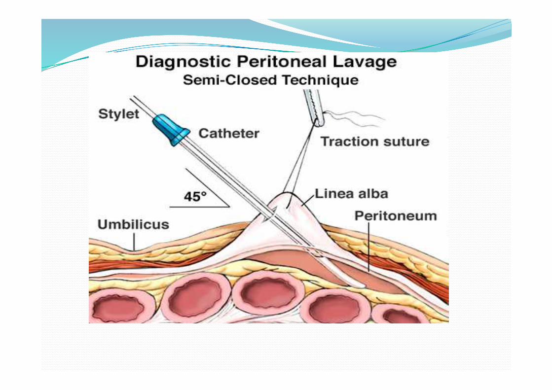
Diagnostic Peritoneal Lavage Largely replaced by FAST and CTIn blunt trauma, used to triage pt who is haemodynamically unstable and has multiple injuries with an equivocal FAST examination
Rosen’s Emergency Medicine, 7th ed. 2009

42
Diagnostic Peritoneal Lavage 1. attempt to aspirate free peritoneal blood
>10 mL positive for intraperitoneal injury2. insert lavage catheter by seldinger, semiopen, or open3. lavage peritoneal cavity with salinePositive test:
RBC count > 100,000/mm3
Rosen’s Emergency Medicine, 7th ed. 2009

43
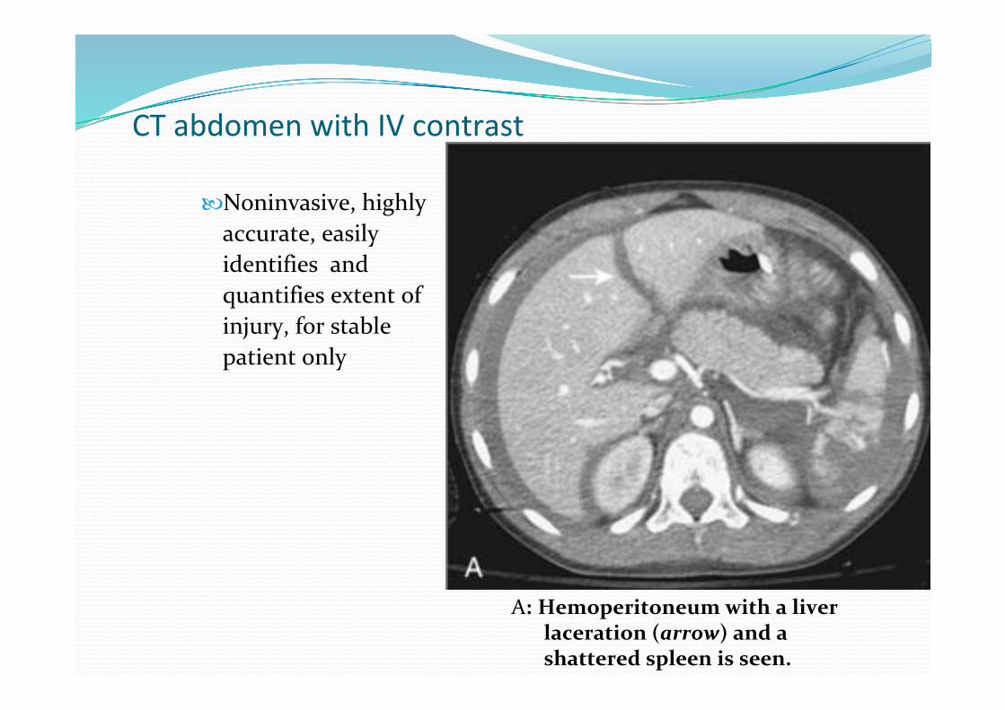
CT abdomen with IV contrast
Noninvasive, highly accurate, easily identifies and quantifies extent of injury, for stable patient only
A: Hemoperitoneum with a liver laceration (arrow) and a shattered spleen is seen.
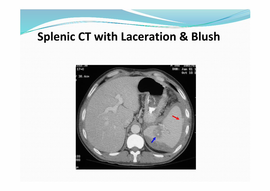
Splenic CT with Laceration & Blush

Contrast Blush ‐ Spleen
• 216 Pts – 7 yrs• 26 Pts – Contrast blush on CT scan
• Lower HgB• More likely to need op (22% vs 4%)
• Not a definite indication for operation, but indicates subset of pts who have active bleeding and may need transfusion and/or operation
Blunt Splenic Injury

AAST Splenic Injury Scale Spleen injury scale: (advance one grade for multiple injuries, up to grade III)
Grade I: Hematoma: subcapsular, < 10% of surface area Laceration: capsular tear, < 1cm parenchymal depth
Grade II: Hematoma: subcapsular, 10‐50% surface area; intraparenchymal, <5cm in
diameter Laceration: 1‐3cm parenchymal depth which does not involve a trabecular vessel
Grade III: Hematoma: subcapsular, >50% surface area or expanding; ruptured subcapsular
or parenchymal hematoma; intraparenchymal hematoma >5cm or expanding Laceration: >3cm parenchymal depth or involving trabecular vessesls
Grade IV: Laceration: laceration involving segmental or hilar vessels producing major
devascularization (>25% of spleen) Grade V: Laceration: completely shattered spleen Vascular: hilar vascular injury which devascularizes spleen

AAST Splenic Injury Scale
17‐yo boy injured on an ATV. Grade I injury with subcapsular fluid occupying less than 10% of spleen’s surface area.

AAST Splenic Injury Scale
17‐yo girl injured in an MVC. Grade II injury with laceration involving less than 3 cm of parenchymal depth

AAST Splenic Injury Scale
18‐yo boy injured playing football. Lacerations involving more than 3 cm of parenchymal depth radiating from splenic hilum ‐grade III laceration

AAST Splenic Injury Scale
16‐yo boy injured playing hockey. Fractured spleen involving more than 25%, Grade IV splenic laceration

AAST Splenic Injury Scale
12‐yo boy pedestrian struck by MV. Fractured spleen with hilar devascularization. Grade V injury.

Treatment


The unstable patient

Stabilizing the patient
IVF (crystalloid, not colloid)TransfusionPRBC/plt/FFP
Recombinant activated factor VIIRewarming if hypothermicCorrection of metabolic abnormalitiesLow tidal volume ventilation recommended (4‐6 ml/kg)

58
Damage Control Surgery
Patients with major exsanguinating injuries may not survive complex proceduresGOAL OF DCS: control hemorrhage, limit GI spillage, to prevent ischemia, infection, with abbreviated laparotomy followed by resuscitation prior to definitive repair

59
Damage Control
Waibel et al. Damage control in trauma and abdominal sepsis. Crit Care Med 2010 38:S421‐430

60
Damage Control0. Initial resuscitation1. Control of hemorrhage and contaminationControl injured vasculature, bleeding solid organsAbdominal packingAutologous blood transfusion after collecting the blood in a cell saver which washes and collects RBCs.
2. back to the ICU for resuscitation3. Definitive repair of injuries4. Definitive closure of the abdomen

61

Definitive repair

Indications for Laparotomy Unstable vital signs with strongly suspected splenic injury
Unstable patients with known or positive FAST/DPL
Unequivocal peritoneal irritation
Minor splenic injury associated with other intra‐abdominal injury requiring laparotomy
Failure of conservative management

Haemodynamically stable patient

Treatment Recommendations based on CT findingsGrade I and II injuries – admit for minimum of > 24 hours with serial exams and HCT – conservative management
Grade III injuries – admit ICU/step down unit, serial HCTS (q 4 ‐6 hrs) for a minimum of 3 times and until stable
Grade III injuries with moderate to large hemoperitoneum ‐Splenic Artery Angio‐embolization (SAE) ASAP with goal to be within 2 hours

Size of HemoperitoneumSmall Hemoperitoneum ‐ perisplenic blood or blood in Morrison’s pouch
Moderate Hemoperitoneum – blood in one or both pericolic gutters
Large hemoperitoneum – blood in one or both gutters with additional blood in pelvis

Grade IV injuries: Urgent SAE with goal to be within 2 hours
Grade V injuries: to the operating room in most circumstances
Grades I – IV: that show CT evidence of blush/pseudoaneurysm or extravasation – Urgent SAE or Laparotomy with goal to be within 2 hours

SPLENIC ARTERY EMBOLIZATION

69
Initial Mangement of a Hemodymically Stable Patient with a Blunt Spleen Injury
CT scan with IV contrast
Angio‐Embolization (within 1‐2 hours)
Grade I & II can admit to Floor
Grade III
Moderate/Large hemoperitoneum
ICU/Step‐down unit* ‐ q 4‐ 6 hr HCTs until stable X 3
Dropping HCTs for 24 hours, or need to transfuse blood
Stable HCTs for 24 hours
Can mobilize patient, transfer to floor, and give diet pending other injuries
Consider the following options:OR, angio, Repeat CT scan
No or Small hemoperitoneum
Blush, Extravisation, or Psuedoaneurysm
Floor patients: should be mobilized, diet advance and daily HCTs until stable for two days.Grade 1 and 2: minimum of 1 floor dayGrade 3‐5: minimum of 3 hospital daysSplenectomy patients require immunizations
Grade IV
Grade V

70
CONSERVATIVE MANAGEMENT
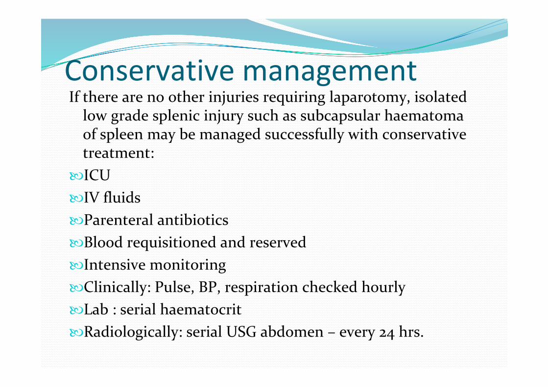
Conservative managementIf there are no other injuries requiring laparotomy, isolated low grade splenic injury such as subcapsular haematoma of spleen may be managed successfully with conservative treatment:
ICUIV fluidsParenteral antibioticsBlood requisitioned and reservedIntensive monitoringClinically: Pulse, BP, respiration checked hourlyLab : serial haematocritRadiologically: serial USG abdomen – every 24 hrs.
The patient kept under observation for 24‐48 hrs.
If patient is stable, haematoma not increasing and shows signs of regression, conservative treatment may be continued.
If haematoma is expanding and there is evidence of intraperitoneal rupture, immediate laparotomy is to be done.

SURGICAL MANAGEMENT•Splenectomy•Partial splenectomy•Spleen salvage

Splenectomy Extensive laceration or Avulsion of splenic pedicle:

Steps of splenectomy
1. Mobilization of the spleen to the midline by division of the splenophrenic ligament superiorly and the splenocolic and splenorenal ligaments at the lower pole

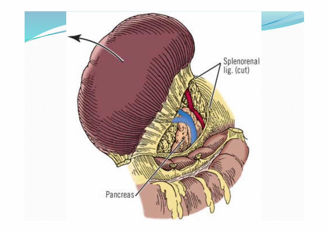

2) The short gastric vessels are then divided between ligatures or clips

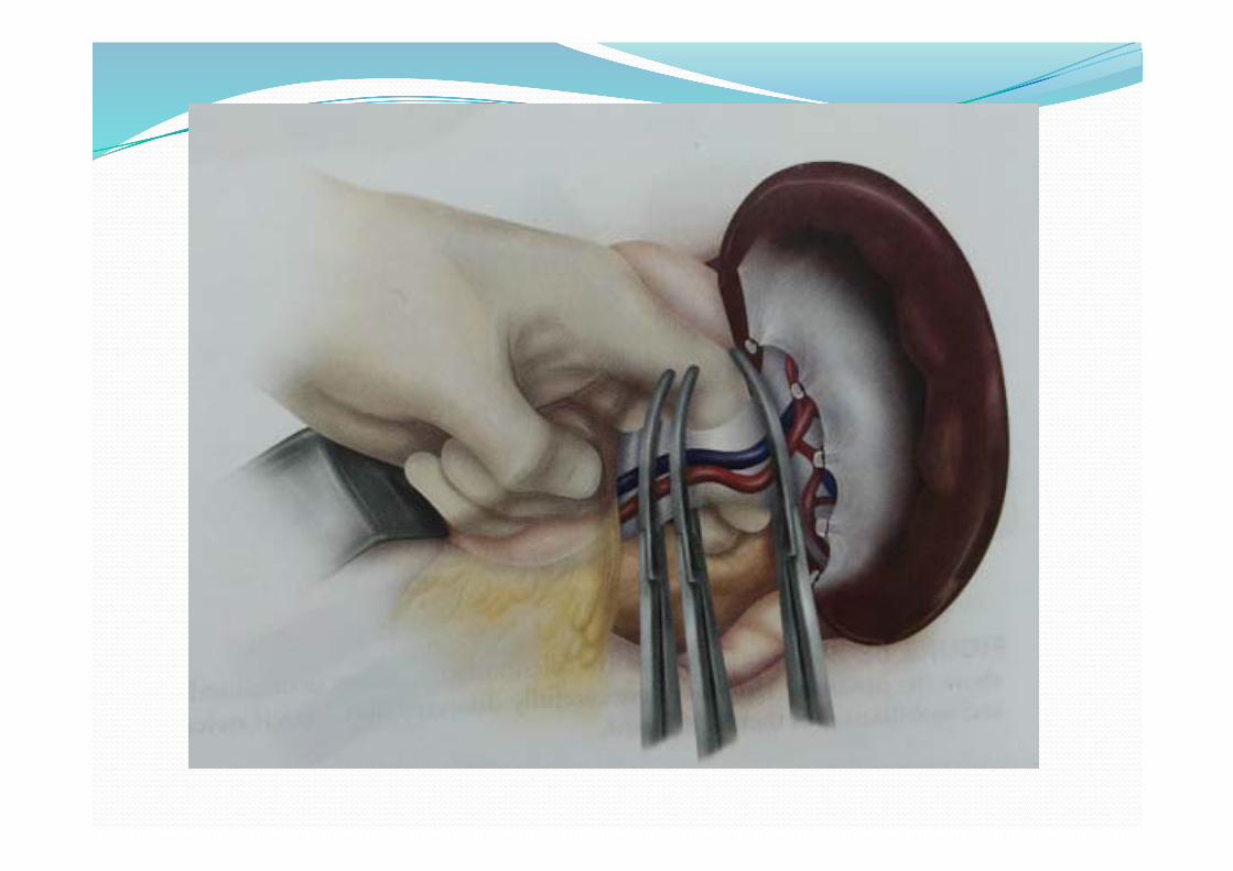
Spleen medialized and hilar dissection performed carefully with isolation of splenic vesselsSplenic hilum clamped en bloc and divided and doubly ligated proximally and once distally or splenic artery and vein are individually ligated.

Splenic slice graftA portion of the spleen – 6 pieces of 40 * 40 * 3mm may be reimplanted within the leaf of greater omentum

Splenic slice graft

Splenic salvage Being practised from 1980, in view of development of Post splenectomy syndrome &OPSI, following splenectomy.
Splenorrhaphy ( repair of spleen)
The importance of spleen with regards to its immune function started the recent trend to conserve the spleen unless it is,
extensively shattered and bleeding is uncontrollabe

Types of splenorrhaphy Topical methodsEnvelopment in meshPledgeted suture repair

Indications Small capsular tear: Bleeding controlled with local haemostatic agents like:Oxidized regenerated cellulose (surgicel) Fibrin glueElectrocauteryArgon beam coagulationApplication of thrombin soaked gelatin foam spongesIf it fails: – bleeding from the splenic tear controlled with a series of mattress sutures ‐ Pledgeted suture repair


Polar tear managed with splenorrhaphy or partial splenectomy followed by approximation of edges with a series of mattress sutures

Placement of the spleen in a mesh
Envelopment of the injured spleen in absorbable mesh, and to compress the splenic tissue and capsule back together, aided by purse–string sutures.

Envelopment of the injured spleen in absorbable mesh
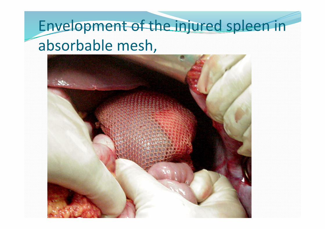
Envelopment of the injured spleen in absorbable mesh,

Role of Laparoscopy
Most useful to evaluate penetrating wounds to thoracoabdominal region in stable pt
Not a substitute for open laparotomy.
Rosen’s Emergency Medicine, 7th ed. 2009

92


Post operative complicationsImmediate: Bleeding from the remaining spleen
Early:Haematemesis Sub‐diaphragmatic abscessLeft basal atelactasisLeft pleural effusionThrombocytosisPancreatitis/abscess/fistula Gastric fistula
Late:Post splenectomy sepsis with S.pneumoniae, N.meningitides,
H.influenzae, E.coliOPSI – opportunist post – splenectomy infection

In‐Hospital Recommendations
DVT prophylaxisSequential compression devices upon admissionStable HCT for 48 ‐ 72 hours and no other contraindications‐ Strongly consider starting chemoprophylaxis (Low molecular weight heparin is preferred)

Post operative prophylaxisPt requires vaccinations prior to dischargeStreptococcus pneumoniae
• Pneumovax 23Haemophilus influenzae type B
• Hib vaccineNeisseria meningitidis
• Quadravalent meningococcal/diphtheria conjugate
• Prophylactic antibiotics controversialMost centers use penicillin
98
Follow up and discharge adviseAvoid contact sports for 2‐4 monthsImmediate treatment of infectionsPneumococcal and meningococcal vaccinations every 5 yearsHib every ten yearsYearly infuenza vaccinations Regular followups (weekly) for those who were placed on conservative treatment.

THANK YOU

REFERENCESBiffl WL, Moore EE. Management guidelines for penetrating abdominal trauma.
Curr Opin Crit Care 2010;16:609‐617 Waibel BH, Rotondo MF. Damage control in trauma and abdominal sepsis. Crit
Care Med. 2010 Sep;38(9 Suppl):S421‐30.Marx: Rosen’s Emergency Medicine, 7th ed. 2009 Mosby The effects of splenic artery embolization on nonoperative management of blunt
splenic injury: a 16‐year experience.Sabe AA, Claridge JA, Rosenblum DI, Lie K, Malangoni MA. J Trauma. 2009
Sep;67(3):565‐72; discussion 571‐2.PMID: 19741401 [ Blunt splenic injuries: dedicated trauma surgeons can achieve a high rate of
nonoperative success in patients of all ages.Myers JG, Dent DL, Stewart RM, Gray GA, Smith DS, Rhodes JE, Root HD, Pruitt
BA Jr, Strodel WE.J Trauma. 2000 May;48(5):801‐5; discussion 805‐6.PMID: 10823522 [PubMed –

Improved outcome of adult blunt splenic injury: a cohort analysis.Rajani RR, Claridge JA, Yowler CJ, Patrick P, Wiant A, Summers JI, McDonald AA, Como JJ,
Malangoni MA.Surgery. 2006 Oct;140(4):625‐31; discussion 631‐2.PMID: 17011910 Improved success in nonoperative management of blunt splenic injuries: embolization of splenic
artery pseudoaneurysms.Davis KA, Fabian TC, Croce MA, Gavant ML, Flick PA, Minard G, Kudsk KA, Pritchard FE.J Trauma. 1998 Jun;44(6):1008‐13; discussion 1013‐5.PMID: 9637156 Use of splenic artery embolization as an adjunct to nonsurgical management of blunt splenic
injury.Liu PP, Lee WC, Cheng YF, Hsieh PM, Hsieh YM, Tan BL, Chen FC, Huang TC, Tung CC.J Trauma. 2004 Apr;56(4):768‐72; discussion 773.PMID: 15187739 Blunt splenic injury in adults: Multi‐institutional Study of the Eastern Association for the
Surgery of Trauma.Peitzman AB, Heil B, Rivera L, Federle MB, Harbrecht BG, Clancy KD, Croce M, Enderson BL,
Morris JA, Shatz D, Meredith JW, Ochoa JB, Fakhry SM, Cushman JG, Minei JP, McCarthy M, Luchette FA, Townsend R, Tinkoff G, Block EF, Ross S, Frykberg ER, Bell RM, Davis F 3rd, Weireter L, Shapiro MB. J Trauma. 2000 Aug;49(2):177‐87; discussion 187‐9.PMID: 10963527 [PubMed ‐ indexed for MEDLINE]