Management of inferior vena cava thrombosis after blunt ... · Inferior vena cava (IVC) thrombosis...
4
Korean J Hepatobiliary Pancreat Surg 2014;18:97-100 http://dx.doi.org/10.14701/kjhbps.2014.18.3.97 Case Report Management of inferior vena cava thrombosis after blunt liver injury Kyung-Yun Kim, Byung-Jun So, and Dong-Eun Park Department of Surgery, Wonkwang University School of Medicine, Iksan, Korea Inferior vena cava (IVC) thrombosis after traumatic liver injury is an extremely rare condition, and only 12 cases have been reported in the English literature since 1911. We report a case of a 26-year-old man who presented with IVC thrombosis after blunt liver injury. IVC thrombosis was incidentally detected by computed tomography 15 days after con- servative management of blunt liver injury. The patient denied any symptoms of thrombophlebitis and did not have any evidence of hypercoagulable state. We placed an IVC filter via the right jugular vein and started the anticoagulation treatment. The patient recovered successfully without operative treatment and IVC thrombosis disappeared completely two months later. We suggest that that the possibility of IVC thrombosis should be considered in patients with a large hematoma of the liver, which may cause compression of the IVC. (Korean J Hepatobiliary Pancreat Surg 2014;18:97-100) Key Words: Liver injury, Hematoma, Inferior vena cava, Thrombosis Received: July 17, 2014; Revised: August 14, 2014; Accepted: August 20, 2014 Corresponding author: Dong-Eun Park Department of Surgery, Wonkwang University School of Medicine, 344-2, Shinyoung-dong, Iksan 570-711, Korea Tel: +82-63-859-1490, Fax: +82-63-855-2386, E-mail: [email protected] This paper was supplied by Wonkwang University 2012. Copyright Ⓒ 2014 by The Korean Association of Hepato-Biliary-Pancreatic Surgery Korean Journal of Hepato-Biliary-Pancreatic Surgery ∙ pISSN: 1738-6349ㆍeISSN: 2288-9213 INTRODUCTION Inferior vena cava (IVC) thrombosis after hepatic trau- ma is an extremely rare condition, and only 12 cases have been reported in the English literature since 1911. 1-8 Various mechanisms for the origin of this condition have been suggested; but there is no well-established evidence and treatment. We herein report a case of IVC thrombosis after blunt liver injury that was managed by IVC filter insertion and anticoagulation therapy. CASE A 26-year-old man was taken to the emergency room of a University Hospital after high- speed motor vehicle collision. The patient complained of bilateral knee pain and right upper quadrant abdominal pain. On arrival, the patient was alert (Glasgow coma scale score of 14) and vital signs were as follows: the patient was normothermic, heart rate was 70 beats/min, respiratory rate was 20/min, and blood pressure was 105/60 mmHg. Physical examina- tion was unremarkable and the findings of the initial blood tests were as follows: hemoglobin 15.0 g/dl, white blood cell count 8,130/mm 3 , platelet count 192,000/mm 3 , aspartate transaminase 491 IU/L, alanine transaminase 495 IU/L, amylase 62 IU/L, and lipase 82 IU/L. Routine trau- ma series X-rays were normal except for multiple frac- tures of the right ribs. However, abdominal computed to- mography (CT) showed hepatic laceration with paren- chymal hematoma in the segments 4, 5, 7, and 8, and moderate amount of intra-abdominal fluid collection (Fig. 1). Because the patient was hemodynamically stable and he did not have any other visceral organ injuries, he was managed conservatively in the intensive care unit. On fol- low-up day 2, there were no changes in the vital signs and there was no decrease in hemoglobin and hematocrit levels. As the patient also did not have any specific symp- toms, we continued the conservative treatment. The fol- low-up abdominal CT on day 5 showed a slight decrease in the size of parenchymal hematoma and intra-abdominal fluid collection (Fig. 2). At this time point, he could walk by himself and could be given a regular diet.
Transcript of Management of inferior vena cava thrombosis after blunt ... · Inferior vena cava (IVC) thrombosis...

Korean J Hepatobiliary Pancreat Surg 2014;18:97-100http://dx.doi.org/10.14701/kjhbps.2014.18.3.97 Case Report
Management of inferior vena cava thrombosisafter blunt liver injury
Kyung-Yun Kim, Byung-Jun So, and Dong-Eun Park
Department of Surgery, Wonkwang University School of Medicine, Iksan, Korea
Inferior vena cava (IVC) thrombosis after traumatic liver injury is an extremely rare condition, and only 12 cases have been reported in the English literature since 1911. We report a case of a 26-year-old man who presented with IVC thrombosis after blunt liver injury. IVC thrombosis was incidentally detected by computed tomography 15 days after con-servative management of blunt liver injury. The patient denied any symptoms of thrombophlebitis and did not have any evidence of hypercoagulable state. We placed an IVC filter via the right jugular vein and started the anticoagulation treatment. The patient recovered successfully without operative treatment and IVC thrombosis disappeared completely two months later. We suggest that that the possibility of IVC thrombosis should be considered in patients with a large hematoma of the liver, which may cause compression of the IVC. (Korean J Hepatobiliary Pancreat Surg 2014;18:97-100)
Key Words: Liver injury, Hematoma, Inferior vena cava, Thrombosis
Received: July 17, 2014; Revised: August 14, 2014; Accepted: August 20, 2014Corresponding author: Dong-Eun ParkDepartment of Surgery, Wonkwang University School of Medicine, 344-2, Shinyoung-dong, Iksan 570-711, KoreaTel: +82-63-859-1490, Fax: +82-63-855-2386, E-mail: [email protected] paper was supplied by Wonkwang University 2012.
Copyright Ⓒ 2014 by The Korean Association of Hepato-Biliary-Pancreatic SurgeryKorean Journal of Hepato-Biliary-Pancreatic Surgery ∙ pISSN: 1738-6349ㆍeISSN: 2288-9213
INTRODUCTION
Inferior vena cava (IVC) thrombosis after hepatic trau-
ma is an extremely rare condition, and only 12 cases have
been reported in the English literature since 1911.1-8
Various mechanisms for the origin of this condition have
been suggested; but there is no well-established evidence
and treatment.
We herein report a case of IVC thrombosis after blunt
liver injury that was managed by IVC filter insertion and
anticoagulation therapy.
CASE
A 26-year-old man was taken to the emergency room
of a University Hospital after high- speed motor vehicle
collision. The patient complained of bilateral knee pain
and right upper quadrant abdominal pain. On arrival, the
patient was alert (Glasgow coma scale score of 14) and
vital signs were as follows: the patient was normothermic,
heart rate was 70 beats/min, respiratory rate was 20/min,
and blood pressure was 105/60 mmHg. Physical examina-
tion was unremarkable and the findings of the initial
blood tests were as follows: hemoglobin 15.0 g/dl, white
blood cell count 8,130/mm3, platelet count 192,000/mm3,
aspartate transaminase 491 IU/L, alanine transaminase 495
IU/L, amylase 62 IU/L, and lipase 82 IU/L. Routine trau-
ma series X-rays were normal except for multiple frac-
tures of the right ribs. However, abdominal computed to-
mography (CT) showed hepatic laceration with paren-
chymal hematoma in the segments 4, 5, 7, and 8, and
moderate amount of intra-abdominal fluid collection (Fig.
1).
Because the patient was hemodynamically stable and he
did not have any other visceral organ injuries, he was
managed conservatively in the intensive care unit. On fol-
low-up day 2, there were no changes in the vital signs
and there was no decrease in hemoglobin and hematocrit
levels. As the patient also did not have any specific symp-
toms, we continued the conservative treatment. The fol-
low-up abdominal CT on day 5 showed a slight decrease
in the size of parenchymal hematoma and intra-abdominal
fluid collection (Fig. 2). At this time point, he could walk
by himself and could be given a regular diet.

98 Korean J Hepatobiliary Pancreat Surg Vol. 18, No. 3, August 2014
Fig. 1. Computed tomography scan on admission showing mul-tiple liver lacerations.
Fig. 2. Follow-up abdominal CT on the follow-up day 5 showing a slight decrease in the size of parenchymal hematoma.
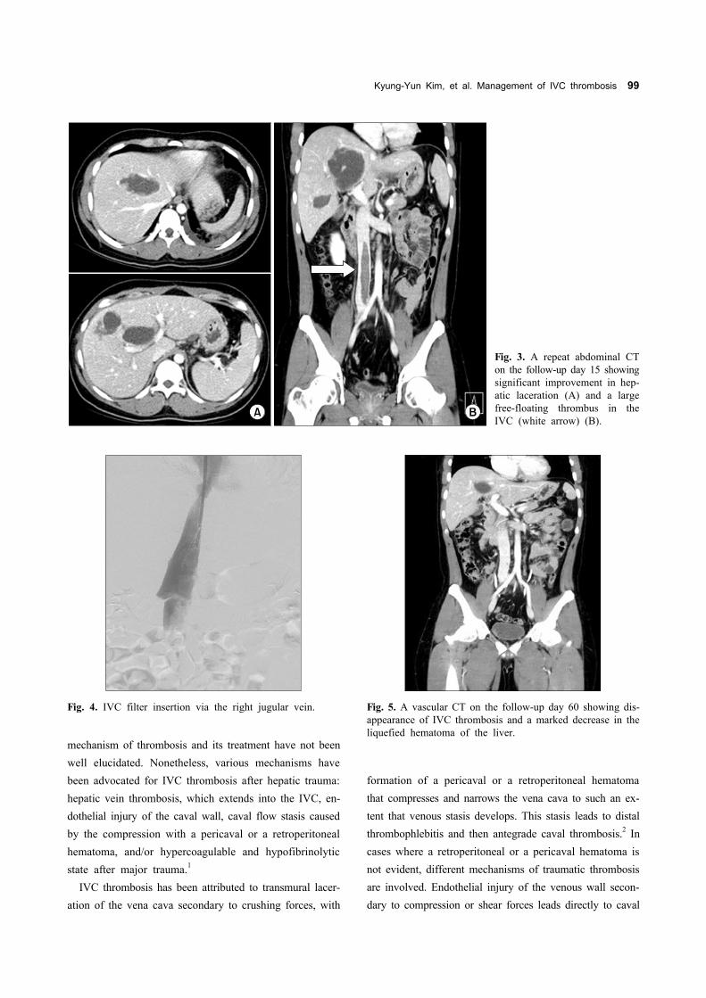
A repeat abdominal CT on day 15 demonstrated consid-
erable improvement in hepatic laceration and hematoma,
but a large thrombus in the IVC (about 10 cm in length,
from L1 to L5 spines) was detected (Fig. 3). The patient
did not show any symptoms and signs of thrombophlebitis
such as bilateral leg edema, skin color changes, and pain
except for vague nonspecific upper abdominal pain. For
evaluation of any hypercoagulable state, the specific blood
test was performed, but no evidence of hypercoagulability
was obtained.
One day later, an IVC filter was placed in the supra-
renal IVC via the right jugular vein to protect against the
risk of pulmonary embolism (Fig. 4). Localized intra-
thrombus lytic therapy was considered; but it was not per-
formed due to concerns with respect to the underlying liv-
er laceration and hematoma. Instead, we started oral anti-
coagulation therapy and performed close observation of
bleeding, but no sign of bleeding was detected. The pa-
tient was discharged from hospital at 1 month after adm-
ission. On follow-up abdominal CT performed 2 months lat-
er, IVC thrombus had disappeared completely (Fig. 5).
DISCUSSION
IVC thrombosis after hepatic trauma is an extremely
rare condition. Since it is a rare entity, the definitive
Kyung-Yun Kim, et al. Management of IVC thrombosis 99
Fig. 4. IVC filter insertion via the right jugular vein.
Fig. 3. A repeat abdominal CT on the follow-up day 15 showingsignificant improvement in hep-atic laceration (A) and a large free-floating thrombus in the IVC (white arrow) (B).
Fig. 5. A vascular CT on the follow-up day 60 showing dis-appearance of IVC thrombosis and a marked decrease in theliquefied hematoma of the liver.
mechanism of thrombosis and its treatment have not been
well elucidated. Nonetheless, various mechanisms have
been advocated for IVC thrombosis after hepatic trauma:
hepatic vein thrombosis, which extends into the IVC, en-
dothelial injury of the caval wall, caval flow stasis caused
by the compression with a pericaval or a retroperitoneal
hematoma, and/or hypercoagulable and hypofibrinolytic
state after major trauma.1
IVC thrombosis has been attributed to transmural lacer-
ation of the vena cava secondary to crushing forces, with
formation of a pericaval or a retroperitoneal hematoma
that compresses and narrows the vena cava to such an ex-
tent that venous stasis develops. This stasis leads to distal
thrombophlebitis and then antegrade caval thrombosis.2 In
cases where a retroperitoneal or a pericaval hematoma is
not evident, different mechanisms of traumatic thrombosis
are involved. Endothelial injury of the venous wall secon-
dary to compression or shear forces leads directly to caval

100 Korean J Hepatobiliary Pancreat Surg Vol. 18, No. 3, August 2014
mural thrombosis resulting in occlusion.2 Hepatic paren-
chymal injury may cause hepatic vein thrombosis that
eventually extends into the IVC; however, this mechanism
is most likely to be present in cases with the Budd−Chiari syndrome. Hypercoagulability associated with sup-
pression of fibrinolysis is a normal physiologic response
after trauma.3 Alterations in the fibrinolytic system are
concurrent with this hypercoagulability. After the initial
hyperfibrinolytic state, the overall fibrinolytic activity is
strikingly reduced. Based on the above-mentioned mecha-
nisms, hypercoagulability is likely to be the triggering fac-
tor for the development of IVC thrombosis.
In the present case, it was difficult to identify the cause
of IVC thrombosis because the CT scan did not show a
retroperitoneal hematoma around the IVC or a thrombus
in the hepatic vein causing caval flow stasis. But, it
showed huge hepatic parenchymal hemorrhage extending
into the retrohepatic portion of the IVC and injury to the
caval wall.
The clinical manifestations of IVC thrombosis after
hepatic trauma are unclear and it unusually presents as
vague, nonspecific abdominal and lower back pain.4 The
symptoms and signs of hepatic trauma are also non-
specific unless there are other associated injuries. In the
present case, there were no distinctive symptoms and
signs in spite of a large thrombus in the IVC. Therefore,
regular interval follow-up CT can be the most valuable
diagnostic tool for detecting IVC thrombosis.
Hamamoto et al. advocated that massive IVC thrombus
prevents the venous drainage from the hepatic veins, re-
sulting in progressive liver congestion, failure, and ulti-
mately death.1 Especially, the condition with massive ex-
tension of IVC thrombus into the suprahepatic level such
as Budd-Chiari syndrome is more dangerous. They sug-
gested that it is important to remove the IVC thrombus
and decompress the congested liver as early as possible
before irreversible liver failure occurs. In contrast, Agos
et al. suggested that timely insertion of an IVC filter for
pulmonary embolism prophylaxis is necessary.5
In our opinion, a massive thrombus extending to the
suprahepatic level needs emergency thrombectomy; other-
wise non-operative treatment such as IVC filter and/or an-
ticoagulation regimen can be chosen depending on the
situation. In the present case, there was a large floating
thrombus in the IVC; but it did not extend to the supra-
hepatic level.
In conclusion, we propose that the possibility of IVC
thrombosis should be considered in patients with severe
hepatic trauma above grade III (AAST liver injury scale)
and regular follow-up CT can be an important diagnostic
method to make an early diagnosis of IVC thrombosis.
The IVC filter with/without anticoagulation treatment is
likely to be the best initial option for prophylaxis of pul-
monary embolism if it has not extended to the supra-
hepatic level such as in Budd-Chiari syndrome.
REFERENCES
1. Hamamoto M, Kobayashi T, Kodama H, Nakamitsu A, Sasaki M, Kuroo Y. Thrombectomy under cardiopulmonary bypass for inferior vena cava thrombosis induced by liver injury. Ann Vasc Dis 2013;6:751-755.
2. Campbell DN, Liechty RD, Rutherford RB. Traumatic thrombo-sis of the inferior vena cava. J Trauma 1981;21:413-415.
3. Kimoto T, Kohno H, Uchida M, Yamanoi A, Yamamoto A, Nagasue N, et al. Inferior vena caval thrombosis after traumatic liver injury. HPB Surg 1998;11:111-116.
4. Nagy KK, Duarte B. Post-traumatic inferior vena caval thrombo-sis: case report. J Trauma 1990;30:218-221.
5. Boggi U, Vistoli F, Del Chiaro M, Signori S, Sgambelluri F, Roncella M, et al. Extracorporeal repair and liver autotransp-lantation after total avulsion of hepatic veins and retrohepatic in-ferior vena cava injury secondary to blunt abdominal trauma. J Trauma 2006;60:405-406.
6. Patel NH, Bradshaw B, Meissner MH, Townsend MF. Posttrau-matic Budd-Chiari syndrome treated with thrombolytic therapy and angioplasty. J Trauma 1996;40:294-298.
7. Knudson MM, Collins JA, Goodman SB, McCrory DW. Thro-mboembolism following multiple trauma. J Trauma 1992;32:2-11.
8. Takeuchi M, Maruyama K, Nakamura M, Chikusa H, Yoshida T, Muneyuki M, et al. Posttraumatic inferior vena caval thrombo-sis: case report and review of the literature. J Trauma 1995;39: 605-608.