Management of Devastating Ocular Trauma- … Trauma Registry from OIF sustained trauma to the head,...
19
Western University Scholarship@Western Surgery Publications Surgery 6-2010 Management of Devastating Ocular Trauma-- Experience of Maxillofacial Surgeons Deployed to a Forward Field Hospital M. J. Ansell Queen Alexandra Hospital, Portsmouth, UK J. Breeze Centre for Defence Medicine, Birmingham, UK Vivian C. McAlister e University of Western Ontario, [email protected] M. D. Williams Eastbourne District General Hospital, East Sussex, UK Follow this and additional works at: hp://ir.lib.uwo.ca/surgerypub Part of the Ophthalmology Commons , and the Surgery Commons Citation of this paper: Ansell, M. J.; Breeze, J.; McAlister, Vivian C.; and Williams, M. D., "Management of Devastating Ocular Trauma--Experience of Maxillofacial Surgeons Deployed to a Forward Field Hospital" (2010). Surgery Publications. Paper 53. hp://ir.lib.uwo.ca/surgerypub/53
Transcript of Management of Devastating Ocular Trauma- … Trauma Registry from OIF sustained trauma to the head,...

Western UniversityScholarship@Western
Surgery Publications Surgery
6-2010
Management of Devastating Ocular Trauma--Experience of Maxillofacial Surgeons Deployed toa Forward Field HospitalM. J. AnsellQueen Alexandra Hospital, Portsmouth, UK
J. BreezeCentre for Defence Medicine, Birmingham, UK
Vivian C. McAlisterThe University of Western Ontario, [email protected]
M. D. WilliamsEastbourne District General Hospital, East Sussex, UK
Follow this and additional works at: http://ir.lib.uwo.ca/surgerypub
Part of the Ophthalmology Commons, and the Surgery Commons
Citation of this paper:Ansell, M. J.; Breeze, J.; McAlister, Vivian C.; and Williams, M. D., "Management of Devastating Ocular Trauma--Experience ofMaxillofacial Surgeons Deployed to a Forward Field Hospital" (2010). Surgery Publications. Paper 53.http://ir.lib.uwo.ca/surgerypub/53

Management of Devastating Ocular Trauma - experience of Maxillofacial
Surgeons deployed to a Forward Field Hospital
Ansell M J 1, Breeze J 2, McAlister VC 3 Williams M D 4
1 Trainee in Maxillofacial Surgery, Queen Alexandra Hospital, Portsmouth
2 Trainee in Maxillofacial Surgery, Centre for Defence Medicine, Birmingham
3 General Surgeon, Canadian Forces Medical Service
4 Consultant Maxillofacial Surgeon, Eastbourne District General Hospital, Kings Drive,
Eastbourne, East Sussex, Deployed Consultant Role 3 Hospital Kandahar
Keywords
Ocular
Trauma
Military
Evisceration
Enucleation
Corresponding Author
Major Mark Ansell RAMC

Abstract
Combat related eye injuries continue to increase in frequency and are generally secondary
to Improvised Explosive Devices. Many ocular injuries are potentially preventable by the
wearing of ballistic eye protection. The management of penetrating eye trauma is normally
outside the routine practice of maxillofacial surgeons in the UK. The aim of this paper is to
describe the surgical techniques used in the modern management of devastating ocular
trauma including selected case examples managed by British military maxillofacial
surgeons deployed to Afghanistan.

Introduction
The incidence of military head, face and neck injuries has increased in modern conflict in
comparison to that seen in the last century. Owens et al [1] reported that 29% of all battle
wounds during Operation Iraqi Freedom (OIF) and Operation Enduring Freedom (OEF)
involved the head and neck compared with 16% during the Vietnamese War. Additionally,
Wade et al [2] reported that 39% of battle-injured patients in the Navy-Marine Corps
Combat Trauma Registry from OIF sustained trauma to the head, face, and neck.
An increasing trend in the incidence of ocular combat injuries has also been well
documented. The reported incidence in the 19th century was 1% [3], which rose to
between 2% and 2.5% during World Wars I and II [3]. Incidences of ocular injury in the 21st
century by US forces reveal an incidence between 6% [1, 2] and 10% [4]. Mader et al [4]
found that blast fragmentation from munitions caused 82% of all injuries in US servicemen
deployed on OIF, the most common single cause of which was the improvised explosive
device (IED) (51% of all injuries).
The ocular surface area represents only 0.27% of the body surface area. However the eye
casualty rate in combat is 20 to 50 times greater than expected based on the body surface
area [3]. Thoraco- abdominal protection provided by modern body armour has played its
part in increasing the survivability of many previously fatal injuries, shifting the area of
wounding to the exposed extremities, including the face and eyes. IED’s produce multiple
high-velocity projectiles that cover a large body surface area but are potentially stopped by
simple eye protection. The benefit of eye protection is demonstrated in Figures 1 and 2,
showing two coalition servicemen injured on a foot patrol by an IED. Figure 1 was wearing
ballistic eye protection and received no significant injury to his eyes. Figure 2, who had
momentarily removed his eye protection, received shrapnel spray to his face with complete

rupture of his left eye and scleral perforation of his right eye. To date no author has
definitively published data comparing the incidence of eye injuries in different coalition
forces wearing different types of eye protection or between those wearing no ocular
protection.
Figure 1: Coalition serviceman hit by an IED on foot patrol wearing issued eye protection
Figure 2: Coalition servicemen in same patrol not wearing eye protection
The British Maxillofacial Cadre has deployed to the multinational hospital in Kandahar for
two separate blocks of one year each, with consecutive consultant surgeons deploying for

two months at a time. The second block was for a 21- month period, between 01 Feb 2007
and 31 Oct 2008, on which this paper is based. Casualties treated included coalition
servicemen, local police, army and civilians as well as hostile forces. Great emphasis is
placed on the multidisciplinary nature of the team, with surgeons representing the
specialities of general surgery, orthopaedic/trauma surgery and maxillofacial surgery.
Neurosurgeons were present for less than half of the 2 year deployment. At no time were
ophthalmic surgeons present and it fell to the maxillofacial surgeons to manage these
casualties. The management of penetrating eye injuries is normally outside the routine
practice of Maxillofacial Surgeons in the UK and therefore the aim of this paper is to
provide an overview of the types of ocular injuries seen and case examples of the surgical
techniques utilised in their management.

Management of ocular trauma in coalition servicemen
Coalition forces are managed on an immediate life, limb or sight saving surgery basis and
then rapidly evacuated for definitive medical care in the UK for British servicemen or
Germany for US and Canadian servicemen. Treatment was generally limited to
debridement of ocular injuries and protection by a Morgan eye shield with the
administration of tetanus toxoid, prophylactic systemic and topical antibiotics prior to
evacuation.

Management of ocular trauma in the indigenous population
For this population group definitive surgery was undertaken in Kandahar to minimise the
risks of infection and sympathetic ophthalmia due to the limited healthcare facilities
available in Afghanistan. Consideration was also given to adequate cosmesis as well as
making the remaining eye safe. If the eye was deemed salvageable then the patient was
referred to the Non Governmental Organisation (NGO) Eye Surgeon in Kabul.
The vast majority of open globes can be repaired without requiring primary enucleation [5].
However if the eye was deemed non-salvageable the following three options were possible
and are illustrated in figure 3.
• Enucleation with immediate prosthesis
• Enucleation with delayed insertion of prosthesis
• Evisceration
Enucleation involves removing both the globe and a segment of the anterior optic nerve,
while preserving all other orbital structures, taking care to preserve the conjunctiva [6].
Evisceration is the removal of the contents of the globe while leaving the sclera and extra-
ocular muscles intact. The evisceration process removes the ocular contents but
preserves the sclera and, in some cases, the cornea. Ideally surgeons should attempt to
avoid enucleation as a primary procedure unless the patient has been properly counselled
and consented [7]. It is better to perform a primary repair allowing the eye to be
eviscerated or enucleated later; however in mass- casualty situations this may be difficult
to achieve.
The goals of evisceration and enucleation are the same (figure 4). Although no firm
consensus exists on the indications for evisceration, most experts agree that a patient with
a blind, painful eye without risk of intraocular malignancy is a good candidate for the
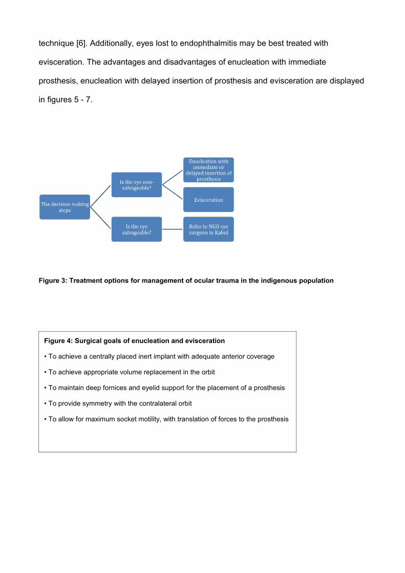
technique [6]. Additionally, eyes lost to endophthalmitis may be best treated with
evisceration. The advantages and disadvantages of enucleation with immediate
prosthesis, enucleation with delayed insertion of prosthesis and evisceration are displayed
in figures 5 - 7.
Figure 3: Treatment options for management of ocular trauma in the indigenous population
Figure 4: Surgical goals of enucleation and evisceration
• To achieve a centrally placed inert implant with adequate anterior coverage
• To achieve appropriate volume replacement in the orbit
• To maintain deep fornices and eyelid support for the placement of a prosthesis
• To provide symmetry with the contralateral orbit
• To allow for maximum socket motility, with translation of forces to the prosthesis
Figure 5: Advantages and disadvantages of enucleation and immediate
repair with prosthesis
Advantages
• Single stage procedure
Disadvantages
• Risk of infection from contaminated orbital content
• Difficulty closing extra ocular muscles over implant
Figure 6: Advantages and disadvantages of delayed staged repair
Advantages
• Ensures orbital content is safe
• Fibrotic reaction in muscles leads to more robust closure over the implant
Disadvantages
• A two stage procedure with possibility of casualty not returning to unit
• Protracted timeline requiring extended theatre and ward bed times
Figure 7: Advantages and
disadvantages of evisceration
Advantages
• To reduce risk of implant extrusion
• To reduce risk of infection
• Better cosmesis with a mobile globe
Disadvantages
• Technically difficult
Case example 1 (coalition serviceman)
This coalition serviceman whose injuries can be seen in Figure 2 had momentarily
removed his eye protection before being injured by a roadside IED. He received shrapnel
spray to his face with complete rupture of his left eye and a scleral perforation of his right
eye. Management was to repair the right eye scleral penetration with prolene sutures and
rapid evacuation for definitive treatment with a US ophthalmic surgeon. Although the left
eye was obviously not salvageable, the decision was made not to enucleate or eviscerate
it at the time of surgery to the right eye as it was felt that such casualties should be given
time to accept the diagnosis allowing appropriate consent.
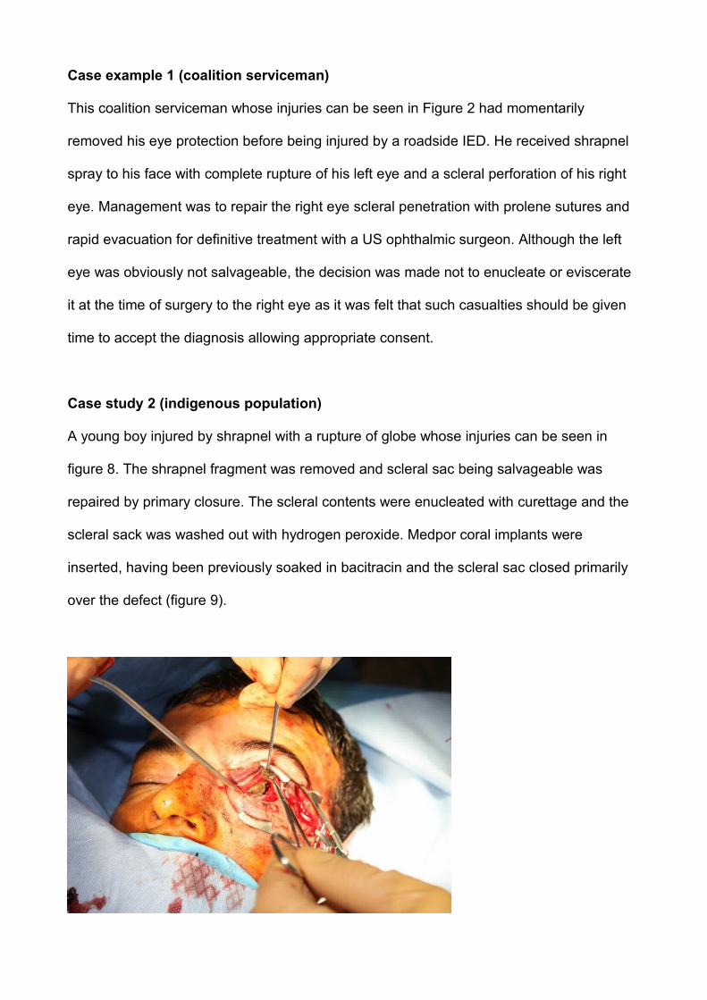
Case study 2 (indigenous population)
A young boy injured by shrapnel with a rupture of globe whose injuries can be seen in
figure 8. The shrapnel fragment was removed and scleral sac being salvageable was
repaired by primary closure. The scleral contents were enucleated with curettage and the
scleral sack was washed out with hydrogen peroxide. Medpor coral implants were
inserted, having been previously soaked in bacitracin and the scleral sac closed primarily
over the defect (figure 9).

Figure 8: Presentation of intraoccular shrapnel injury
Figure 9: Post insertion of medpor coral implants
Case study 3 (indigenous population)
This case demonstrates an Afghan Army soldier hit by shrapnel from an IED resulting in
extensive globe injuries that were obviously impossible to salvage. The eye was
enucleated, leaving the extra ocular muscles with optic nerve divided and tied off (Figure
10). The intra-conal space was washed with Betadine, hydrogen peroxide and saline and
packed with iodoform ribbon gauze. Finally the eye lids were repaired and the fornices
preserved with a Morgan eye shield.

Figure 10: Presentation post enucleation
The patient returned to theatre one week later where the pack was removed. Coral
implants were inserted with the extra ocular muscles closed over the implant and the
conjunctiva closed over muscles (figure 11).
Figure 11: Implants in situ

Discussion
Perforated globe injuries sustained in combat generally result in poor visual and
anatomical outcomes despite surgical intervention [8]. There is little evidence to favour the
techniques of enucleation and evisceration over one another. Enucleations are generally
carried out more commonly than eviscerations in both military and civilian practice.
However a recent survey of 297 UK consultant ophthalmologists [9] found two- thirds
preferred evisceration over enucleation but reasons for this were not given. On the
question of choice of implant, 12% preferred natural coral implants, coincident with the
material available and used in theatre by deployed British maxillofacial surgeons. Savar et
al [5] looked at 660 open globe injuries over a 5- year period admitted to a civilian trauma
centre in the US; 51 underwent enucleation and only 4 had eviscerations. These
proportions were similar to those found in the 21- month period that Oral and Maxillofacial
surgeons deployed to Kandahar in which 19 eye enucleations and 3 eviscerations were
performed.
The most serious complication of both enucleation and evisceration is the risk of
developing sympathetic ophthalmia. This is a rare bilateral pan-uveitis in which there is a
painful red eye, visual loss and a history of an open globe injury. The risk of sympathetic
ophthalmia has been quoted as 0.14% in a large scale civilian study of penetrating eye
injuries in South Africa [10], and in less than 1/500 cases of open globe injury [11]. Colyer
et al [8] reported no episodes of sympathetic ophthalmia in 25 enucleations on US
servicemen evacuated from Iraq to the Walter Reed hospital between 2003- 2006. Freidlin
et al [12] in 2006 published a case report about a 21-year-old US soldier who received a
penetrating eye injury while fighting in Iraq and was treated with evisceration. Sympathetic
ophthalmia developed, which responded well to steroid treatment. They stated that it was
the first case of sympathetic ophthalmia after a war injury reported since World War II. In
the time period covered in this article neither evisceraton or enucleation resulted in post-
operative complications. We would therefore conclude that both techniques appear safe
but feel that overall evisceration carries better cosmesis than enucleation.
The management of devastating ocular trauma is addressed in the head and neck
component of the Military Operational Surgical Training (MOST) course. This new course,
delivered entirely by consultant surgeons is designed to teach all newly deploying
surgeons the theoretical and practical concepts behind the surgical techniques currently
required on deployment. These techniques may be outside of the routine civilian practice
of that surgeon, as is the management of devastating ocular trauma by OMFS surgeons.
Both enucleation and evisceration are taught by consultant ophthalmic surgeons based on
cadaveric dissection.
The increased use of ballistic eye protection remains the greatest way of reducing
preventable penetrating eye injuries but its uptake amongst servicemen remains variable.
Cotter and La Piana modelled data from the Vietnam War and proposed that standard US
Army 2 mm thick defence goggle would have been prevented 52% of eye injuries [13].
Mader et al [4] analysed 207 eye injuries sustained by US servicemen and commented
that polycarbonate ballistic eyewear could have prevented many, but not all, of the injuries.
Colyer et al [8] found that during 2003 - 2006, of 61 US servicemen evacuated to the US
with perforating eye injuries, only 43% were wearing eye protection. Servicemen complain
that eye protection degrades their vision due to misting, there is a restricted field of view
from the frames and that the lenses scratch easily [7]. Further work needs to be
undertaken to increase compliance in the wearing of eye protection. Enforced use of eye
protection in US military convoys in Iraq in 2004 reduced the incidence of eye injuries to
0.5% from a conflict wide incidence of 6% [14], making a strong case for the mandatory

use of military eye protection by all deployed UK servicemen.
Conclusion
The management of penetrating eye trauma is normally outside the routine practice of
maxillofacial surgeons in the UK. However improved pre- deployment training and the
combined experience of a cadre that has undertaken two long- term deployments has
resulted in a large number of surgical interventions for devastating ocular trauma without
complications on long- term follow up. Although there is little in the literature to suggest
enucleation is more effective than evisceration, the former is undertaken more commonly
in both military and civilian environments and was reflected in the proportions performed in
our deployment. Many ocular wounds are still potentially preventable and greater
emphasis must be placed on exploring reasons why deployed UK servicemen do not
always wear ballistic eye protection.
Acknowledgements
The cases discussed were treated by the third author, however, other maxillofacial
consultants; Mr A Gibbons, Mr N McKenzie and Mr A Monaghan also treated similar cases
and their figures have been included in the statistics.
References
[1] Owens BD, Kragh JF, Wenke JC, Macaitis J, Wade CE, Holcomb JB. Combat wounds
in Operation Iraqi Freedom and Operation Enduring Freedom. J Trauma 2008; 64: 295-
299.
[2] Wade AL, Dye JL, Mohrle CR, Galarneau MR. Head, face, and neck injuries during
Operation Iraqi Freedom II: results from the US Navy-Marine Corps Combat Trauma
Registry. J Trauma 2007; 63: 836- 840.
[3] Wong TY, Seet MB, Ang CL. Eye injuries in twentieth century warfare: a historical
perspective. Surv Ophthalmol 1997; 41: 433- 459.
[4] Mader TH, Carroll RD, Slade CS, George RK, Ritchey JP, Neville SP. Ocular war
injuries of the Iraqi insurgency, January–September 2004. Ophthalmology 2006; 113: 97-
104.
[5] Savar A, Andreoli MT, Kloek CE, Andreoli CM. Enucleation for open globe injury. Am J
Ophthalmol 2009; 147(4): 595- 600.
[6] Ophthalmic Care of the Combat Casualty. M.D. Allen B. Thach (Editor), Walter Reed
Army Medical Center Borden Institute.
[7] Blanch R, Scott R. Ocular Blast Injury. J Royal Army Med Corps 2009; 155 (4): 279-284
[8] Colyer MH, Chun DW, Bower KS, Dick JS, Weichel ED. Perforating globe injuries
during operation Iraqi Freedom. Ophthalmology 2008; 115(11): 2087-93.
[9] Viswanathan P, Sagoo MS, Olver JM. UK national survey of enucleation and orbital
implant trends. Br J Ophthalmol 2007; 91: 616- 619.
[10] Du Toit N, Motala MI, Richards J, Murray ADN, Maitra S. The risk of sympathetic
opthalmia following evisceration for penetrating eye injuries at Grotte Schuur Hospital. Br J
Ophthalmol 2008; 92: 61-63.
[11] Albert DM, Diaz-Rohena R. A historical review of sympathetic ophthalmia and its
epidemiology. Surv Ophthalmol 1989: 34: 1-14.

[12] Freidlin J, Pak J, Tessler H, Putterman A, Goldstein D. Sympathetic Ophthalmia After
Injury in the Iraq War. Ophthal Plast Recon Surg 2006; 22 (2): 133-134.
[13] Cotter F, La Piana F G. Eye casualty reduction for eye armour. Mil Med 1991; 156:
126–8.
[14] Gondusky JS, Reiter MC. Protecting military convoys in Iraq: An examination of battle
injuries sustained by a mechanised battalion during operation Iraqi Freedom II. Mil Med
2005; 170; 546- 549.