Management of Complex Cases Orthopaedic Surgery
11
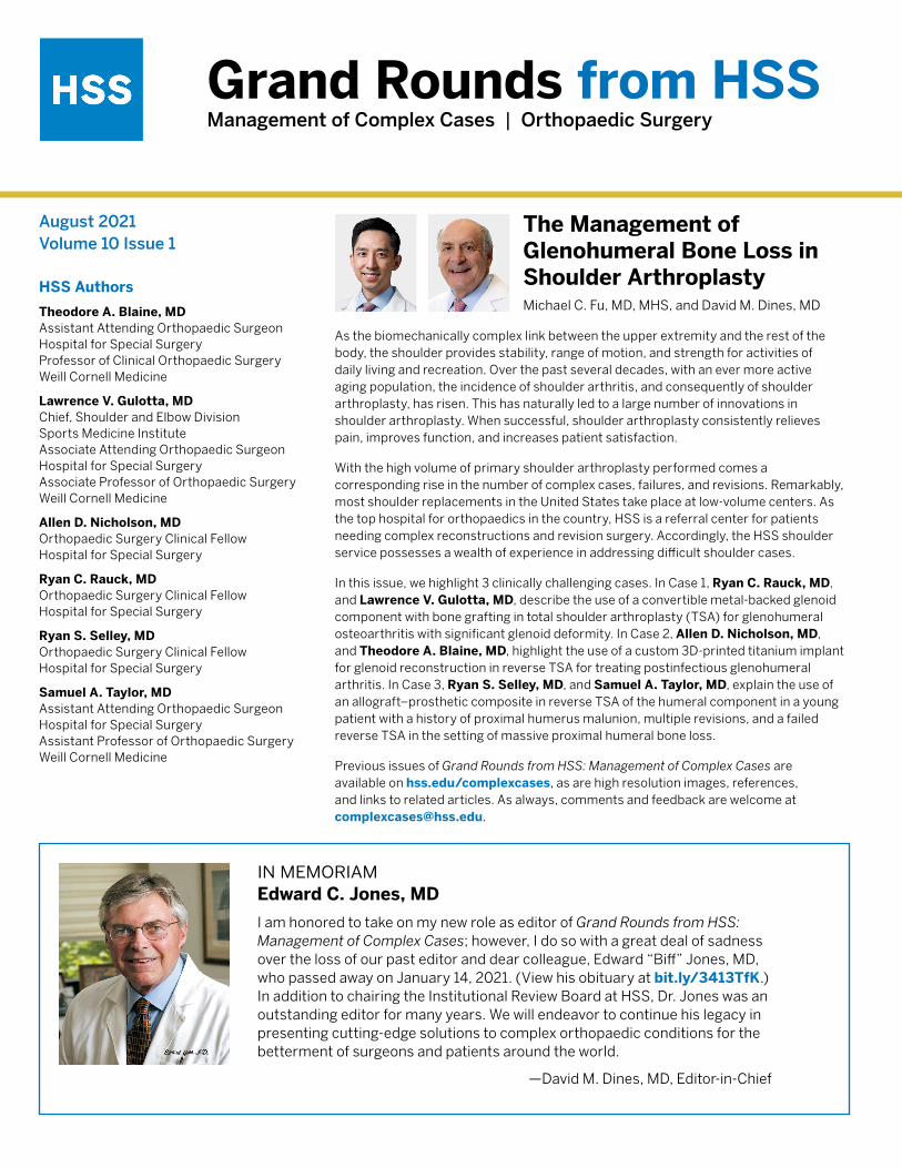
Grand Rounds from HSS Management of Complex Cases | Orthopaedic Surgery August 2021 Volume 10 Issue 1 HSS Authors Theodore A. Blaine, MD Assistant Attending Orthopaedic Surgeon Hospital for Special Surgery Professor of Clinical Orthopaedic Surgery Weill Cornell Medicine Lawrence V. Gulotta, MD Chief, Shoulder and Elbow Division Sports Medicine Institute Associate Attending Orthopaedic Surgeon Hospital for Special Surgery Associate Professor of Orthopaedic Surgery Weill Cornell Medicine Allen D. Nicholson, MD Orthopaedic Surgery Clinical Fellow Hospital for Special Surgery Ryan C. Rauck, MD Orthopaedic Surgery Clinical Fellow Hospital for Special Surgery Ryan S. Selley, MD Orthopaedic Surgery Clinical Fellow Hospital for Special Surgery Samuel A. Taylor, MD Assistant Attending Orthopaedic Surgeon Hospital for Special Surgery Assistant Professor of Orthopaedic Surgery Weill Cornell Medicine The Management of Glenohumeral Bone Loss in Shoulder Arthroplasty Michael C. Fu, MD, MHS, and David M. Dines, MD As the biomechanically complex link between the upper extremity and the rest of the body, the shoulder provides stability, range of motion, and strength for activities of daily living and recreation. Over the past several decades, with an ever more active aging population, the incidence of shoulder arthritis, and consequently of shoulder arthroplasty, has risen. This has naturally led to a large number of innovations in shoulder arthroplasty. When successful, shoulder arthroplasty consistently relieves pain, improves function, and increases patient satisfaction. With the high volume of primary shoulder arthroplasty performed comes a corresponding rise in the number of complex cases, failures, and revisions. Remarkably, most shoulder replacements in the United States take place at low-volume centers. As the top hospital for orthopaedics in the country, HSS is a referral center for patients needing complex reconstructions and revision surgery. Accordingly, the HSS shoulder service possesses a wealth of experience in addressing difficult shoulder cases. In this issue, we highlight 3 clinically challenging cases. In Case 1, Ryan C. Rauck, MD, and Lawrence V. Gulotta, MD, describe the use of a convertible metal-backed glenoid component with bone grafting in total shoulder arthroplasty (TSA) for glenohumeral osteoarthritis with significant glenoid deformity. In Case 2, Allen D. Nicholson, MD, and Theodore A. Blaine, MD, highlight the use of a custom 3D-printed titanium implant for glenoid reconstruction in reverse TSA for treating postinfectious glenohumeral arthritis. In Case 3, Ryan S. Selley, MD, and Samuel A. Taylor, MD, explain the use of an allograft–prosthetic composite in reverse TSA of the humeral component in a young patient with a history of proximal humerus malunion, multiple revisions, and a failed reverse TSA in the setting of massive proximal humeral bone loss. Previous issues of Grand Rounds from HSS: Management of Complex Cases are available on hss.edu/complexcases, as are high resolution images, references, and links to related articles. As always, comments and feedback are welcome at [email protected]. in memoriam Edward C. Jones, MD I am honored to take on my new role as editor of Grand Rounds from HSS: Management of Complex Cases; however, I do so with a great deal of sadness over the loss of our past editor and dear colleague, Edward “Biff” Jones, MD, who passed away on January 14, 2021. (View his obituary at bit.ly/3413TfK.) In addition to chairing the Institutional Review Board at HSS, Dr. Jones was an outstanding editor for many years. We will endeavor to continue his legacy in presenting cutting-edge solutions to complex orthopaedic conditions for the betterment of surgeons and patients around the world. —David M. Dines, MD, Editor-in-Chief
Transcript of Management of Complex Cases Orthopaedic Surgery
Grand Rounds from HSSManagement of Complex Cases | Orthopaedic Surgery
August 2021Volume 10 Issue 1
HSS Authors
Theodore A. Blaine, MD Assistant Attending Orthopaedic Surgeon Hospital for Special Surgery Professor of Clinical Orthopaedic Surgery Weill Cornell Medicine
Lawrence V. Gulotta, MD Chief, Shoulder and Elbow Division Sports Medicine Institute Associate Attending Orthopaedic Surgeon Hospital for Special Surgery Associate Professor of Orthopaedic Surgery Weill Cornell Medicine
Allen D. Nicholson, MD Orthopaedic Surgery Clinical Fellow Hospital for Special Surgery
Ryan C. Rauck, MD Orthopaedic Surgery Clinical Fellow Hospital for Special Surgery
Ryan S. Selley, MD Orthopaedic Surgery Clinical Fellow Hospital for Special Surgery
Samuel A. Taylor, MD Assistant Attending Orthopaedic Surgeon Hospital for Special Surgery Assistant Professor of Orthopaedic Surgery Weill Cornell Medicine
The Management of Glenohumeral Bone Loss in Shoulder ArthroplastyMichael C. Fu, MD, MHS, and David M. Dines, MD
As the biomechanically complex link between the upper extremity and the rest of the body, the shoulder provides stability, range of motion, and strength for activities of daily living and recreation. Over the past several decades, with an ever more active aging population, the incidence of shoulder arthritis, and consequently of shoulder arthroplasty, has risen. This has naturally led to a large number of innovations in shoulder arthroplasty. When successful, shoulder arthroplasty consistently relieves pain, improves function, and increases patient satisfaction.
With the high volume of primary shoulder arthroplasty performed comes a corresponding rise in the number of complex cases, failures, and revisions. Remarkably, most shoulder replacements in the United States take place at low-volume centers. As the top hospital for orthopaedics in the country, HSS is a referral center for patients needing complex reconstructions and revision surgery. Accordingly, the HSS shoulder service possesses a wealth of experience in addressing difficult shoulder cases.
In this issue, we highlight 3 clinically challenging cases. In Case 1, Ryan C. Rauck, MD, and Lawrence V. Gulotta, MD, describe the use of a convertible metal-backed glenoid component with bone grafting in total shoulder arthroplasty (TSA) for glenohumeral osteoarthritis with significant glenoid deformity. In Case 2, Allen D. Nicholson, MD, and Theodore A. Blaine, MD, highlight the use of a custom 3D-printed titanium implant for glenoid reconstruction in reverse TSA for treating postinfectious glenohumeral arthritis. In Case 3, Ryan S. Selley, MD, and Samuel A. Taylor, MD, explain the use of an allograft–prosthetic composite in reverse TSA of the humeral component in a young patient with a history of proximal humerus malunion, multiple revisions, and a failed reverse TSA in the setting of massive proximal humeral bone loss.
Previous issues of Grand Rounds from HSS: Management of Complex Cases are available on hss.edu/complexcases, as are high resolution images, references, and links to related articles. As always, comments and feedback are welcome at [email protected].
in memoriamEdward C. Jones, MD
I am honored to take on my new role as editor of Grand Rounds from HSS: Management of Complex Cases; however, I do so with a great deal of sadness over the loss of our past editor and dear colleague, Edward “Biff” Jones, MD, who passed away on January 14, 2021. (View his obituary at bit.ly/3413TfK.) In addition to chairing the Institutional Review Board at HSS, Dr. Jones was an outstanding editor for many years. We will endeavor to continue his legacy in presenting cutting-edge solutions to complex orthopaedic conditions for the betterment of surgeons and patients around the world.
—David M. Dines, MD, Editor-in-Chief

Case 1 Case presented by Ryan C. Rauck, MD, and Lawrence V. Gulotta, MD
Addressing Significant Glenoid Deformity in a Younger Patient with Glenohumeral Osteoarthritis
Case Report A 56-year-old man presented with several years of right shoulder pain after a failed trial of conservative therapy. An energetic man who wanted to return to his prior active lifestyle, he had undergone left shoulder hemiarthroplasty but continued to have functional limitations on that side. He hoped for better results on the right side.
On examination, range of motion in the right shoulder was 140° of forward elevation, 20° of external rotation, and internal rotation to his lumbar spine. Significant crepitus was noted. His rotator cuff strength demonstrated 5/5 supraspinatus, 5/5 external rotation, and 5/5 subscapularis.
Radiographs of the right shoulder showed end-stage glenohumeral osteoarthritis with significant glenoid retroversion (B3 Walch classification) [1] and posterior humeral head subluxation (Fig. 1). A computed tomography (CT) scan to assist with preoperative planning showed excessive glenoid wear, with 25° of retroversion (Fig. 2).
The patient underwent an anatomic total shoulder replacement using a convertible glenoid baseplate with bone grafting to correct significant retroversion. A standard deltopectoral approach with the patient in the beach chair position was used. A lesser tuberosity osteotomy was performed and the humeral head was cut in 30° of retroversion. The cut humeral head was taken to the back table to be used for autograft on the back of the glenoid baseplate.
Exposure of the glenoid was notable for significant retroversion, consistent with preoperative imaging. A guide pin was placed down the glenoid vault in the appropriate orientation. Reaming was then performed so that the anterior 50% of the baseplate was flush with native bone. The cut humeral head was reamed with the central screw boss and then cut in half and shaped to fit the posterior aspect of the glenoid (Fig. 3). The humeral head autograft was placed on the posterior glenoid and the baseplate impacted into place and secured with the central compression screw. Two locking screws were placed superiorly and inferiorly, providing excellent fixation. The polyethylene liner was impacted into
place, followed by finishing preparation of the humerus and repair of the lesser tuberosity osteotomy.
The patient had an excellent recovery. After immobilization in a sling for 4 weeks, he began progressive physical therapy to regain motion and strength. At the 4-year follow-up examination, his right shoulder demonstrated 170° of forward elevation, 60° of external rotation, and internal rotation to T8. His rotator cuff strength was 5/5. Radiographs showed that the right humeral head was well centered, the glenoid version was neutral, and the bone graft had incorporated on the posterior glenoid (Fig. 4).
Discussion Significant glenoid deformity presents a challenge to the surgeon managing a patient with glenohumeral osteoarthritis with an anatomic shoulder arthroplasty. Depending on the degree of deformity, treatment may include eccentric reaming using a posteriorly augmented polyethylene glenoid component or bone grafting using a convertible glenoid baseplate.
Given the degree of retroversion in this case, eccentric reaming would have sacrificed too much bone for glenoid fixation, and a standard posterior augment would not have corrected the glenoid version. Historically, anatomic shoulder replacement with a metal-backed glenoid had poor survivorship [2], but the advent of locking screws and improved polyethylene and porous materials have renewed interest in its use. In this case, a convertible glenoid had the benefits of allowing bone grafting of the posterior glenoid to correct the retroversion, while allowing secure fixation of the glenoid component. (Few studies have examined long-term outcomes of this procedure.) Younger patients undergoing anatomic shoulder replacement may require a revision in their lifetime [3, 4]; a convertible glenoid may allow for easier transition to a reverse shoulder replacement, if that is required. ■
Images on page 3 and 4
REFERENCES:
1. Walch G, Badet R, Boulahia A, Khoury A. Morphologic study of the glenoid in primary glenohumeral osteoarthritis. J Arthroplasty. 1999;14(6):756–760.
2. Gauci MO, Bonnevialle N, Moineau G, Baba M, Walch G, Boileau P. Anatomical total shoulder arthroplasty in young patients with osteoarthritis: all-polyethylene versus metal-backed glenoid. Bone Joint J. 2018;100-B(4):485–492.
3. Sperling JW, Cofield RH, Rowland CM. Neer hemiarthroplasty and Neer total shoulder arthroplasty in patients fifty years old or less. Long-term results. J Bone Joint Surg Am. 1998;80(4):464–473.
4. Denard PJ, Raiss P, Sowa B, Walch G. Mid- to long-term follow-up of total shoulder arthroplasty using a keeled glenoid in young adults with primary glenohumeral arthritis. J Shoulder Elbow Surg. 2013;22(7):894–900.
2 | Grand Rounds August 2021 | Volume 10 Issue 1

Case 1: Addressing Significant Glenoid Deformity in a Younger Patient with Glenohumeral Osteoarthritis Case Images
Figure 1
Anteroposterior (AP) (left) and axillary (right) radiographs of the right shoulder demonstrate glenohumeral osteoarthritis with a B3 glenoid deformity.
Figure 2
Axial CT image demonstrates 25° of glenoid retroversion.
3 | Grand Rounds August 2021 | Volume 10 Issue 1

Case 1: Addressing Significant Glenoid Deformity in a Younger Patient with Glenohumeral Osteoarthritis Case Images Continued
Figure 3
Humeral head autograft (left) and placement on the posterior glenoid (right).
Figure 4
AP (left) and axillary (right) radiographs at 4-year follow-up demonstrate right anatomic shoulder replacement with metal backed convertible glenoid, restoration of neutral glenoid version, and incorporation of the bone graft.
4 | Grand Rounds August 2021 | Volume 10 Issue 1

Case 2 Case presented by Allen D. Nicholson, MD, and Theodore A. Blaine, MD
Treating Massive Glenoid Bone Deficiency with a Custom Metal Glenoid Implant
Case Report A 63-year-old man presented for evaluation after several years of increasing right shoulder pain. While serving in the Vietnam War, he had sustained a shrapnel injury to his right shoulder and developed proximal humerus osteomyelitis, requiring multiple irrigation and debridement procedures. He subsequently developed severe glenohumeral arthritis and underwent several years of conservative management including nonsteroidal anti-inflammatory drugs and steroid injections.
Radiographs showed severe glenohumeral arthritis (Fig. 1). His range of motion was forward elevation 70°, external rotation −10°, internal rotation to the side with pain throughout the motion arc. Infections workup showed erythrocyte sedimentation rate of 10 mm/hr, C-reactive protein level of 1.12 mg/L, and white blood cell count of 5.3 × 10⁹/L. He elected to proceed with shoulder arthroplasty as a staged procedure. A custom molded antibiotic spacer was placed as a hemiarthroplasty, and the patient completed a 6-week course of antibiotics. Intraoperative cultures showed no growth, and a reverse shoulder arthroplasty was planned as the second stage of the procedure.
Computed tomography with 3D reconstruction was used preoperatively and showed bony deficiency with a compromised glenoid vault (Fig. 2). The Comprehensive® Vault Reconstruction System (VRS) (Zimmer Biomet, Warsaw, IN) was planned with computer-aided design (CAD) software to create a custom titanium implant. The software allowed for planning of exact screw trajectories to obtain the best purchase in the remaining scapular bone.
The previous incision was used with a standard deltopectoral approach to place the VRS glenoid baseplate over the anterior rim of the glenoid, which was secured with a central 6.5 × 45 mm nonlocking screw and 4 peripheral 4.75 mm locking screws. A 41 mm glenosphere was impacted onto the taper with maximal offset inferiorly. The humeral bone was deficient from prior osteomyelitis, and 2 cerclage wires were placed to prevent extension of a 2.5 cm cortical defect in the proximal humerus. A 10 × 122 mm cemented humeral prosthesis was implanted in 20° of
retroversion with a standard tray and +0 polyethylene. The components were reduced and the subscapularis was repaired with braided nylon sutures.
At 6 months postoperatively, the patient had no pain, a stable implant (Fig. 3), and greatly improved range of motion, with forward elevation to 110° and external rotation to 30°. The implant remains intact and stable 4 years postoperatively.
Discussion Glenoid deficiency in primary and revision shoulder arthroplasty creates difficulty in positioning the glenoid component in poor bone stock and achieving adequate fixation [1]. Glenoid baseplate fixation failure, bone graft resorption, or inadequate component positioning can lead to increased baseplate stresses and early failure [2]. 3D printing technology has allowed the manufacture of implants that match glenoid bone deformity, which can allow for restoring the joint line, increasing bone–metal contact, stabilizing the baseplate with flanges, and determining preoperatively the ideal length and trajectory of screw fixation. Zimmer Biomet received US Food and Drug Administration 510(k) clearance in 2016 to market the VRS device for use in patients with significant bone loss and is the only implant of this type available in the United States [3].
VRS reconstruction has shown promise in 2 small retrospective case series. Bodendorfer et al reported on 12 shoulders treated with the VRS at average 30 months’ follow-up [4]. Median improvement in American Shoulder and Elbow Surgeons (ASES) scores was 45 points (P < .02), with 20° forward elevation (P < .009), 40° external rotation (P < .014), and 2 spinal levels of internal rotation (P < .002), with no signs of radiographic loosening or complications. Rangarajan et al reported 1-year results of 19 patients treated with the VRS, finding a 47-point improvement in ASES scores (P < .001) and 71° forward elevation (P < .001), with complications in 4 patients [2]. Our case is consistent with outcomes reported in those case series, with 40° improvement in both forward elevation and external rotation and a stable implant 4 years postoperatively, with no reported complications. ■
Images on page 6 and 7
REFERENCES:
1. Mourad W, Wiater JM, Wiater BP, Martusiewicz A. Baseplate options for reverse total shoulder arthroplasty. Curr Rev Musculoskelet Med. 2020;13(6):769–775.
2. Rangarajan R, Blout CK, Patel VV, Bastian SA, Lee BK, Itamura JM. Early results of reverse total shoulder arthroplasty using a patient-matched glenoid implant for severe glenoid bone deficiency. J Shoulder Elbow Surg. 2020;29(7S):S139–S148.
3. Dines DM, Gulotta L, Craig EV, Dines JS. Novel solution for massive glenoid defects in shoulder arthroplasty: a patient-specific glenoid vault reconstruction system. Am J Orthop. 2017;46(2):104–108.
4. Bodendorfer BM, Loughran GJ, Looney AM, et al. Short-term outcomes of reverse shoulder arthroplasty using a custom baseplate for severe glenoid deficiency. J Shoulder Elbow Surg. 2021;30(5):1060–1067.
5 | Grand Rounds August 2021 | Volume 10 Issue 1

Case 2: Treating Massive Glenoid Bone Deficiency with a Custom Metal Glenoid Implant Case Images
Figure 1
X-rays of the right shoulder show severe glenohumeral arthritis.
Figure 2
CT with 3D reconstruction shows deficient glenoid vault.
6 | Grand Rounds August 2021 | Volume 10 Issue 1

Case 2: Treating Massive Glenoid Bone Deficiency with a Custom Metal Glenoid Implant Case Images Continued
Figure 3
X-rays at 6 months postoperatively show a stable implant.
7 | Grand Rounds August 2021 | Volume 10 Issue 1

Case 3 Case presented by Ryan S. Selley, MD, and Samuel A. Taylor, MD
Allograft-Prosthetic Composite Proximal Humerus Reconstruction in Reverse Shoulder Arthroplasty for Massive Bone Loss
Case Report A 38-year-old woman presented to HSS in 2019 with a poorly functioning reverse total shoulder arthroplasty (rTSA) and pain at rest and with activity. After a malunion following a proximal humerus fracture in 2003, she had undergone several surgical procedures, including a valgus-producing osteotomy of the proximal humerus, removal of hardware, anatomic total shoulder arthroplasty (TSA) with an all-polyethylene glenoid, and finally rTSA with a cemented humeral stem in 2016 (Fig. 1). Symptomatic management had failed in the 3 years prior.
Examination revealed numerus extensile healed incisions and visible atrophy of her anterior deltoid, pectoralis major, supraspinatus, and infraspinatus. There was marked tenderness over the bicipital groove and deltoid insertion. Active forward flexion (FF) was limited to 60° and external rotation (ER) to 20°.
Workup for infection showed a mildly elevated (27 mm/hr) erythrocyte sedimentation rate, no leukocytosis, normal C-reactive protein level, and negative aspiration cultures. Magnetic resonance imaging suggested prosthesis loosening, and a computed tomography (CT) scan (Fig. 2) showed significant metadiaphyseal bone stock deficiency.
The patient underwent revision rTSA of the humeral component only, using an allograft-prosthetic composite (APC), rotator cuff repair, and biceps tenodesis. The proximal humerus was exposed via an extended deltopectoral approach, and biopsies and frozen sections were obtained. With no evidence of infection, a single-stage revision proceeded.
The humeral prosthesis was grossly loose proximally but fixed distally, and the surrounding proximal humeral bone was of poor quality and largely nonviable. Metal cerclage wires were removed, as were the prosthesis and 9 cm of proximal humeral bone, with a transverse cut made 1.5 cm distally to the area of bone loss. The stem was extracted with a combination of the Midas Rex drill and narrow flexible osteotomes.
The allograft was then fashioned for use; 9 cm of the proximal humerus was matched to the defect for placement of a revision stem. Of note, the allograft was prepared
on the back table to accept the inlay humeral prosthesis and then transferred to the patient to allow for the stem to bypass the allograft–humeral diaphysis interface. The APC was trialed, and satisfied with our length and proximal articulation we fixed an anterior plate both proximally into the allograft and distally into the remaining humeral shaft (Fig. 3). Next, the final implant was trialed and a distal screw was removed in order to seat the implant. Satisfied with our length and motion (FF 140°, ER 70°), we cemented the humerus and seated the final components. Final fluoroscopy demonstrated an appropriate intramedullary cement mantle, with some cement extrusion distally out of a breach in the humeral diaphysis incurred during cement removal (Fig. 4). The cement was left in situ to avoid additional morbidity of removal. The posterior rotator cuff tendons (teres minor and infraspinatus) were repaired to the allograft rotator cuff tendon stumps to empower ER.
The patient adhered to our postoperative protocol: weeks 0–6, use sling with distal range of motion (ROM) and Codman pendulum exercises only; weeks 6–12, discontinue sling, with a goal of FF 140° and ER 45°; weeks 12+, continue pursuit of ROM, strength, and endurance. Her postoperative course was uncomplicated aside from feelings of fullness around the area of extramedullary cement extravasation. Radial nerve function was normal. At 1-year follow-up, she reported satisfaction with her outcome, reporting occasional soreness with activity but no resting or nighttime pain. She achieved active forward elevation to 120°, ER to 20°, and internal rotation to the greater trochanter.
Discussion Failed rTSA has catastrophic implications. Proximal humeral bone loss is an uncommon clinical dilemma, with management options including revision with a tumor prosthesis versus APC. Given our patient’s young age and the probability of future revisions, we opted for APC, a technique adopted from treatments for musculoskeletal tumor resection [1], to preserve as much bone stock as possible. Our case illustrates a pitfall of this operation: cement extravasation from the distal humerus occurring through a breach in the cortex created during cement removal. Fortunately, no substantive deficit occurred as a result. Other potential pitfalls of the procedure
include establishing the appropriate length and offset while reapproximating tissue attachments to minimize the risk of dislocation and maximize function [2]. When performed successfully, APC reconstruction is a reasonable option for cases of severe proximal humeral bone loss, showing good functional results with relatively low revision rates and durable results to 10 years [3-7]. A study of upper and lower extremity patients with massive bone loss showed greater durability and lower revision rates with APC than with megaprosthesis reconstruction [8]. ■
Images on page 9 and 10
REFERENCES:
1. Jensen KL, Johnston JO. Proximal humeral reconstruction after excision of a primary sarcoma. Clin Orthop Relat Res. 1995;(311):164-175.
2. Sanchez-Sotelo J, Wagner ER, Houdek MT. Allograft-prosthetic composite reconstruction for massive proximal humeral bone loss in reverse shoulder arthroplasty. JBJS Essent Surg Tech. 2018;8(1):e3.
3. Abdeen A, Hoang BH, Athanasian EA, Morris CD, Boland PJ, Healey JH. Allograft-prosthesis composite reconstruction of the proximal part of the humerus: functional outcome and survivorship. J Bone Joint Surg Am. 2009;91(10):2406-2415.
4. Sanchez-Sotelo J, Wagner ER, Sim FH, Houdek MT. Allograft-prosthetic composite reconstruction for massive proximal humeral bone loss in reverse shoulder arthroplasty. J Bone Joint Surg Am. 2017;99(24):2069-2076.
5. Reif T, Schoch B, Spiguel A, et al. A retrospective review of revision proximal humeral allograft-prosthetic composite procedures: an analysis of proximal humeral bone stock restoration. J Shoulder Elbow Surg. 2020;29(7):1353-1358.
6. Boileau P, Raynier JL, Chelli M, Gonzalez JF, Galvin JW. Reverse shoulder-allograft prosthesis composite, with or without tendon transfer, for the treatment of severe proximal humeral bone loss. J Shoulder Elbow Surg. 2020;29(11):e401-e415.
7. El Beaino M, Liu J, Lewis VO, Lin PP. Do early results of proximal humeral allograft-prosthetic composite reconstructions persist at 5-year followup? Clin Orthop Relat Res. 2019;477(4):758-765.
8. Gautam D, Arora N, Gupta S, George J, Malhotra R. Megaprosthesis versus allograft prosthesis composite for the management of massive skeletal defects: a meta-analysis of comparative studies. Curr Rev Musculoskelet Med. 2021;14(3):255-270.
8 | Grand Rounds August 2021 | Volume 10 Issue 1

Case 3: Allograft-Prosthetic Composite Proximal Humerus Reconstruction in Reverse Shoulder Arthroplasty for Massive Bone Loss Case Images
Figure 1
Initial X-rays show rTSA with a well-positioned glenosphere component and proximal humeral bone loss with radiolucency surrounding the humeral stem cement mantle.
Figure 23-dimensional CT image with post-scan processing to highlight the prosthesis shows a well-fixed glenoid baseplate but near circumferential proximal humeral bone loss at the metadiaphysis.
9 | Grand Rounds August 2021 | Volume 10 Issue 1

Case 3: Allograft-Prosthetic Composite Proximal Humerus Reconstruction in Reverse Shoulder Arthroplasty for Massive Bone Loss Case Images Continued
Figure 3A Figure 3B
Intraoperative photographs show (a) the humeral defect after component explantation and (b) the final humeral prosthesis with allograft and plate fixation.
Figure 4
Postoperative X-rays show APC proximal humerus reconstruction.
10 | Grand Rounds August 2021 | Volume 10 Issue 1

Grand Rounds from HSSManagement of Complex Cases
HSS Editorial BoardEditorDavid M. Dines, MDAttending Orthopaedic Surgeon Clinical Professor of Orthopaedic SurgeryWeill Cornell Medicine
ConsultantsEric A. Bogner, MDAttending RadiologistProfessor of Clinical RadiologyWeill Cornell Medicine
Michael C. Fu, MD, MHSAssistant Attending Orthopaedic SurgeonInstructor in Orthopaedic SurgeryWeill Cornell Medicine
BoardFriedrich Boettner, MDAssistant Attending Orthopaedic SurgeonInstructor in Orthopaedic SurgeryWeill Cornell Medicine
Alexander P. Hughes, MDAssociate Attending Orthopaedic Surgeon Associate Professor of Orthopaedic SurgeryWeill Cornell Medicine
Bryan T. Kelly, MD, MBASurgeon-in-Chief and Medical DirectorChief Emeritus, Sports Medicine InstituteAttending Orthopaedic SurgeonCo-Director, Center for Hip PreservationProfessor of Orthopaedic SurgeryWeill Cornell Medicine
Robert G. Marx, MD, MSc, FRCSCAttending Orthopaedic SurgeonProfessor of Orthopaedic Surgeryand Public HealthWeill Cornell Medicine
Carolyn M. Sofka, MD, FACRAttending RadiologistDirector of EducationDepartment of Radiology and ImagingProfessor of Radiology Weill Cornell Medicine
Laura Robbins, DSWSenior Vice PresidentEducation Institute & Global PartnershipsAssociate ProfessorGraduate School of Medical SciencesClinical Epidemiology andHealth Services Research Weill Cornell Medicine
Joy Jacobson, MFADirector, Academic PublicationsManaging Editor, HSS JournalHSS Education Institute
Design/ProductionMarcia EnnisSenior Creative DirectorEducation Marketing & Digital CommunicationsHSS Education Institute
Randy HawkeAssociate DirectorEducation Marketing & Digital CommunicationsHSS Education Institute
Produced by Education Marketing & Digital Communications
©2021 Hospital for Special Surgery. 535 East 70th Street, New York, NY 10021. Hospital for Special Surgery, HSS and the HSS logo are trademarks or registered trademarks of Hospital for Special Surgery in the United States and other countries.
HSS Education Institute
11 | Grand Rounds August 2021 | Volume 10 Issue 1