
Management of Citrate Toxicity in Pediatric Patients · PTH Response to Hypocalcemia •...
51
Management of Citrate Toxicity in Pediatric Patients by Edward Wong, M.D. Director, Hematology Associate Director, Transfusion Medicine Children’s National Medical Center Center for Cancer and Blood Disorders Division of Laboratory Medicine Departments of Pediatrics and Pathology George Washington School of Medicine and Health Sciences Washington, DC 20010
-
Upload
nguyenkhanh -
Category
Documents
-
view
216 -
download
0
Transcript of Management of Citrate Toxicity in Pediatric Patients · PTH Response to Hypocalcemia •...
Management of Citrate Toxicity in
Pediatric Patients
by
Edward Wong, M.D.
Director, Hematology
Associate Director, Transfusion Medicine
Children’s National Medical Center
Center for Cancer and Blood Disorders
Division of Laboratory Medicine
Departments of Pediatrics and Pathology
George Washington School of Medicine and Health Sciences
Washington, DC 20010
Relevant Disclosures
• None
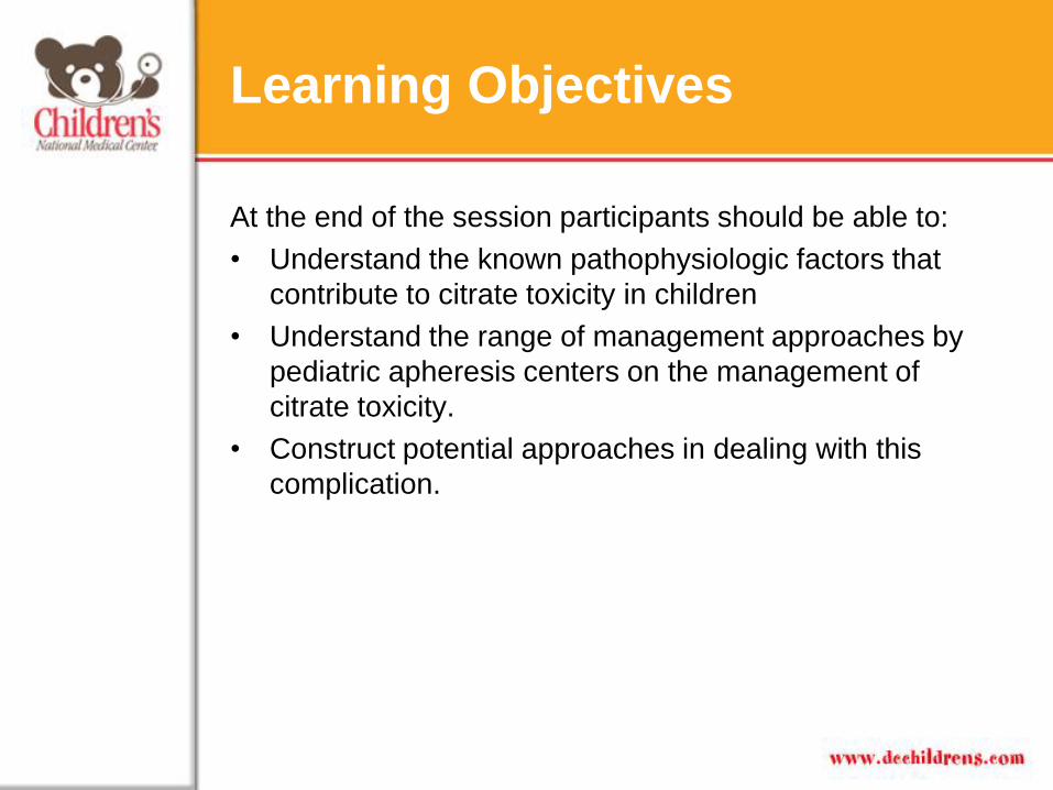
Learning Objectives
At the end of the session participants should be able to:
• Understand the known pathophysiologic factors that
contribute to citrate toxicity in children
• Understand the range of management approaches by
pediatric apheresis centers on the management of
citrate toxicity.
• Construct potential approaches in dealing with this
complication.

What is Citrate?
• Definition: salt or ester of citric acid
– Chemical formula: C6H5O73- or C3H5O(COO)3
3-
• Intermediate in the Krebs cycle
• Capable of binding many cations
– Postulated by Sabbatani (1901) to bind calcium and to inhibit blood
coagulation by lowering calcium levels in blood (Sabbatani,1902)
• NMR/X-ray diffraction studies (Glusker, 1980)
– 3 dimensional analysis of citrate binding to Ca2+ (tridentate binding)
Ca2+
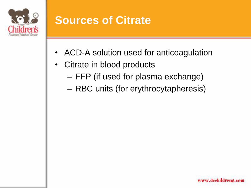
Sources of Citrate
• ACD-A solution used for anticoagulation
• Citrate in blood products
– FFP (if used for plasma exchange)
– RBC units (for erythrocytapheresis)
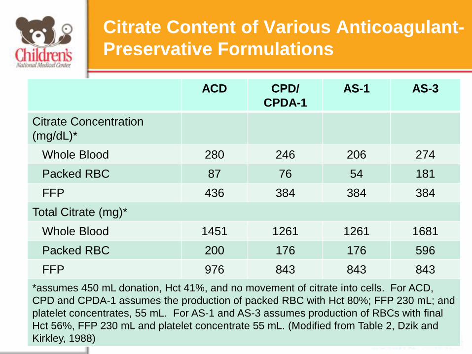
Citrate Content of Various Anticoagulant-
Preservative Formulations
ACD CPD/
CPDA-1
AS-1 AS-3
Citrate Concentration
(mg/dL)*
Whole Blood 280 246 206 274
Packed RBC 87 76 54 181
FFP 436 384 384 384
Total Citrate (mg)*
Whole Blood 1451 1261 1261 1681
Packed RBC 200 176 176 596
FFP 976 843 843 843
*assumes 450 mL donation, Hct 41%, and no movement of citrate into cells. For ACD,
CPD and CPDA-1 assumes the production of packed RBC with Hct 80%; FFP 230 mL; and
platelet concentrates, 55 mL. For AS-1 and AS-3 assumes production of RBCs with final
Hct 56%, FFP 230 mL and platelet concentrate 55 mL. (Modified from Table 2, Dzik and
Kirkley, 1988)
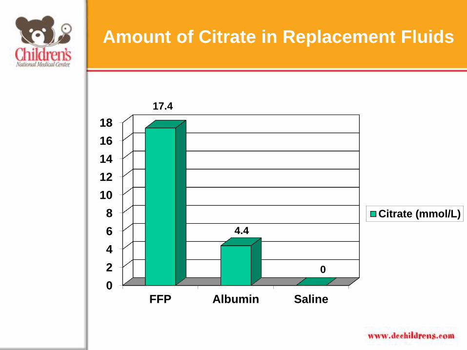
Amount of Citrate in Replacement Fluids
0
2
4
6
8
10
12
14
16
18
FFP Albumin Saline
Citrate (mmol/L)
17.4
4.4
0

Citrate Levels: Normal Levels
Normal Adult Concentrations of Citrate and Ionized Calcium
Concentration Citrate* Ionized Calcium
mg/dL 0.9-2.5 4.5-5.4
mEq/L 0.14-0.39 2.3-2.7
mmol/L 0.047-0.130 1.1-1.4
*slightly higher in children and patients with renal/liver disease
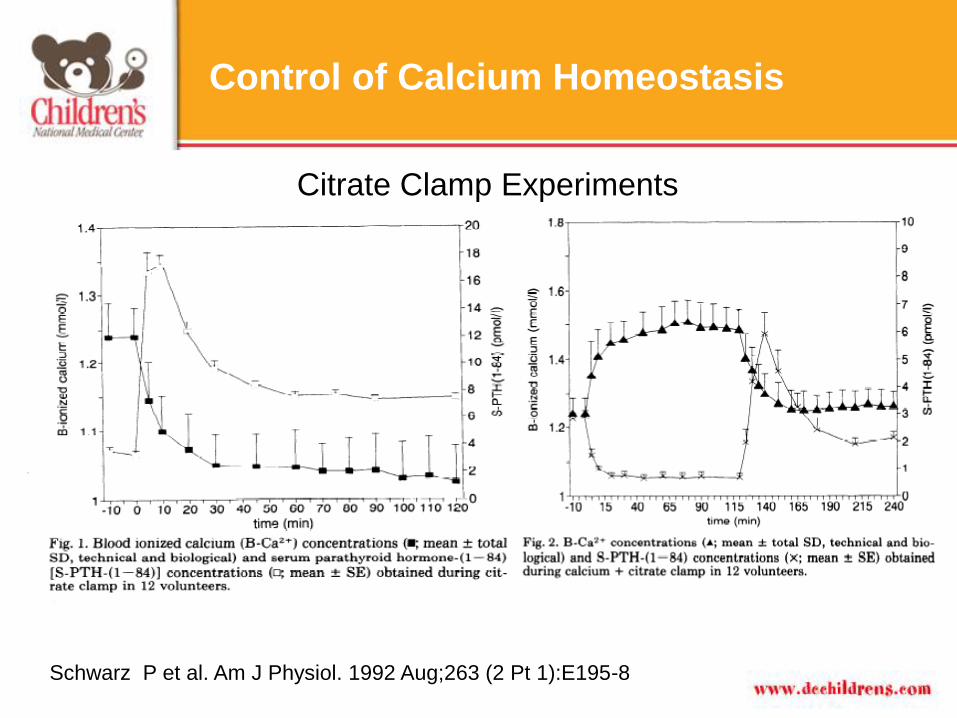
Control of Calcium Homeostasis
Schwarz P et al. Am J Physiol. 1992 Aug;263 (2 Pt 1):E195-8
Citrate Clamp Experiments
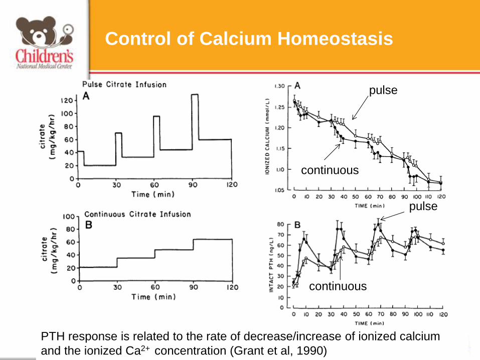
Control of Calcium Homeostasis
PTH response is related to the rate of decrease/increase of ionized calcium
and the ionized Ca2+ concentration (Grant et al, 1990)
pulse
continuous
pulse
continuous
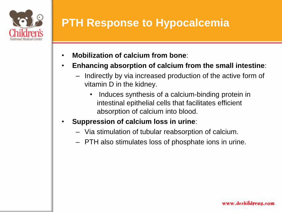
PTH Response to Hypocalcemia
• Mobilization of calcium from bone:
• Enhancing absorption of calcium from the small intestine:
– Indirectly by via increased production of the active form of
vitamin D in the kidney.
• Induces synthesis of a calcium-binding protein in
intestinal epithelial cells that facilitates efficient
absorption of calcium into blood.
• Suppression of calcium loss in urine:
– Via stimulation of tubular reabsorption of calcium.
– PTH also stimulates loss of phosphate ions in urine.

Therapeutic Apheresis Citrate Related
Complications in Children
• Published experience in children is limited
• Most publications relate to hematopoietic stem cell
collection
• Limited information on the risk of complications in
children, including normal physiologic response to
electrolyte disturbances

Factors Affecting The Development of
Citrate Toxicity
• Type of Replacement Fluid
– Duration of procedure
– Rate of infusion
• Type of Anticoagulation
• Individual Variation
– Metabolism by liver and other tissues
– Elimination by the kidney
• Underlying Medical Conditions
• Age
– Especially newborns within the first 3 days of life,
(Nincsoy et al., 1982)
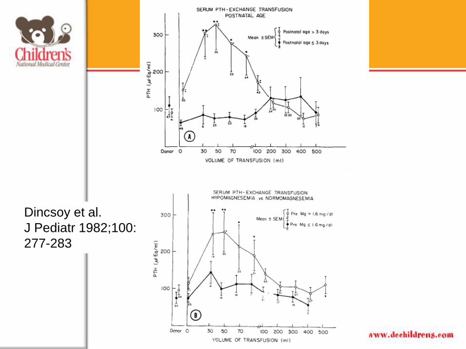
Dincsoy et al.
J Pediatr 1982;100:
277-283
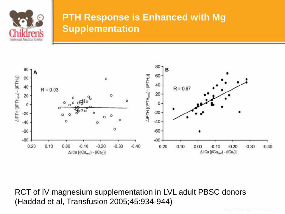
PTH Response is Enhanced with Mg
Supplementation
RCT of IV magnesium supplementation in LVL adult PBSC donors
(Haddad et al, Transfusion 2005;45:934-944)
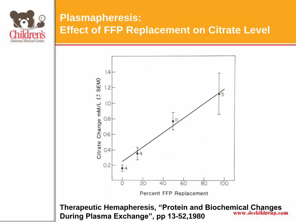
Plasmapheresis:
Effect of FFP Replacement on Citrate Level
Therapeutic Hemapheresis, “Protein and Biochemical Changes
During Plasma Exchange”, pp 13-52,1980
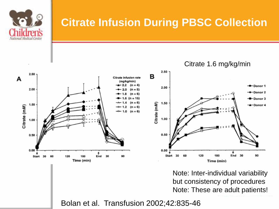
Citrate Infusion During PBSC Collection
Note: Inter-individual variability
but consistency of procedures
Note: These are adult patients!
Citrate 1.6 mg/kg/min
Bolan et al. Transfusion 2002;42:835-46
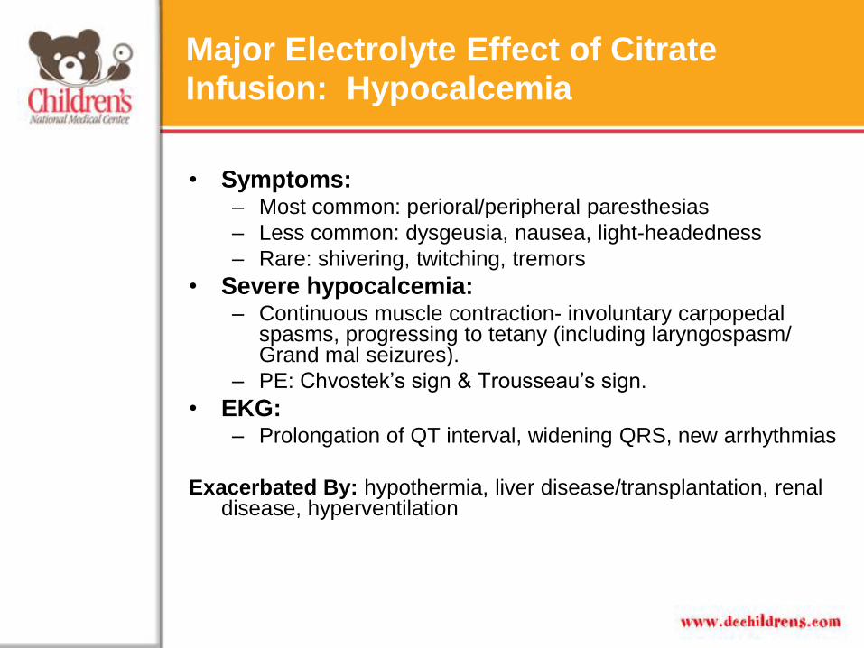
Major Electrolyte Effect of Citrate Infusion: Hypocalcemia
• Symptoms: – Most common: perioral/peripheral paresthesias
– Less common: dysgeusia, nausea, light-headedness
– Rare: shivering, twitching, tremors
• Severe hypocalcemia: – Continuous muscle contraction- involuntary carpopedal
spasms, progressing to tetany (including laryngospasm/ Grand mal seizures).
– PE: Chvostek’s sign & Trousseau’s sign.
• EKG: – Prolongation of QT interval, widening QRS, new arrhythmias
Exacerbated By: hypothermia, liver disease/transplantation, renal disease, hyperventilation
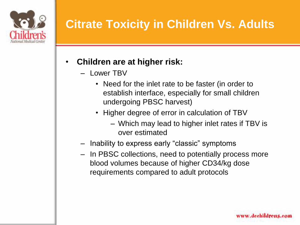
Citrate Toxicity in Children Vs. Adults
• Children are at higher risk:
– Lower TBV
• Need for the inlet rate to be faster (in order to
establish interface, especially for small children
undergoing PBSC harvest)
• Higher degree of error in calculation of TBV
– Which may lead to higher inlet rates if TBV is
over estimated
– Inability to express early “classic” symptoms
– In PBSC collections, need to potentially process more
blood volumes because of higher CD34/kg dose
requirements compared to adult protocols
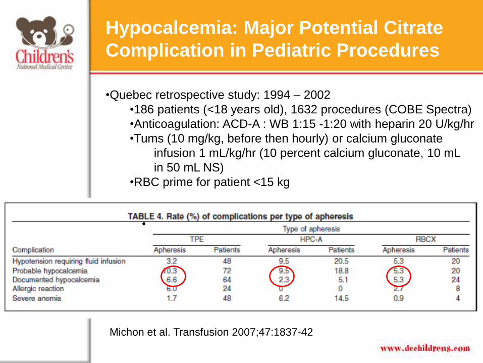
Hypocalcemia: Major Potential Citrate
Complication in Pediatric Procedures
Michon et al. Transfusion 2007;47:1837-42
•Quebec retrospective study: 1994 – 2002
•186 patients (<18 years old), 1632 procedures (COBE Spectra)
•Anticoagulation: ACD-A : WB 1:15 -1:20 with heparin 20 U/kg/hr
•Tums (10 mg/kg, before then hourly) or calcium gluconate
infusion 1 mL/kg/hr (10 percent calcium gluconate, 10 mL
in 50 mL NS)
•RBC prime for patient <15 kg
•
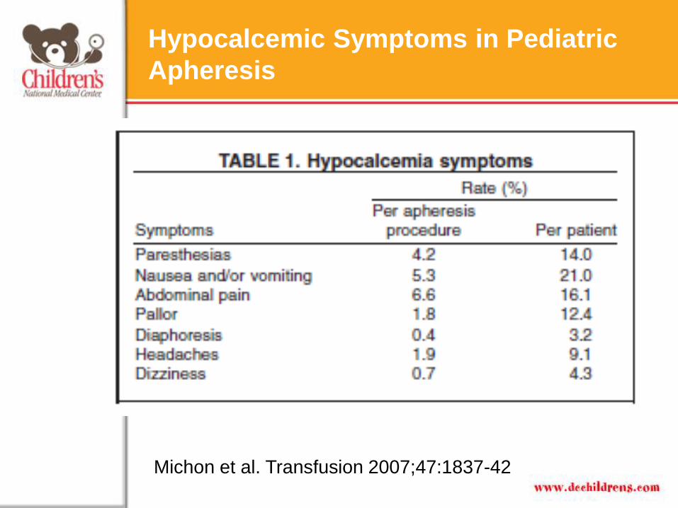
Hypocalcemic Symptoms in Pediatric
Apheresis
Michon et al. Transfusion 2007;47:1837-42
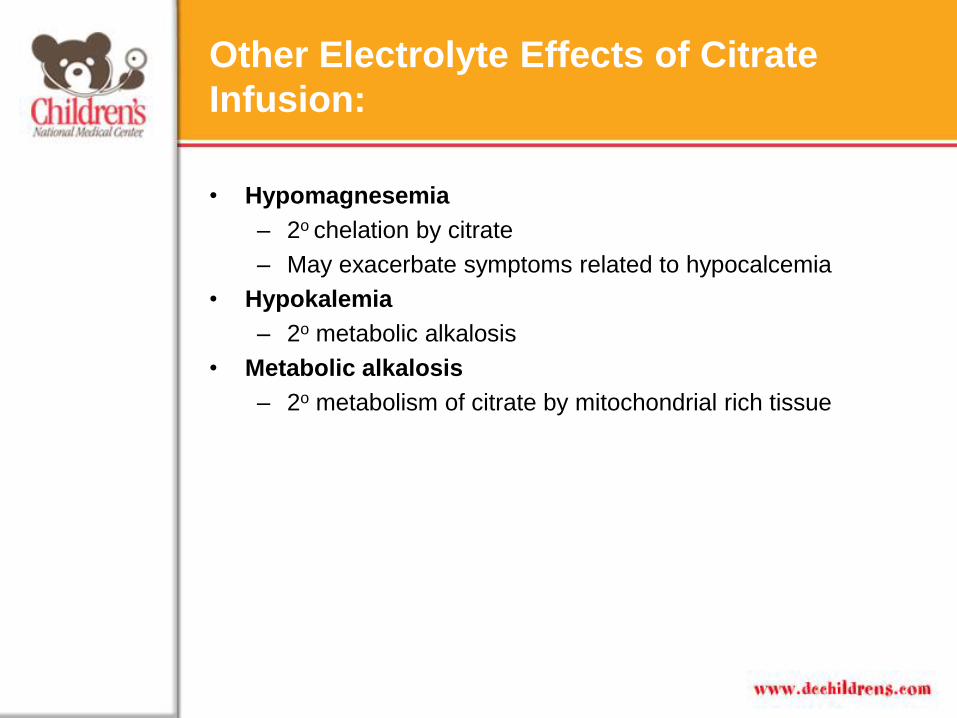
Other Electrolyte Effects of Citrate
Infusion:
• Hypomagnesemia
– 2o chelation by citrate
– May exacerbate symptoms related to hypocalcemia
• Hypokalemia
– 2o metabolic alkalosis
• Metabolic alkalosis
– 2o metabolism of citrate by mitochondrial rich tissue

Hypokalemia
• Symptoms: Musculoskeletal = weakness, cramps,
paresthesias, paralysis; GI = nausea, anorexia, vomiting,
diarrhea; CNS = lethargy, confusion.
• Physical findings: hyporeflexia
• Lab findings: low urine specific gravity; low serum K+ (<3.5
mEq/L requires therapy).
• EKG findings: flat or inverted T waves; ST depression.
Less commonly: QT Long, wide QRS, U wave.
Hypokalemia
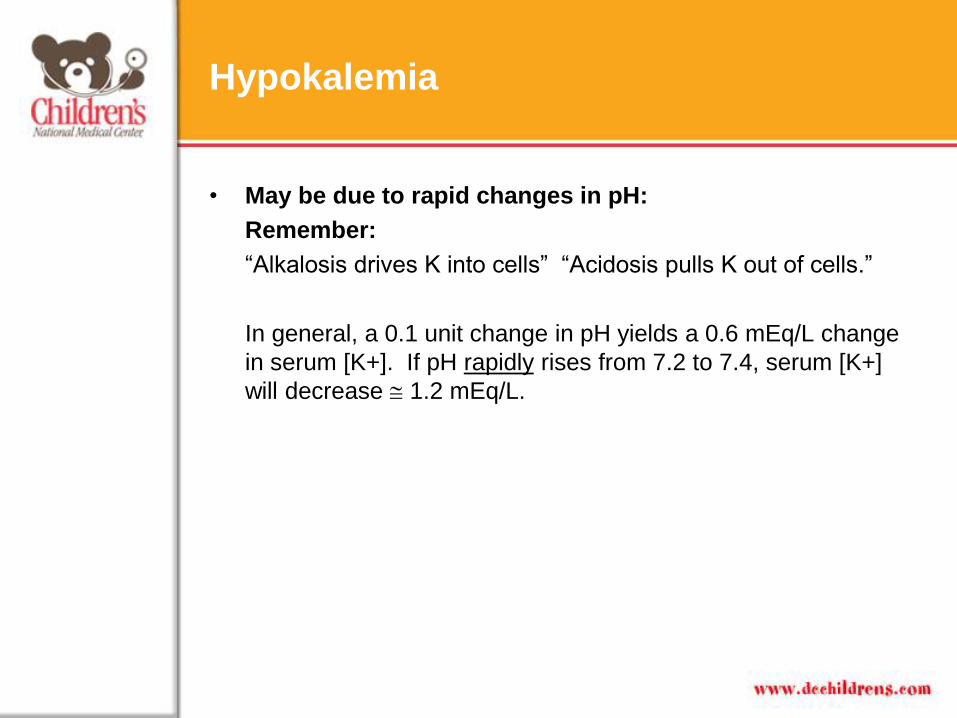
• May be due to rapid changes in pH:
Remember:
“Alkalosis drives K into cells” “Acidosis pulls K out of cells.”
In general, a 0.1 unit change in pH yields a 0.6 mEq/L change
in serum [K+]. If pH rapidly rises from 7.2 to 7.4, serum [K+]
will decrease 1.2 mEq/L.
Hypokalemia: how much K+ to give?
• Under normal circumstances: ~ 2 meq K/100 mL
• Thus, under ordinary conditions where a patient has a normal
cardiovascular status, and normal renal function, adequate
electrolytes will be provided using an intravenous fluid
containing ¼ normal saline (Na = approx. 35 mEq/l), with 20
mEq of potassium per liter.
• At CNMC, if hypokalemia is present, we will ask primary
service to add K to the maintenance fluids during the
procedure or add 10-20 meq K/L in our calcium gluconate
solution, patient weight/hr.
– Max: 0.5 meq/Kg/hr
ex. 20 kg patient, at 20 mL/hr with 10-20 meq K+/L will
receive only 2-4 meq/20 kg/hr = 0.1-0.2 meq/kg/hr
Caveat: Need to monitor
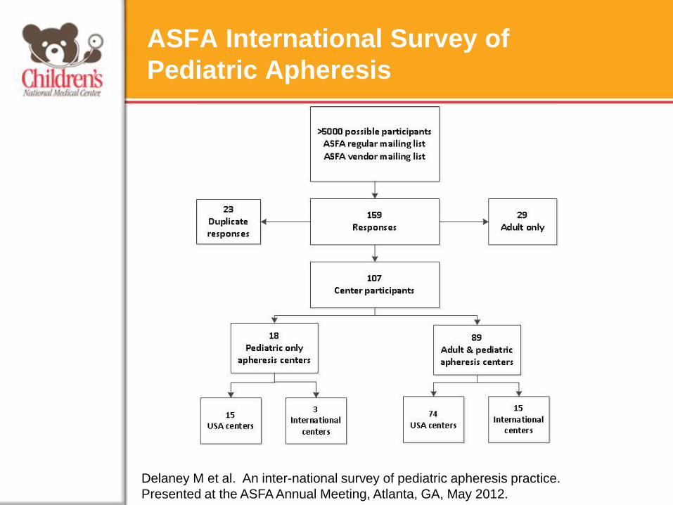
ASFA International Survey of
Pediatric Apheresis
Delaney M et al. An inter-national survey of pediatric apheresis practice.
Presented at the ASFA Annual Meeting, Atlanta, GA, May 2012.

ASFA International Survey of Pediatric Apheresis
Type of Procedure % of Responders (Anticoagulation)
TPE and RCE 78.3% (Citrate)
14.2% (Citrate/Heparin)
PBSC collection 65.7% (Citrate)
28.6% (Citrate/Heparin)
ECP 19.4% (Citrate)
11.1% (Heparin/Citrate)
63.8% (Heparin)
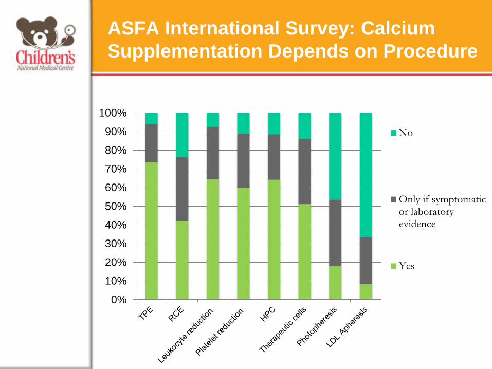
ASFA International Survey: Calcium
Supplementation Depends on Procedure
0%
10%
20%
30%
40%
50%
60%
70%
80%
90%
100%
No
Only if symptomaticor laboratoryevidence
Yes
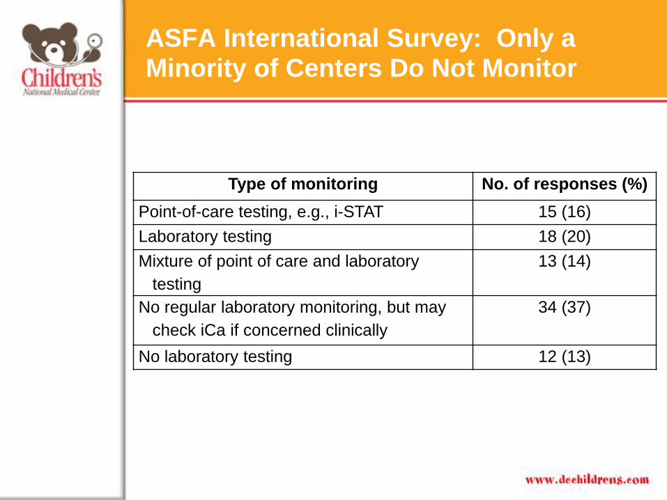
ASFA International Survey: Only a Minority of Centers Do Not Monitor
Type of monitoring No. of responses (%)
Point-of-care testing, e.g., i-STAT 15 (16)
Laboratory testing 18 (20)
Mixture of point of care and laboratory
testing
13 (14)
No regular laboratory monitoring, but may
check iCa if concerned clinically
34 (37)
No laboratory testing 12 (13)

ASFA International Survey of Pediatric
Apheresis: Of Those That Routinely Use
Calcium Supplementation
0.0
10.0
20.0
30.0
40.0
50.0
60.0
70.0
80.0
TPE, albuminreplacement
TPE, plasmareplacement
RCE HPC
Re
sp
on
din
g C
en
ters
(%
)
Calcium gluconate
Calcium chloride
Calcium carbonate
n=98 n=101 n=81 n=85
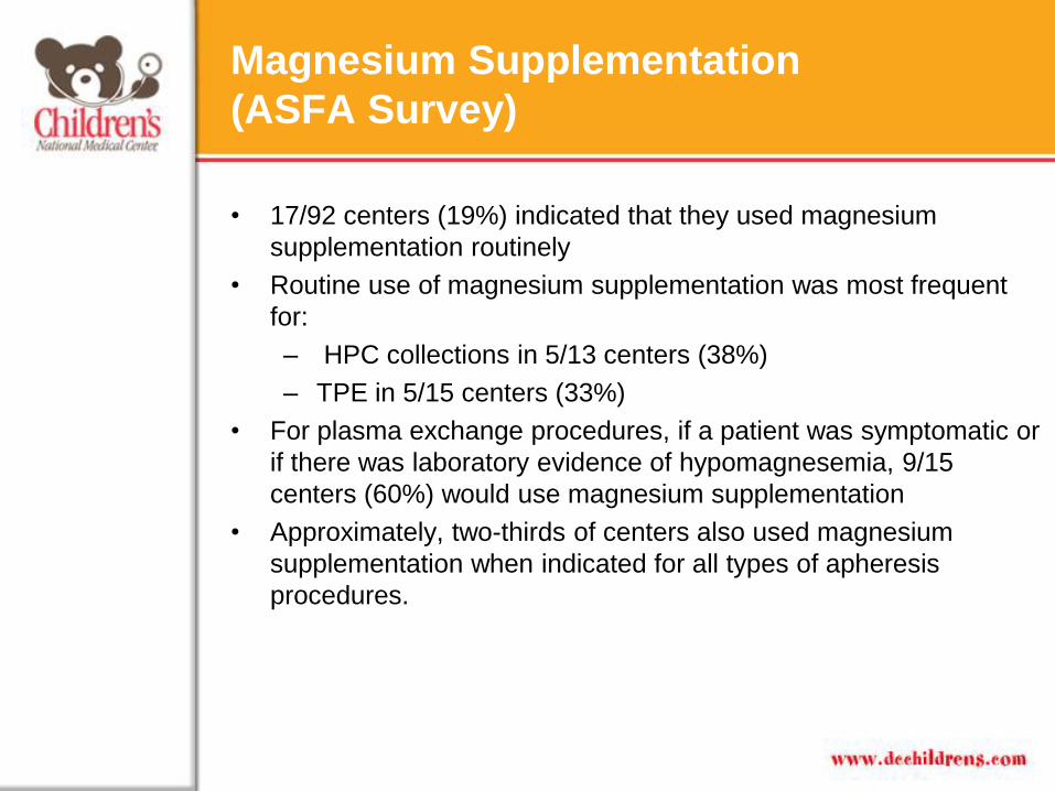
Magnesium Supplementation
(ASFA Survey)
• 17/92 centers (19%) indicated that they used magnesium
supplementation routinely
• Routine use of magnesium supplementation was most frequent
for:
– HPC collections in 5/13 centers (38%)
– TPE in 5/15 centers (33%)
• For plasma exchange procedures, if a patient was symptomatic or
if there was laboratory evidence of hypomagnesemia, 9/15
centers (60%) would use magnesium supplementation
• Approximately, two-thirds of centers also used magnesium
supplementation when indicated for all types of apheresis
procedures.
Potassium Supplementation
(ASFA Survey)
• Potassium supplementation was used for TPE by 19/91 (21%)
centers
• Potassium supplementation was used approximately 6 times
more frequently with albumin replacement than with FFP
replacement.
• However, once a patient became symptomatic, approximately
40% of respondents (n=19) used potassium supplementation,
regardless of the type of replacement solution.
Maneuvers to Minimize Hypocalcemia
• PO Calcium carbonate
– Pros:
• IV not needed (Especially with current electrolyte
shortages)
– Cons:
• Potentially not well tolerated
• Cannot, if NPO, especially in the younger children
Maneuvers to Minimize Hypocalcemia
• Calcium carbonate regimens (selected):
– 10 mg/kg initially, then every hr (Michon et al, 2007)
– For children >11 years, 2 extra strength Tums (300 mg
calcium carbonate) pre procedure, then 2 every 30 min to 1 hr
(max 10, CNMC unpublished)
• Tums smoothies may be used (CNMC)
• Other
– Oral calcium gluconate 10%, 100 – 200 mg/kg/hr (Urban et al
1997, pts <20 kg, 2 BVs processed)
– Isotonic sports drink (adults, Kishimoto et al, 2002)
Maneuvers to Minimize Hypocalcemia
• Continuous IV infusion
– Procedures using only ACD-A (or ACD-A/heparin)
– Calcium gluconate
• 1 mL/kg/hr (10 mL of 10% calcium gluconate in 50
mL NS) (Michon et al, 2007),
• 20 mg/mL calcium gluconate in NS, run at patient
weight (kg)/hr (CNMC)
– Calcium chloride
• Administer 0.6 mg elemental calcium ion per every
mL of ACD-A (1.2 mmol calcium/10 mmol citrate,
Bolan et al, 2004)
• Intermittent IV: calcium gluconate bolus (100 mg/10 mL
ampule, adults) over several minutes with development of
symptoms only (rare reports in children with little details on
concentration of calcium gluconate used)
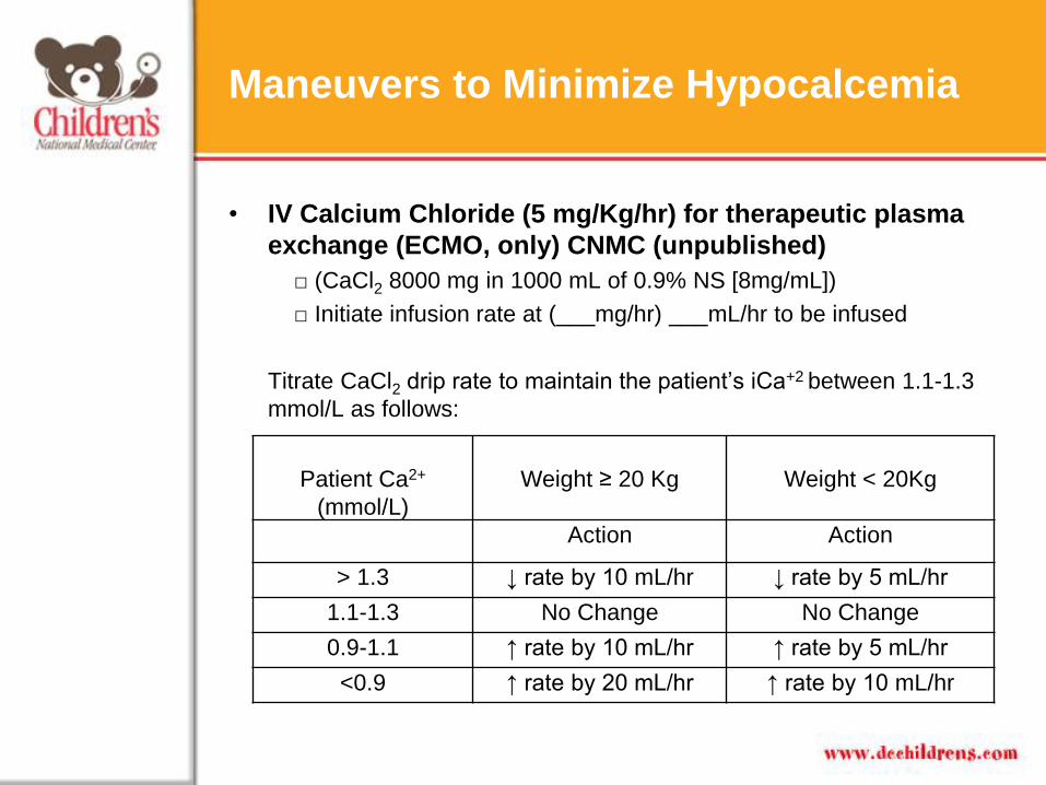
• IV Calcium Chloride (5 mg/Kg/hr) for therapeutic plasma
exchange (ECMO, only) CNMC (unpublished)
□ (CaCl2 8000 mg in 1000 mL of 0.9% NS [8mg/mL])
□ Initiate infusion rate at (___mg/hr) ___mL/hr to be infused
Titrate CaCl2 drip rate to maintain the patient’s iCa+2 between 1.1-1.3
mmol/L as follows:
Maneuvers to Minimize Hypocalcemia
Patient Ca2+
(mmol/L)
Weight ≥ 20 Kg
Weight < 20Kg
Action Action
> 1.3 ↓ rate by 10 mL/hr ↓ rate by 5 mL/hr
1.1-1.3 No Change No Change
0.9-1.1 ↑ rate by 10 mL/hr ↑ rate by 5 mL/hr
<0.9 ↑ rate by 20 mL/hr ↑ rate by 10 mL/hr

• Electrolyte Supplementation in Albumin Replacement
Solution During Plasma exchange
– Kim (2010)
• Especially with > 0.8 mL ACD-A /min/L TBV
• Albumin replacement, 2-3 mL 10% calcium gluconate
can be added to each 250 mL bottle.
• FFP replacement, 2-3 mL 10% calcium gluconate
bolus (over 10 minutes/continuous infusion can be
given with each 100 mL FFP)
– Krishnan RG & Coulthard MG (2007)
• Supplemented albumin replacement by adding 2
mmol/l calcium chloride and 0.8 mmol/l magnesium
sulphate
Maneuvers to Minimize Hypocalcemia
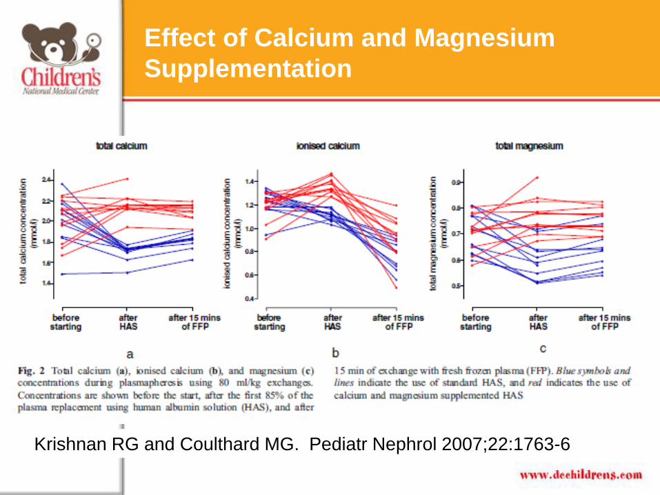
Effect of Calcium and Magnesium
Supplementation
Krishnan RG and Coulthard MG. Pediatr Nephrol 2007;22:1763-6
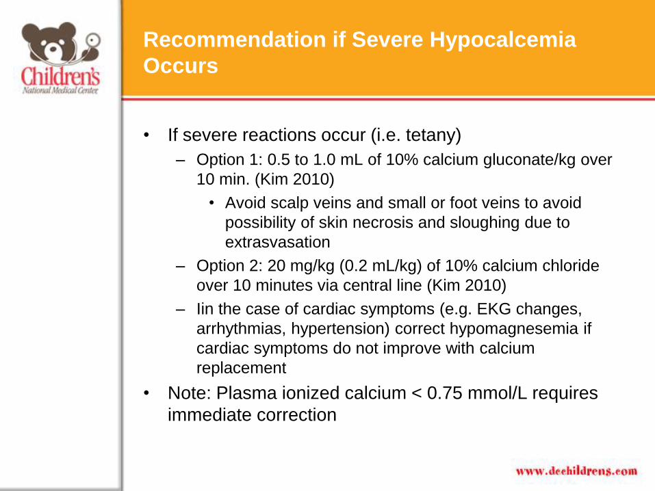
• If severe reactions occur (i.e. tetany)
– Option 1: 0.5 to 1.0 mL of 10% calcium gluconate/kg over
10 min. (Kim 2010)
• Avoid scalp veins and small or foot veins to avoid
possibility of skin necrosis and sloughing due to
extrasvasation
– Option 2: 20 mg/kg (0.2 mL/kg) of 10% calcium chloride
over 10 minutes via central line (Kim 2010)
– Iin the case of cardiac symptoms (e.g. EKG changes,
arrhythmias, hypertension) correct hypomagnesemia if
cardiac symptoms do not improve with calcium
replacement
• Note: Plasma ionized calcium < 0.75 mmol/L requires
immediate correction
Recommendation if Severe Hypocalcemia
Occurs
• Another caveat: Cardiac Patients:
– The heart denervated by either transplantation or
pharmacological blockade is extremely sensitive to
lowered ionized calcium (Corbascio and Smith, 1967;
Smith and Hurley, 1969)
Maneuvers to Minimize Hypocalcemia
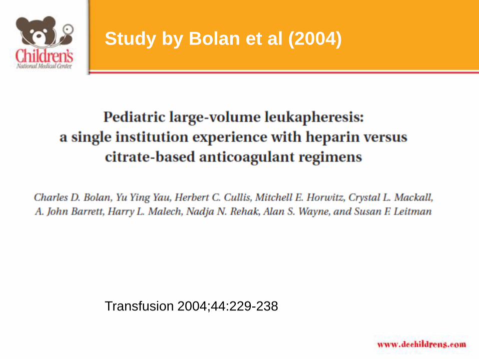
Study by Bolan et al (2004)
Transfusion 2004;44:229-238
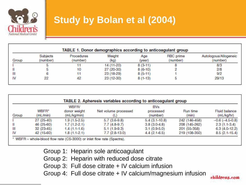
Study by Bolan et al (2004)
Group 1: Heparin sole anticoagulant
Group 2: Heparin with reduced dose citrate
Group 3: Full dose citrate + IV calcium infusion
Group 4: Full dose citrate + IV calcium/magnesium infusion

• Magnesium sulfate infusion: (Bolan et al, 2004)
– Created by pharmacy
• Magnesium solution: 6 mL (24 meq) of 50 percent
magnesium sulfate (American Pharmaceutical
Partners) to a final volume of 98.6 mL of half normal
saline, providing 3 mg of magnesium ion per mL
– Administered at 0.18 mg of magnesium ion per
mL of ACD-A (0.6 mmol magnesium/10 mmol
citrate).
• Calcium solution: were prepared from 10-percent
vials of calcium chloride to a final concentration of
2 mg calcium ion per mL (two 10-mL CaCl2 vials
added to 250 mL of half normal saline).
– Administered at 0.6 mg of calcium ion per mL of
ACD-A (1.2 mmol calcium/10 mmol citrate)
Maneuvers to Minimize Hypomagnesemia

• Example:
– ACD-A inlet flow rate of 2 mL/min on the Spectra,
A) Want: 0.6 mg Ca2+ ion per 1 mL ACD-A
Have: Ca2+ solution at 2 mg/mL
Know: 2 mL/min (ACD-A) x 60 min/hr = 120 mL/hr (ACD-A)
120 mL/hr (ACD-A) x 0.6 mg Ca2+ ion/mL ACD-A =
72 mg Ca2+ needed/hr
You have Ca2+ solution at 2 mg/mL
Therefore you need 72/2 or 36 mL/hr Ca2+ solution/hr!
• .
Maneuvers to Minimize Hypomagnesemia
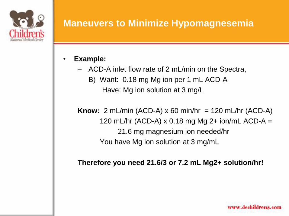
• Example:
– ACD-A inlet flow rate of 2 mL/min on the Spectra,
B) Want: 0.18 mg Mg ion per 1 mL ACD-A
Have: Mg ion solution at 3 mg/L
Know: 2 mL/min (ACD-A) x 60 min/hr = 120 mL/hr (ACD-A)
120 mL/hr (ACD-A) x 0.18 mg Mg 2+ ion/mL ACD-A =
21.6 mg magnesium ion needed/hr
You have Mg ion solution at 3 mg/mL
Therefore you need 21.6/3 or 7.2 mL Mg2+ solution/hr!
Maneuvers to Minimize Hypomagnesemia
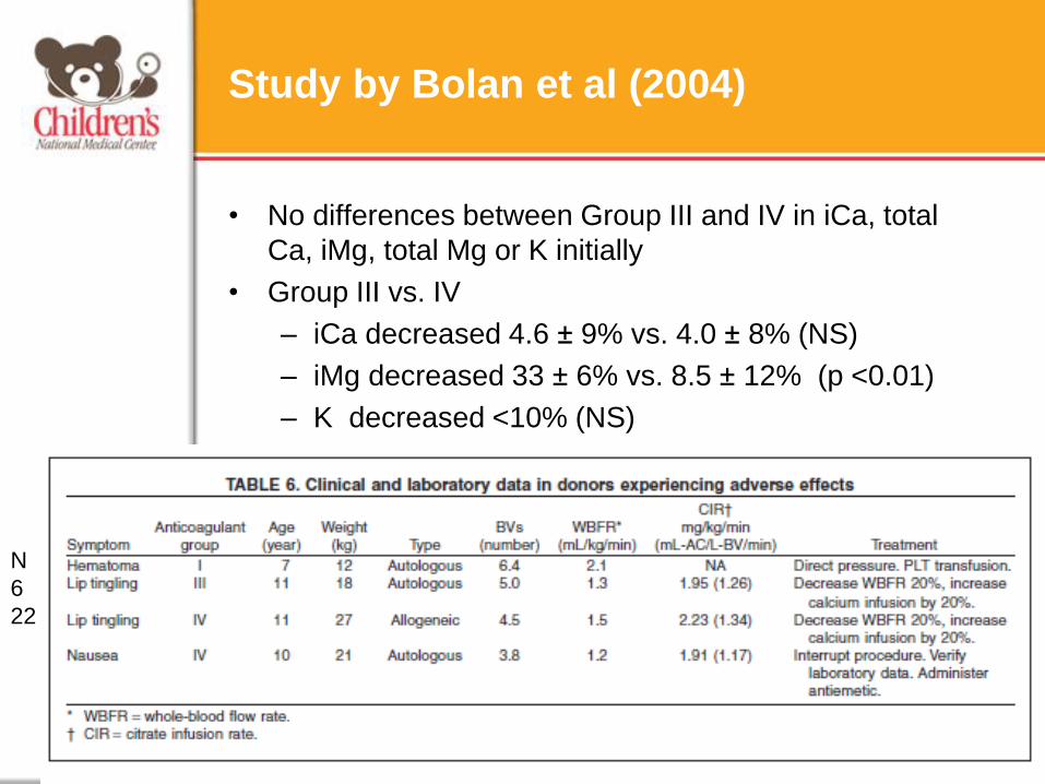
Study by Bolan et al (2004)
• No differences between Group III and IV in iCa, total
Ca, iMg, total Mg or K initially
• Group III vs. IV
– iCa decreased 4.6 ± 9% vs. 4.0 ± 8% (NS)
– iMg decreased 33 ± 6% vs. 8.5 ± 12% (p <0.01)
– K decreased <10% (NS)
N
6
22
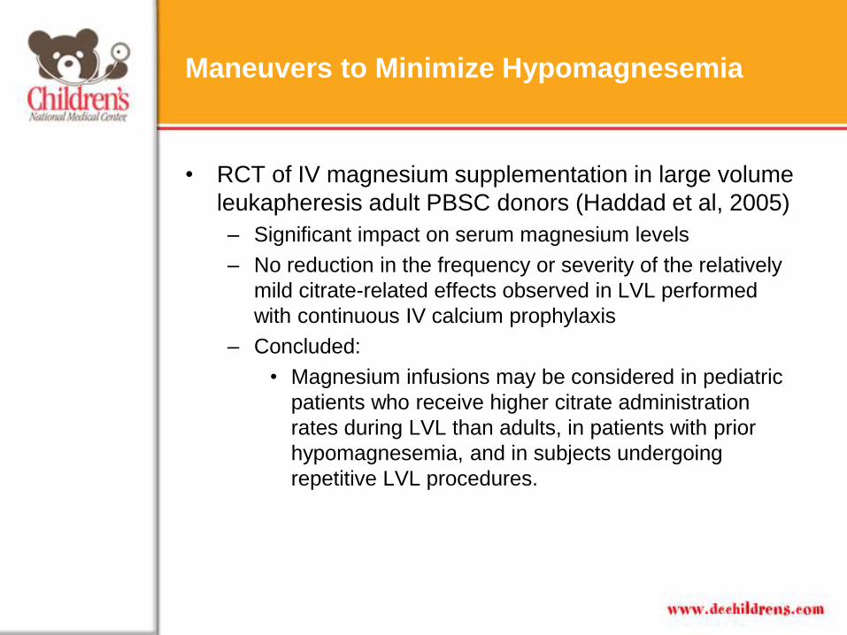
• RCT of IV magnesium supplementation in large volume
leukapheresis adult PBSC donors (Haddad et al, 2005)
– Significant impact on serum magnesium levels
– No reduction in the frequency or severity of the relatively
mild citrate-related effects observed in LVL performed
with continuous IV calcium prophylaxis
– Concluded:
• Magnesium infusions may be considered in pediatric
patients who receive higher citrate administration
rates during LVL than adults, in patients with prior
hypomagnesemia, and in subjects undergoing
repetitive LVL procedures.
Maneuvers to Minimize Hypomagnesemia
Conclusions
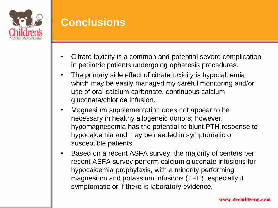
• Citrate toxicity is a common and potential severe complication
in pediatric patients undergoing apheresis procedures.
• The primary side effect of citrate toxicity is hypocalcemia
which may be easily managed my careful monitoring and/or
use of oral calcium carbonate, continuous calcium
gluconate/chloride infusion.
• Magnesium supplementation does not appear to be
necessary in healthy allogeneic donors; however,
hypomagnesemia has the potential to blunt PTH response to
hypocalcemia and may be needed in symptomatic or
susceptible patients.
• Based on a recent ASFA survey, the majority of centers per
recent ASFA survey perform calcium gluconate infusions for
hypocalcemia prophylaxis, with a minority performing
magnesium and potassium infusions (TPE), especially if
symptomatic or if there is laboratory evidence.

References
1. Bolan CD, Yau YY, Cullis HC, Horwitz ME, Mackall CL, Barrett AJ, Malech HL,
Rehak NN, Wayne AS, Leitman SF. Pediatric large-volume leukapheresis: a
single institution experience with heparin versus citrate-based anticoagulant
regimens. Transfusion. 2004 Feb;44(2):229-38.
2. Bolan CD, Wesley RA, Yau YY, Cecco SA, Starling J, Oblitas JM, Rehak NN,
Leitman SF. Randomized placebo-controlled study of oral calcium carbonate
administration in plateletpheresis: I. Associations with donor symptoms. Transfusion. 2003 Oct;43(10):1403-13.
3. Bolan CD, Cecco SA, Wesley RA, Horne M, Yau YY, Remaley AT, Childs RW,
Barrett AJ, Rehak NN, Leitman SF. Controlled study of citrate effects and
response to i.v. calcium administration during allogeneic peripheral blood
progenitor cell donation.Transfusion. 2002 Jul;42(7):935-46
4. Corbascio AN, Smith NT. Hemodynamic effects of experimental hypercitremia.
Anesthesiol 1967;28:510.
5. Delaney M, Capocelli KE, Eder AF, Schneiderman J, Schwartz J, Sloan SR,
Wong ECC, Kim HC. An international survey of pediatric apheresis practice.
Presented at the ASFA Annual Meeting, Atlanta, GA, May 2012.
6. Dincsoy MY, Tsang RC, Laskarzewski P, Chen MH, Chen IW, Lo D, Donovan
EF. The role of postnatal age and magnesium on parathyroid hormone
responses during "exchange" blood transfusion in the newborn period.J
Pediatr. 1982 Feb;100(2):277-83.
6. Glusker JP. Citrate conformation and chelation: enzymatic implications Acc
Chem Res 1980;13:345-352.
7. Gorlin JB. Therapeutic plasma exchange and cytapheresis in pediatric patients.
Transfus Sci 1999;21:21–39.
8. Grant FD, Conlin PR, Brown EM. Rate and concentration dependence of
parathyroid hormone dynamics during stepwise changes in serum ionized
calcium in normal humans. J Clin Endocrinol Metab. 1990 Aug;71(2):370-8.
9. Haddad S, Leitman SF, Wesley RA, Cecco S, Yau YY, Starling J, Rehak NN,
Bolan CD. Placebo-controlled study of intravenous magnesium
supplementation during large-volume leukapheresis in healthy allogeneic
donors.Transfusion. 2005 Jun;45(6):934-44.
10. Kim HC. Therapeutic apheresis in pediatric patients. In: McLeod BC,
Szczepiorkowski ZM, Weinstein R, Winters JL, editors. Apheresis: Principles
and Practice. Bethesda, MD: AABB Press; 2010. pp 445–464.
11. Kishimoto M, Ohto H, Shikama Y, Kikuta A, Kimijima I, Takenoshita S.
Treatment for the decline of ionized calcium levels during peripheral blood
progenitor cell harvesting. Transfusion. 2002 Oct;42(10):1340-7.
12. Krishnan RG and Coulthard MG. Minimising changes in plasma calcium and
magnesium concentrations during plasmapheresis. Pediatr Nephrol
2007;22:1763-6.
13. Mollison PL. The introduction of citrate as an anticoagulant for transfusion and
of glucose as a red cell preservative. Br J Haematol. 2000 Jan;108(1):13-8.
References
References
14. Sabbatani L. Arch Ital Biol 1901; 36 :397
15. Schwarz P, Sørensen HA, Transbøl I, McNair P. Regulation of acute
parathyroid hormone release in normal humans: combined calcium and citrate
clamp study. Am J Physiol. 1992 Aug;263(2 Pt 1):E195-8
16. Smith NT, Hurley EJ.Citrate infusion in dogs following cardiac
autotransplantation: studies on cardiovascular effects. Arch Surg 1969;98:44


