MANAGEMENT OF CA HYPOPHARYNX - Assiut University lectures/management of cancer... · patients with...
65
MANAGEMENT OF CA HYPOPHARYNX
Transcript of MANAGEMENT OF CA HYPOPHARYNX - Assiut University lectures/management of cancer... · patients with...
MANAGEMENT OF CA
HYPOPHARYNX
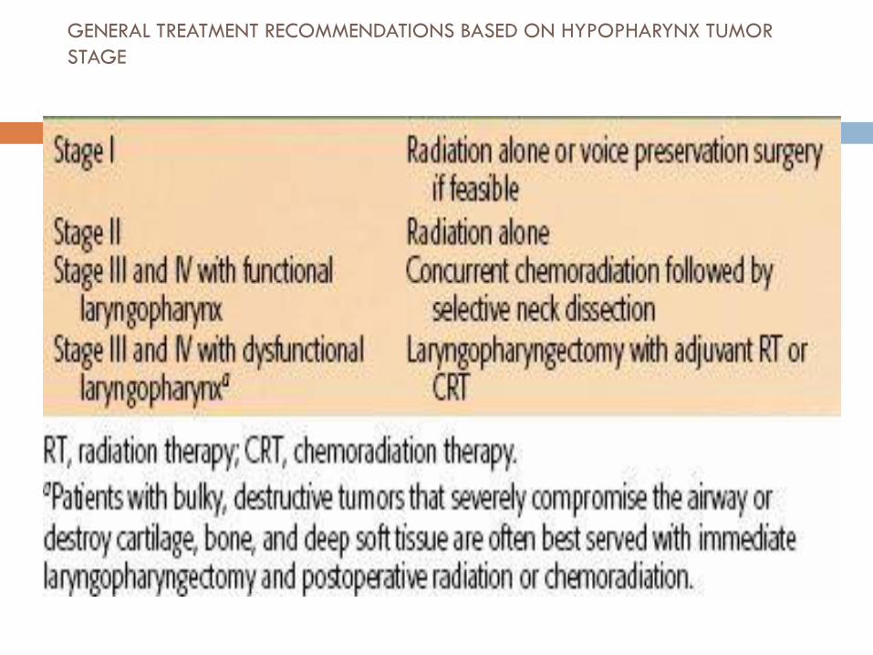
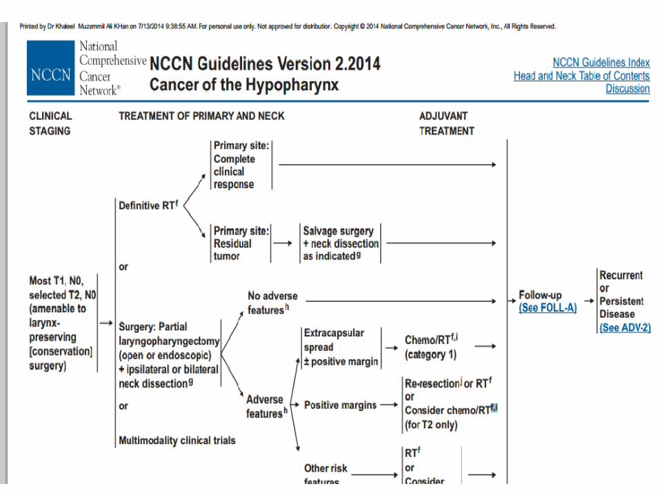
GENERAL TREATMENT RECOMMENDATIONS BASED ON HYPOPHARYNX TUMOR
STAGE
For patients presenting with early-stage definitive
radiotherapy alone or voice-preserving surgery are
viable and acceptable treatment options.
The vast majority of patients, however, present with
stage III or IV disease and warrant multimodality
treatment.
Primary Surgery
T1 and T2 Tumors
Indications for primary surgical management of
patients with early cancers of the hypopharynx
History of previous H&N radiation
Organ conservation approaches possible
Those who refuse radiation

Role of surgeon in Ca hypo pharynx patients
who undergo non operative treatment approaches
Performing
Endoscopic biopsy
Detailed assessment of tumor extent
Methods to secure the airway (tracheotomy or laser
debulking)
Methods to ensure adequate nutrition (gastrostomy)
Multidisciplinary oncologic follow-up after nonoperative
treatment
Selected T1 and T2 hypopharynx cancers may lend
themselves to surgical excision.
Favorable subsites include
Upper pyriform sinus
Posterior pharyngeal wall.
The standard supraglottic laryngectomy
encompasses the aryepiglottic fold and may be
extended to include part of the arytenoids, the
base of the tongue, and the upper pyriform sinus.
Relative contraindications to organ conservation surgery for
hypopharynx cancers
Cartilage invasion
Vocal fold fixation
Postcricoid invasion
Deep pyriform sinus invasion
Extension beyond the larynx
In recent years, advancements in organ preservation
surgery have included the use of
Transoral laser microsurgery
Transoral robotic surgery.
For selected cases, these approaches can achieve
oncologic tumor removal, while limiting normal tissue
disruption, thereby potentially avoiding
tracheostomy and the use of feeding tubes

Recent results have demonstrated that
appropriately selected T1 or T2 lesions can achieve
negative margins by transoral laser microsurgery or
transoral robotic surgery.
Oncologic outcomes appear similar to open
surgical approaches using this technique and are
likely accompanied by lower rates of permanent
gastrostomy tube or tracheostomy placement

FIVE-YEAR ONCOLOGIC OUTCOMES FOR TRANSORAL MICROLASER
SURGERY FOR
T1 OR T2 HYPOPHARYNGEAL TUMORS

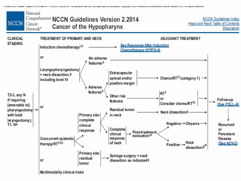
T3 or T4 Resectable Tumors
Most T3 and T4 hypopharynx cancers that are
treated surgically will require total laryngectomy
with efforts to preserve a posterior strip of the
hypopharynx spanning the oropharynx to the
esophagus.
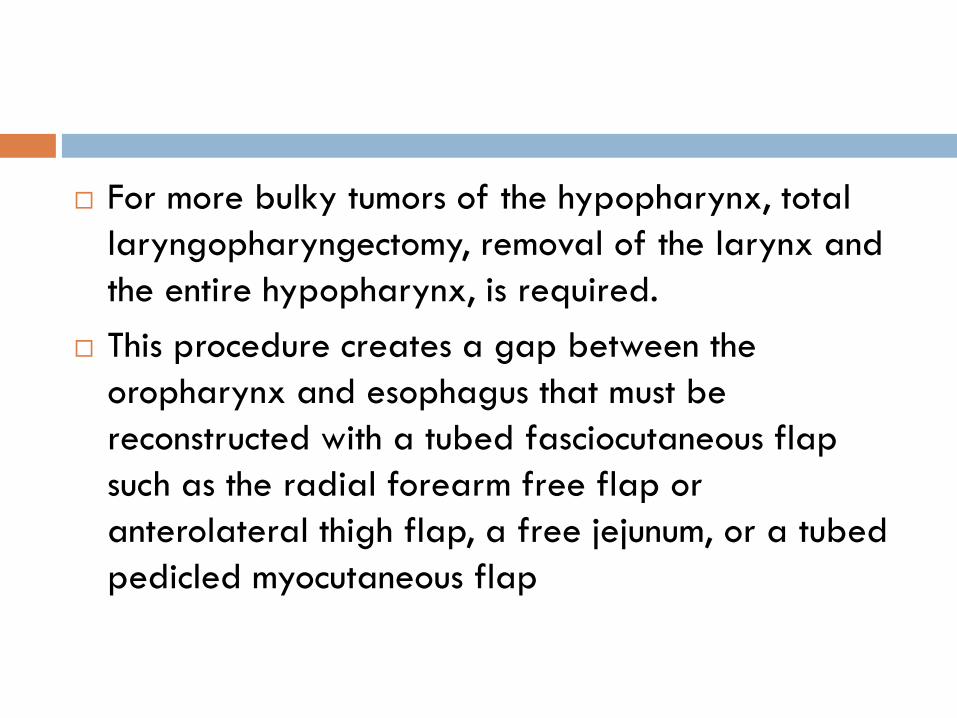
For more bulky tumors of the hypopharynx, total
laryngopharyngectomy, removal of the larynx and
the entire hypopharynx, is required.
This procedure creates a gap between the
oropharynx and esophagus that must be
reconstructed with a tubed fasciocutaneous flap
such as the radial forearm free flap or
anterolateral thigh flap, a free jejunum, or a tubed
pedicled myocutaneous flap

Types of surgeries in T3 T4 Ca
hypopharynx
For small medial and
anterior PF sinus lesions
Removes false cords
,epiglottis
,areyepiglottic folds ,
PF sinus but True vocal
cords are preserved
For more advanced
hypopharyngeal lesions
Total laryngectomy with
removal of variable
amount of pharyngeal
wall.
Partial laryngo pharyngectomy Total laryngo pharyngectomy

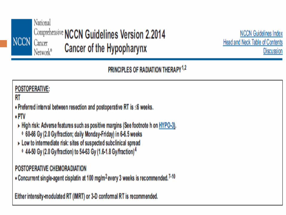
Postoperative Radiation Therapy
Most advanced hypopharynx cancers that are
treated with initial surgical resection
Postoperative radiation therapy can be added in
an effort to enhance locoregional control rates

Indications for postoperative radiation
T4 primary tumors
Close or positive microscopic margins
Cartilage or bony invasion
More than one metastatic lymph node
Extracapsular extension (ECE)
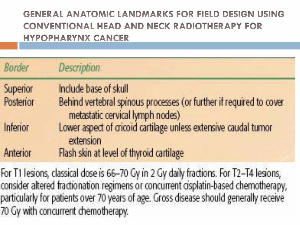
Conventional therapy involves the use of a shrinking-field technique to deliver 54 to 63 Gy to all areas at risk and a boost to 60 to 66 Gy to regions of ECE or positive margins. The entire cervical nodal chain from the skull base to the clavicle bilaterally should be included
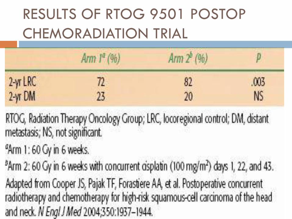
RESULTS OF RTOG 9501 POSTOP
CHEMORADIATION TRIAL

RESULTS OF EORTC POSTOPERATIVE CHEMORADIATION TRIAL

Definitive Radiation Therapy
T1 and T2 Tumors
Curative radiation therapy (RT) is generally the
preferred treatment option for patients with T1 or
T2 hypopharynx tumors.
Affords good potential for organ preservation
without compromise in clinical outcome
A classical course of radiation therapy for
hypopharynx cancer lasts 6 to 7 weeks, with
treatment delivered 5 days per week

Conventional treatment involves a shrinking-field
technique that initiates with opposed lateral fields
encompassing the primary tumor and upper neck
lymphatics with a matched anterior field to
complete treatment of the lower neck
One of the most common worldwide fractionation
regimens involves the delivery of 2 Gy daily
fractions to 70 Gy over 7 weeks

Digitally reconstructed radiograph depicting a classical lateral field designed to encompass the
T2 pyriform sinus cancer . plus bilateral cervical lymphatics from skull base to cricoid, with a
matching anterior low-neck field to extend the lymphatic coverage to the level of the clavicle

Due to the high likelihood of subclinical nodal
metastases even in the clinically N0 neck, patients
traditionally receive comprehensive radiation to
encompass nodal regions from the skull base to the
clavicle .
Due to the varying thicknesses of the head and
neck, custom compensators or wedges should be
used for the lateral fields to improve dose
homogeneity .
Shrinking field techniques to spare direct spinal
cord dose after approximately 45 Gy, as well as
final mucosal reductions after 54 to 60 Gy, are
often appropriate with posterior neck boosting, with
electrons to supplement posterior chain nodal
dosing without excessive dose to the spinal cord
Altered fractionation techniques
Hyperfractionation (e.g., 1.1–1.4 Gy twice daily)
Accelerated fractionation (e.g., 6 fraction per week
or concomitant boost regimens).
Improved locoregional control rates for H&N
cancer patients
A recent meta-analysis examined 15 trials that
compared conventional fractionation to altered
fractionation, either
Hyperfractionation or accelerated fractionation
Small but statistically significant survival benefit of
3.4% at 5 years with altered fractionation.
The benefit was higher with hyperfractionation
compared to accelerated fractionation and was
more pronounced for patients younger than age 50.
Early T-stage hypopharynx patients with N0 or N1
neck disease can be considered for treatment with
radiation alone or concurrent radiation plus
chemotherapy.
In this setting, gross disease should receive 70 Gy
and the contralateral neck (N0) should receive 50
to 54 Gy
T1N0 lesions, patients may achieve 5-year disease-
specific survival (DSS) on the order of 90%.
T2N0 lesions may achieve DSS above 70%.
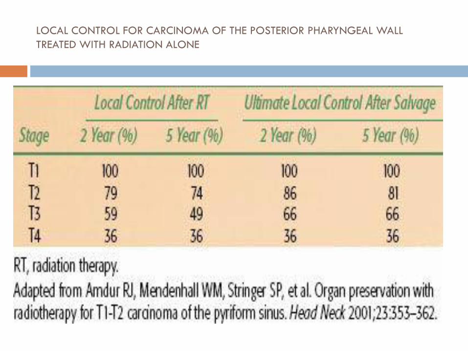
LOCAL CONTROL FOR CARCINOMA OF THE POSTERIOR PHARYNGEAL WALL
TREATED WITH RADIATION ALONE

CAUSE-SPECIFIC AND OVERALL SURVIVAL FOR CARCINOMA
OF THE PYRIFORM SINUS TREATED WITH RADIATION ALONE

IMRT
IMRT in H&N cancer as a means of diminishing
normal tissue toxicities
Particularly xerostomia resulting from irradiation of
major salivary glands.
Excellent candidates for IMRT include patients with
unilateral T1 to T3 primary lesions with N2b or less
neck disease.
A recent randomized trial of conventional
radiotherapy versus IMRT in patients with T1-4N0-3
oropharyngeal and hypopharyngeal tumors at high
risk for xerostomia highlighted the benefits of IMRT
for parotid sparing
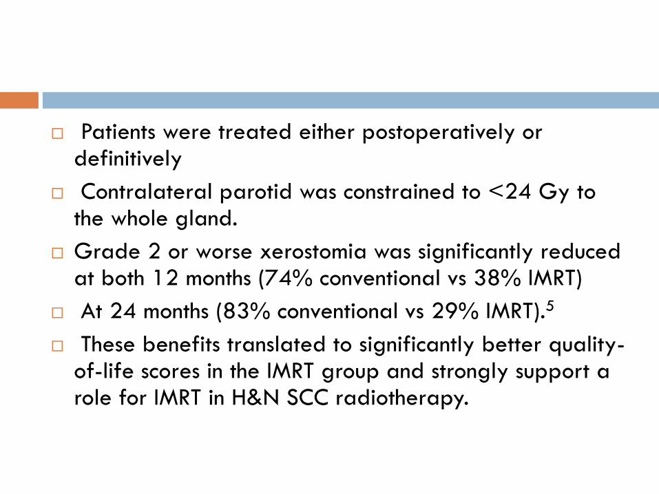
Patients were treated either postoperatively or definitively
Contralateral parotid was constrained to <24 Gy to the whole gland.
Grade 2 or worse xerostomia was significantly reduced at both 12 months (74% conventional vs 38% IMRT)
At 24 months (83% conventional vs 29% IMRT).5
These benefits translated to significantly better quality-of-life scores in the IMRT group and strongly support a role for IMRT in H&N SCC radiotherapy.
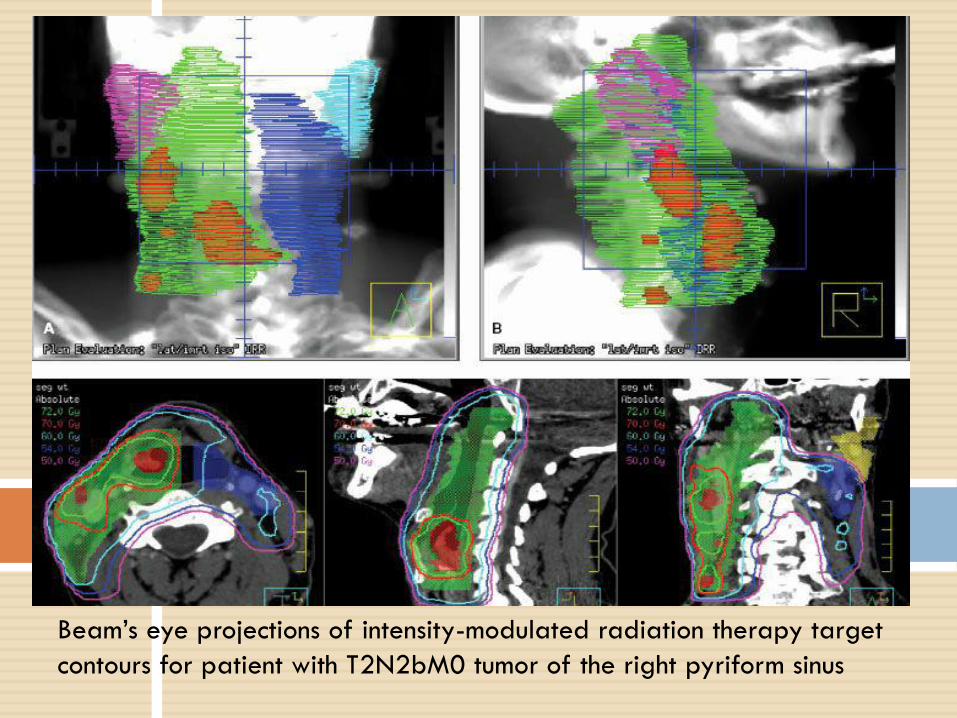
Beam’s eye projections of intensity-modulated radiation therapy target
contours for patient with T2N2bM0 tumor of the right pyriform sinus
T3 and T4 Tumors
Hypopharynx cancer patients who are technically
resectable may not undergo primary surgery.
These include age (e.g., patients over 70 to 80
years old),
Presence of significant medical comorbidities, or
patient unwillingness to accept total laryngectomy.
Curative-intent radiation or chemoradiation is often
pursued in these setting
Conventional radiation therapy commonly involves a
shrinking three-field technique to deliver approximately
70 Gy in 2-Gy daily fractions to areas of gross disease
and 50 to 60 Gy to areas of microscopic disease.
If patients are scheduled to undergo postradiotherapy
neck dissection, then gross nodal disease can be limited
to 60 to 63 Gy
If patients are not candidates for postradiotherapy
neck dissection, then gross nodal disease should be
carried to 70 Gy
In patients with adequate performance status,
concurrent chemoradiation strategies using
platinum-based chemotherapy should be
considered.
Once-daily radiation regimens without concurrent
chemotherapy may be quite reasonable for
hypopharynx patients over 70 years of age or for
those patients with modest performance status
Molecular-targeted therapies in the
treatment of H&N cancer patients
Another alternative to concomitant chemotherapy or
accelerated fractionation is the more recent
introduction of molecular-targeted therapies in the
treatment of H&N cancer patients
The most mature clinical dataset in H&N cancer
involves the use of EGFR inhibitors such as
cetuximab (monoclonal antibody against the EGFR)
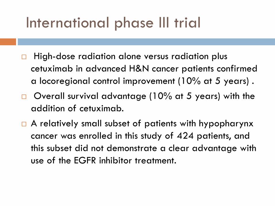
International phase III trial
High-dose radiation alone versus radiation plus
cetuximab in advanced H&N cancer patients confirmed
a locoregional control improvement (10% at 5 years) .
Overall survival advantage (10% at 5 years) with the
addition of cetuximab.
A relatively small subset of patients with hypopharynx
cancer was enrolled in this study of 424 patients, and
this subset did not demonstrate a clear advantage with
use of the EGFR inhibitor treatment.

Data from the NCDB Benchmark Reports addressing 16,136 cases diagnosed in 2000 to 2008 reveal the combination of radiation and chemotherapy to be the most common initial treatment overall for stage II (32.6%), stage III (47.8%), and stage IV (43.8%) disease.
Over 50% of stage III and stage IV cases received initial treatment with chemotherapy in some form—either alone or in combination with radiation or surgery.

It has been reported that approximately 35% to
45% of patients with advanced hypopharyngeal
tumors treated with concurrent chemoradiotherapy
utilizing IMRT can be expected to live 5 years, with
laryngeal preservation in approximately two-thirds
of survivors .

The stage-specific first course of treatment for hypopharyngeal cancer in the United States from
2000 to 2008 is presented with unknown stage excluded. S, surgery; XRT, radiotherapy; Ch,
chemotherapy. (From National Cancer Data Base. Commission on Cancer. American College of
Surgeons. Benchmark Reports

ONCOLOGIC OUTCOMES FOR PATIENTS UNDERGOING CONCURRENT
CHEMORADIOTHERAPY AND INTENSITY-MODULATED RADIATION THERAPY FOR
ADVANCED HYPOPHARYNGEAL CANCERS

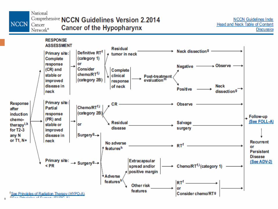
Induction Chemotherapy and Sequential
(Chemo)radiation
For patients with locoregionally advanced H&N
cancer.
EORTC conducted a randomized trial for patients
with tumors that would require total laryngectomy
as the surgical approach.
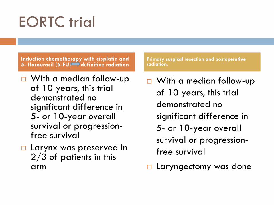
EORTC trial
With a median follow-up of 10 years, this trial demonstrated no significant difference in 5- or 10-year overall survival or progression-free survival
Larynx was preserved in 2/3 of patients in this arm
With a median follow-up
of 10 years, this trial
demonstrated no
significant difference in
5- or 10-year overall
survival or progression-
free survival
Laryngectomy was done
Induction chemotherapy with cisplatin and 5- florouracil (5-FU) definitive radiation
Primary surgical resection and postoperative radiation.

Five-year survival in the TPF arm was 52% versus 42% receiving PF, while no increased rates of gastric feeding tubes or tracheostomieswere noted between groups
TAX-324, which utilized similar induction
chemotherapy arms (TPF vs. PF), followed by
concurrent chemoradiotherapy with carboplatin.
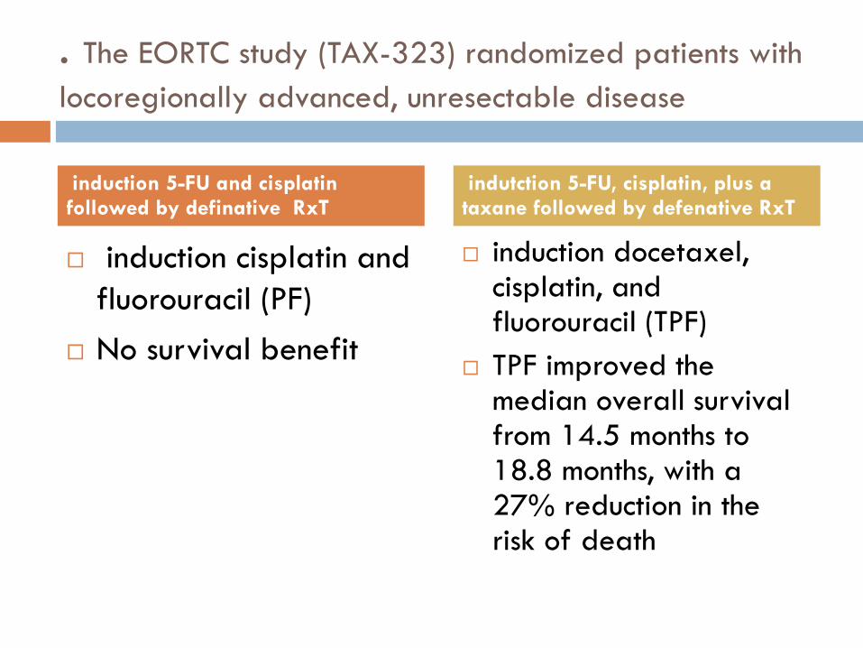
. The EORTC study (TAX-323) randomized patients with
locoregionally advanced, unresectable disease
induction cisplatin and
fluorouracil (PF)
No survival benefit
induction docetaxel, cisplatin, and fluorouracil (TPF)
TPF improved the median overall survival from 14.5 months to 18.8 months, with a 27% reduction in the risk of death
induction 5-FU and cisplatinfollowed by definative RxT
indutction 5-FU, cisplatin, plus a taxane followed by defenative RxT

Post radiotherapy Neck Dissection
N0 or N1 patients treated with primary
radiation or chemoradiation
Adjuvant neck dissection not necessary
N2 or N3 neck disease
Adjuvant neck dissection necessary
Detailed imaging of the neck 12 weeks postradiation with FDG-PET can serve as a valuable guide to help select those patients warranting adjuvant neck dissection.
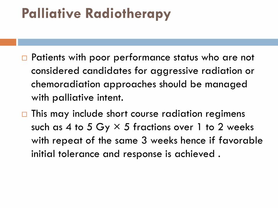
Palliative Radiotherapy
Patients with poor performance status who are not
considered candidates for aggressive radiation or
chemoradiation approaches should be managed
with palliative intent.
This may include short course radiation regimens
such as 4 to 5 Gy × 5 fractions over 1 to 2 weeks
with repeat of the same 3 weeks hence if favorable
initial tolerance and response is achieved .
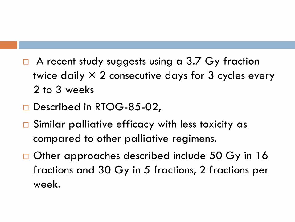
A recent study suggests using a 3.7 Gy fraction
twice daily × 2 consecutive days for 3 cycles every
2 to 3 weeks
Described in RTOG-85-02,
Similar palliative efficacy with less toxicity as
compared to other palliative regimens.
Other approaches described include 50 Gy in 16
fractions and 30 Gy in 5 fractions, 2 fractions per
week.
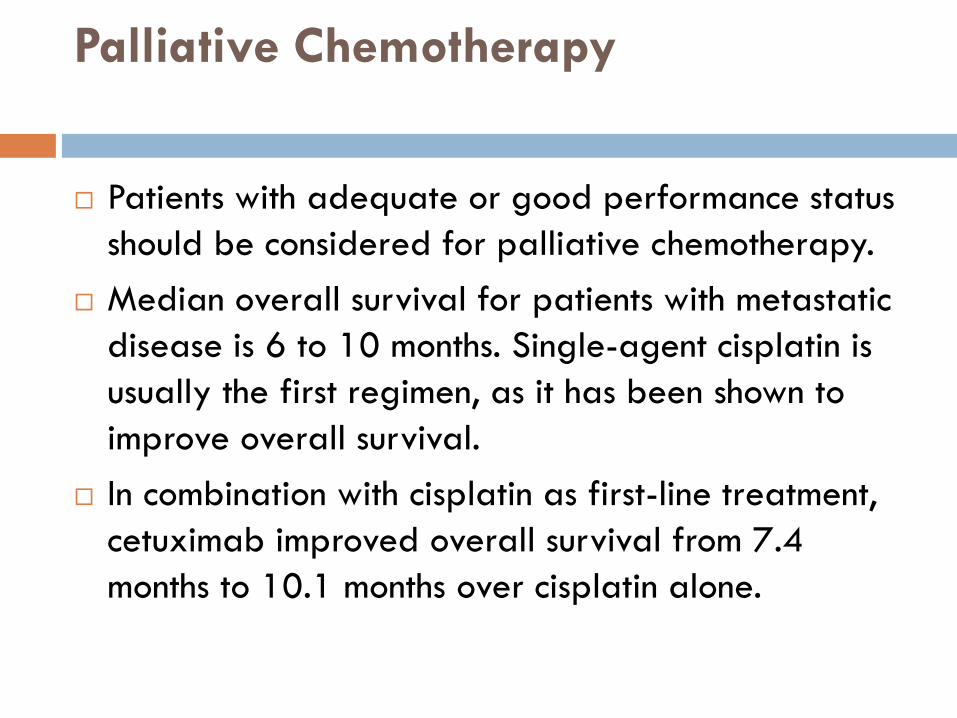
Palliative Chemotherapy
Patients with adequate or good performance status
should be considered for palliative chemotherapy.
Median overall survival for patients with metastatic
disease is 6 to 10 months. Single-agent cisplatin is
usually the first regimen, as it has been shown to
improve overall survival.
In combination with cisplatin as first-line treatment,
cetuximab improved overall survival from 7.4
months to 10.1 months over cisplatin alone.

MANAGEMENT OF RECURRENCE
After completion of treatment, patients should be
followed closely for signs of recurrent or persistent
disease. If recurrence is suspected, this should be
confirmed by biopsy.
If biopsy is confirmatory
Patient should undergo complete restaging to
assess the extent of disease.

Recurrent patients who initially received comprehensive
H&N radiation have traditionally not been considered
good candidates for repeat high-dose radiation in light of normal tissue tolerances.
However, two recent prospective RTOG studies have demonstrated that reirradiation to the H&N is feasible.
With the advent of highly conformal radiation delivery techniques, selected patients may benefit from reirradiation approaches in conjunction with systemic chemotherapy.
Many patients with recurrent disease, however, are
not good candidates for aggressive surgery or
salvage radiation therapy and are best served with
systemic chemotherapy or best supportive care
approaches .
COMPLICATIONS
CHEMORADIATION Therapy
Mucositis
Fatigue
Loss of taste acuity
Radiation dermatitis
Xerostomia
Nausea
Low counts
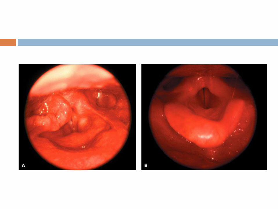
Majority of patients who receive high-dose radiation across major segments of the larynx and hypopharynxwill manifest some degree of edema, mucosal congestion, and eventual fibrosis .

Long term followup
During the first 6 months after treatment, patients
should be followed every 4 to 6 weeks with clinical
examination, including fiberoptic
nasopharyngoscopy.
Recommended guidelines include a follow-up visit
every 1 to 3 months during the first year, every 2 to
4 months for the second year, every 4 to 6 months
for years 3 through 5, and every 6 to 12 months
thereafter .
If the patient received comprehensive H&N radiation, the serum thyroid-stimulating hormone level should be measured every 6 to 12 months.
Imaging evaluation of the neck, most commonly with CT or MRI scan, are obtained at 3- to 6-month intervals during the first 2 years or as indicated based on clinical findings
Functional imaging with FDG-PET can sometimes prove valuable to help differentiate post treatment fibrosis from persistent or recurrent disease.
Conclusion
Despite an aggressive approach in the overall management of hypopharynx cancer patients, ultimate cure rates remain quite poor.
There are relatively few early-stage patients; and for many advanced-stage patients, it is difficult to achieve long-term control.
Even for those patients with excellent response to therapy, there exists a continuous risk for the development of second malignancies, particularly of the upper aerodigestive track with long-term follow-up.

Post treatment patients often require aggressive
speech and swallow therapy to maximize their
functional outcome.
There is significant interest in the incorporation of
molecular targeted therapies in combination with
traditional cytotoxic therapy and radiation in an
effort to improve outcomes.