Management of Acute Shoulder Injuries - sportmedab.ca K2A Edm/2016 K2A Speaker P… · Dr. Connie...
63
Dr. Connie Lebrun MDCM, MPE, CCFP (SEM), Dip. Sport Med, FACSM Glen Sather Sports Medicine Clinic Professor, Department of Family Medicine Faculty of Medicine & Dentistry University of Alberta, Edmonton, Alberta Management of Acute Shoulder Injuries
Transcript of Management of Acute Shoulder Injuries - sportmedab.ca K2A Edm/2016 K2A Speaker P… · Dr. Connie...

Dr. Connie Lebrun MDCM, MPE, CCFP (SEM), Dip. Sport Med, FACSMGlen Sather Sports Medicine ClinicProfessor, Department of Family Medicine Faculty of Medicine & DentistryUniversity of Alberta, Edmonton, Alberta
Management of Acute Shoulder Injuries
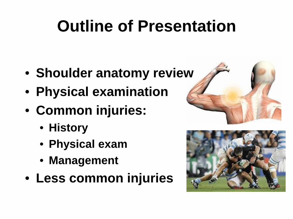
Outline of Presentation
• Shoulder anatomy review• Physical examination • Common injuries:
• History• Physical exam• Management
• Less common injuries

Shoulder Anatomy
Four joints:
1) Sternoclavicular2) Acromioclavicular3) Glenohumeral joint:
ball and socket joint 4) Scapulothoracic
NB: Glenoid covers only 25% of humerus

Static Stabilizers
• Glenoid labrum: anterior > posterior support
• Glenohumeralligaments: superior, coracohumeral, middle, and inferior
• Joint capsule

Dynamic Stabilizers
Deep layer • Rotator cuff:
Supraspinatus, Infraspinatus, Teres minor and Subscapularis
Superficial layer• Deltoid, Teres major, Long
head of Biceps, Pectoralismajor and minor
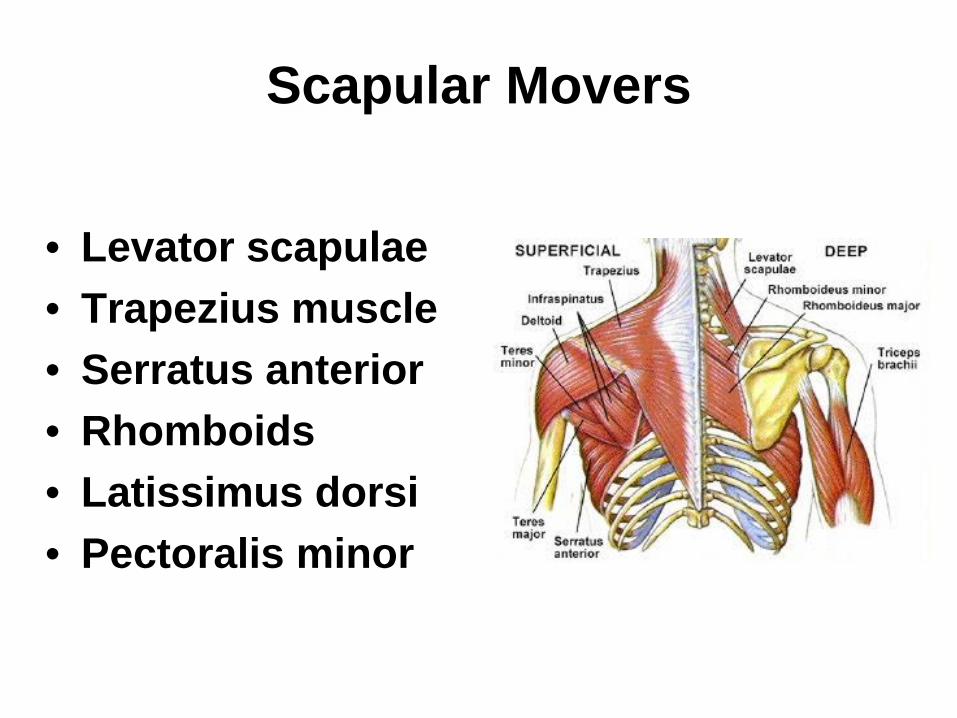
Scapular Movers
• Levator scapulae• Trapezius muscle• Serratus anterior• Rhomboids• Latissimus dorsi• Pectoralis minor
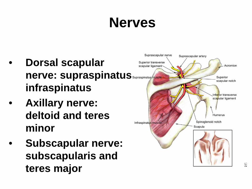
Nerves
• Dorsal scapular nerve: supraspinatus, infraspinatus
• Axillary nerve: deltoid and teresminor
• Subscapular nerve: subscapularis and teres major

Diagnosis
• History• Physical
Examination• Provocative Tests• Diagnostic Tests
(including imaging)
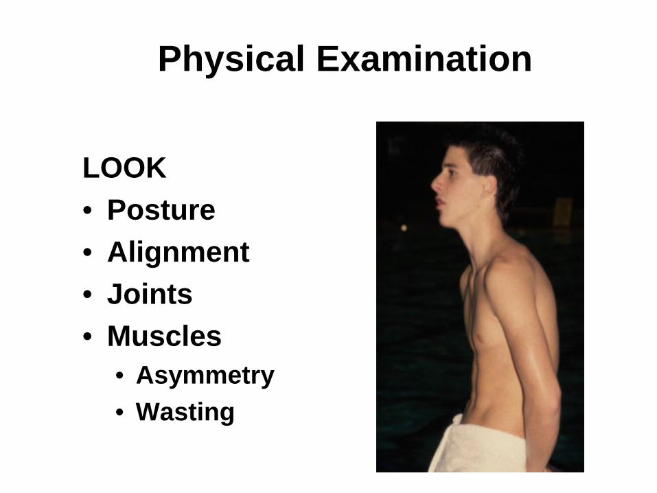
Physical Examination
LOOK• Posture• Alignment• Joints• Muscles
• Asymmetry• Wasting
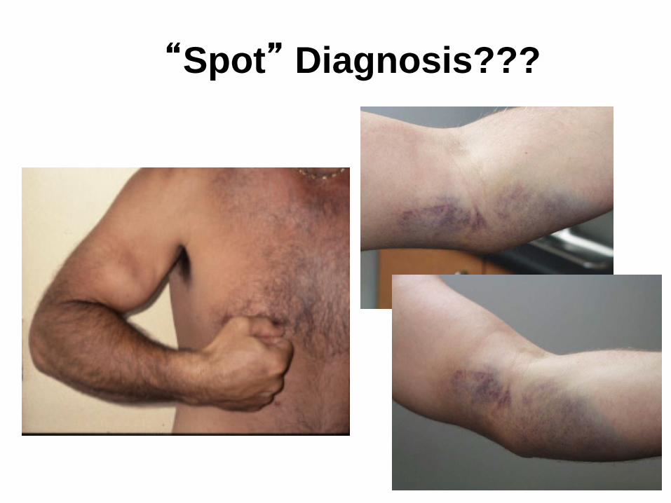
“Spot” Diagnosis???

Physical Examination
FEEL• Joints• Muscle• Tendons• Crepitus

Physical Examination
• Palpation:• Humeral
head• Clavicle• Acromion• AC joint• Scapula

Physical Examination
MOVE• GH joint
• Forward flexion• “Scaption”• Abduction• Rotation: IR/ER
• At side• 90° abduction
• Combined

Physical Examination
MOVE:• Scapula
• True weakness i.e. long thoracic nerve
• Poor scapular control:• Rehabilitation for
stabilization!

Physical Examination
NEUROVASCULAR EXAM• Motor• Sensory• Reflexes• Pulses• Capillary refill• NB: NECK EXAM
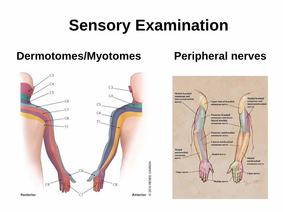
Sensory Examination
Dermotomes/Myotomes Peripheral nerves

Special Tests
• Cervical spine:• Spurling test• L’Hermitte sign
• Thoracic outlet• Adson’s, Allen’s• Hyperabduction test• Roos’ test

Physical Examination
ROTATOR CUFF:• S – Supraspinatus• I – Infraspinatus• T - Teres minor• S - Subscapularis• (Biceps)
NB: Need to distinguishpain-mediated weaknessversus true weakness
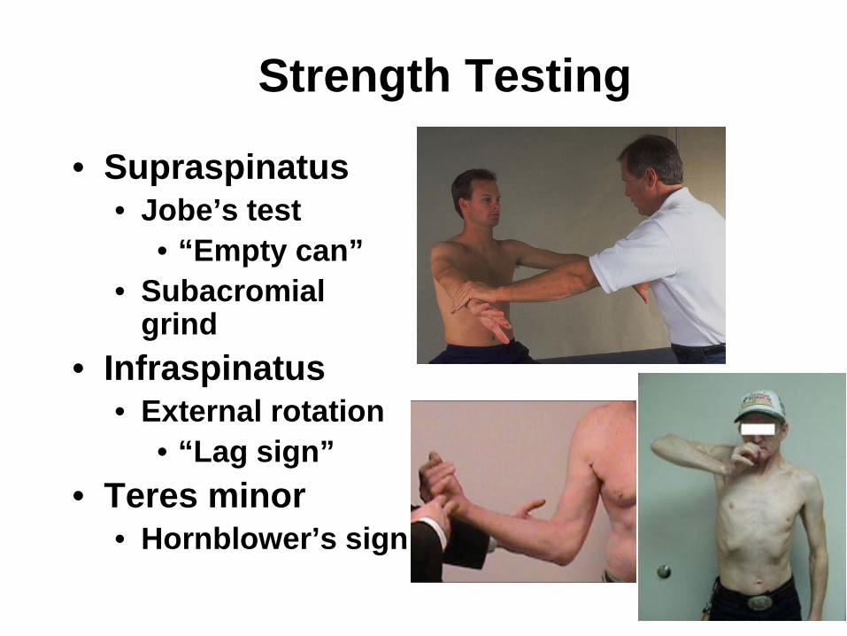
Strength Testing
• Supraspinatus• Jobe’s test
• “Empty can”• Subacromial
grind• Infraspinatus
• External rotation• “Lag sign”
• Teres minor• Hornblower’s sign
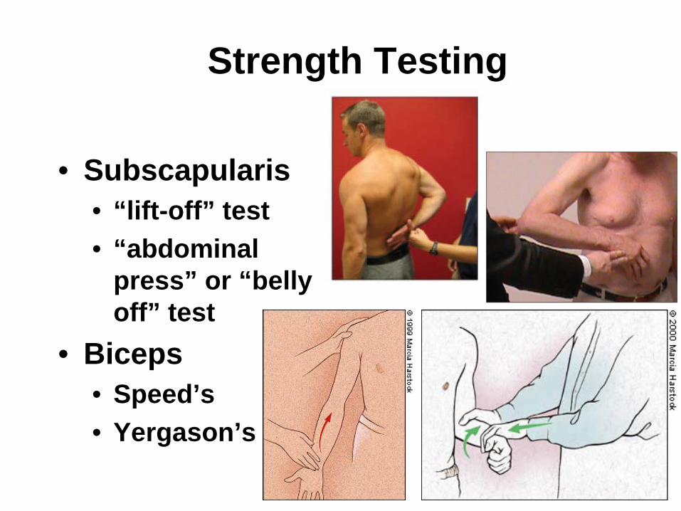
Strength Testing
• Subscapularis• “lift-off” test• “abdominal
press” or “belly off” test
• Biceps• Speed’s • Yergason’s

Physical Examination
SPECIAL TESTS:• Cross-arm
adduction• “scarf” sign• AC joint primarily
• Impingement tests:• Neer’s sign• Hawkin’s sign
Presenter
Presentation Notes
Also painful arc?

Physical Examination
SPECIAL TESTS:• “Drop-arm” test• Apley’s scratch test
• Subacromialinjection test –diagnostic?
Presenter
Presentation Notes
Also painful arc?

Physical Examination
INSTABILITY TESTS:• Load and shift
(+1,+2,+3)• Sulcus (inferior
instability)• Apprehension test• Relocation test• “Surprise” sign
Presenter
Presentation Notes
Jerk test?

Physical Examination
• Posterior instability• Jerk test

Acute Shoulder Injuries
• Acute glenohumeral dislocation:• Anterior• Posterior• (Inferior)
• Acromioclavicular separation• Clavicular fractures • Sternoclavicular subluxation

Somewhat age-dependent
• Young athletes:• Physeal injuries
• <25 y.o.:• GH dislocations• AC separations
• 40-60 y.o.:• Rotator cuff disease
• >60 y.o.:• Rotator cuff disease
• Osteoarthritis
“Little-Leaguer’s” shoulder

Serious Conditions
• Missed fractures• Missed posterior dislocations• Missed vascular or neurologic injury
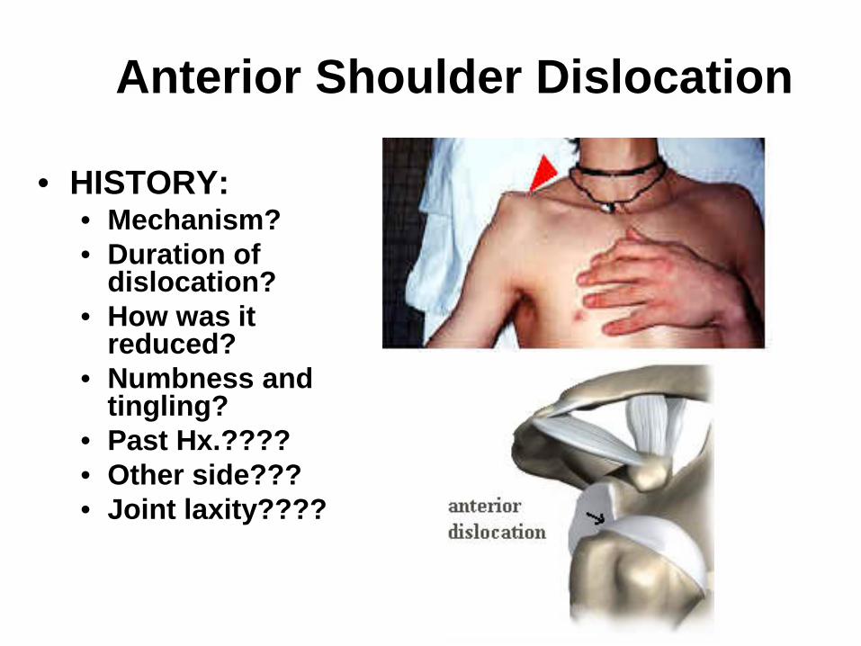
Anterior Shoulder Dislocation
• HISTORY:• Mechanism?• Duration of
dislocation?• How was it
reduced?• Numbness and
tingling? • Past Hx.???? • Other side???• Joint laxity????
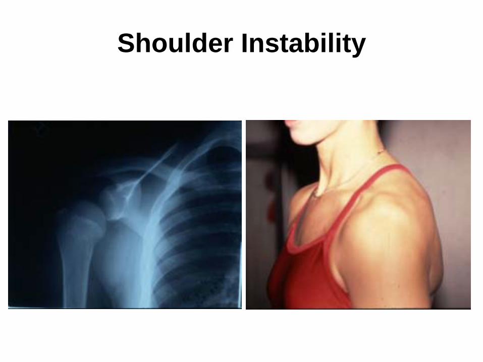
Shoulder Instability
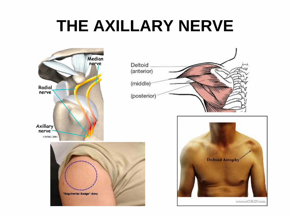
THE AXILLARY NERVE
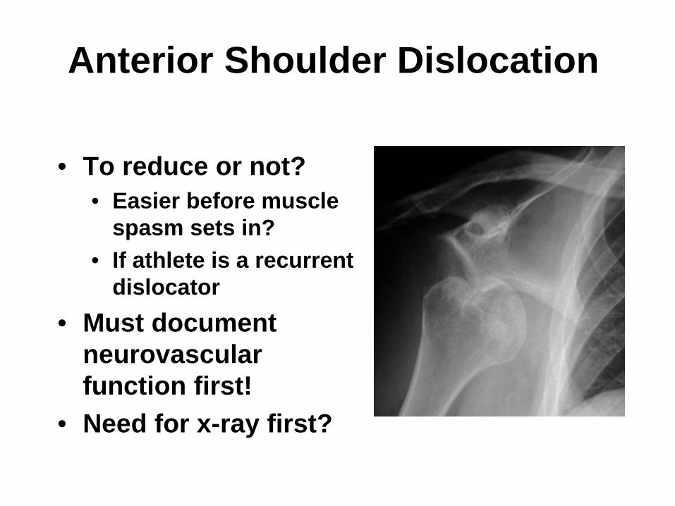
Anterior Shoulder Dislocation
• To reduce or not?• Easier before muscle
spasm sets in?• If athlete is a recurrent
dislocator• Must document
neurovascular function first!
• Need for x-ray first?
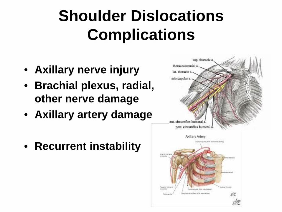
Shoulder Dislocations Complications
• Axillary nerve injury• Brachial plexus, radial,
other nerve damage• Axillary artery damage
• Recurrent instability

Shoulder Dislocations Complications
• Associated fractures:• Humerus
• Head• Neck• Greater tuberosity
• Glenoid• Coracoid

To reduce or not?
Hippocratic methodTraction-countertraction Stimpson method
External rotation method Patient self-reduction

Anterior Shoulder Dislocation
• After reduction:• Immobilize or not?• For how long?• Position? IR vs ER?• Bracing?• When to refer?
• Younger athlete• Contact or collision
sport• Traumatic dislocation

Acromioclavicular Separations
• THE HISTORY:• Mechanism: Fall
onto lateral shoulder/arm
• Sudden anterior pain +/- deformity

Classification of AC Separations

Type IV AC Separation

Type IV AC Separation
• AP projection

Type IV AC Separation
• Axillary views

Acromioclavicular Separation
• Grades I, II, III:• Conservative Rx• No need for x-ray
views with weights• Grade IV, V:
• Wait and see?• Grade VI:
• May need surgery

Acromioclavicular Osteolysis
• Sub-acute injury• Often in weight-
lifters, overhead or shoulder-dominentathletes
• Frequently bilateral
• Treatment:• Injection• Surgery (Mumford
procedure)

Sternoclavicular Injuries

Sternoclavicular Injuries
• Posterior dislocation may be medical emergency!
• NB trachea, esophagus, great vessels
• Anterior/superior dislocation more of a management issue

Sternoclavicular Injuries

Sternoclavicular Injuries

Sternoclavicular Injuries

Sternoclavicular Injuries
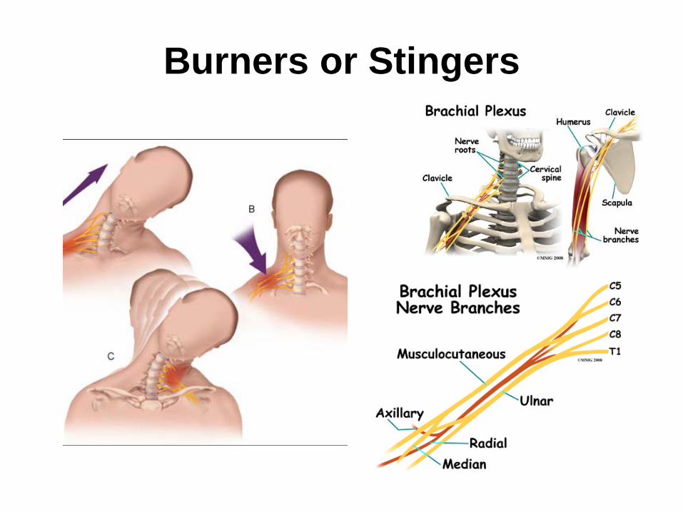
Burners or Stingers

Clavicle Fractures
• Mechanism:• Fall onto shoulder (87%)• Direct blow (7%)• Fall onto outstretched
hand (FOOSH injury)

Clavicle Fractures
• Associated injuries:
• Brachial plexus• Vascular injuries• Rib fractures• Scapular fractures• Pneumothorax

• Figure-of-8 bandage
• ‘out of favor”• Sling for comfort
Clavicle Fractures

Operative Management
• Neurovascular injury• Severe chest injuries• Open fractures• Uncontrolled deformity• Cosmetic reasons?• Non-union

Vascular Issues
• Subclavian• Axillary
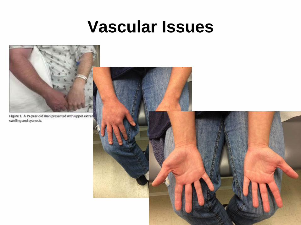
Vascular Issues

Vascular Issues
• Diagnosis• Ultrasound
Doppler imaging
• Anticoagulation

Scapular Fractures
• Potential damage to suprascapularnerve
• Traction• Hematoma• Nerve conduction
studies not helpful – not positive for 4-6 weeks

Acute Rotator Cuff Disease
• HISTORY:• Sudden pain, usually
associated with traumatic event
• No prior shoulder symptoms
• Dramatic decrease in function
• Intense pain• WEAKNESS

When to Order X-rays
• Immediately:• ANY Decreased ROM• ANY Trauma• Suspected OA• When the patient just:
“doesn’t fit”• Follow-up:
• Pain not responsive to conservative management
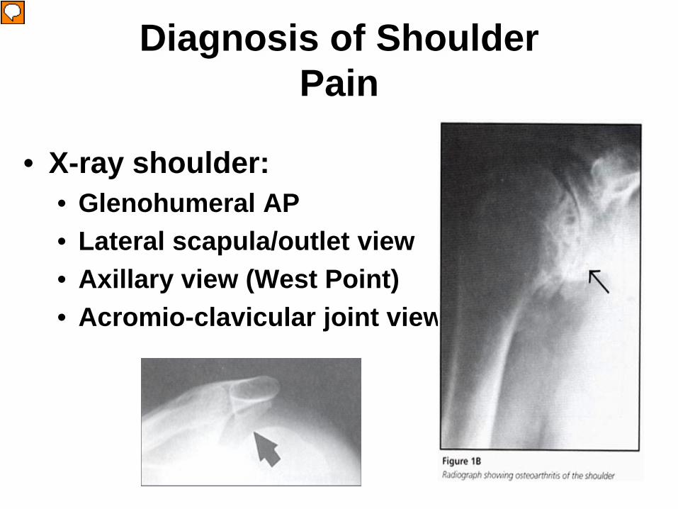
Diagnosis of Shoulder Pain
• X-ray shoulder:• Glenohumeral AP • Lateral scapula/outlet view• Axillary view (West Point)• Acromio-clavicular joint view
Presenter
Presentation Notes
Stryker Notch view?
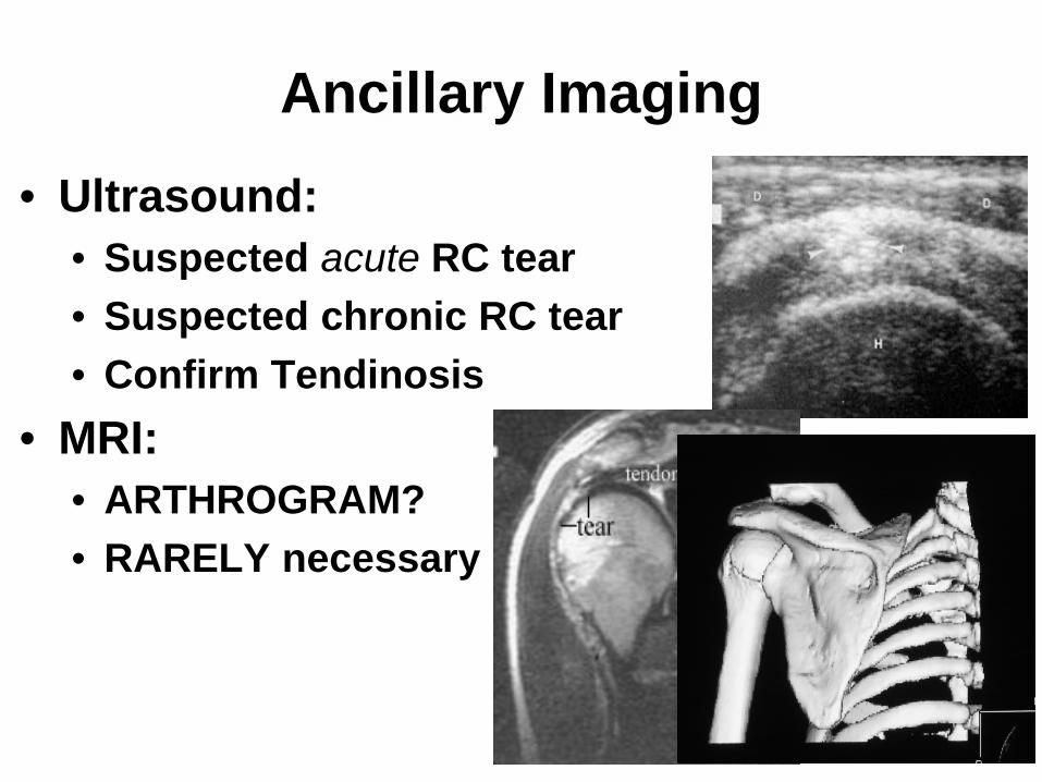
Ancillary Imaging• Ultrasound:
• Suspected acute RC tear• Suspected chronic RC tear• Confirm Tendinosis
• MRI:• ARTHROGRAM?• RARELY necessary

Principles of Shoulder Treatment
• Restore full ROM • Strengthen the
shoulder girdle and stabilizers of the shoulder
• Improve function