Management of Acute Renal Failure•Unasyn/enrofloxacin • Careful dosing of enrofloxacin in cats...
45
NICOLE GUMA, DVM, DACVECC, DACVIM (SAIM) MANAGEMENT OF ACUTE KIDNEY INJURY
Transcript of Management of Acute Renal Failure•Unasyn/enrofloxacin • Careful dosing of enrofloxacin in cats...

N I C O L E G U M A , D V M , D A C V E C C , D A C V I M ( S A I M )
MANAGEMENT OF ACUTE KIDNEY INJURY

DEFINITION OF AKI
AKI
•Reflects the broad spectrum of acute diseases of the kidney
Stages
•Can be imperceptible clinically at early stages to severe and requiring renal replacement therapy
Human criteria
•Absolute increase in creatinine of > 0.3mg/dL
•Increase of creatinine by 50%
•Reduction in UOP
REVERSIBLE
•Near normal renal function and small changes in creatinine represent large changes in GFR
•Important to recognize EARLY

CAUSES OF AKI
Pyelonephritis
Infectious diseases
Ureteral obstruction Systemic Diseases
NephrotoxicantsIntrinsic
Ischemic injury

TYPES OF AKI
PreRenal
• Polyuric >2-3ml/kg/hr
• Oliguric <1ml/kg/hr
• Anuric <0.5ml/kg/hr
Intrinsic Renal
Postrenal

MANIFESTATIONS OF AKI
Abnormal fluid balance
Electrolyte disorders
Metabolic acidosis
Uremic intoxication
Systemic complications

HYPERTENSION OF RENAL DISEASE
Blood pressure
• Product of cardiac output and SVR
Hypertension is multifactorial
• Altered Na excretion leads to volume expansion
• Altered RAAS/increased catecholamines leads to increased SVR

CLINICAL PRESENTATION
History
Acute onset
GI signs
Variable urine output
Physical examination
Dehydration
Signs of uremia/hyp
ertension
Enlarged/painful kidneys

DIAGNOSTICS
CBC/CHEM/blood
gas/SDMA
Urinalysis/UPC
Urine culture
Systolic blood
pressureImaging
Infectious disease testing
EG test Novel
biomarkersCVP
Renal biopsy

IRIS AKI STAGING CRITERIA
AKI Stage Serum creatinine
(mg/dL)
Clinical Description
Stage I <1.6 Evidence of renal injury
nonazotemic increase
in creatinine;
>0.3mg/dL in 48hrs
Stage II 1.7-2.5 Mild AKI—evidence of
AKI and mild static or
progressive azotemia
Stage III 2.6-5.0 Moderate to severe
AKI: documented AKI
and increasing
severities of azotemia
and functional renal
failure
Stage IV 5.1-10.0
Stage V >10.0
Sub stages would include (NO) for nonoliguric and (O) for oliguric and on
the basis of the need for RRT

TREATMENT
Antidote
• 4-methylpyrazole or ethanol for EG
• Very short window of opportunity to intervene (5-8 hrs dogs, 3 cats)
Fluid therapy
• Replace dehydration deficit, ongoing losses, maintenance
• Correct acid/base and electrolyte abnormalities
• Avoid fluid overload—associated with poor prognosis
• Enteral water
• NE or E tube
• Consider maintenance fluids after rehydration with replacement fluids
• 0.45% NaCl + 2.5% dextrose

ADDITIONAL TREATMENT
Antacids:
• Famotidine 0.5mg/kg IV BID
• Pantoprazole 0.7-1mg/kg IV SID-BID
• Carafate ¼-1g PO TID-QID
Antiemetics:
• Cerenia 1mg/kg SQ
• Anzemet 0.6mg/kg IV or Ondansetron 0.1-0.5mg/kg IV BID
• Metoclopramide 0.5-2mg/kg/d CRI
KCL supplementation
• If not oliguric/anuric—maintenance rates
• Can become profoundly hypokalemic and require supplementation at high rates
• If <3.0mEq/L give 0.5mEq/kg/hr (diluted 1:1 with saline) over 4 hours then recheck and supplement accordingly—use syringe pump
Antibiotics
• Unasyn 22mg/kg IV TID
• Doxycycline 5mg/kg IV BID
• Fluoroquinolone
• Ideally based on UCS

ADDITIONAL TREATMENT
•Indicated if BP > 160-180mmHg
•ACEi?
•Can be deleterious in the acute setting
•Calcium channel blocker
•Amlodipine 0.2-0.75mg/kg/d (can give rectally)
•Hydralazine 2.5mg/cat PO, 0.5-3mg/kg PO Dog (1hr onset)
Antihypertensives
•ALOH only if eating/with food 30-90mg/kg/d divided
•Renagel, lanthanum
Phosphate binders
•Ideally enteral if can tolerate
•Place NE/NG or Etube if not voluntarily eating, avoid syringe/force feeding
•IV nutrition PRN (procalamine, TPN)
Nutrition

DRUG DOSE ALTERATIONS
Drug Standard
dose
Method to
adjust
Iris stage 2
Creat 1.4-2.0
Iris stage 3
Creat 2.1-5.0
Iris stage 4
Creat > 5
Atenolol 0.25mg/kg q
12hr
Dose/interval 0.19mg/kg q
12-24 hour
0.125mg/kg
q 12-24hr
0.06mg/kg q
24hr
Enrofloxacin 5-10mg/kg q
24hr
Dose/interval 1.25-5mg/kg
q 24hr
1.25-5mg/kg
q 24-48hr
(not reco in
cats
0.8-3mg/kg q
24-48 hour
(not reco in
cats)
Reglan 1-2mg/kg/d
CRI
Dose 1mg/kg/d
CRI
0.5mg/kg/d
CRI
0.25mg/kg/d
CRI
Prazosin 1-4mg/dog
q 12-24hr
Dose None 1-2mg/dog
q 12-24hr
0.75-
1.5mg/dog q
12-24 hr
Spironolactone 1-2mg/kg q
12hr
Dose/interval 0.5-1mg/kg
q 24hr
0.25mg/kg q
24hr
Not
recommend
ed
Tramadol 3-5mg/kg/q
8-12hr
Dose/interval 1-3mg/kg q
12hr
1-3mg/kg q
12hr
1-2mg/kg q
24hr

MONITORING
•Heart rate, pulses, RR, CRT
•Body temperature
•BP
•Body weight TID-QID!!
Vital signs
•Often Ucath required
•Minimize CAI
•Monitor Ins/outs every 2-4 hours
•Can also weigh bedding (1gm = 1ml)
Urine output
•Indirect measure of volume status
•Blood sampling
•Avoid if hemodialysis a possibility
CVP Measurements

MANAGING OLIGURIA
• Physiologic
• Pathologic
• Fluid challenge if not overhydrated, ins and outs monitoring
• If anuric but euhydrated = insensible losses only (22ml/kg/d)
• Avoid over hydration = poor prognosis
Assess volume/hydration status
• Conversion to polyuria does NOT always equal increased GFR
Medical management

FOR OLIGURIA
Mannitol Lasix Dextrose
Diltiazem Dopamine? Fendolapam?
Peritoneal dialysis
Hemodialysis

FOR ANURIA
Lasix Diltiazem IV dextrose Peritoneal dialysis
Hemodialysis

MANNITOL
Osmotic diuretic
Increases RBF, GFR, solute excretion
Increases osmotic tubular flow
Scavenges oxygen free radicals
Only if volume replete
• Not if overhydrated/anuric
• Hypotensive
• CHF
Dose:
• 0.25-1g/kg as a bolus over 20 minutes
• If effective UOP increases in 1 hour
• CRI can be considered at 1-2mg/kg/min—use filter, dilute 1:1 to 10% solution
• Avoid doses > 2-4gm/kg/d (can cause AKI)

LASIX
•Natriuresis/diuresis
•Increases tubular flow rate
Inhibits active Na/Cl reabsorption in distal nephron
•Increased renal blood flow
Causes vasodilation
Dose = 2-6mg/kg
•CRI induces more reliable diuresis with lower dosing than intermittent boluses
•Can result in hypovolemia
As a CRI = 0.25-1mg/kg/hr

IV DEXTROSE
• Metabolized by the cells (unlike mannitol)
• Acts as an osmotic diuretic
Hyperosmolar solution 20%
• 2-10ml/min for 10-15 min then 1-5ml/min for total daily dose of 22-66ml/kg
Dose of 20%
Monitor for glucosuria

DILTIAZEM
MOA
•Causes pre-glomerular dilation
•Natriuresis independent of GFR
Studies
•Used in human kidney transplant patients/cyclosporine
•Used in 11 of 18 dogs with leptospirosis induced ARF with furosemide CRI
•Rate of reduction of creatinine 1.76 times faster than for control—not ss
Dose
•0.1-0.5mg/kg IV bolus
•Then 1-5ug/kg/min CRI until serum creatinine normal or stable
•Used conjunction with Lasix CRI, beneficial alone?

DOPAMINE
MOA
•Stimulates DA-1, DA-2, B, Alpha receptors
• Increases RBF and UOP in normal dogs, GFR unchanged
•DA-1 receptor found in cats
Humans
•No longer used in humans
•Negative effects identified
Dogs/cats
•Little to no documentation on efficacy in dogs/cats
•No longer in vogue/recommended

FENDOLOPAM
Selective DA-1 agonist
•Renoprotective in humans
•Vasodilates renal vessels
Retrospective study AKI dogs/cats
•Angell
•Administration of 0.8ugkg/min dog and 0.5ug/kg/min cat well tolerated
•No improvement in survival to discharge, Length of hospital stay or creatinine

PERITONEAL/HEMODIALYSIS
Oliguria or anuria
Fluid overload Hyperkalemia
Progressive azotemia
Dialyzable toxin

PEARLS OF MANAGING SPECIFIC AKI CASES

LEPTOSPIROSIS
• Spirochete
• Reservoir hosts—raccoon, voles, skunks, dogs, pigs, cattle, rats
• Leptospira interrogans (icterohaemorrhagiae, Canicola, Pomona, Bratislava, Autumnalis), Leptospira kirschneri (grippotyphosa)
• L Pomona results in more severe disease (50% survival compared to 80%)
• Common in warm/tropical areas
• Urban areas increasing in prevalence
• >20% may be chronic healthy carriers

LEPTOSPIROSIS
• Factors predisposing• Stagnant water, high rainfall
• Rodent/wild-life exposure
• Spring/fall
• Rare but is reported in cats
• Can remain viable in soil for weeks to months
• Inactivated by UV radiation/freezing
• Clinical signs same as for AKI BUT• Can see elevated liver enzymes, hyperbilirubinemia = big red
flag
• Thrombocytopenia seen in 58% of dogs
• Azotemia noted in 80-90%
• Can be coagulopathic (6-50%)
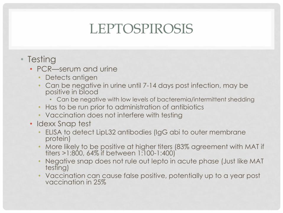
LEPTOSPIROSIS
• Testing• PCR—serum and urine
• Detects antigen• Can be negative in urine until 7-14 days post infection, may be
positive in blood• Can be negative with low levels of bacteremia/intermittent shedding
• Has to be run prior to administration of antibiotics• Vaccination does not interfere with testing
• Idexx Snap test• ELISA to detect LipL32 antibodies (IgG abi to outer membrane
protein)• More likely to be positive at higher titers (83% agreement with MAT if
titers >1:800, 64% if between 1:100-1:400)• Negative snap does not rule out lepto in acute phase (Just like MAT
testing)• Vaccination can cause false positive, potentially up to a year post
vaccination in 25%

LEPTOSPIROSIS
• Point of Care Zoetis test (Witness Lepto)• Tests anti-Leptospira IgM antibodies to canicola, grippotyphosa,
icterohaemerrhagiae and pomona• Detects antibody 4 days post infection• Witness lepto greater detection of antibodies at day 7 than MAT• Sensitivity 93.5%, Sensitivity 98%• Can detect antibodies that have been generated due to vaccination
within 6 months• Only 25% of dogs will be positive at 3 months post vaccination
• Available April 20-27• Negative test does not rule out leptospirosis
• Microscopic agglutination test• Paired titers 2-3 weeks apart• 4 fold increase in titers consistent with active infection• Non-vaccinal serovar of >1:800 • Antibiotics can blunt response

LEPTOSPIROSIS
• Urinary catheter essential• Prevents contamination of hospital and zoonotic risk
• Allows quantification of urine output closely
• Oliguria
• Profound polyuria
• Treatment• Standard therapy as for AKI
• Unasyn @ 22mg/kg IV TID, Doxycycline 5mg/kg IV q 12hr
• Doxycycline 5mg/kg PO BID x 2 weeks needed to resolve carrier phase
• Usually given after 2 weeks of amoxicillin or Clavamox
• No longer leptospiremic after 24 hours of IV antibiotics

LEPTOSPIROSIS
• Oliguria managed medically or with dialysis
• Often profound polyuria follows
• Prognosis• 85% survival but can require considerable supportive care• If polyuric can discharge on SQF if unable to afford continued care
• Careful monitoring of hydration status still recommended
• Chronic renal disease possible outcome with recovery
• Zoonotic disease• Barrier isolation—wear gloves, gown, goggles• Discuss risk with owners• Clean with bleach (10%), iodine based disinfectant, accelerated
hydrogen peroxide, quaternary ammonium solutions
• Vaccination post infection?• Wait until recovered x 2 months• Vaccinate house mates• Environmental control

PYELONEPHRITIS
• Routes of infection
• Ascends from the lower urinary tract
• Breakdown in barrier for prevention of ascending infection
• Hematogenous
• Predisposing factors
• Urinary specific
• Systemic illness

PYLEONEPHRITIS
• Organisms• Ecoli most common• Staphylococcus, Streptococcus, Enterococcus• Klebsiella, Pseudomonas, Enterobacter, Proteus, fungi
• Diagnosis:• Asymptomatic to critically ill/septic
• May have renomegaly/fever—absence does not rule out• Blood work may be normal or show leukocytosis/AKI• UA (via cystocentesis or pyelocentesis)—pyuria, bacteriuria, positive
culture• Antibiotic responsive • Ultrasound
• Kidneys may be small/chronic or enlarged
• Echogenic material in collecting system
• Dilation of renal pelvis/proximal ureter• Can also be seen with CKD, diuresis, urinary/ureteral obstruction

COMPARING ULTRASOUND IMAGES OF CKD, PYELO, URETERAL OBSTRUCTIONS
• Feline study
• Renal pelvic dilation seen in all groups
• If > 13mm attributed to UO• Greatest transverse pelvic diameter
• CKD 1.7 +/- 2.6mm), Pyelo 3.2 +/- 3.1mm, UO 10.5 +/- 5.5mm
• Over lap between them all• 30% of normal cats had renal pelvic dilation
• 66% of CKD cats had pelvic dilation
• 84.6% of pyelonephritis cats had renal PD
• 100% of ureteral obstruction cats
• Ureteral dilation• No normal cats
• 6% of CKD cats
• 46.2% of pyelonephritis cats
• 81.8% of ureteral obstruction cats
• Get normal baseline for CKD cats so can monitor going forward

PYELONEPHRITIS
• Imaging recommended to document renal/ureteral
pelvic dilation, concurrent diseases (stones, CKD
etc)
• May be useful to monitor this going forward to document
treatment duration
• Culture negative pyelonephritis rare
• Pyelocentesis necessary
• Risks—hemorrhage, urine leakage
• Only if clinically warranted

PYELONEPHRITIS
• Treatment/monitoring• Supportive as previously outlined
• Initial empiric therapy broad spectrum
• Unasyn/enrofloxacin• Careful dosing of enrofloxacin in cats if azotemic
• De-escalate antibiotics based on culture/sensitivity
• Perform UA 1 week into therapy and ideally repeat ultrasound after 3-4 weeks of therapy.
• Continue antibiotics for 2 weeks beyond resolution of renal pelvic dilation if applicable
• Usually 4-6 weeks needed
• Repeat culture 3-5 days after discontinuation of antibiotics then at 1 month, 3 months, and 6 months

URETERAL OBSTRUCTIONS
• Technically post-renal cause for azotemia• Can cause kidney injury with chronicity
• Causes:• Stones
• Dried blood clots, strictures (25% of cats), tumors (<5% dogs/cats)
• Obstructive pyonephrosis
• Cats• >90% Calcium oxalate
• Usually sterile
• Do not dissolve
• Dogs• 50% of the time struvite 50% calcium oxalate
• 50% of the time associated with pyelonephritis

URETERAL OBSTRUCTIONS
• Complete ureteral obstruction
• Increase pressure in renal pelvis reduced renal blood flow
by 60% in 24 hours and 80% within 2 weeks
• Partial obstructions may preserve renal function for a bit longer
and recovery may be better than complete obstruction
• Majority of cases are partial based on pyelography
• GFR may normalize after several weeks post relief of obstruction
if partial
• Renomegaly not sensitive to make a diagnosis of
obstruction
• Imaging necessary to make diagnosis

DIAGNOSIS URETERAL OBSTRUCTION
• Preferably a combination of radiographs and
ultrasound
• Rads document stone size, number location and presence
of concurrent nephroliths
• Ultrasound—identifies hydronephrosis, hydroureter and location
• If no stone or tumor is found, stricture
• In a recent study 60% of cats with ureteral stricture had peri-
ureteral hyperechoic tissue at the site on AUS
• Sensitivity for rads in cats 81%, dogs 88%, and for AUS 77% in
cats and 100% in dogs
• In combination the sensitivity of both is 90% in cats

URETERAL OBSTRUCTIONS
• Systemic illness common
• Dogs often dysuric, uncommon in cats
• Most dogs have concurrent pyelonephritis and
cystitis
• 77%
• Less common to have concurrent UTI in cats (33%)
• Dogs commonly have neutrophilia and 44% have
thrombocytopenia (< 40 K sometimes)
• Sepsis, ITP

URETERAL OBSTRUCTION
• Medical management
• Should be initiated immediately following diagnosis
• Always attempted for 24-48 hours unless:
• Already overhydrated, oliguric, anuric
• Hyperkalemic
• IV fluid therapy
• Monitor body weight, electrolytes, hydration status, ideally
central venous pressures closely
• Fluid overload common
• Protocol used by Dr. Berent
• Maintenance fluids at 50-60ml/kg/d with 0.45% NaCL + 2.5%
dextrose + replacement fluid to correct dehydration deficit and
promote diuresis (45-75ml/kg/d)

MEDICAL MANAGEMENT URETERAL OBSTRUCTION
• Mannitol
• IV bolus at 0.25-0.5g/kg over 20-30 minutes then
• 1mg/kg/min for 24 hours
• Use a filter, do not use if overhydrated, cardiac compromise
• If after 24 hours no improvement, discontinue CRI
• Alpha 2 adrenergic antagonist
• Spasmolytic
• Tamsulosin (alpha 1a/1d adrenergic antagonist) used to expel distal ureteral stones <5mm in people
• Study in dogs comparing alpha adrenergic antagonists (tamsulosin, prazosin, experimental B-2/B-3 adrenergic agonists, calcium channel blocker and a phosphodiesterase inhibitor• Experimental b-adrenergic agonist best, tamsulosin next best
• Prazosin had little effect and worsened ureteral spasming at high doses

URETERAL OBSTRUCTIONS
• Amitriptyline
• Urinary smooth muscle relaxer mediated by opening of voltage gated potassium channels• Evaluated in cats with urethral obstructions
• Extrapolated to ureteral obstructions
• 1mg/kg PO/day, not documented to be efficacious
• Glucagon
• Out of favor
• Relaxes ureteral smooth muscle
• Abstract showed improved urine output in oliguric cats but no short or long term benefit in ureteral obstruction
• Side effects
• Majority of the time medical management fails
• 10% success rate

INTERVENTIONAL/SURGICAL MANAGEMENT OF URETERAL
OBSTRUCTIONS
• Ureteral stents—canine
• SUBs—feline
• See Dr. Garnett’s lecture
• Prognosis:
• Dependent on chronicity of obstruction, cause, response to
medical management, need for surgical intervention,
timing of intervention
• Recovery can take weeks to months

PROGNOSIS FOR AKI
•Dogs—53-60%
•Cats—50%Species Mortality
•Leptospirosis 85% survival
•50% of cats with nephrotoxicity survive
•75% of cats with ischemic induced AKI survived
•40-60% of patients treated with RRT for AKI survive
Based on cause:
•Dog--Creat >10mg/dL, hypocalcemia, anemia, decreased UOP, hyperphosphatemia, comorbid disorders
•Cat—hyperkalemia, hypoalbuminemia, decreased bicarb, level of azotemia not associated with px
Negative prognostic indicators
•50% of patients treated medically have CKD, 50% have complete renal recovery (d/c)
Long term prognosis of those that survive