Case Report Acute Abdominal Pain Secondary to Chilaiditi ...

Management of a patient with acute abdominal pain
College of Surgeons of Sri Lanka 2007 AUTHORS Dr M Ganesharatnam FRCS(Eng), Consultant Surgeon Dr GS De Silva FRCS. Consultant Surgeon Dr DS Liyanarachchi FRCS, Consultant Surgeon Dr Semaka Jayasekera MS, Consultant Surgeon Dr Aloka Pathirana MS, FRCS Senior Lecturer in Surgery

Acute abdominal pain / National Guidelines CSSL
78
Contents
1. Introduction 75 2. Clinical evaluation 76 3. Initial management 82 4. Category A ( conditions that may respond to initial
management, requiring subsequent referral to a specialized unit )
4.1 Renal/Ureteric colic 87
4.2 Biliary colic 88
4.3 Gastritis 89
4.4 UTI 90
4.5 Irritable bowel syndrome 91
5. Category B (conditions that may have to be transferred to a specialized unit after initial management)
5.1 Intestinal colic
(except when due to gastroenteritis) 92
5.2 Cholecystitis 93
5.3 Pancreatitis 94
5.4 Acute appendicitis 95
5.5 Perforated viscus 96
5.6 Strangulated hernia 97
5.7 Torsion of testis 98
6. References 99
7. Annexures 1. Insertion of NG tube 100
2. Insertion of catheter 101

National Guidelines CSSL / Acute abdominal pain
79
SECTION 1 Introduction Definition sudden onset abdominal pain severe enough to seek immediate medical attention (and/or make him/her deviate from normal day to day activities) One of the commonest reasons for seeking medical attention in the out patient department (need evidence). If not properly managed, could lead to significant morbidity and mortality. Age and sex is an important consideration in the diagnosis. Although investigations are helpful, management should be guided by clinical judgment. Investigations recommended should be performed according to availability, in the institution. All patients presenting with acute abdominal pain should be assessed by a medical officer. For management purposes we have divided the conditions commonly causing abdominal pain in to two categories (A and B). Category A, has conditions which could be managed in a center without a specialist and referred subsequently. The conditions under category B, are more serious ones, which may need to be transferred to a center with a specialist after the initial management. Traumatic causes of abdominal pain and abdominal pain specific to the paediatric age group are not dealt with in this document.

Acute abdominal pain / National Guidelines CSSL
80
SECTION 2 Clinical evaluation will consist of the following. History Examination Investigations Observation Treatment – may commence before investigations are performed. Causes (see figure 1) Colicky
Ureteric colic Intestinal colic - gastroenteritis, acute
appendicitis, intestinal obstruction, (hernia, adhesions, volvulus etc. chronic constipation)
Biliary colic Non-colicky
Gastritis UTI Appendicitis Cholecystitis Pancreatitis Strangulated hernia Perforated viscus Torsion of testis Irritable bowel syndrome (rarely)

National Guidelines CSSL / Acute abdominal pain
81
Non-surgical conditions
Gynae conditions (eg. Ruptured ectopic pregnancy, twisted ovarian cyst)
Medical conditions (eg. Ketoacidosis, basal pneumonia, porphyria)
Rare conditions Ruptured aortic aneurysm
Figure 1. Different types of abdominal pain

Acute abdominal pain / National Guidelines CSSL
82
History
1. age 2. sex 3. main site (region and depth) 4. radiation 5. onset/duration 6. type of pain / character 7. severity 8. periodicity / frequency 9. special times of occurrence (after meals, time of day
etc.) 10. aggravating and relieving factors 11. Associated symptoms –eg. Faintishness (particularly
in females if a period of amenorrhoea is present) Past history – similar episodes, trauma, surgery or interventions, medical conditions Drug history – Non-steroidal anti-inflammatory drugs (NSAIDS), steroids, anti-coagulants, antiplatelet drugs Menstrual history – LRMP Social history – smoking, alcohol, substance abuse Systemic enquiry –change in bowel habits, urinary symptoms etc. Last meal / drink Possible intake of unhygienic food/drink Allergies Examination General – hydration (see box), pallor, “Ill look”, degree of distress (lying still or moving about), elevated temperature CVS – pulse, BP

National Guidelines CSSL / Acute abdominal pain
83
RS – rate, chest movements, air entry, added sounds Abdomen
• Inspection Movement with respiration ‘Cough test’ (aggravation of pain with
coughing – site of pain more evident) Distention Shape – asymmetry, scaphoid Visible peristalsis - pulsations Umbilicus – hernial orifices, genitalia
• Palpation – guarding, rigidity, • tenderness (site of maximum
tenderness – eg. McBurney’s point), rebound tenderness, lumps. “Murphy’s
sign” • Percussion – liver dullness, free fluid • Auscultation – bowel sounds (absence or exaggerated)
Peritonism – presence of tenderness, rebound tenderness and guarding – this is seen with perforation of viscus, inflammation or blood within the peritoneal cavity. Different organs within the peritoneal cavity, gives rise to maximum tenderness in different regions of the anterior abdominal wall. A guide is given in figure 2. Rarely – signs of peritoneal irritation absent (eg. In Mesenteric ischaemia and intestinal obstruction). Signs may be masked in immuno-suppressed patients and those who are heavily sedated.

Acute abdominal pain / National Guidelines CSSL
84
Box 1
Symptoms and signs of dehydration Thirst Reduced passage of urine Loss of skin turgor Sunken eyes Tachycardia Hypotension (late sign)

National Guidelines CSSL / Acute abdominal pain
85
Figure 2. Regions of the abdomen
Acute abdominal pain / National Guidelines CSSL
86
SECTION 3 Initial management In most instances of acute abdominal pain, management may have to precede investigations. This is particularly true in patients who are ill or in severe pain. In these instances relief of pain, correction of dehydration etc. is more important than investigations to find the cause. Admit (unless pain has settled) Observe vital signs in a– Primary care unit or similar set up Keep nil orally IV access / fluids Blood for – FBC, RBS, Amylase, U&E (if clinically indicated) Analgesics – depending on severity NSAID – suppositories – contra-indicated in renal failure, asthma, gastritis
Opioids – Tramadol Suppositories – may cause vomiting
(particularly in females) Morphine/ Pethidine – if administered should
monitor the patient.(Note: Morphine should be avoided in biliary colic and pancreatitis) Paracetamol – suppositories useful if available
Antispasmodics (for intestinal or biliary colic) Nasogastric tube insertion (see annexure 1) – if suspected of having intestinal obstruction Catheterization (see annexure 2) – if acute retention or dehydrated as in shock
National Guidelines CSSL / Acute abdominal pain
87
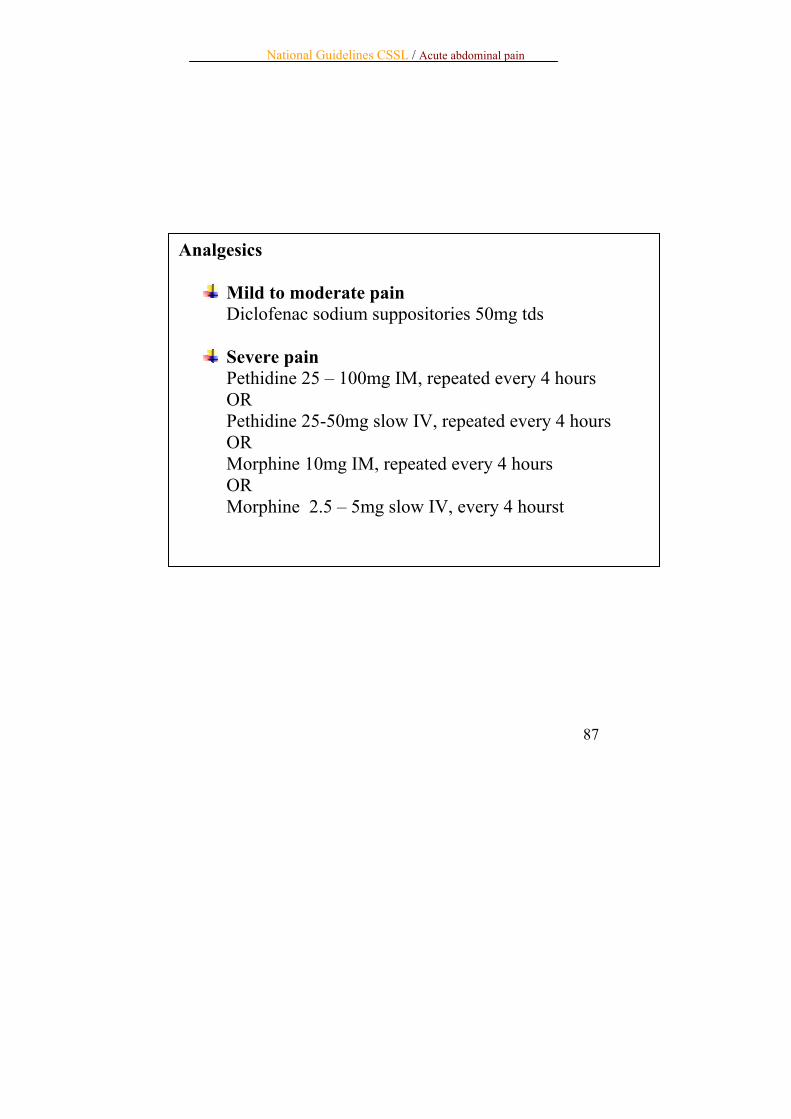
Analgesics
Mild to moderate pain Diclofenac sodium suppositories 50mg tds
Severe pain Pethidine 25 – 100mg IM, repeated every 4 hours OR Pethidine 25-50mg slow IV, repeated every 4 hours OR Morphine 10mg IM, repeated every 4 hours OR Morphine 2.5 – 5mg slow IV, every 4 hourst

Acute abdominal pain / National Guidelines CSSL
88
Cautions and contra-indications to non-steroidal anti-inflammatory drugs(NSAID)
Cautions
in the elderly, allergic disorders renal, cardiac and hepatic impairment
Contra-indications
hypersensitivity to aspirin or any other NSAID (attacks pf asthma, urticaria, rhinitis or angiooedema precipitated by NSAID)
during pregnancy and lactation coagulation defects
previous or active peptic ulceration Monitor
• Temperature • Pulse • BP • Respiration • Input/Output • Abdominal signs – girth

National Guidelines CSSL / Acute abdominal pain
89
Initial Investigations X ray abdomen – supine AP CXR erect (or Lateral decubitus of abdomen) UFR Urine for HCG (if indicated) USS abdomen if clinically indicated – renal colics, gynae pathology, cholecystitis, pancreatitis Testicular Doppler – if torsion suspected and facility is available Definitive management would depend on the provisional diagnosis. Senior opinion or referral to a center with facilities should be considered depending on the clinical diagnosis and the severity. If the patient responds to the initial management, he/she may be discharged and subsequently referred to a specialized unit. This applies to the clinical conditions described in category A. If the patient is to be transferred, the following details should be provided – summary of history, examination, investigations and treatment given with the time being indicated clearly. A responsible person should accompany the patient. Monitoring should continue and resuscitation facilities must be available during transfer.

Acute abdominal pain / National Guidelines CSSL
90
Category A ( conditions that may respond to initial management, requiring subsequent referral to a specialized unit )
1. Renal/Ureteric colic 2. Biliary colic 3. Gastritis 4. UTI 5. Irritable bowel syndrome
Category B (conditions that may have to be transferred to a specialized unit after initial management)
8. Intestinal colic (except when due to gastroenteritis) 9. Cholecystitis 10. Pancreatitis 11. Acute appendicitis 12. Perforated viscus 13. Strangulated hernia 14. Torsion of testis

National Guidelines CSSL / Acute abdominal pain
91
SECTION 4 Category A ( conditions that may respond to initial management, requiring subsequent referral to a specialized unit ) 4.1. Renal/Ureteric colic
Clinical features Investigations Management Sudden onset severe pain Loin to groin (or vice versa) radiation – may radiate to upper thigh, penis, scrotum Associated with vomiting Moves about in pain May have associated urinary symptoms Examination Minimal signs May have tenderness in the iliac fossa, lumbar region and/or renal angle
UFR – predominantly red cells X ray KUB – after bowel preparation USS KUB
Pain relief - Diclofenac sodium suppositories (if not contraindicated) Pethidine if no response to above Adequate fluid intake Follow up – necessary (if stone detected). Refer to a specialized unit
Acute abdominal pain / National Guidelines CSSL
92
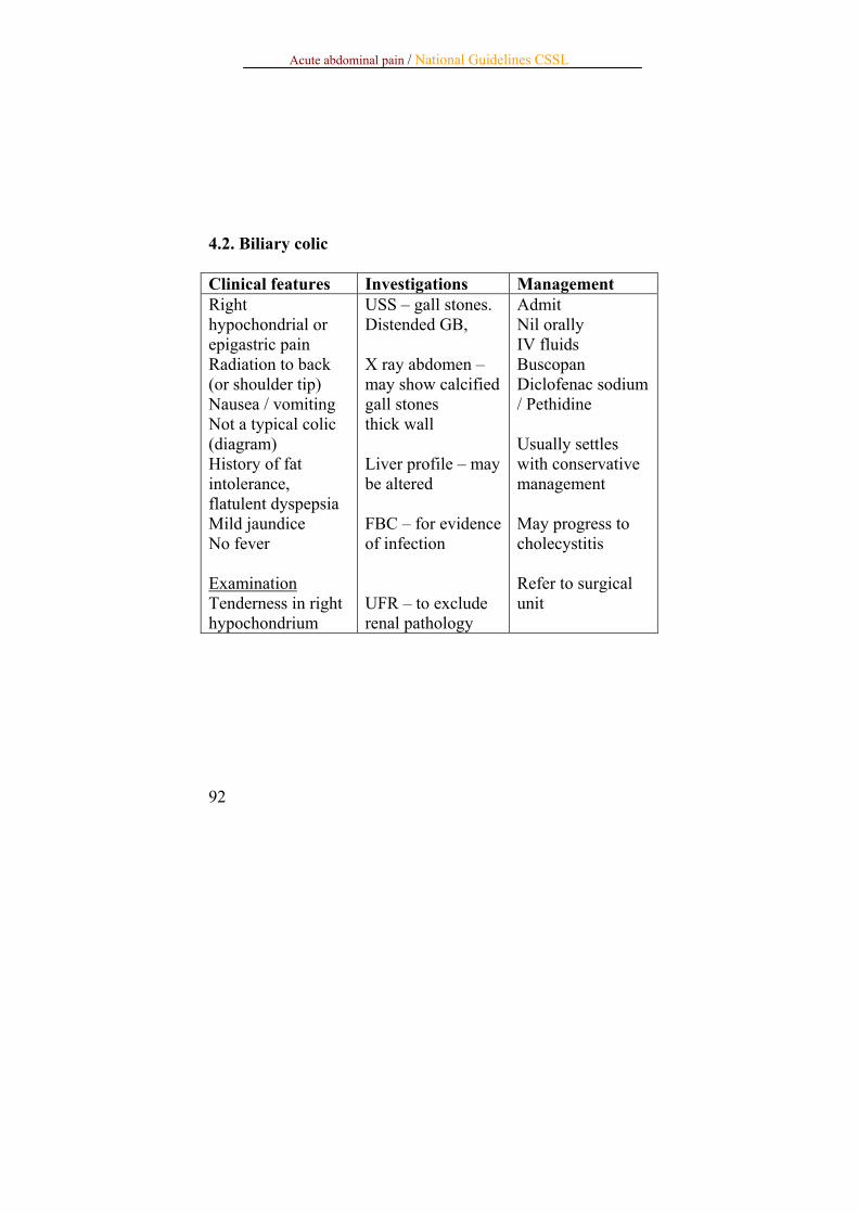
4.2. Biliary colic Clinical features Investigations Management Right hypochondrial or epigastric pain Radiation to back (or shoulder tip) Nausea / vomiting Not a typical colic (diagram) History of fat intolerance, flatulent dyspepsia Mild jaundice No fever Examination Tenderness in right hypochondrium
USS – gall stones. Distended GB, X ray abdomen – may show calcified gall stones thick wall Liver profile – may be altered FBC – for evidence of infection UFR – to exclude renal pathology
Admit Nil orally IV fluids Buscopan Diclofenac sodium / Pethidine Usually settles with conservative management May progress to cholecystitis Refer to surgical unit

National Guidelines CSSL / Acute abdominal pain
93
4.3. Gastritis Clinical features Investigations Management Burning epigastric pain Distension – after meals NSAID intake, food intolerance, alcohol, steroids History of gastro-esophageal reflux disease (GERD), dyspeptic symptoms Localized tenderness only Myocardial infarction may mimic the clinical features of gastritis
S Amylase ECG – to exclude myocardial infarction UGIE – If age over 40 years or symptoms are recurrent
Antacids – should have prompt response H2 receptor antagonists (H2RA) OR Proton pump inhibitors (PPI) should be given (if severe, these may be commenced intravenously) If symptoms are recurrent, refer to a specialised unit
Acute abdominal pain / National Guidelines CSSL
94
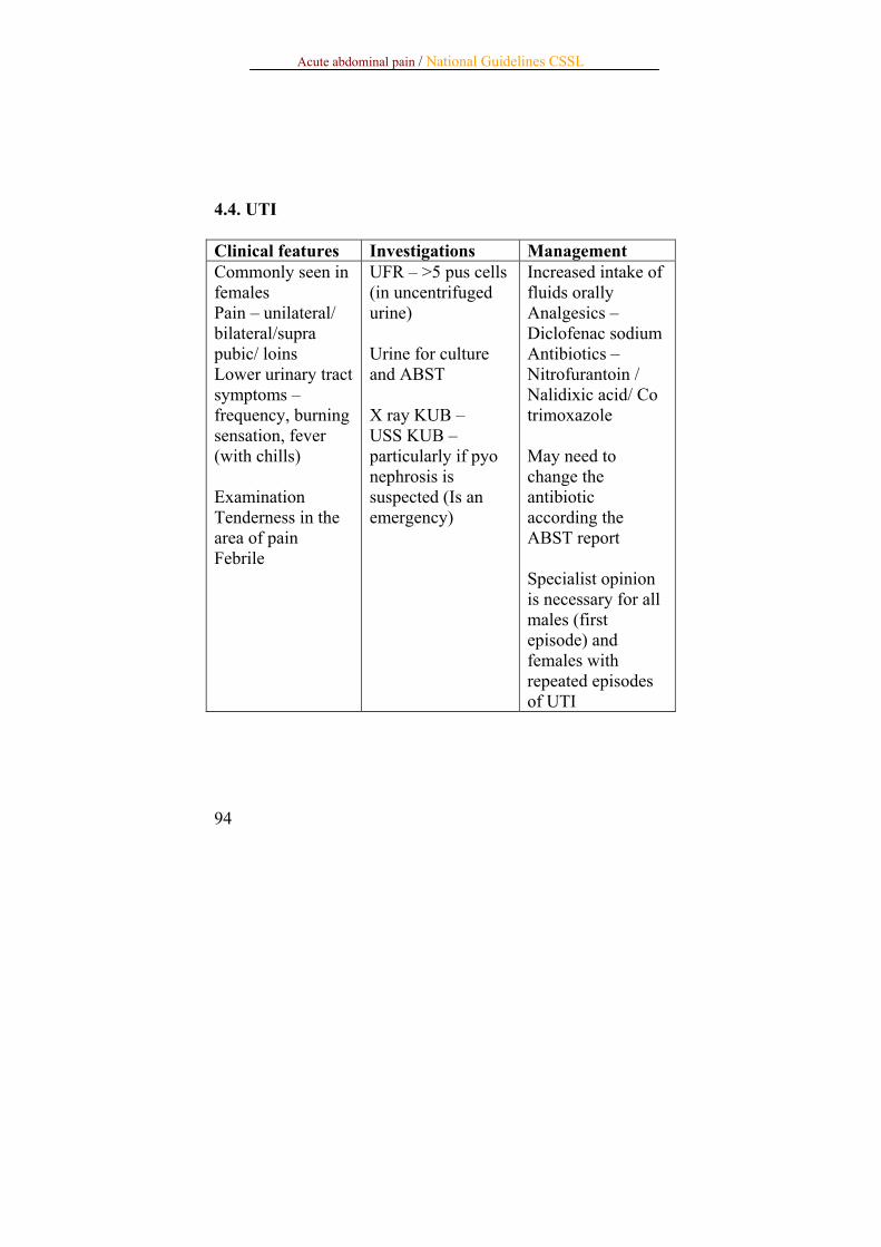
4.4. UTI Clinical features Investigations Management Commonly seen in females Pain – unilateral/ bilateral/supra pubic/ loins Lower urinary tract symptoms – frequency, burning sensation, fever (with chills) Examination Tenderness in the area of pain Febrile
UFR – >5 pus cells (in uncentrifuged urine) Urine for culture and ABST X ray KUB – USS KUB – particularly if pyo nephrosis is suspected (Is an emergency)
Increased intake of fluids orally Analgesics – Diclofenac sodium Antibiotics – Nitrofurantoin / Nalidixic acid/ Co trimoxazole May need to change the antibiotic according the ABST report Specialist opinion is necessary for all males (first episode) and females with repeated episodes of UTI

National Guidelines CSSL / Acute abdominal pain
95
4.5. Irritable bowel syndrome Clinical features Investigations Management Periodic pain Associated with bowel symptoms Examination Patient not ill
Exclude – inflammatory bowel disease, intestinal obstruction ESR, Stools FR, faecal occult blood May need – Double contrast barium enema, colonoscopy (electively)
Reassure Symptomatic treatment – (eg – antispasmodics for colics) Identify and avoid precipitating factors (eg. Milk)

Acute abdominal pain / National Guidelines CSSL
96
SECTION 5 Category B (conditions that may have to be transferred to a specialized unit after initial management) 5.1. Intestinal colic Clinical features Investigations Management Sudden onset pain Site – circum umbilical (small bowel) or hypogastrium (large bowel) Vomiting Diarrhoea (in gastroenteritis) Constipation Abdominal distension Dehydration – level should be assessed Lumps, ascites, scars of previous laparotomy Hernial orifices need to be checked (particularly for femoral hernia in females) DER – empty rectum, tumour, hard faeces
X ray abdomen supine AP – distended bowel loops USS – if mass is suspected U & E RBS FBC
Nil orally NG tube – if vomiting or gross distension+ IV fluids – type, volume, rate depending on level of dehydration Catheter – if close monitoring is needed Surgical referral is mandatory (except in patients having gastroenteritis) If evidence of possible strangulation of bowel – urgent surgical referral is indicated.

National Guidelines CSSL / Acute abdominal pain
97
5.2. Cholecystitis Clinical features Investigations Management Right hypochondrial or epigastric pain – may be referred to the right shoulder / back Hyperaesthesia in the region of the inferior angle of right scapula (Boas sign) Vomiting Fever Low grade icterus may be present Murphy’s sign
Ultra sound scan of abdomen FBC LFT X ray of GB area (particularly if USS is not available) CXR – erect PA (to exclude basal pneumonia / perforated peptic ulcer) Amylase (to exclude pancreatitis) UFR
Nil orally IV fluids Diclofenac sodium suppositories Pethidine (if pain is severe) Monitor – for evidence of peritonitis Antibiotics – ciprofloxacin or cefuroxime IV (if diabetic/immuno compromised – add metronidazole) Early surgical referral – particularly if deteriorating

Acute abdominal pain / National Guidelines CSSL
98
5.3. Pancreatitis Clinical features Investigations Management Sudden onset Severe pain Epigastric – predominantly Radiates to back Vomiting Pain reduced when bending forwards History of alcohol, gall stones Examination Ill looking – in pain Tenderness, guarding and marked rigidity in the epigastrium Free fluid may be present Liver dullness present
Serum Amylase (four fold rise) CXR – PA (to exclude a perforated viscus) Late presentation – Serum lipase If confirmed – need to assess severity FBC LDH Blood urea RBS Blood gas Serum calcium US Scan CT – if severe
Nil orally IV fluids NG tube Analgesics – Pethidine Antibiotics – broad spectrum (if severe attack) Look out for complications (eg. MODS) in severe cases Obtain surgical opinion May need laparotomy – if diagnosis is in doubt

National Guidelines CSSL / Acute abdominal pain
99
5.4. Acute appendicitis Clinical features Investigations Management Circumumbilical pain – later shifting to RIF Anorexia Nausea / Vomiting Fever (low grade – unless perforated) Examination Maximum tenderness/guarding/ rigidity in the iliac fossa Tenderness and guarding would be generalized if appendix has perforated
UFR – to exclude UTI WBC/DC – neutrophil leucocytosis Urine for HCG – in females to exclude ectopic pregnancy USS abdomen – particularly in females – when diagnosis is in doubt Laparoscopy – in females when diagnosis is in doubt
Nil orally IV fluids Analgesics – Diclofenac sodium suppositories Monitor – pulse, BP, respiration Broad spectrum antibiotics should be given after confirming the diagnosis Definitive treatment - appendicectomy

Acute abdominal pain / National Guidelines CSSL
100
5.5. Perforated viscus Clinical features Investigations Management Sudden onset severe pain Generalized History of peptic ulcer disease/ NSAID ingestion/ diverticular disease/ bowel malignancy Examination Febrile Board like rigidity Absent bowel sounds Free fluid Impaired liver dullness
CXR PA – erect (if patient cannot be kept erect , X ray abdomen lateral decubitus view) Serum Amylase (to exclude Pancreatitis) FBC
U & E RBS
Nil orally NG Tube IV fluids Analgesics – Pethidine or Morphine Antibiotics – broad spectrum plus metronidazole Monitor – Pulse, BP, resp, UOP Optimize before surgery Definitive treatment - surgery

National Guidelines CSSL / Acute abdominal pain
101
5.6. Strangulated hernia Clinical features Investigations Management Previous history of hernia Symptoms and signs of intestinal obstruction preceding the persistent severe pain Examination Irreducible hernia – tender Tachycardia
FBC RBS ECG (if >40 years of age)
Nil orally IV fluids Analgesics – Narcotic Avoid forceful manipulation Needs surgery If the patient is to be transferred for surgery, place an ice pack on hernia, elevate foot end
Acute abdominal pain / National Guidelines CSSL
102
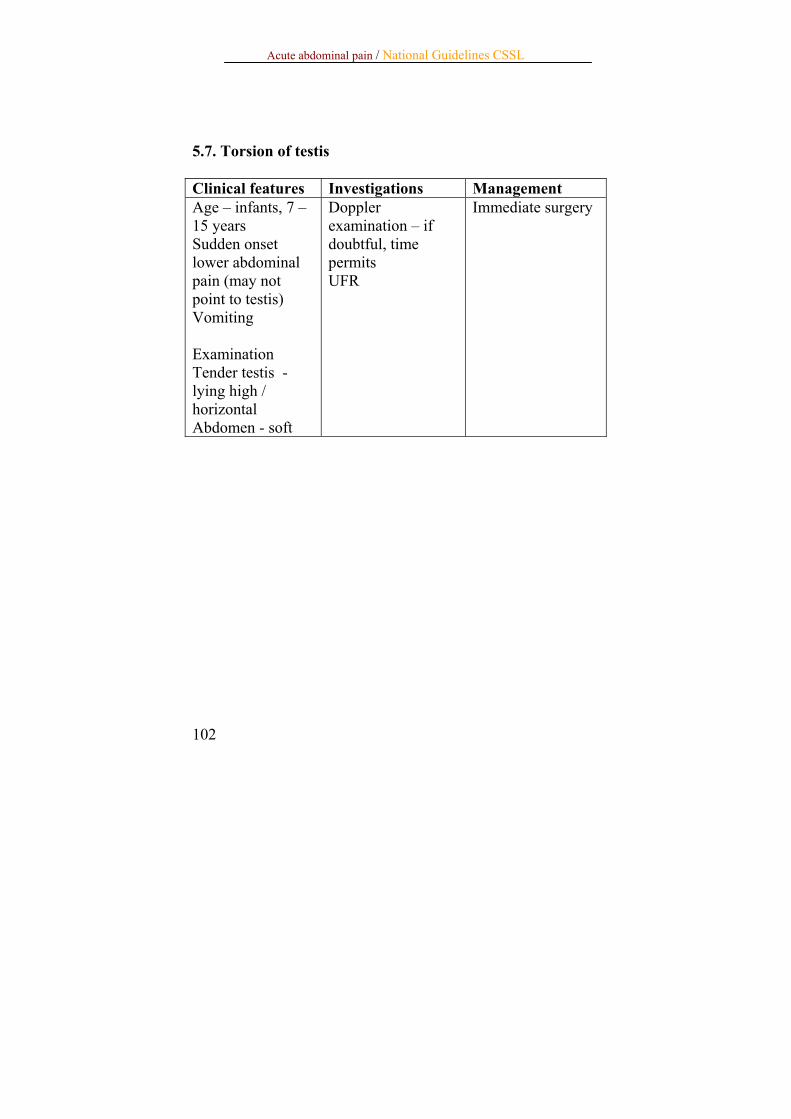
5.7. Torsion of testis Clinical features Investigations Management Age – infants, 7 – 15 years Sudden onset lower abdominal pain (may not point to testis) Vomiting Examination Tender testis - lying high / horizontal Abdomen - soft
Doppler examination – if doubtful, time permits UFR
Immediate surgery

National Guidelines CSSL / Acute abdominal pain
103
Section 6. References Bailey and Love’s Short Practice of Surgery – 23rd Edition British National Formulary An introduction to the symptoms and signs of surgical disease – Norman L Browse 29th January 2007

Acute abdominal pain / National Guidelines CSSL
104
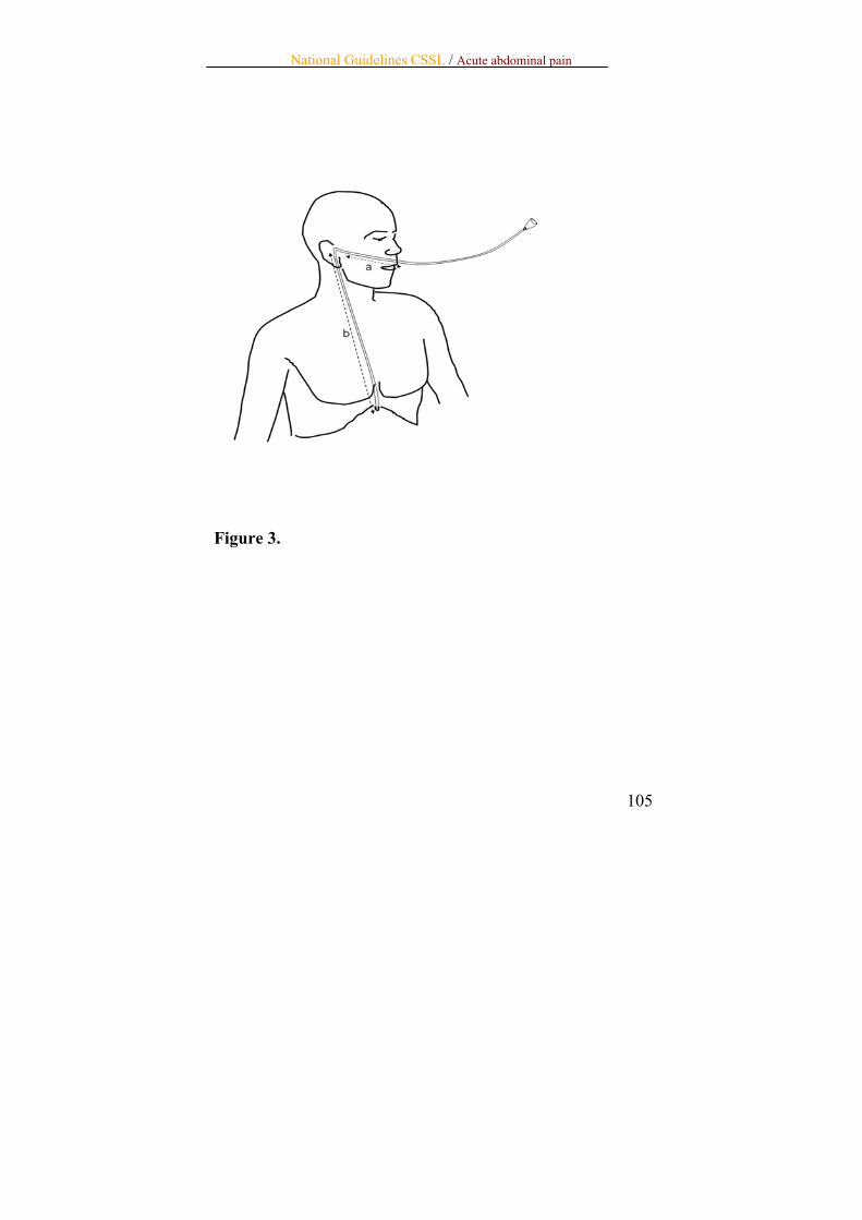
Annexure 1 Insertion of a Nasogastric tube
1. Explain the procedure to the patient and obtain consent. 2. Select a Nasogastric tube of appropriate size. (It is
helpful to stiffen the tube by placing it in a freezer compartment of a refrigerator)
3. Measure the length of the tube to be inserted (see diagram 3) – from the nostrils to the tragus and from the tragus to the xiphoid process(a+b)
4. Lubricate the nostril and the tip of the tube with 2% Lignocaine gel
5. Select the nostril which appears patent. 6. Pass the tube slowly and gently along the floor of the
nasal cavity. 7. Ask the patient to swallow, when he feels the tip of the
tube in the throat. This opens the upper oesophageal sphincter and facilitates the passage of the tube in to the oesophagus.
8. Push the tube in, until the mark (a+b). 9. Check the correct position by instilling air with a
syringe, and auscultating over the stomach for a hissing sound. Appearance of gastric contents through the tube is also confirmatory of the correct position.
10. The tube has to be secured with a plaster attached to the face. It is important not to allow the tube to exert pressure on the nostril, but lie horizontal to the upper lip. This is to avoid pressure necrosis of the nostril skin.
National Guidelines CSSL / Acute abdominal pain
105
Figure 3.

Acute abdominal pain / National Guidelines CSSL
106
Annexure 2 Technique of urethral catheterization of a male
Explain the procedure to the patient Aseptic technique is important Select an appropriate catheter – Size 14F is adequate
for an average male Wear gloves and retract the prepuce – clean the prepuce,
glans and penis with an anti-septic solution Sterile drape should be placed around the penis 2% Lignocaine gel is introduced in to the urethra, using
the nozzle provided in the tube (if new) or with a 2cc syringe (without the needle)
Retain the gel in thee urethra for at least 2-3 minutes (may need to compress the glans)
Insert the catheter by gradually stripping the polythene covering – should avoid direct contact with the catheter – penis should be held slightly stretched
AVOID FORCEFUL INSERTION Pass the catheter until urine starts flowing through it,
and until the shoulder of the catheter is at the external meatus– it is useful to connect a drainage bag prior to complete insertion. If urine does not flow freely, pressing the supra-pubic area would be useful
Inflate the balloon of the catheter with the appropriate volume of sterile water – ONLY AFTER YOU ARE SATISFIED THAT THE CATHETER TIP IS WELL WITHIN THE BLADDER
Pull back the catheter to ensure that it is secure within the bladder