Mamta Desai, BS, MBACLS, CIC Director of Epidemiology and...
44
Mamta Desai, BS, MBA, CLS, CIC Director of Epidemiology and Infection Prevention CLINICAL MICROBIOLOGY
Transcript of Mamta Desai, BS, MBACLS, CIC Director of Epidemiology and...

Mamta Desai, BS, MBA, CLS, CICDirector of Epidemiology and Infection Prevention
CLINICAL MICROBIOLOGY

Objectives
• Describe role of the clinical laboratory in infection prevention; emphasis on microbiology
• Describe factors that can adversely affect reliable lab results
• Discuss the importance of the gram stain
• Discuss the interpretation, use and importance of the antibiogram
• Discuss common pathogens that may contribute to HAIs
• Understand laboratory testing methods for confirming infections
©2013 Pomona Valley Hospital Medical Center 2

Microbiology and Infection Prevention
Microbiology has two important functions related to infections
• Clinical: diagnosis and management of infections
• Epidemiological : understand infectious microbes in patients (and populations), to find sources and routes of transmission necessary for prevention efforts
©2013 Pomona Valley Hospital Medical Center 3

Clinical Microbiology
Physician’s perspective:– What’s growing?– What antibiotic can be used?
• Determined either by predictive value of the organism type (e.g. gram negative bacillus) or by complete result with sensitivities
IP or Epidemiologist’s perspective:– Surveillance for determining clusters/outbreaks and assessing
trends– Need to know organism so IP can implement proper
transmission-based precautions as needed in a timely fashion
©2013 Pomona Valley Hospital Medical Center 4

Primary Rules on Microbiology Cultures
• Rule 1: No Lab Test is 100% Accurate
• Rule 2: Positive Cultures Do Not Make an Infection
©2013 Pomona Valley Hospital Medical Center 5

Assessing Accuracy of Lab Results
Rule #1: No lab test is 100% accurate 100% of the time
Many factors can affect accuracy of laboratory tests
1. Collection Error▫ How was specimen collected, handled, transported, preserved prior to arrival
in the lab?2. Lab Error▫ Were correct agar plates used? Was the specimen incubated at correct
temp? Lab protocols followed? Skill of the micro tech? Accuracy of biochemical and instrument system?
3. Reporting Error▫ Accurate result transcription in computer systems? Did results get
communicated to the doctor accurately?
©2013 Pomona Valley Hospital Medical Center 6

Assessing Accuracy of Lab Results
Rule #2: Positive Culture Does Not Mean Infection
Bacteria Must Invade Tissue To Cause An Infection
• For some tests such as polymerase chain reaction (PCR), because an organism is present does not mean it is viable (transmissible)
• Pseudo-outbreaks due to lab contamination of samples can occur
©2013 Pomona Valley Hospital Medical Center 7

What might indicate invasion into Tissue???
©2013 Pomona Valley Hospital Medical Center 8

White Blood Cell (WBC)
• PMNs ( polymorphonuclear leukocytes) made in bone marrow; provide general response to threat
• Neutrophils (~50-60% wbcs) are first line of response to infection; may also be called ‘segs’
• Eosinophils (1-7% wbcs); allergic reactions and parasites)
• Basophils (<1%); allergic reactions, help mediate strength of immune response)
• Left shift: presence of immature neutrophils (called ‘ bands’ or ‘stabs’) in blood count; are indicative of acute infection or inflammatory process
©2013 Pomona Valley Hospital Medical Center 9
www.rnceus.com/cbc/cbcdiff.htm

Lymphocytes & Monocytes
•Lymphocytes ( lymphs) mature in the lymphatic portion of the immune system
•Include pathogen-specific immune response (B cells, T cells)•Increase may be indicative of viral infection
•Monocytes (or macrophages) phagocyte function (or eat) cellular debris and foreign pathogens from the immune system
©2013 Pomona Valley Hospital Medical Center 10
www.rnceus.com/cbc/cbcdiff.htm
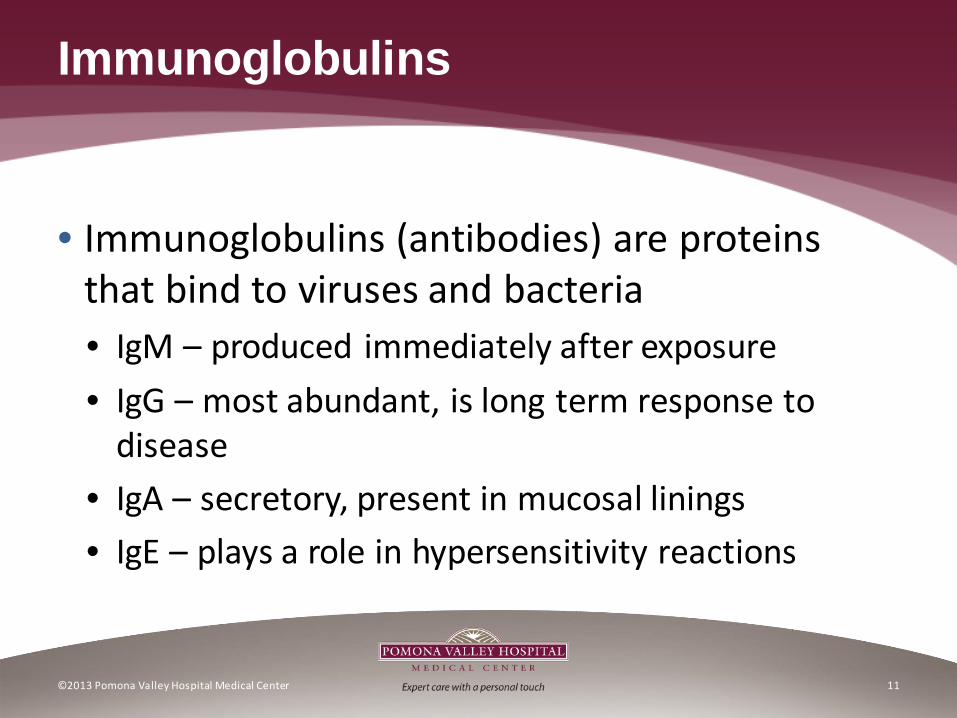
Immunoglobulins
• Immunoglobulins (antibodies) are proteins that bind to viruses and bacteria• IgM – produced immediately after exposure• IgG – most abundant, is long term response to
disease• IgA – secretory, present in mucosal linings• IgE – plays a role in hypersensitivity reactions
©2013 Pomona Valley Hospital Medical Center 11

What is Gram Stain?
• Method of classifying bacteria into 2 large groups: positive (+) and negative (-)
• Differentiates bacteria by the chemical and physical properties of their cell walls
• Helpful in guiding initial empiric therapy• results should get to physician ASAP
©2013 Pomona Valley Hospital Medical Center 12

Bacterial Groups
1 3
GRAM POSITIVE
(BLUE)
GRAM
NEGATIVE
BACILLI
GRAM
NEGATIVE
COCCI
GRAM
POSITIVE
BACILLI
GRAM
POSITIVE
COCCI
GRAM NEGATIVE
(Pink)
E coli Neisseria Clostridium Streptococci Staphylococci
Gram stain identifies four basic groups of bacteria:
1. Gram positive cocci (Staphylococcus, Streptococcus, Enterococcus)2. Gram negative cocci (Neisseria, Moraxella)3. Gram positive bacilli (Clostridium, Listeria, Corynebacterium)4. Gram negative bacilli (Pseudomonas, Escherichia coli, Haemophilus, Bacteroides)

©2013 Pomona Valley Hospital Medical Center 14

1 5
15
Sputum Gram StainQuality of sputum specimen:• Squamous epithelial cells (SEC)
• <10 excellent, no appreciable contamination• 10-25 equivocal but acceptable• >25 reject due to unacceptable levels of oral contamination
• WBC• <10 no infection (or poor immune response)• 10-25 equivocal• >25 purulence indicates presence of infection
• Bacteria

16
Common Lower Respiratory Tract Pathogens
• Community-acquired pneumonia (CAP) ▫ S. pneumoniae• H. influenzae• Mycoplasma
16
• Hospital-acquired, most often ICU or ventilator-associated • Pseudomonas aeruginosa• Stenotrophomonas
maltophilia
• Either CAP or hospital-acquired pneumonia• Staphylococcus aureus (MRSA or MSSA)
⇑ mortality; must be recognized quickly• Moraxella catarhallis (most often CAP)
Note: Yeast is NOT usually an infecting organism for pneumonia or other lower respiratory tract infections unless it constitutes >90% of organisms in a specimen and specimen is not contaminated with oral flora

17
Cerebrospinal Fluid (CSF) Bacteria
• Meningitis due to gram negative rods or Staphylococcus usually associated with predisposing factors such as trauma
• Adult, most common: Strep pneumo (gram positive cocci in pairs)• generates increased WBC response
• Meningococcemia: gram stain showing gram-negative diplococci is diagnostic
• a single case is a true infection emergency

18
Onset of Symptoms
Patient presents for medical evaluation Lumbar Puncture (LP)
Bacterial Viral (aseptic)
Meningitis
CSF clearnormal or elevated proteinnormal glucoseno organisms on gram stain
CSF cloudyelevated proteindecreased glucoseWBC; positive neutrophilsorganisms on gram stain

Blood Cultures
• A single blood culture consists of two bottles • Bottles designed to recover aerobes and anaerobes • Irrelevant which bottle has growth or
if both or only one bottle has growth
• Adults: low numbers of bacteria in blood ( ≤30/mL)• Can lead to negative gram stain and false negative• Volume is important; usual 4 bottles/40cc blood• Less blood needed for children due to larger number of bacteria
per cc of blood/don’t normally have anaerobes
©2013 Pomona Valley Hospital Medical Center 19

2 0
Blood Culture Common Commensals
Partial list of common commensals• Coag neg staphylococci• Diphtheroids• Bacillus• Proprionibacteria• Viridans strep• Aerococcus• Micrococcus
For these bacteria to be interpreted as causing infection, two sets of blood cultures are required PLUS specific signs and symptoms such as fever; refer to your NHSN definitions and for a more comprehensive list

2 1
21
Common Pathogens of Deep and Organ Space SSI
• Anaerobic (does not require O 2 for growth)• B. fragilis• Clostridium• Peptostreptococcus• Propionibacterium (septic arthritis, endocarditis,
suture sites for craniotomy)
• Aerobic examples• Staphylococcus• Streptococcus• Gram negative rods (GNR)

2 2
22
Common UTI Pathogens
• Gram negatives• E. coli: Causes 80% of all UTI• Proteus, Klebsiella, Enterobacter, Pseudomonas,
Gardnerella cause 5-10%• Gram positives
• Staph, Enterococcus, Staph saprophyticus, 10-20%• Positive leukocyte esterase and/or nitrite found on a UA can be helpful in
determining infection status.
• Increased WBC in urine w/ negative cultures may indicate infection w/ chlamydia or gonorrhea.
Presence of yeast are not part of the NHSN definition for a urinary tract infection

23
23
Common Bowel Flora
• Normal mix of bacterial flora keeps numbers of yeast, C. difficile, and other potential pathogens in the gut in check
• With altered flora, yeast, C. difficile, pseudomonas species, VRE, and others can proliferate
Of note: Stool samples contain digestive enzymes; enzymes continue to work after collection, necessitating addition of a preservative and/or prompt processing of specimens

Antibiotics Resistance
• Emerges when some or all of a species/subspecies of bacteria survive exposure to an antibiotic• Can be intrinsic or transferred • Multi-drug resistance organisms
(MDRO) - resistant to multiple antibiotic agents; defined by organism type/specific agents
©2013 Pomona Valley Hospital Medical Center 24
Kirby-Bauer Disk Diffusion Susceptibility
Plate

Sensitivity Testing: Dilution in liquid broth
• Tubes containing increasing antibiotic concentrations
• Incubation during 18 hr at 37°C
©2013 Pomona Valley Hospital Medical Center 25
0 (Control) 0,25 0,50 1 2 4 8 mg/l
Bacterial growth

©2013 Pomona Valley Hospital Medical Center 26

©2013 Pomona Valley Hospital Medical Center 27

What is Antibiogram ?
©2013 Pomona Valley Hospital Medical Center 28
An antibiogram is an overall profile of antimicrobial susceptibility testing results of a specific microorganism to a battery of antimicrobial drugs. ... Only results for antimicrobial drugs that are routinely tested and clinically useful should be presented to clinician
Used for Clinical decision making

©2013 Pomona Valley Hospital Medical Center 29

©2013 Pomona Valley Hospital Medical Center 30

31

32
32
Extended Spectrum Beta-lactamase (ESBL)-producing Gram-negative Bacteria
• Cephalosporins: class of antibiotics developed to combat emergence of β-Lactamase producing GNR
• Resistance to cephalosporins began in ~1990s • ESBLs now resistant to 3rd generation Cephalosporins (eg:
cefotaxime, ceftazidime, ceftriaxone) and monobactams (e.g.: aztreonam)
• ESBL remain susceptible to cephamycins (cefoxitin, cefotetan, cefmetazole) and carbenapenems (meropenem, imipenem)

33
ESBL (continued)
• Carbapenemsare the last β-Lactam antibiotic class for treatment of ESBL infections • e.g. imipenem, meropenem, doripenem, ertapenem
• New Delhi metallo-beta-lactamase 1 (ndm-1) CRE detected in 2008; susceptible only to polymyxins and tigecycline
• Carbapenemase-resistant Enterobacteriaceae (CRE) beginning to emerge, leaving few treatment options• Seen in 47 states by Feb 2014
See 2013 CDC guidance for management of CRE infected patients atwww.cdc.gov/hai/organisms/cre

CRE – Reportable to LA PHD
©2013 Pomona Valley Hospital Medical Center 34

35
35
Hepatitis A Viral Markers
• Hepatitis A Virus (HAV)
– HAV, total – current or past HAV
– HAV, IgM – definitive diagnosis of active HAV infection
All Hepatitis (acute and chronic) are reportable communicable diseases via local public health
Acute hepatitis A requires immediate notification

Hep A – Outbreak CA
©2013 Pomona Valley Hospital Medical Center 36
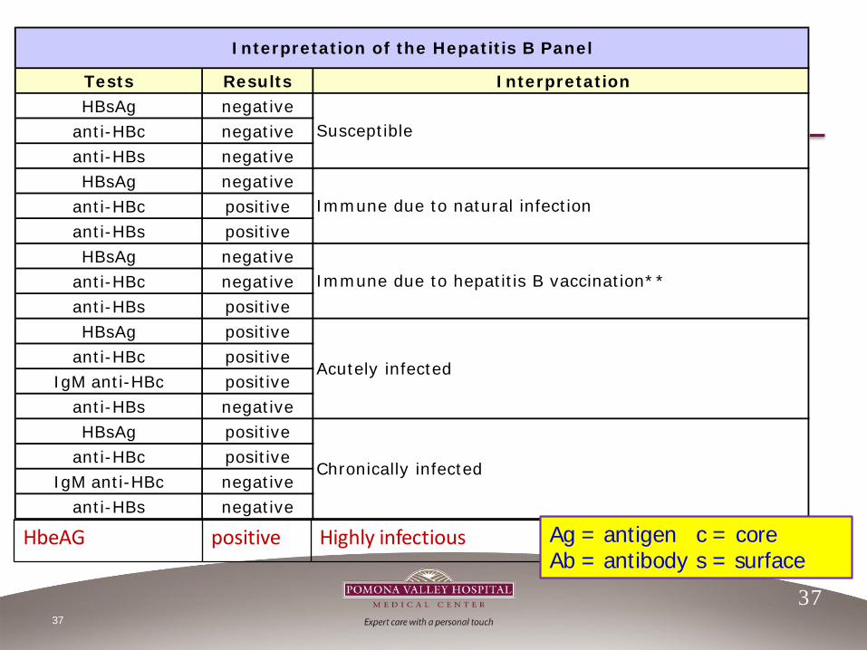
37
37
Tests Results InterpretationHBsAg negative
anti-HBc negativeanti-HBs negativeHBsAg negative
anti-HBc positiveanti-HBs positiveHBsAg negative
anti-HBc negativeanti-HBs positiveHBsAg positive
anti-HBc positiveIgM anti-HBc positive
anti-HBs negativeHBsAg positive
anti-HBc positiveIgM anti-HBc negative
anti-HBs negative
Interpretation of the Hepatitis B Panel
Immune due to hepatitis B vaccination**
Susceptible
Immune due to natural infection
Acutely infected
Chronically infected
HbeAG positive Highly infectious Ag = antigenAb = antibody
c = cores = surface

38
38
Hepatitis C Viral Markers • Hepatitis C Virus (HCV)• Anti-HCV
– Presence of antibodies to the virus, indicating exposure to HCV
– Active vs. chronic vs. resolved • HCV RIBA (recombinant immunoblot assay)
– Confirmatory test of antibodies to the virus– Demonstrates if HCV was true positive (present or past is
unanswered)
All Hepatitis (acute and chronic) are reportable communicable diseases via local public health

Laboratory Tests of Interest to IP
• Acid Fast Bacillus (AFB) test of sputum for diagnosis of TB • First morning specimen or bronch lavage are best• Rarely negative smear, positive culture (must follow up exposures)• Specimens must be at least 8hrs apart from each other
• Direct fluorescent antibody (DFA) tests for identification of respiratory viruses such as legionella
• Rapid diagnostic testing: provides quick diagnosis • HIV: detects antibiodies, has high sensitivity/specificity but because
of false positives, confirmatory testing should be done• Influenza: very fast antigen detection; false positives 51-82% of
time, so should not be used alone• Strep: antigen detection w/ 95% sensitivity; will also detect carriers
39
Nucleic Acid Amplification Tests (NAAT)
•Molecular technique that detects viruses or bacterium• Polymerase chain reaction (PCR) assays amplify gene segments
specific to organism of interest; available for a number of bacterial and viral pathogens• Uses alternating step and temperature cycle process to detect
molecules• Highly sensitive; may not indicate viability of organism• Expensive but getting cheaper, more rapid
• Ligase chain reaction (LCR) uses DNA polymerase (enzymes that build DNA and an enzyme that helps repair DNA. Because two targets are used, the test has greater specificity
• Newer, faster, expensive, less versatile, best for use with a single target
4 0

• Serology testing to look for that demonstrate exposure/infection • Indicates patient immunity• Testing can also look for antigens
• Antibiotic susceptibility testing performed on bacterial cultures to test the susceptibility or resistance to specific antimicrobial agents (see Kirby Bauer, Slide 22)
• Viral load testing for HIV, HCV
• Microscopic evaluation for fungal infections such as wet mounts for vaginal organisms, CSF, skin
• Antigen tests for cryptococcalmeningitis
41
Laboratory Tests of Interest to IP - continued

Role of Microbiology in HAI Prevention
Microbiology support is critical to • Outbreak management • Performing additional tests for epidemiologic analyses • Infection surveillance• Knowledge of new microbes or unusual resistance• Design of antibiotic formulary (antibiogram)• Interpretation of microbiological results• Education of health care staff
42

©2013 Pomona Valley Hospital Medical Center 43