
Mammography: Fundamental Principles, … Fundamental Principles, Equipment Design & Siting Kalpana...
61
AAPM 2012 Summer School on Medical Imaging using Ionizing Radiation Mammography: Fundamental Principles, Equipment Design & Siting Kalpana M. Kanal, Ph.D., DABR Associate Professor Director, Resident Physics Education Affiliate Faculty, Harborview Injury Prevention Center Adjunct Associate Professor, Oral Medicine Department of Radiology, University of Washington Seattle, Washington
Transcript of Mammography: Fundamental Principles, … Fundamental Principles, Equipment Design & Siting Kalpana...

AAPM 2012 Summer School on Medical Imaging using Ionizing Radiation
Mammography: Fundamental Principles, Equipment Design & Siting
Kalpana M. Kanal, Ph.D., DABRAssociate Professor
Director, Resident Physics Education Affiliate Faculty, Harborview Injury Prevention Center
Adjunct Associate Professor, Oral Medicine
Department of Radiology, University of WashingtonSeattle, Washington
AAPM 2012 Summer School on Medical Imaging using Ionizing Radiation
Disclosures
No disclosures
AAPM 2012 Summer School on Medical Imaging using Ionizing Radiation
Educational Objectives
• Understand the physics of digital detector technology• Recognize that vendors use varying detector
technology in FFDM systems• Appreciate the advantages and disadvantages of digital
mammography systems • Radiation Dose in FFDM systems• Economics of FFDM systems

AAPM 2012 Summer School on Medical Imaging using Ionizing Radiation
Full-Field Digital Mammography (FFDM) versus Screen-Film Mammography (SFM)• Wide dynamic range (1000:1) compared with SFM (40:1)• Dynamic image manipulation• Ability to post-process• Soft-copy read accompanied by computer-aided-diagnosis
(CAD)• 3D imaging
Radiographics 2004:24,1749
AAPM 2012 Summer School on Medical Imaging using Ionizing Radiation
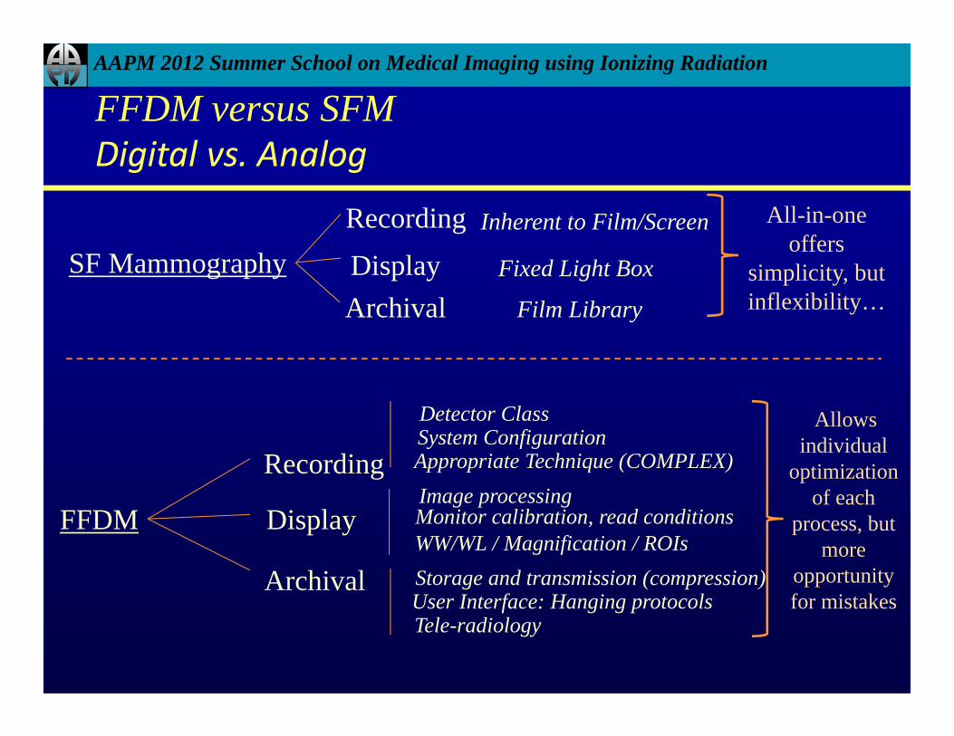
SF MammographyRecordingDisplayArchival
All-in-one offers
simplicity, but inflexibility…
Inherent to Film/Screen
Fixed Light Box
Film Library
FFDM
Recording
Detector ClassSystem ConfigurationAppropriate Technique (COMPLEX)
DisplayImage processingMonitor calibration, read conditionsWW/WL / Magnification / ROIs
Archival Storage and transmission (compression)User Interface: Hanging protocolsTele-radiology
Allows individual
optimization of each
process, but more
opportunity for mistakes
FFDM versus SFMDigital vs. Analog

AAPM 2012 Summer School on Medical Imaging using Ionizing Radiation
SFM vs. FFDM
SFM: Half mAs, Automatic exposure control, Double mAs
FFDM: Same technique factors as SFM, W/L adjusted
Radiographics 2004:24,1750

AAPM 2012 Summer School on Medical Imaging using Ionizing Radiation
SFM vs. FFDM
Radiographics 2004:24,1751SFM FFDM
AAPM 2012 Summer School on Medical Imaging using Ionizing Radiation
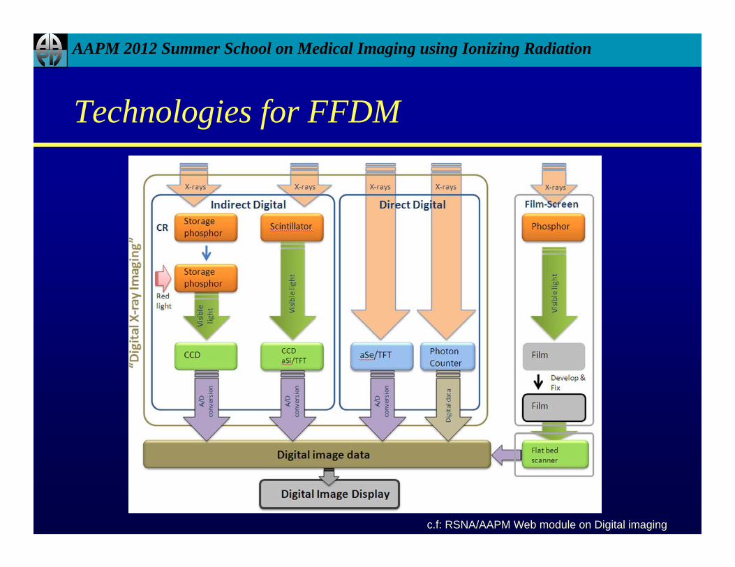
Technologies for FFDM
c.f: RSNA/AAPM Web module on Digital imaging

AAPM 2012 Summer School on Medical Imaging using Ionizing Radiation
Technologies for FFDM - Indirect Capture
• A scintillator such as cesium iodide (CsI) absorbs x-rays and generates a light scintillation
• Detected by an array of photodiodes or charge-coupled devices (CCDs)
• Resolution degradation
c.f: Bushberg, Third edition, pg. 266

AAPM 2012 Summer School on Medical Imaging using Ionizing Radiation
Technologies for FFDM - Direct Capture
• X-ray photons are captured by a photoconductor such as amorphous selenium (a-Se), which converts the absorbed x-rays directly into a electron-hole pair
• Spatial resolution limited to pixel size
c.f: Bushberg, Third edition, pg. 266

AAPM 2012 Summer School on Medical Imaging using Ionizing Radiation
Vendor Approaches - FFDM systems
• Indirect– A single flat-panel scintillator and an amorphous silicon (a-Si)
diode array – GE – Slot scanning with scintillators and CCD arrays – Fischer
Imaging, not commercially available now– Photostimulable phosphor plates - Fuji
• Direct– A flat-panel amorphous selenium (a-Se) array – Hologic,
Siemens– A dual-layer a-Se system using direct optical switching
technology - Fuji Aspire HD
AAPM 2012 Summer School on Medical Imaging using Ionizing Radiation
FDA and Digital Mammography
• FDA Office of Device Evaluation
• Clears FFDM for sale in US
• Approves monitors and printers for sale in US
• FDA Office of Communication, Education and Radiation Programs
• Writes and enforces MQSA regulations
• Issues MQSA certificates

AAPM 2012 Summer School on Medical Imaging using Ionizing Radiation
FDA and Digital Mammography
• FDA approved, cleared, or accepted the following FFDM for use in mammography facilities as indicated by date:– Konica Minolta Xpress CR System on 12/23/11 – Agfa CR System on 12/22/11 – Fuji Aspire CR System on 12/8/11 – Giotto Image 3D-3DL FFDM System on 10/27/11 – Fuji Aspire HD FFDM System on 9/1/11 – GE Senographe Care FFDM System on 10/7/11 – Planmed Nuance Excel FFDM System on 9/23/11 – Planmed Nuance FFDM System on 9/23/11
AAPM 2012 Summer School on Medical Imaging using Ionizing Radiation
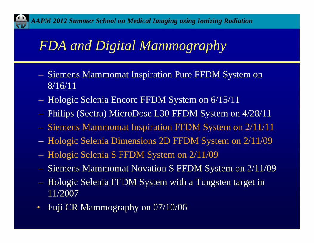
FDA and Digital Mammography
– Siemens Mammomat Inspiration Pure FFDM System on 8/16/11
– Hologic Selenia Encore FFDM System on 6/15/11 – Philips (Sectra) MicroDose L30 FFDM System on 4/28/11 – Siemens Mammomat Inspiration FFDM System on 2/11/11 – Hologic Selenia Dimensions 2D FFDM System on 2/11/09 – Hologic Selenia S FFDM System on 2/11/09 – Siemens Mammomat Novation S FFDM System on 2/11/09 – Hologic Selenia FFDM System with a Tungsten target in
11/2007 • Fuji CR Mammography on 07/10/06
AAPM 2012 Summer School on Medical Imaging using Ionizing Radiation
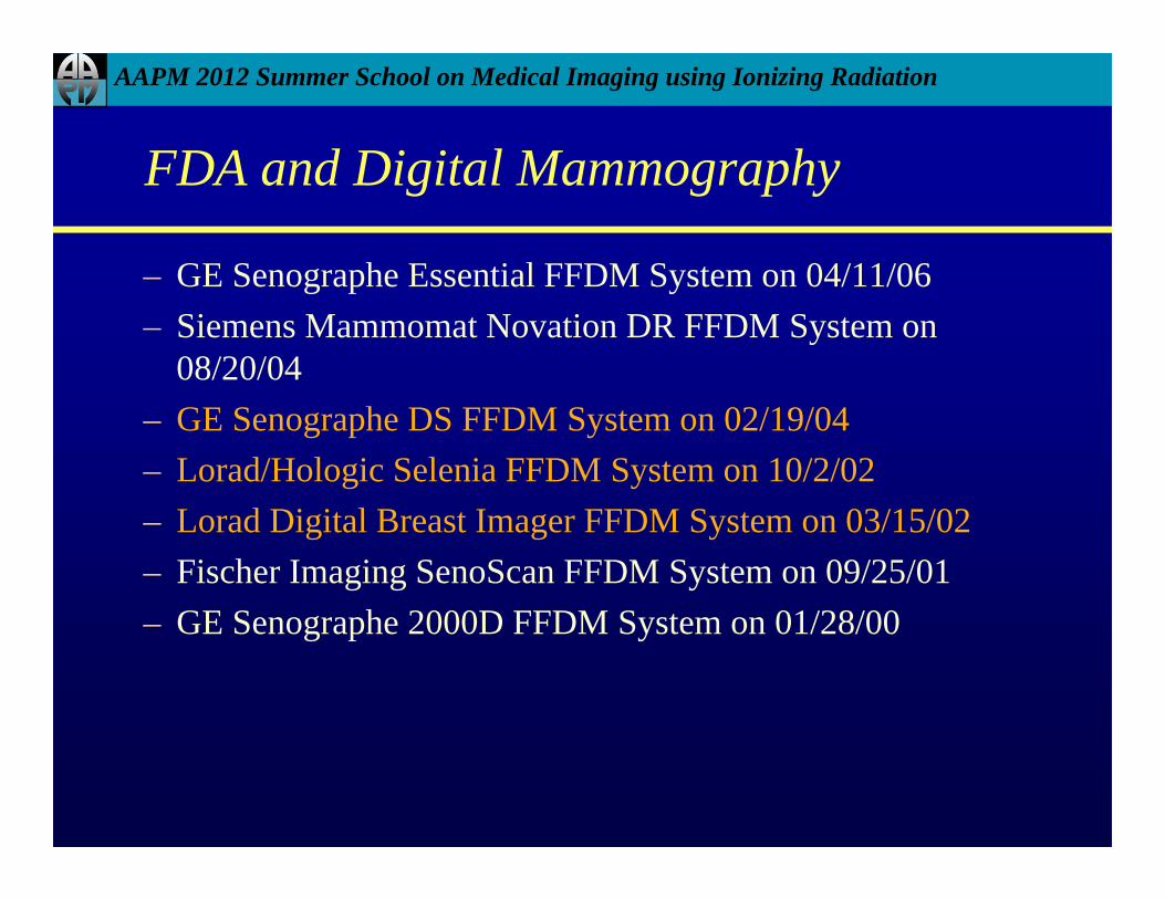
FDA and Digital Mammography
– GE Senographe Essential FFDM System on 04/11/06 – Siemens Mammomat Novation DR FFDM System on
08/20/04 – GE Senographe DS FFDM System on 02/19/04 – Lorad/Hologic Selenia FFDM System on 10/2/02 – Lorad Digital Breast Imager FFDM System on 03/15/02– Fischer Imaging SenoScan FFDM System on 09/25/01 – GE Senographe 2000D FFDM System on 01/28/00
AAPM 2012 Summer School on Medical Imaging using Ionizing Radiation
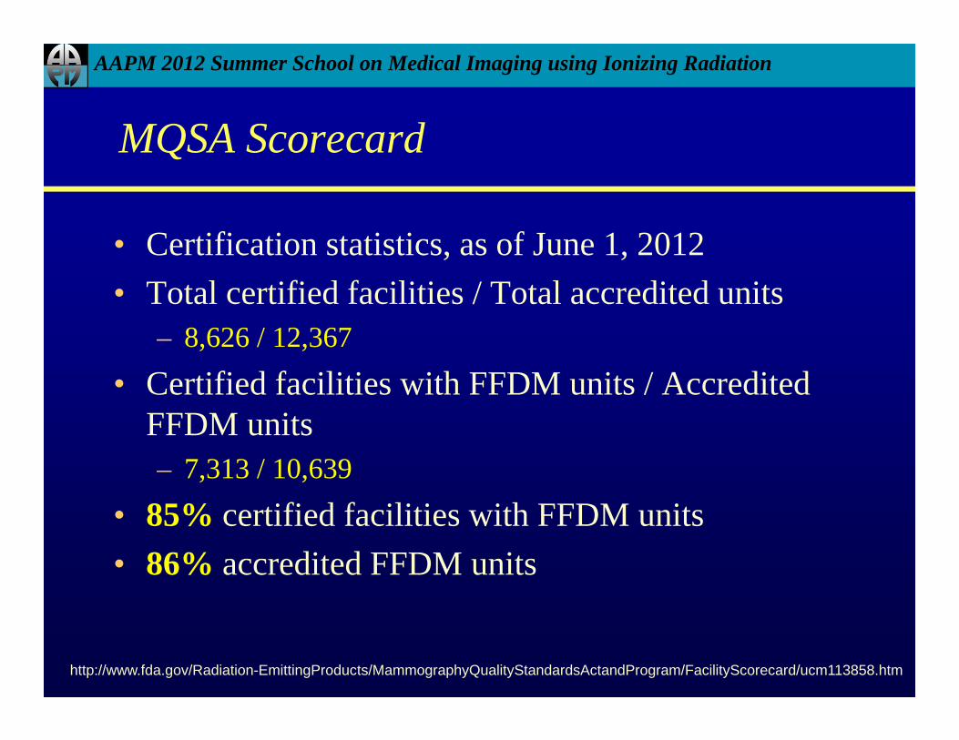
MQSA Scorecard
• Certification statistics, as of June 1, 2012 • Total certified facilities / Total accredited units
– 8,626 / 12,367
• Certified facilities with FFDM units / Accredited FFDM units– 7,313 / 10,639
• 85% certified facilities with FFDM units• 86% accredited FFDM units
http://www.fda.gov/Radiation-EmittingProducts/MammographyQualityStandardsActandProgram/FacilityScorecard/ucm113858.htm
AAPM 2012 Summer School on Medical Imaging using Ionizing Radiation
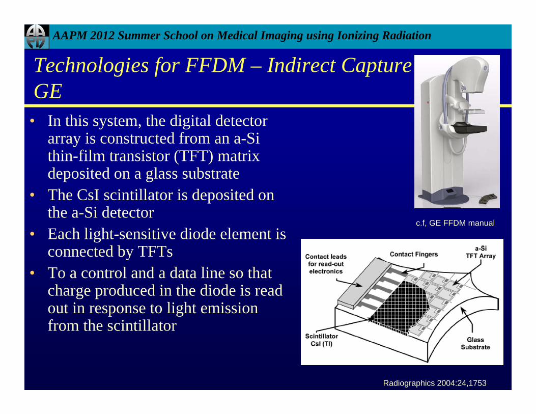
Technologies for FFDM – Indirect CaptureGE• In this system, the digital detector
array is constructed from an a-Si thin-film transistor (TFT) matrix deposited on a glass substrate
• The CsI scintillator is deposited on the a-Si detector
• Each light-sensitive diode element is connected by TFTs
• To a control and a data line so that charge produced in the diode is read out in response to light emission from the scintillator
Radiographics 2004:24,1753
c.f, GE FFDM manual

AAPM 2012 Summer School on Medical Imaging using Ionizing Radiation
Technologies for FFDM – Indirect CaptureGE
2000D DS Essential
Detector size 19.2 x 23.0 19.2 x 23.0 24.0 x 30.7
Pixel size 100 µm 100 µm 100 µmLimiting Spatial
Resolution 5 lp/mm 5 lp/mm 5 lp/mm
Image size1914 x 2294
pixels (9 MB)1914 x 2294
pixels (9 MB)
2394 x 3062 pixels (14 MB)
Bit Depth 14 14 14
AAPM 2012 Summer School on Medical Imaging using Ionizing Radiation
Technologies for FFDM – Indirect CaptureGE –
• Advantages and Disadvantages• Bonding between CsI and a-Si ensures minimal light loss• Strong signal from the Si diode array yields higher detective
quantum efficiency• Detector is linear over a wide range (105)
• Limiting factor is the large pixel size (100 m)• Smaller pixel sizes improve spatial resolution but at the cost of
increased image noise and decreased SNR for the same breast dose
• Possibility of ghosting in images
AAPM 2012 Summer School on Medical Imaging using Ionizing Radiation
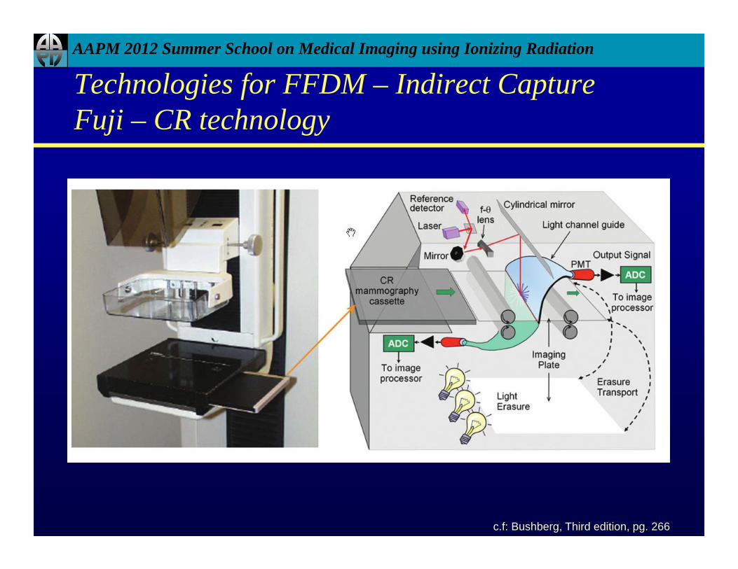
Technologies for FFDM – Indirect CaptureFuji – CR technology
c.f: Bushberg, Third edition, pg. 266

AAPM 2012 Summer School on Medical Imaging using Ionizing Radiation
• Fuji FCRm, Dual-side reader
Detector size
18 x 2424 x 30
Pixel size 50 µm
Image size 3328 x 4096 pixels (24 MB)
Spatial Resolution 10 lp/mm
Dynamic Range 14 bits
http://www.fujimed.com/
Technologies for FFDM – Indirect CaptureFuji – CR technology
AAPM 2012 Summer School on Medical Imaging using Ionizing Radiation
• Advantages and Disadvantages• Film-screen cassettes can be replaced by CR cassettes without
replacing the entire system• Both small and large cassettes can be accommodated by the reader• Dual side reader, 50 m pixel size
• Effective pixel size influenced by phosphor thickness, light diffusion within phosphor, laser light scatter & diameter of laser beam
• Technologist time on processing of images• Noise associated with the low collection efficiency of emitted light
Technologies for FFDM – Indirect CaptureFuji – CR technology

AAPM 2012 Summer School on Medical Imaging using Ionizing Radiation
• A narrow slot-detector and a narrow fan beam of x-rays are scanned synchronously across the full field of view to cover the entire breast
• System consists of phosphor (thallium-activated CsI) with fiberoptic coupling to a CCD
Radiographics 2004:24,1752
Technologies for FFDM – Indirect CaptureFischer – SenoScan (not available now)

AAPM 2012 Summer School on Medical Imaging using Ionizing Radiation
Technologies for FFDM – Indirect CaptureFischer – SenoScan (not available now)

AAPM 2012 Summer School on Medical Imaging using Ionizing Radiation
• Advantages and Disadvantages• Compact detector that is less expensive compared to others• Excellent scatter rejection due to small volume of breast
exposed at any time• No grid needed therefore less dose• Longer compression since scan times are longer (approx. 6
sec)• Powerful tubes, elaborate signal readout and image
reconstruction required
Technologies for FFDM – Indirect CaptureFischer – SenoScan (not available now)

AAPM 2012 Summer School on Medical Imaging using Ionizing Radiation
Comparison – Indirect Capture
GE DS Fischer Seno Fuji FCRm
Detector size19.2 x 23.0 21 x 1
18 x 2424 x 30
Pixel size 100 µm 25 or 50 µm 50 µmLimiting Spatial
Resolution 5 lp/mm13 lp/cm at 2510 lp/cm at 50
10 lp/mm
Image matrix1914 x 2294
pixels 4096 x 56253328 x 4096

AAPM 2012 Summer School on Medical Imaging using Ionizing Radiation
• a-Se, photoconductor is deposited directly onto the a-Si TFT substrate enabling direct capture
• The a-Se detector directly converts x-rays to electron-hole pairs
• The a-Si TFT converts the electron-hole pairs to electronic signal
http://www.hologic.com/wh/digisel.htm
Technologies for FFDM – Direct CaptureHologic – Selenia

AAPM 2012 Summer School on Medical Imaging using Ionizing Radiation
http://www.hologic.com/wh/digisel.htm
Detector size 24.0 x 29.0
Pixel size 70 µm
Image size 3328 x 4096 pixels (24 MB)
Spatial Resolution > 7 lp/mm
Dynamic Range 14 bits
Technologies for FFDM – Direct CaptureHologic – Selenia

AAPM 2012 Summer School on Medical Imaging using Ionizing Radiation
• Advantages and Disadvantages• Advantage is that the detector response function maintains its
sharpness even with increasing thickness
• Potential weaknesses are the need for high biasing voltage, drifting of the dark signal and cost of detector
• Inherent sharpness of detector may also increase the severity of aliasing artifacts associated with undersampling on any digital detector
Technologies for FFDM – Direct CaptureHologic – Selenia
AAPM 2012 Summer School on Medical Imaging using Ionizing Radiation
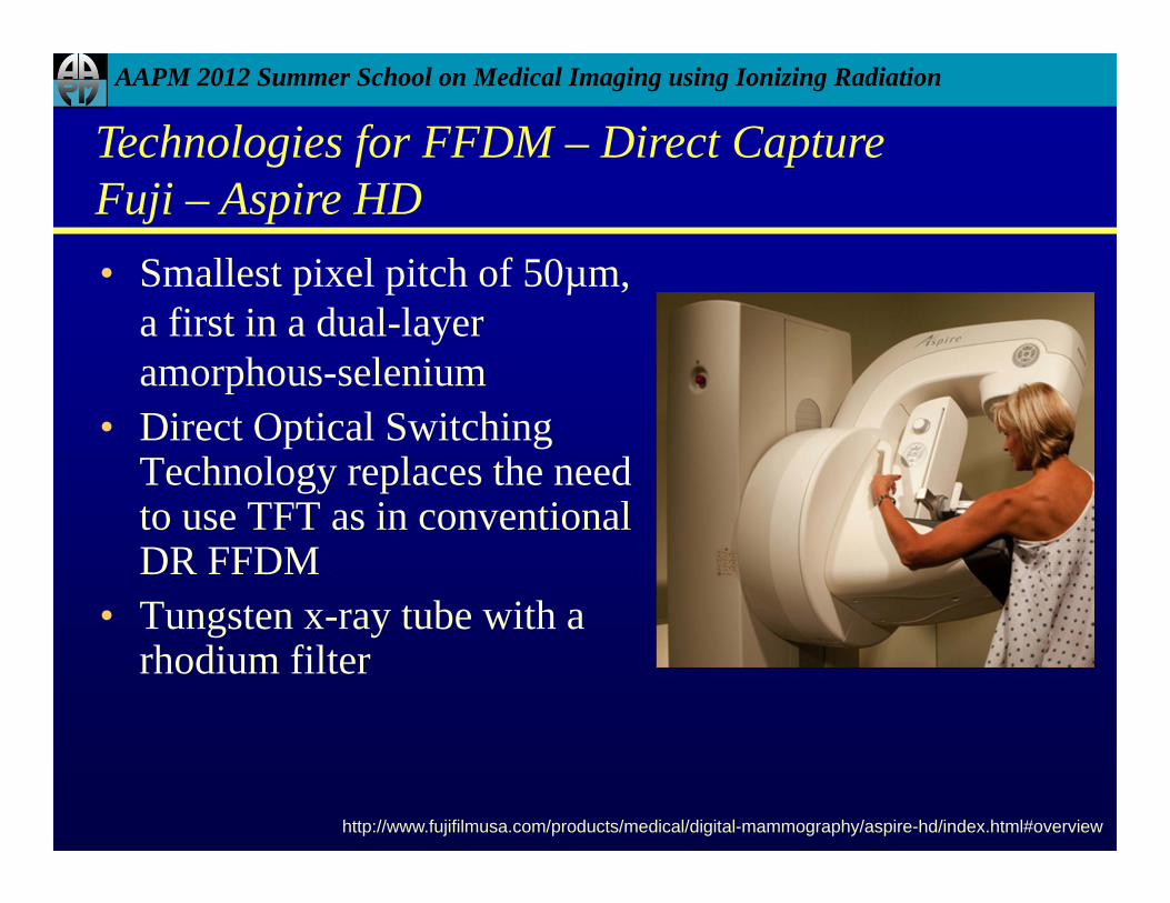
• Smallest pixel pitch of 50µm, a first in a dual-layer amorphous-selenium
• Direct Optical Switching Technology replaces the need to use TFT as in conventional DR FFDM
• Tungsten x-ray tube with a rhodium filter
Technologies for FFDM – Direct CaptureFuji – Aspire HD
http://www.fujifilmusa.com/products/medical/digital-mammography/aspire-hd/index.html#overview

AAPM 2012 Summer School on Medical Imaging using Ionizing Radiation
Detector size 24.0 x 30.0
Pixel size 50 µm
Image size 3328 x 4096 pixels (24 MB)
Spatial Resolution > 7 lp/mm
Dynamic Range 14 bits
Technologies for FFDM – Direct CaptureFuji – Aspire HD
http://www.fujifilmusa.com/products/medical/digital-mammography/aspire-hd/index.html#overview
AAPM 2012 Summer School on Medical Imaging using Ionizing Radiation
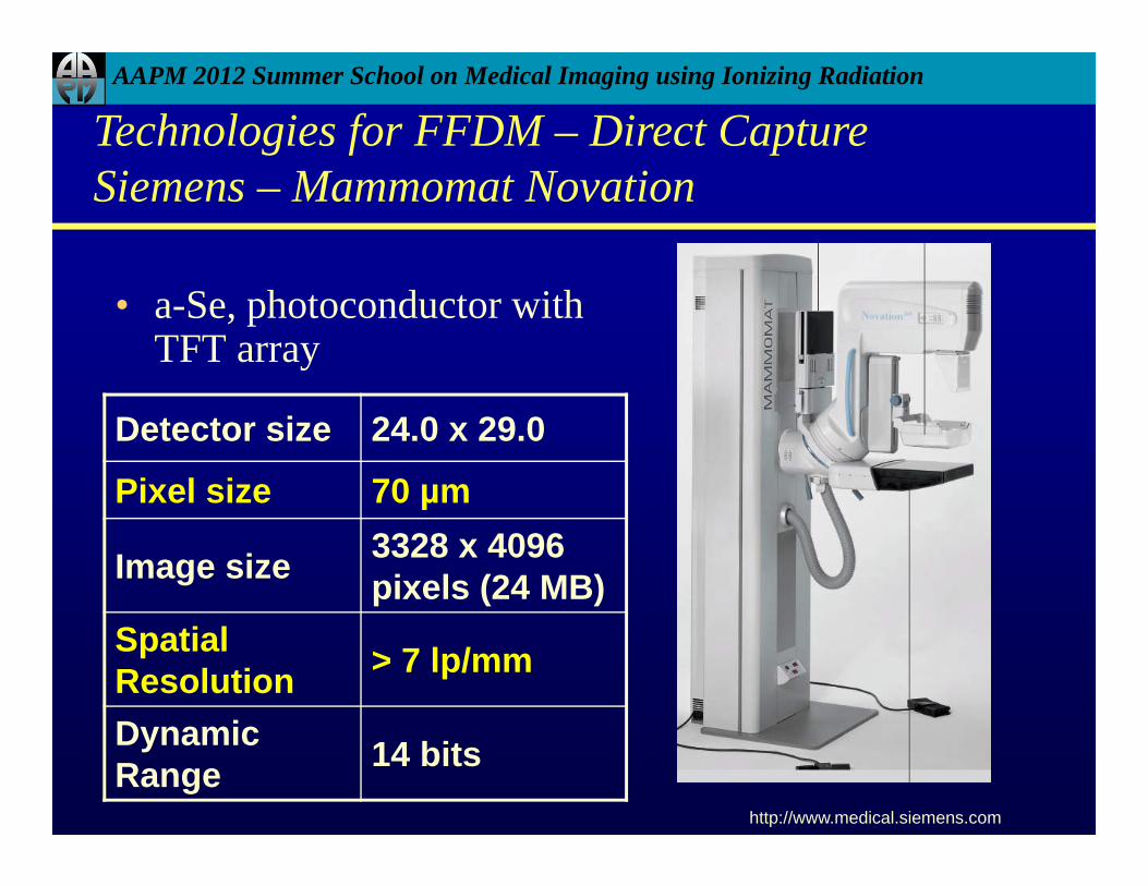
• a-Se, photoconductor with TFT array
http://www.medical.siemens.com
Technologies for FFDM – Direct CaptureSiemens – Mammomat Novation
Detector size 24.0 x 29.0
Pixel size 70 µm
Image size 3328 x 4096 pixels (24 MB)
Spatial Resolution > 7 lp/mm
Dynamic Range 14 bits

AAPM 2012 Summer School on Medical Imaging using Ionizing Radiation
Comparison – Direct Capture
HologicSelenia
Siemens Novation Fuji Aspire HD
Detector size24 x 29 24 x 29 24 x 30
Pixel size 70 µm 70 µm 50 µmLimiting Spatial
Resolution > 7 lp/mm > 7 lp/mm 10 lp/mm ?
Image matrix 3328 x 40962560 x 3328
3328 x 40963328 x 4096

AAPM 2012 Summer School on Medical Imaging using Ionizing Radiation
Planmed
• 85 µm pixel size • Amorphous selenium (a-Se)
direct-conversion detector • Two detector sizes - 17x24
cm (Planmed Nuance) and 24x30 cm (Planmed Nuance Excel)
• Tungsten tube, Ag/Rh filters
http://www.planmed.com
AAPM 2012 Summer School on Medical Imaging using Ionizing Radiation
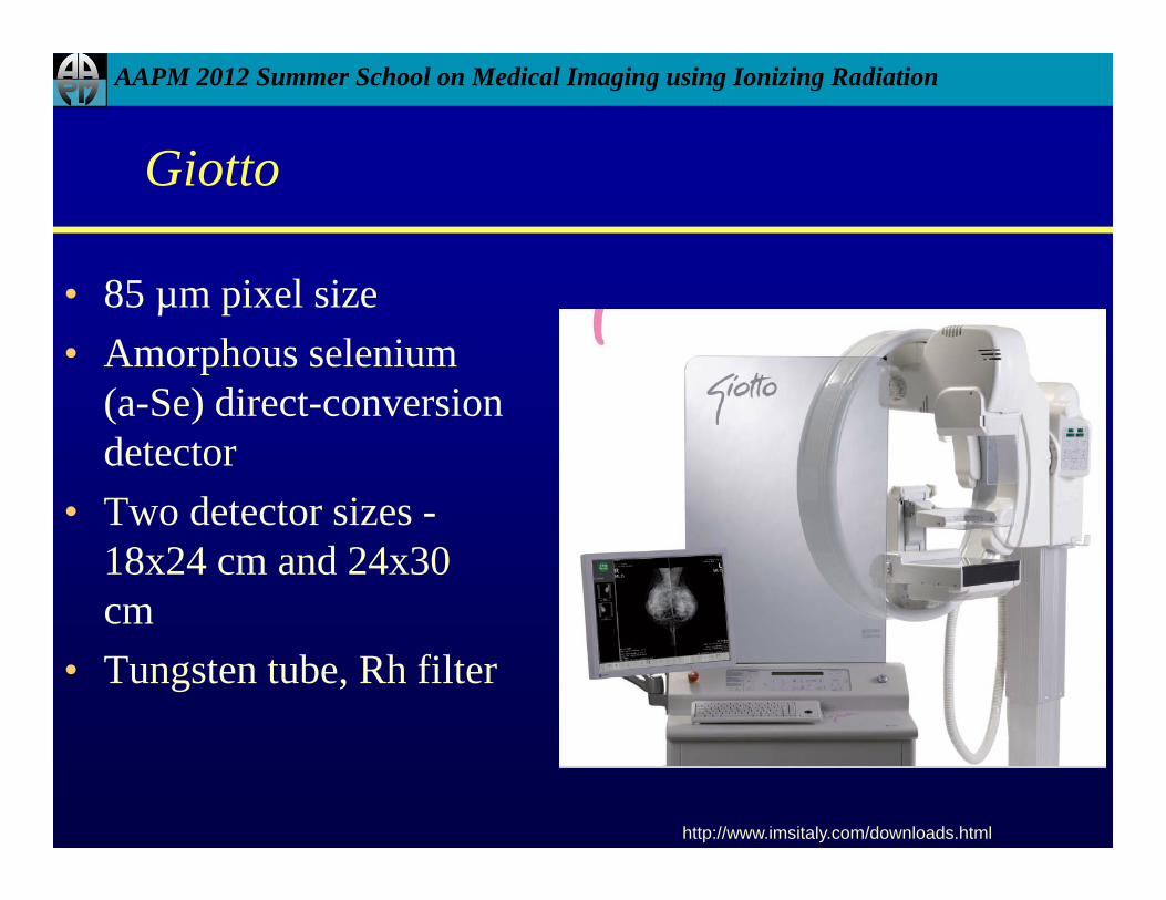
Giotto
• 85 µm pixel size • Amorphous selenium
(a-Se) direct-conversion detector
• Two detector sizes -18x24 cm and 24x30 cm
• Tungsten tube, Rh filter
http://www.imsitaly.com/downloads.html

AAPM 2012 Summer School on Medical Imaging using Ionizing Radiation
Technologies for FFDM
http://www.hologic.com/oem/pdf/DROverviewR-007_Nov2000.pdf
Fuji/Kodak/
Agfa/Philips
Fischer (Hologic)
GE
Hologic
Siemens
Planmed
Giotto
Mammo
Direct
a-Se/optical switching
Fuji Aspire
HD
AAPM 2012 Summer School on Medical Imaging using Ionizing Radiation
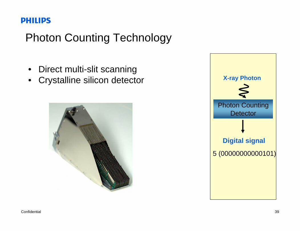
Photon Counting Technology
• Slides are Courtesy of Dr. Eric Berns and Philips

June 6, 2012 38
Digital MammographyTechnology Overview
Digital Mammography
Direct conversion Indirect conversion
Analog-to-Digital
TrueDigital
Analog-to-Digital
Delayedprocessing
Non-delayedprocessing
a-SeleniumPhoton countingCrystalline silicon
CR – storagephosphor
a-silicon
Confidential 39
Photon Counting Technology
• Direct multi-slit scanning• Crystalline silicon detector X-ray Photon
Digital signal
Photon CountingDetector
5 (00000000000101)
June 6, 2012 40
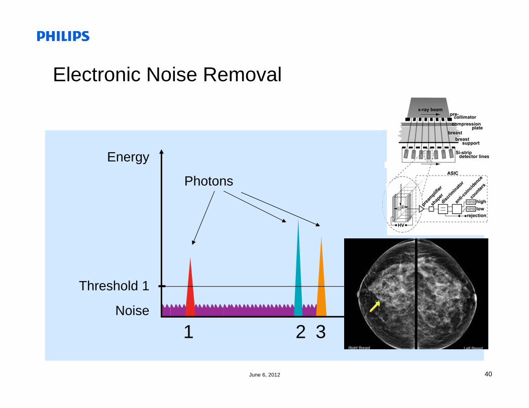
Photons
Energy
Threshold 1
1 2 3Noise Time
Electronic Noise Removal

June 6, 2012 41photo courtesy Philips by Digital Mammography
Multi-slit detector module scansthe breast
Module contains50 um detector elements, 21 detector lines
No anti-scattergrid required

June 6, 2012 42
Photon Counting in PracticeMicroDose

June 6, 2012 43
DQE – A Measure of Dose Efficiency
Monnin et al, Med. Phys. 34, 906-14 (2007)
DQ
E(0
)
PHILIPS MicroDosePhoton Counting
Monnin et al., Med. Phys. (34) 2007
June 6, 2012 44
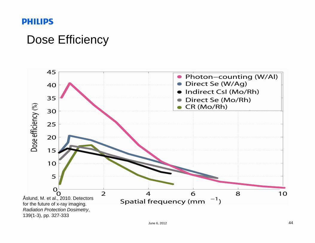
Dose Efficiency
Åslund, M. et al., 2010. Detectors for the future of x-ray imaging. Radiation Protection Dosimetry, 139(1-3), pp. 327-333

AAPM 2012 Summer School on Medical Imaging using Ionizing Radiation
Modulation Transfer Function (MTF)
• MTF is a measure of signal transfer over a range of frequencies and quantifies spatial resolution
Yaffe - Radiology 2005:234,353
Type 1 – CsI (TFT)
Type 2 – CsI (CCD)
Type 3 – CR
Type 4 – a-Se
http://www.hologic.com/wh/pdf/R-LM-016_Radiology_Management.pdf

AAPM 2012 Summer School on Medical Imaging using Ionizing Radiation
• Bloomquist et al - DMIST trial
Bloomquist – Medical Physics 2006:33 (3), 719
Modulation Transfer Function (MTF)
AAPM 2012 Summer School on Medical Imaging using Ionizing Radiation
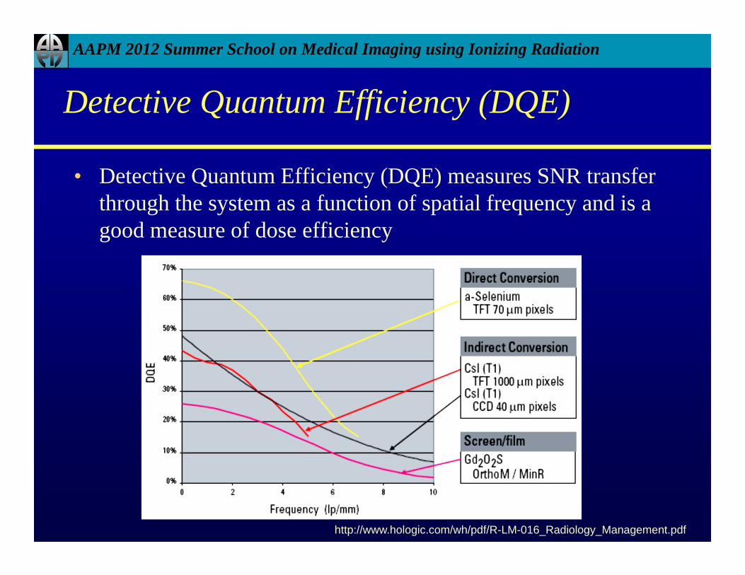
Detective Quantum Efficiency (DQE)
• Detective Quantum Efficiency (DQE) measures SNR transfer through the system as a function of spatial frequency and is a good measure of dose efficiency
http://www.hologic.com/wh/pdf/R-LM-016_Radiology_Management.pdf
AAPM 2012 Summer School on Medical Imaging using Ionizing Radiation
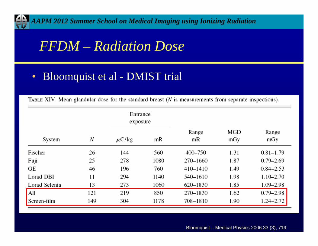
FFDM – Radiation Dose
• Bloomquist et al - DMIST trial
Bloomquist – Medical Physics 2006:33 (3), 719
AAPM 2012 Summer School on Medical Imaging using Ionizing Radiation
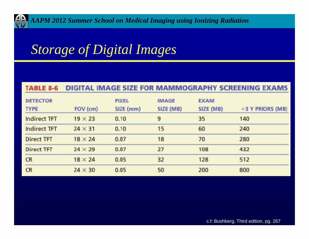
Storage of Digital Images
c.f: Bushberg, Third edition, pg. 267
AAPM 2012 Summer School on Medical Imaging using Ionizing Radiation
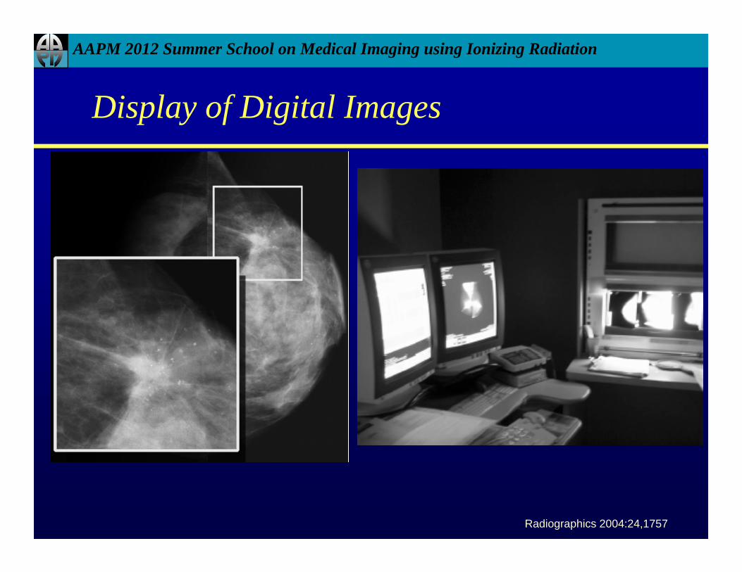
Display of Digital Images
Radiographics 2004:24,1757

AAPM 2012 Summer School on Medical Imaging using Ionizing Radiation
Economics of FFDM
• SFM systems cost well under $100,000• FFDM systems cost in the range of $300,000 -
$450,000

AAPM 2012 Summer School on Medical Imaging using Ionizing Radiation
FFDM Reimbursement
www.cms.gov

AAPM 2012 Summer School on Medical Imaging using Ionizing Radiation
Expected Benefits of FFDM
• The costs of FFDM systems should be compared along with the inherent benefits of the digital technology prior to the purchase:– Reduced recall rates– Increased patient throughput– Increased early detection of breast cancer– Decreased false-negative biopsy results– Decreasing film and processing costs– Increasing the caseload of each mammography room

AAPM 2012 Summer School on Medical Imaging using Ionizing Radiation
Clinical Trials and Phantom Studies
• Larger screening study screened 49,500 women• Digital Mammographic Imaging Screening Trial
(DMIST), funded by NCI and conducted by ACRIN (http://www.acrin.org/6652_protocol.html)

AAPM 2012 Summer School on Medical Imaging using Ionizing Radiation
Advantages and Disadvantages
• Advantages– Optimize post-processing of images– Permit computer-aided detection to improve the
detection of lesions– Storage of images easier
• Disadvantages– Image display and system cost– Limiting spatial resolution is inferior to film, 5-13
lp/mm vs. 20 lp/mm– Superior contrast resolution

AAPM 2012 Summer School on Medical Imaging using Ionizing Radiation
Siting Requirements
• Room dimensions and power requirements needed depend on vendor equipment
• Breast support provides adequate primary barrier for radiation
• Typically 2 sheets or 28 mm of gypsum wallboard (sheetrock) provide adequate secondary shielding
• Technologist protected by lead shield, 0.3mm lead• Wood doors attenuate less than gypsum wallboard,
may need metal doors or solid-core wood doors• “X-ray on” light typically required on the door (in
outside room/hallway)

AAPM 2012 Summer School on Medical Imaging using Ionizing Radiation
Example – One Vendor

AAPM 2012 Summer School on Medical Imaging using Ionizing Radiation
Educational Objectives
• Understand the physics of digital detector technology• Recognize that vendors use varying detector
technology in FFDM systems• Appreciate the advantages and disadvantages of digital
mammography systems • Radiation Dose in FFDM systems• Economics of FFDM systems

AAPM 2012 Summer School on Medical Imaging using Ionizing Radiation
TAKE HOME POINTS
• Different technologies exist for digital systems – indirect and direct
• Commercially available FFDM systems vary in technology
• Many advantages exist for FFDM in comparison to FSM• Dose is lower with FFDM compared to SFM
AAPM 2012 Summer School on Medical Imaging using Ionizing Radiation
Resources
• Digital Mammography: An overview – Dr. Mahesh(Radiographics 2004;24:1747-1760)
• Fundamentals of Digital Mammography Primer – Dr. Smith(Hologic Inc)
• Digital Mammography – Pisano and Yaffe (Radiology 2005; 234:353-262)
• Bloomquist and Yaffe – Med Phys 33 (3), 2006• MHRA report 05037: Comparitive Specifications of Full Field
Digital Mammography Systems• http://www.fda.gov/Radiation-
EmittingProducts/MammographyQualityStandardsActandProgram/FacilityCertificationandInspection/ucm114148.htm
AAPM 2012 Summer School on Medical Imaging using Ionizing Radiation
THANK YOU