
Farm Animals Which animals live on a farm? Why are farm animals important? Let’s learn!

HAL Id: hal-00903024https://hal.archives-ouvertes.fr/hal-00903024
Submitted on 1 Jan 2006
HAL is a multi-disciplinary open accessarchive for the deposit and dissemination of sci-entific research documents, whether they are pub-lished or not. The documents may come fromteaching and research institutions in France orabroad, or from public or private research centers.
L’archive ouverte pluridisciplinaire HAL, estdestinée au dépôt et à la diffusion de documentsscientifiques de niveau recherche, publiés ou non,émanant des établissements d’enseignement et derecherche français ou étrangers, des laboratoirespublics ou privés.
MALT structure and function in farm animalsElisabeth Liebler-Tenorio, Reinhard Pabst
To cite this version:Elisabeth Liebler-Tenorio, Reinhard Pabst. MALT structure and function in farm animals. VeterinaryResearch, BioMed Central, 2006, 37 (3), pp.257-280. �10.1051/vetres:2006001�. �hal-00903024�

257Vet. Res. 37 (2006) 257–280© INRA, EDP Sciences, 2006DOI: 10.1051/vetres:2006001
Review article
MALT structure and function in farm animals
Elisabeth M. LIEBLER-TENORIOa*, Reinhard PABSTb
a Friedrich-Loeffler-Institute, Federal Research Institute for Animal Health, Naumburger Str. 96 a, 07743 Jena, Germany
b Medical School Hanover, Centre of Anatomy, Carl-Neuberg-Str. 1, 30625 Hanover, Germany
(Received 17 May 2005; accepted 15 September 2005)
Abstract – Mucosa-associated lymphoid tissue (MALT) is defined as an organized lymphoid tissuein the mucosa that samples antigens. The morphological characteristics that distinguish MALT fromlymphoid infiltrates are discussed. MALT has been extensively investigated in laboratory animals,while knowledge in cattle, sheep, goats, pigs and horses that are summarized under the term farmanimals in this review is fragmentary. Literature data about the distribution, morphology, functionand involvement in infectious diseases of MALT in farm animals are described. The understandingof specific features of MALT in other species than laboratory animals is important for comparativeresearch, in order to understand pathological and immunological processes in the respective speciesand as a potential route of vaccination of mucosal surfaces.
MALT / NALT / tonsils / BALT / GALT
Table of contents
1. Introduction...................................................................................................................................... 2582. Conjunctiva-associated lymphoid tissue (CALT)............................................................................ 2613. Nose-associated lymphoid tissue (NALT)....................................................................................... 2614. Lymphoid tissues of the Waldeyer’s ring ........................................................................................ 261
4.1. Bovine ..................................................................................................................................... 2624.1.1. Composition of the Waldeyer’s ring............................................................................ 2624.1.2. Pharyngeal tonsils ........................................................................................................ 2634.1.3. Palatine tonsils ............................................................................................................. 2634.1.4. Differences between tonsils and other MALT structures ............................................ 2634.1.5. Role of tonsils in infection........................................................................................... 263
4.2. Ovine/caprine .......................................................................................................................... 2644.2.1. Composition of the Waldeyer’s ring............................................................................ 2644.2.2. Pharyngeal tonsils ........................................................................................................ 2644.2.3. Tubal tonsils................................................................................................................. 2644.2.4. Palatine tonsils ............................................................................................................. 2644.2.5. Role of tonsils in infection........................................................................................... 264
4.3. Porcine..................................................................................................................................... 2654.3.1. Composition of the Waldeyer’s ring............................................................................ 2654.3.2. Tonsils of the soft palate .............................................................................................. 265
* Corresponding author: [email protected]
Article published by EDP Sciences and available at http://www.edpsciences.org/vetres or http://dx.doi.org/10.1051/vetres:2006001

258 E.M. Liebler-Tenorio, R. Pabst
4.3.3. Lingual tonsil................................................................................................................2654.3.4. Differences between tonsils and other MALT structures.............................................2654.3.5. Role of tonsils in infection ...........................................................................................266
4.4. Equine ......................................................................................................................................2664.4.1. Composition of the Waldeyer’s ring ............................................................................2664.4.2. Pharyngeal tonsils.........................................................................................................2664.4.3. Lingual tonsil................................................................................................................2664.4.4. Role of tonsils in infection ...........................................................................................266
5. Larynx- and trachea-associated lymphoid tissue (LTALT) .............................................................2666. Bronchus-associated-lymphoid tissue (BALT) ................................................................................267
6.1. Bovine ......................................................................................................................................2676.2. Ovine/caprine...........................................................................................................................2686.3. Porcine .....................................................................................................................................2686.4. Equine ......................................................................................................................................268
7. Gastric-MALT..................................................................................................................................2698. Gut-associated lymphoid tissue (GALT)..........................................................................................269
8.1. Bovine ......................................................................................................................................2698.1.1. Distribution, ontogeny and amount .............................................................................2698.1.2. Histology and lymphocyte subsets ...............................................................................2718.1.3. Morphology and function of the lymphoepithelium ....................................................271
8.2. Ovine/caprine...........................................................................................................................2718.2.1. Distribution, ontogeny and amount .............................................................................2718.2.2. Histology and lymphocyte subsets ...............................................................................2728.2.3. The IPP as a primary lymphoid organ ..........................................................................272
8.3. Porcine .....................................................................................................................................2728.3.1. Distribution, ontogeny and amount .............................................................................2728.3.2. Histology and lymphocyte subsets ...............................................................................2738.3.3. Morphology and function of the lymphoepithelium ....................................................273
8.4. Equine ......................................................................................................................................2738.4.1. Distribution and amount ..............................................................................................2738.4.2. Histology .....................................................................................................................2748.4.3. Morphology of the lymphoepithelium..........................................................................274
9. Conclusions ......................................................................................................................................274
1. INTRODUCTION
Organized mucosa-associated lymphoidtissue (MALT) which is widely distributedin mucosal surfaces is an essential part ofthe mucosal immune system. MALT is theinitial inductive site for mucosal immunity:antigens are sampled from mucosal sur-faces and cognate naïve B- and T-lym-phocytes stimulated. MALT structures arethe origin of lymphocyte trafficking tomucosal effector sites. MALT containslymphatics which transport immune cellsand antigens to regional lymph nodes thatcan therefore be called part of the inductive
sites of mucosa and augment the immuneresponses.
MALT is located at strategical sites toallow efficient antigen sampling frommucosal surfaces. Based on the anatomicallocalization, MALT structures can be sub-divided into [17]: • conjunctiva-associated lymphoid tissue(CALT),• lacrimal drainage-associated lymphoidtissue (LADLT),• salivary gland-associated lymphoid tissue/duct-associated lymphoid tissue (SALT/DALT),
MALT in farm animals 259
• nose or nasopharynx-associated lym-phoid tissue (NALT),• lymphoid tissues of Waldeyer’s ring,• larynx-associated lymphoid tissue(LTALT),• bronchus-associated lymphoid tissue(BALT), • gastric mucosa-associated lymphoid tis-sue (gastric MALT),• gut-associated lymphoid tissue (GALT).
Only lymphoid tissues in the mucosa thatfulfill certain morpholocial and functionalcriteria should be called MALT [17].
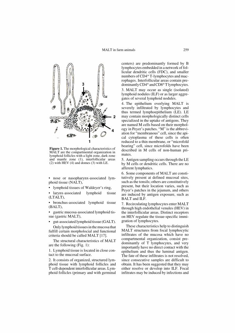
The structural characteristics of MALTare the following (Fig. 1):1. Lymphoid tissue is located in close con-tact to the mucosal surface.2. It consists of organized, structured lym-phoid tissue with lymphoid follicles andT cell-dependent interfollicular areas. Lym-phoid follicles (primary and with germinal
centers) are predominantly formed by Blymphocytes embedded in a network of fol-licular dendritic cells (FDC), and smallernumbers of CD4+ T-lymphocytes and mac-rophages. Interfollicular areas contain pre-dominantly CD4+ and CD8+ T lymphocytes.3. MALT may occur as single (isolated)lymphoid nodules (ILF) or as larger aggre-gates of several lymphoid nodules. 4. The epithelium overlying MALT isseverely infiltrated by lymphocytes andthus termed lymphoepithelium (LE). LEmay contain morphologically distinct cellsspecialized in the uptake of antigens. Theyare named M cells based on their morphol-ogy in Peyer’s patches. “M” is the abbrevi-ation for “membranous” cell, since the api-cal cytoplasma of these cells is oftenreduced to a thin membrane, or “microfoldbearing” cell, since microfolds have beendescribed in M cells of non-human pri-mates. 5. Antigen sampling occurs through the LEby M cells or dendritic cells. There are noafferent lymphatics. 6. Some components of MALT are consti-tutively present at defined mucosal sites,such as the tonsils; others are constitutivelypresent, but their location varies, such asPeyer’s patches in the jejunum, and othersare induced by antigen exposure, such asBALT and ILF.7. Recirculating lymphocytes enter MALTthrough high endothelial venules (HEV) inthe interfollicular areas. Distinct receptorson HEV regulate the tissue-specific immi-gration of lymphocytes.
These characteristics help to distinguishMALT structures from focal lymphocyticinfiltrates of the mucosa which have nocompartmental organization, consist pre-dominantly of T lymphocytes, and veryimportantly have no direct contact with theepithelium and thus the luminal antigen.The fate of these infiltrates is not resolved,since consecutive samples are difficult toobtain. It has been suggested that they mayeither resolve or develop into ILF. Focalinfitrates may be induced by infections and
Figure 1. The morphological characteristics ofMALT are the compartmental organization inlymphoid follicles with a light zone, dark zoneand mantle zone (1), interfollicular areas(2) with HEV (4) and domes (3) with LE.

260 E.M. Liebler-Tenorio, R. Pabst
careful examination is necessary to distin-guish for example ILF in the large intestineof pigs from granulomas induced byOesophagostomum dentatum [39] orBALT from pneumonic lesions [6] or gas-tric MALT from gastric lesions [34].
MALT has been intensely investigatedin laboratory rodents, since MALT plays amayor role in the protection of mucosal bar-riers and also in allergic reactions. Investi-gations involving human tissues are limitedto biopsy or resection material. Thereforedata obtained in laboratory rodents areoften extrapolated to humans, althoughmice, rats and rabbits are quite distinct indevelopment, lifespan, environment (germfree, SPF), nutrition, and physiology fromhumans, and other species such as farm ani-mals might be better as animal models.
The distribution, occurrence, morphol-ogy, ontogeny and evolution of MALT varybetween species. In the following the cur-rent knowledge will be summarized for cat-tle, sheep, goats, pigs and horses whichwere arbitrarily combined under the term“farm animals” to distinguish them fromthe commonly used small laboratory ani-mal species. Differences between speciesmight be caused by anatomical and physi-ological characteristics, since MALT struc-tures are always strategically located at
sentinel positions for optimal antigen sam-pling. The appearance of MALT is alsoinfluenced by differences in antigen expo-sure, e.g. due to management practices,since its development is often antigen-driven. Knowledge about MALT in farmanimals is important for the following rea-sons:• to recognize the normal structures tounderstand the physiology, • to localize MALT, e.g. to collect it fordiagnostic tests [121],• to distinguish normal MALT fromhyperreactive, activated, altered MALT [6,34, 72],• to learn about the pathogenesis of infec-tions/host reactions [104, 105, 114, 117],• to determine if inductive sites for thelocal application of vaccines are present [7,59, 116],• to investigate MALT structure and func-tion under comparative aspects [37].
MALT has not been described at all sitesinitially listed in each of the farm animalspecies which does, however, not implythat it does not exist. The current knowl-edge of the distribution of MALT in cattle,sheep, goats, pigs and horses is summarizedin Table I.
Table I. MALT structures described in cattle, sheep/goats, pigs and horses.
CALT NALT Waldeyer’s ring DALT SALT LDALT
LTALT BALT Gastric MALT
GALT
Nasopharynx Oropharynx
Bovine + nd T. pharyngeaT. tubaria
T. lingualisT. veli palat.T. palatina
nd + (+) nd +
Ovine / caprine
+ + T. pharyngeaT. tubaria
T. lingualisT. veli palat.T. palatina
nd + (+) nd +
Porcine + nd T. pharyngeaT. tubaria
T. lingualisT. veli palat.
nd + (+) + +
Equine + + T. pharyngeaT. tubaria
T. lingualisT. veli palat.T. palatina
nd + (+) nd +
+: constitutively present; nd: not examined / not described; (+): not constitutively present.

MALT in farm animals 261
2. CONJUNCTIVA-ASSOCIATED LYMPHOID TISSUE (CALT)
CALT was investigated in a comparativestudy for cattle, sheep and pigs [24]. Inthese species, CALT is formed by variablenumbers of ILF which are predominantlylocalized along the palpebral surface of theforniceal conjunctiva [24]. In goats, ILF andaggregated lymphoid nodules are present inthe upper, lower and third eyelid [7]. CALThas been described in humans, but not inmice or rats [24].
Primary and secondary lymphoid folli-cles were found in the lymphoid nodules ofCALT with secondary lymphoid folliclesmore common in sections with higher num-bers of lymphoid follicles [24]. Lymphoidnodules are covered by areas of LE, but mor-phologically distinct M cells were not iden-tified [24]. Some of the flattened epithelialcells in the LE of goats show preferentialuptake of ferritin and are functionally com-parable to M cells [7]. The potential of theconjunctiva to internalize bacteria was dem-onstrated for Salmonella abortusovis in lambs[113]. Infection which was locally restrictedor systematically propagated depending onthe dose of bacteria was induced in lambsby conjunctival inoculation. Thus CALTshould be included as a potential site for theapplication of vaccines [113].
In sheep, CALT has increasingly receivedinterest for diagnosis of scrapie, since it isa site where peripheral lymphoid tissue canbe easily harvested in live animals withoutclinical side effects. Comparative samplingfrom the third eyelid, tonsil and mandibularlymph node has revealed the highest yieldof lymphoid follicles in biopsies of the thirdeyelid [121].
3. NOSE-ASSOCIATED LYMPHOID TISSUE (NALT)
NALT is well described and character-ized in small laboratory animals [55, 119,132]. It is present as paired lymphoid aggre-gates in the floor of the nasal cavity at the
entrance to the pharyngeal duct [55]. Infarm animals, no comparable aggregatesare found at these sites, but ILF have beendescribed in horses and sheep [76, 77, 120].ILF are most likely also present in the nasalmucosa of other farm animal species, buthave not been investigated yet. A practicalreason for this may be that the nasal mucosais difficult to dissect from the nasal cavityand the preparation for histology involvesdecalcification steps which further inhibitimmunohistochemical characterization ofthe immune cells.
In sheep and horses, NALT was madevisible by acetic acid treatment [77, 120]. Insheep, ILF with well developed germinalcentres and LE with M cells were concen-trated posterior to the opening of the Eus-tachian tube [120]. In the horse, ILF werefound consistently at defined sites in thenasal cavity: in the nasal vestibule, the mid-dle and ventral meatus, the caudal ventralconchae and the nasopharyngeal walls ([77],Fig. 2D). ILF were already present in 10 to11 month old fetuses and neonates. Therewas a marked increase in the number of ILFin young adult horses and an age relateddecrease in horses of more than 10 years.Lymphoid nodules in the anterior nasophar-ynx were present beneath polypoid epithe-lial protruberances, those in the caudalnasopharynx were associated with crypts[76]. The amount of lymphoid tissue in thenasal cavity was smaller than in the lym-phoid tissues of Waldeyer’s ring [77].
4. LYMPHOID TISSUES OF THE WALDEYER’S RING
In contrast to small laboratory rodents,the lymphoid tissues of Waldeyer’s ring arewell developed in farm animals and humans[55]. Therefore interest has centered onthem instead of NALT. Lymphoid tissuesof Waldeyer’s ring guard the nasal, oral andauditory passages into the pharynx. Theyare formed by large aggregates of lymphoidnodules termed tonsils that occur constitu-tively at distinct anatomical sites in the

262 E.M. Liebler-Tenorio, R. Pabst
pharynx of each species and ILF that varyin number (Fig. 2). In the oropharynx, thelingual tonsil, the palatine tonsil and thetonsil, of the soft palate are present; inthe nasopharynx, the pharyngeal and tubaltonsils are present.
Tonsils are important for inducingimmunity at mucosal sites. Some pathogenshave, however, developed mechanisms toovercome tonsillar defenses and may usethem as the port of entry, replication andcolonization [48]. Several pathogens areable to persist asymptomatically within thetonsils making the identification of carriersdifficult in disease control and elimination.Therefore tonsils are highly important tis-sues for diagnostic investigations of infec-tious diseases.
The pharyngeal and tubal tonsils are themain targets for nasal vaccines which areattractive, because of the relative accessi-bility and high permeability of tonsils andthe microenvironmental conditions with
less acidic pH, lower levels of enzymaticactivity and no ruminal digestion [120].Although not widely used, nasal vaccina-tion may provide a practical alternative tooral vaccination to induce mucosal immuneresponses. The intranasal inoculation ofsheep with Pasteurella hemolytica causeda significant increase in the size of BALT,a significant increase in numbers of BALTstructures and had a protective effectagainst colonization [32, 135].
4.1. Bovine
4.1.1. Composition of the Waldeyer’s ring
In the bovine, a moderate amount of lym-phoid tissue is present in Waldeyer’s ring([78, 118], Fig. 2A). The oropharynx is pro-tected by the lingual tonsil, the palatine ton-sil and the tonsil of the soft palate. Thelingual tonsil consists of many cryptolym-phatic units (CLU) which extend from theroot of the tongue along the lateral pharynx
Figure 2. Distribution of lymphoid tissues of Waldeyer’s ring (1–pharyngeal tonsil, 2–tubal tonsil,3–tonsil of the soft palate, 4–(entrance to) palatine tonsil, 5–lingual tonsil), LTALT (6) and NALT(7) in cattle (A), sheep (B), pig (C) and horse (D), median (a) and transversal section (b, plane indi-cated), modified from [118].

MALT in farm animals 263
to the epiglottis. CLU are epithelial cryptswhich are surrounded by one or more lym-phoid follicles and interfollicular areas. Thepalatine tonsils are global masses of lym-phoid tissue embedded in the submucosa ofthe lateral wall of the oropharynx. In thepharynx, only the openings where the epi-thelial crypts enter the lymphoid tissue arevisible. The tonsil of the soft palate consistsof a few CLU in the oral aspect of the softpalate. The pharyngeal tonsils are formedby two ridges of lymphoid tissue not pene-trated by epithelial crypts in the membra-nous part of the nasal septum. The tubaltonsils are found within the pharyngealopenings of the auditory tube. The pharyn-geal tonsil and the palatine tonsils havebeen examined in detail because theyaccount for the major part of the lymphoidtissue of Waldeyer’s ring in the bovine.
4.1.2. Pharyngeal tonsils
The pharyngeal tonsils are initiallyformed by 8 to 14 parallel rugae separatedby furrows which develop at 95 days of ges-tation [116]. Primary lymphoid folliclesoccur at 5 months of gestation. Theyincrease in number and size in the late fetaland postnatal period. Patches of microvil-lus-bearing cells interpreted to be M cellsare found in the ciliated epithelium at 5 to6 months of gestation. The pharyngeal ton-sil is not fully developed at birth, but dif-ferentiates after antigen contact [116]. Thetypical compartmental organization and thedevelopment of germinal centers and LEdevelops 2 to 4 weeks after birth [116]. Thelow number of germinal centers and intraep-ithelial lymphocytes in calves may contrib-ute to their increased susceptibility toinfections [116]. The size of the pharyngealtonsils decreases in animals over 7 years ofage. Especially the size of lymphoid folli-cles is reduced and there is a relativeincrease in the proportion of T lymphocytes[116].
4.1.3. Palatine tonsils
The palatine tonsils are also not fullydeveloped at birth, but differentiate further
after antigen contact [78]. In 3 week oldcalves, the palatine tonsils are still unorgan-ized, but they are well organized in 2 monthold calves [78]. In adult cattle, numeroussecondary lymphoid follicles with distinctlight, dark and mantle zones are distin-guishable [126]. Within germinal centersfollicular dendritic cells were identified bytransmission electron microscopy [126].
4.1.4. Differences between tonsils and other MALT structures
There are subtle differences between thedifferent tonsils and other MALT structuresas far as homing and recirculation of lym-phocytes are concerned [96]. PNAd isexpressed in a high percentage of HEV inthe pharyngeal and palatine tonsil and muchless in HEV of Peyer’s patches, whereasMADCAM-1 is expressed in few HEV ofthe palatine tonsil, an intermediate percent-age of HEV in the pharyngeal tonsil and ahigh percentage of HEV in Peyer’s patches.Since PNAd binds especially to CD62L onnaïve T-helper cells, their proportion ishigher in tonsils than in Peyer’s patches.MADCAM-1, in contrast, binds preferen-tially to B7 memory T helper cells and thustheir proportion is higher in Peyer’s patchesthan in tonsils.
4.1.5. Role of tonsils in infection
Bovine tonsils have been investigated inthe context of several infectious diseases.The tonsils are infected early in the courseof Bovine Herpes Virus 1 (BHV1)-infec-tion. Viral antigen is present in the tonsillarepithelium and lymphoid tissue and causesnecrosis/apoptosis of the tonsillar epithe-lium and lymphoid tissue in neonates,calves and adult cows [81, 117, 128]. Ton-sils are also important for diagnosingasymptomatic carriers of BHV1, since thevirus becomes latent in the lymphoid tissueof the tonsil and can be reactivated byimmunsuppression causing renewed shed-ding [94, 129]. In Bovine Virus DiarrheaVirus (BVDV) infection, the tonsils are theinitial site of infection in acute postnatal

264 E.M. Liebler-Tenorio, R. Pabst
infection [74]. In Foot and Mouth Disease(FMD), another important reportable dis-ease of cattle, the virus persists and is shedfor extended time periods (> 28 days) fromtonsils, while it is cleared much earlier fromepithelial sites and blood [136]. Mycobac-terium bovis was cultured from tonsils withand without lesions in about 50% of tuber-culin reactor cattle [20]. The palatine tonsilsare more frequently affected than the pha-ryngeal tonsil indicating oral uptake of thepathogen. Tonsils have been shown to bereservoirs for Mannheimia hemolytica[33]. Infected calves can shed the pathogenfrom the tonsils for several weeks and canharbor it for long periods without shedding.In asymptomatic carriers, shedding can beinduced by BHV1 infection [33].
The examples above demonstrate thattonsils are important in the pathogenesis ofseveral infectious diseases of cattle some ofwhich are even reportable. Tonsils are notonly important tissues to diagnose overtdiseases, but also to detect clinically inap-parent carriers.
4.2. Ovine/caprine
4.2.1. Composition of the Waldeyer’s ring
The lymphoid tissues of Waldeyer’s ringin sheep and goats are even less developedthan in cattle (Fig. 2B). Its main constitu-ents are the pharyngeal tonsil, a ridge at thecaudal part of the membranous nasal sep-tum, the palatine tonsils formed by 3 to6 fossulae with crypts in the same locationas the bovine palatine tonsils and the tubaltonsils at the pharyngeal opening of theauditory tube [118]. The lingual tonsil con-sists of a small amount of diffuse lymphoidinfiltrates at the dorsal root of the tongue.A few CLU in the oral mucosa underneaththe tongue in goats form the sublingual ton-sil. The tonsil of the soft palate consists ofa small amount of diffuse lymphoid tissue.
The typical organization of MALT withdistinct lymphoid compartments, lymphoidfollicles with germinal centers and LE has
been described for the pharyngeal, palatineand tubal tonsils only [22]. In addition,dense aggregates of lymphoid tissue occurin all of the above sites and scattered lym-phoid cells are universally present [22].Tonsils reach maximal size in the first twoyears of life and regress afterwards.
4.2.2. Pharyngeal tonsils
A more detailed investigation of the sur-face of the pharyngeal tonsils has revealedthat the ridges of lymphatic tissue arecrossed by deep furrows. There are patchyareas with non-ciliated, microvillous bear-ing cells that are ultrastructurally similar toM cells at other mucosal sites and show apreferential uptake of carbon particles [22,23].
4.2.3. Tubal tonsils
The tubal tonsils are composed of indi-vidual lymphoid nodules with lymphoidfollicles containing B lymphocytes andFDC, parafollicular areas containing CD4+,CD8+ and γδ T lymphocytes and dome-likeaccumulation of lymphocytes [120]. LEwith a mixture of ciliated and microvillousbearing cells interpreted to be M cellsoccurs in the center of the epithelium abovedomes [120].
4.2.4. Palatine tonsils
The sequential formation of LE with Mcells was investigated in the palatine tonsilsof 1 to 21 day old sheep [83]. Initially M cellprecursors and a local MHCII expressionwere observed in the epithelium. This wasfollowed by the immigration of MHCII+
dendritic cells into the epithelium andfinally the immigration of lymphocytesforming the LE.
4.2.5. Role of tonsils in infection
Tonsils are important in several infec-tious diseases of sheep and goats. They area port of entry for Chlamydia psittaci induc-ing seroconversion without infection of the

MALT in farm animals 265
genital tract [53]. Apparently healthy sheepmay harbor pathogens like Salmonella sp.and Pasteurella hemolytica in their tonsils[5, 46]. Tonsils have gained increasinginterest over the last decade, since testingfor scrapie was intensified and PrPsc accu-mulation has been reported in lymphoid tis-sues including tonsils early, in preclinicaldisease [115, 124]. Thus testing of tonsillartissue allows preclinical screening for scrapiein healthy sheep and live-animal confirma-tion in suspect cases of scrapie [84].
4.3. Porcine
4.3.1. Composition of the Waldeyer’s ring
Lymphoid tissues of the Waldeyer’s ringare well developed in swine ([48, 118],Fig. 2C). In the nasopharynx, the pharyn-geal tonsil is present as a patch on themedian roof of the nasopharynx and thetubal tonsils that form patches at the pha-ryngeal opening of the auditory tube. Epi-thelial crypts extend into these tonsils. Inthe oropharynx, there are two symmetrical,very large patches of lymphoid tissue withepithelial crypts on the ventral part of thesoft palate. They form the tonsils of the softpalate, but are sometimes referred to as pal-atine tonsils which are missing in the por-cine species. The lingual tonsil is welldeveloped and consists of lymphoid nod-ules that accumulate in villous-like epithe-lial papillae and a few CLU. The tonsilshave often been included when the porcineimmune system has been investigated.Flow cytometry has demonstrated that ton-sils are particularly rich in B lymphocytes,have moderate numbers of αβ-T lym-phocytes and low numbers of CD8+ and γδT lymphocytes compared to lymph nodes,PBL and spleen [133].
4.3.2. Tonsils of the soft palate
The tonsils of the soft palate consist oflymphoid follicles with B-lymphocytes andscattered CD4+ and CD8+ T lymphocytesand interfollicular areas with CD4+ andCD8+ T lymphocytes [54]. Multiple epithe-
lial crypts extend into the tonsil and branchextensively. Patches of LE characterizedby high numbers of intraepithelial lym-phocytes within the layer of non-kerati-nized epithelial cells, M cells and gobletcells are present within crypts [13]. The fre-quency and size of LE varies among andwithin animals [111]. The intraepitheliallymphocytes in the LE are B and T lym-phocytes. Within the T lymphocyte subset,CD4+ T lymphocytes are the most frequentfollowed by γδ and CD8+ T lymphocytes[111]. M cells are not uniform, but have var-iable surface morphology [13]. The LE isactive in the uptake of macromolecules[127]. The lymph leaves the tonsil throughparafollicular sinuses that drain into effer-ent lymphatics [12].
4.3.3. Lingual tonsil
Immature lymphoid follicles are observedin the lingual tonsil at day 77 of gestation[58]. They increase in size, but a develop-ment of germinal centers was not observedneither in germfree nor in conventional pigs[57].
4.3.4. Differences between tonsils and other MALT structures
Tonsils of the soft palate and pharyngealtonsil differ in the expression of vascularaddressins and epithelial cytokines [18].VCAM-1 is expressed on HEV of the pha-ryngeal tonsil, but not in the tonsil of thesoft palate. In the pharyngeal tonsil highlevels of the epithelial cytokine CCL28 andlow levels of CCL25 were found. In con-trast, the levels of CCL28 are very low inthe tonsils of the soft palate. Differentialexpression of adhesion molecules in the dif-ferent tonsils were detected both in pigsand cattle [18, 97]. Unfortunately differentaddressins were investigated in the studies,thus not allowing a comparison between thespecies. The differential expression of adhe-sion molecules may contribute to the heter-ogeneity of lymphocyte homing describedin pigs (for review see [87]).

266 E.M. Liebler-Tenorio, R. Pabst
4.3.5. Role of tonsils in infection
Many pathogens target porcine tonsils.Classical swine fever virus is detected in thetonsils early during infection [125]. In pigsnaturally infected with pseudorabies virus,virus latency was demonstrated in the ton-sils [107]. The earliest infection and repli-cation of FMD virus occurs in the pharynxincluding the tonsils [4]. Pigs congenitallyinfected with porcine respiratory and repro-ductive syndrome virus can support virusreplication for extended periods in the ton-sils and lymph nodes [110]. Although rep-lication occurs at low levels, virus is easilytransmitted to sentinel pigs [110]. Lesionsin postweaning multisystemic wasting syn-drome which is associated with porcine cir-covirus 2 infection, affect lymphoid organsincluding tonsils [108]. Tonsillar carriershave been described for Mycoplasma hyo-synoviae, Streptococcus suis, Salmonellasp. and Yersinia pseudotuberculosis [40,82, 112, 130]. Therefore tonsils are veryimportant tissues for diagnostic tests.
4.4. Equine
4.4.1. Composition of the Waldeyer’s ring
The lymphoid tissues of Waldeyer’s ringare particularly well developed in the horse([118], Fig. 2D). The lingual tonsil isformed by a collection of CLU at the rootof the tongue. The tonsil of the soft palateforms an about 4 by 2.5 cm ridge on the oralaspect of the soft palate. The palatine tonsilsextend as two symmetrical, 10 to 12 cmlong and 2 cm wide ridges in the lateralpharynx from the basis of the epiglottis tothe tongue. The pharyngeal tonsils arepresent in the dorsal nasopharynx at the endof the nasal septum as CLU. Diffuse lym-phoid tissue and ILF around the pharyngealopenings of the auditory tube are calledtubal tonsils.
The pharyngeal tonsils and the lingualtonsil have been investigated in detail.
4.4.2. Pharyngeal tonsils
In 1 to 2 year old, healthy horses the pha-ryngeal tonsils consist of organized lym-phoid nodules in the lamina propria [59].Superficial folds and deep indentations ofthe epithelium reach into the lymphoid tis-sue. The pseudostratified, columnar, cili-ated epithelium of the nasopharynx ismultifocally replaced by patches of LEcharacterized by severe infiltrates of lym-phocytes, the absence of goblet cells and thepresence of non-ciliated, microvillus-bear-ing cells [59]. Some of these cells havebeen identified as M cells based on theirultrastructural features, positive reactionfor vimentin and a distinct pattern of lectinbinding [62].
4.4.3. Lingual tonsil
The lingual tonsil was investigated for itspossible role in oral infections, e.g. Strep-tococcus equi [60, 61]. Several layers ofwell organized lymphoid tissue are found inthe CLU. Variably sized patches of LE werefound in the crypts. Although no M cellswere identified based on morphologicalcharacteristics, it is suggested that the LE ofthe lingual tonsil represents the functionalcounterpart to the LE of the pharyngeal ton-sil [60, 61].
4.4.4. Role of tonsils in infection
Equine tonsils may serve as reservoirsfor pathogens, e.g. Streptococcus zooepi-demicus. They are, however, most likely theinductive site to protect the upper respira-tory tract against infections like influenzaand Streptococcus sp. and are thus interest-ing targets for vaccines [62, 132].
5. LARYNX- AND TRACHEA-ASSOCIATED LYMPHOID TISSUE (LTALT)
Larynx-associated lymphoid tissue hasbeen described on the epiglottis, in the

MALT in farm animals 267
vestibulum laryngis and on the plica ary-epiglottica of cattle, sheep, pigs and horses[118]. CLU found in two deep furrows atthe base of the epiglottis of pigs and sheepform the tonsilla paraepiglottica. Accumu-lations of lymphoid nodules on the proces-sus vocales of cattle are termed tonsillaglottica. In horses, lymphoid nodules at thelaryngeal inlet are already present innine month old fetuses, a marked increaseof size is seen in neonates and young adulthorses and an age-related reduction inhorses over ten years [77]. In the trachea,lymphoid nodules have been described inhorses only [76]. They occur in horses overtwo years and decline in number from ros-tral to mid trachea. Lymphoid nodules inthe larynx and trachea of the horse arelocated beneath a polypoid protuberance ofthe surface epithelium and have the typicalorganization of MALT [76]. In additioninfiltrates of unorganized lymphoid tissueoccur.
6. BRONCHUS-ASSOCIATED-LYMPHOID TISSUE (BALT)
In contrast to the tonsils of Waldeyer’sring and GALT, BALT is not present beforebirth in cattle, sheep, goats, pigs and horses[6, 10, 77]. BALT is not constitutivelypresent in farm animals and humans, but isvery dynamic in these species [45, 88]. Ithas been suggested that this is typical inspecies with a well developed Waldeyer’sring, while species with little lymphatic tis-sue at Waldeyer’ring, like mice, rats or rab-bits, have constitutively large amounts ofBALT [55]. The occurrence and develop-ment in farm animals is antigen dependentand it may be severely enlarged in certainrespiratory tract infections which cause so-called cuffing pneumonia indicating thepotential for local inflammation to inducelymphoid tissue in airways. BALT struc-tures are strategically placed in the lungs atsites where they are optimally impacted byinhaled antigens.
BALT is important in farm animals,since respiratory tract infections are a com-mon and economically important problem.So far, methods for immunization againstthis disease complex, which often has mul-tifactorial etiology, have not been uni-formly successful. It is recognized thatrespiratory immunity is best correlated withlocal immune responses and the lymphoidtissue within the lung contributes to theseresponses. Therefore knowledge aboutBALT should help to develop methods tostimulate local respiratory immunity.
6.1. Bovine
BALT is not found in neonatal calves,increasing numbers of lymphoid nodulesand lymphoid aggregates are seen in calvesfrom 4 months to 18 months of age and thereis an age-related reduction in numbers inolder cattle [6]. BALT is more frequentlypresent in cranial than in caudal lung lobes.Although the term bronchus-associated isused, it occurs without preference at all air-way levels. Lymphoid tissue is locatedunder the epithelium of larger bronchi, inthe submucosa of small bronchi and extend-ing from the epithelium to adventitia inbronchioles. In bronchioles, it is frequentlylocated adjacent to an arteriole. Both organ-ized lymphoid nodules with primary andsecondary lymphoid follicles and unorgan-ized aggregates of lymphocytes can beseen. They may be different developmentalstages of the same structures [6, 35].
In calves with morphologic signs ofenzootic pneumonia, lymphoid nodulesincrease from about 3 to 20 [6]. They arepredominantly associated with bronchiolesand regionally increased in the craniallobes.
LE with non-ciliated epithelial cells wasidentified in pneumonic calves only [6].Uptake of ink by non ciliated epithelial cellsand macrophages in the LE was observed,however, in 2 to 8 week-old clinicallyhealthy calves [42]. It is unclear, if theseanimals had histological signs of preceding

268 E.M. Liebler-Tenorio, R. Pabst
pneumonia, or if areas of active LE weremore accurately detected by the tracer.Thus problems with identifying M cells inBALT may be due to variations of morphol-ogy and labeling patterns in the differentcompartments of the mucosal immunesystem.
6.2. Ovine/caprine
BALT is not present in neonatal goats,appears in 50% of 1 month old goats andmarkedly increases between 1 month and1 year [10]. No lymphoid nodules, but onlydense aggregates of lymphoid cells wereidentified in healthy sheep from 6 monthsto 9 years around bronchi and bronchioles,more frequently in small bronchi and bron-chioles, predominantly below the muscula-ris [22]. They did not have compartmentalorganization or specialized LE.
BALT is more organized in antigen-challenged ovine lungs [52, 135]. More andlarger BALT structures were described ingoats with not specified pneumonia, whilein chronic pneumonia only the number oflymphoid nodules was increased [10].BALT may acquire the typical features ofMALT in sheep and goats with lung infec-tions [104, 105]. Hyperplastic BALTresulting in cuffing pneumonia has beenrecognized in mycoplasma infections for along time. In goats, Mycoplasma agalac-tiae and Mycoplasma bovis which causemoderate bronchointerstitial pneumoniawithout macroscopic lesions, and Myco-plasma mycoides sp. and Mycoplasma cap-ricola which cause marked pulmonaryconsolidation induce hyperplasia of BALT[104, 105]. Hyperplastic BALT is highlyorganized with secondary lymphoid folli-cles containing increased numbers of IgG+
B lymphocytes and aggregates of CD4+
T-lymphocytes. There is an overall increaseof T-lymphocytes due to an increase ofCD4+ T lymphocytes, and an increase ofmacrophages and dendritic cells [105]. Theinterpretation, if these BALT structures arebeneficial or should be considered aslesions is unclear. They might be important
to control the spreading of Mycoplasma sp.in the lung and to the blood, although thislocal immune reaction does not preventclinical disease. On the contrary, lymphoidhyperplasia may contribute to disease bycompressing small airways.
6.3. Porcine
Data about incidence vary from 33% inhealthy pigs [89], 80 to 100% in 4 month oldSPF pigs [28] and 100% in conventionallyraised 11 to 13 week-old crossbred pigs[49]. There is no BALT in germ free pigs[51]. BALT is formed by single lymphoidfollicles without distinct compartments thatbulge into the airways. Eighty-two percentare located on bronchioles, 10% on respi-ratory bronchi and 8% on bronchi. LE ispresent, but there are no distinct M cells[49].
A significant increase of BALT struc-tures per lung area was observed after infec-tion with Actinobacillus pleuropneumoniae,especially if the infection had been pre-ceded by oral immunization [28, 86].BALT might have been the entry site forlive or attenuated Actinobacillus pleurop-neumoniae applied as a vaccine in aerosolform which protected the pigs from aconsiderable dose of these bacteria in anexposure [44]. After infection of pigs withMycoplasma hyopneumonia, hyperplasiaof BALT is the most significant change. Itresults in the development of highly organ-ized BALT structures as described in goats[114]. The activation of lymphoid tissue ismost likely due to the release of proinflam-matory and immunoregulatory cytokinesinduced by the infection [106].
6.4. Equine
The respiratory tract of horses is partic-ularly at risk for infection, since large airvolumes pass through the respiratory tractof the horse (100 000 L per 24 h in an adulthorse) [29] and thus large amounts ofinert and infectious particles may be carried

MALT in farm animals 269
into the lung depending upon housing andenvironmental conditions. Therefore sev-eral surveys of the immune system of theequine respiratory tract were done [16, 47,76, 77].
BALT is not present in fetuses and neo-natal foals, but develops antigen-dependentin older horses [16, 77]. In adult horses from2 to 16 years of different breeds, BALT wasdetected in 7 of 20 horses, with an individ-ually varying frequency of lymphoid nod-ules [77]. Unorganized infiltrates of closelypacked lymphocytes predominanted andonly a few organized lymphoid noduleswere seen in small intrapulmonary bronchi[76]. In thoroughbreds, BALT formed byorganized lymphoid nodules and unorgan-ized lymphoid aggregates is well developedin 12 week-old foals. Reduced numbers arefound at 1 year of age and they are mostlyabsent at 2 years of age [16]. Differences inthe amount and activity of BALT have beendiscussed as a cause for the increased fre-quency of infections in young horses [8,16]. Dysfunctions of the immune systemmay contribute to the chronic inflammatoryprocesses in horses, such as heaves, recur-rent airway obstruction and COPD.
7. GASTRIC-MALT
Gastric MALT in farm animals is uniquein pigs. Individual lymphoid nodules arepresent in the submucosa and lamina pro-pria of the lesser curvature of the gastriccardia and of the cardiac fundic diverticu-lum [34]. Gastric MALT was initially dis-cussed as evidence of gastritis in experimentalHelicobacter pylori infection in pigs [56].The following studies in non-infected,healthy pigs revealed that gastric MALTdevelops in fetal pigs and is present at birthlike the other MALT structures of the gas-trointestinal tract [31]. In piglets, they arefound as small inactive discrete homoge-nous encapsulated aggregates of lym-phocytes deep in the submucosa [34].Activation of gastric MALT can be inducedby colonization of piglets with Helico-
bacter pylori, but not by enteric bacterial orviral infections [34]. Large activated gastricMALT consisting of several lymphoid fol-licles with germinal centers can be found insows exposed to a microbe-rich environ-ment. The gastric epithelium is devoid ofparietal and goblet cells in these areas andreleases deep crypts with areas of LEbetween the lymphoid follicles. GastricMALT nodules resemble lymphoglandularcomplexes described in the colon of pigs[80]. Since gastric MALT has been describedparticularly after Helicobacter pylori infec-tion in humans, the porcine stomach is ofinterest for comparative investigations.
8. GUT-ASSOCIATED LYMPHOID TISSUE (GALT)
GALT is the part of the MALT that hasbeen the best investigated, since it is animportant entry site for antigens and infec-tious agents. Numerous studies were con-ducted to describe its distribution (Fig. 3)and to investigate the uptake of soluble andparticulate material or infectious agents,despite difficulties using large animals inthese experiments. LE of GALT may be thespecific target of pathogens or may becomeinfected as part of the mucosal surface.Infections may cause severe macroscopiclesions in GALT (fibrinous to erosive toulcerative enteritis, button ulcers). Altera-tions of GALT do not cause localizedlesions only, but affect the defenses of theentire gastrointestinal barrier, since reducednumbers of plasma cell precursors and primedT lymphocytes are produced. In sheep,GALT in the ileum was intensely investi-gated as a potential bursal equivalent [37].
8.1. Bovine
8.1.1. Distribution, ontogeny and amount
GALT is present in cattle as patches inthe jejunum (JPP), one patch in the ileum(IPP), a patch in the colon adjacent to the

270 E.M. Liebler-Tenorio, R. Pabst
ileocecal opening, a patch in the proximalloop of the ascending colon, several smallpatches in the rectum along the anal ring andILF in the small and large intestine (Fig. 3A).
Bovine GALT already develops in thefetus [19, 30]. JPP can be recognized in 5,the IPP in 6 to 7, and colonic lymphoid tis-sue in 6 month old fetuses. The number ofJPP increases during fetal life, with up to76 JPP in late term fetuses [30]. Carlens[19] reports between 24 to 49 JPP thatdevelop in the fetus and remain for theentire life. Especially the IPP grows mark-edly during the fetal and neonatal periodreaching up to 3 m of length. In contrast toall other GALT structures, IPP undergoesan age-dependent involution and is replacedby a few ILF in animals over two years [19].
On average 8.6% of the small and 7.8%of the large intestine are covered by PP inthree month old calves [69]1. JPP of differ-ent sizes, spread irregularly in an antime-senteric position along the entire smallintestine contribute about one third and theIPP about two thirds of GALT in the smallintestine. In the large intestine, the majorpart is formed by the patch in the proximalcolon which extends circularly and is 8 to30 cm long. At the end of the patch there is
Figure 3. Distribution of Peyer’s patches and ILF in the small and large intestine of cattle (A) sheep(B), pig (C) and horse (D) modified from [118].
1 Liebler E., Untersuchungen zur Anzahl,Verteilung und Ausdehnung der schleimhaut-eigenen Solitärfollikel und Peyerschen Platten imDünndarm des Kalbes unter besonderer Berück-sichtigung ihrer Oberfläche, Vet. Med. Thesis, Vet-erinary School Hanover, 1985.

MALT in farm animals 271
a continuous change to ILF which can befound over the next 2 m of large intestine.
8.1.2. Histology and lymphocyte subsets
GALT in the different parts of the intes-tine has the typical organization of MALTstructures, but there are distinct regionaldifferences [66]. JPP have small pear-shaped lymphoid follicles and large inter-follicular areas and domes. The IPP haslong oval lymphoid follicles and smallinterfollicular areas and domes. Withincreasing age, lymphoid follicles with epi-thelial crypts occur particularly in the IPP[41]. In the large intestine, lymphoid nod-ules are found in the lamina propria or formlymphoglandular complexes (LGC) [69].LGC are characterized by one or more lym-phoid follicles in the submucosa and one ormore epithelial crypts extending from themucosal surface into lymphoid tissue [69].
Lymphocyte subpopulations do not dif-fer between JPP and the IPP in fetal intes-tine [134]. In newborne calves differencesbecome evident. In JPP and LGC, IgG- andIgA-mRNA expression and many T lym-phocytes (predominantly CD4+) are presentwithin lymphoid follicles; in the IPP fewerCD4+ and CD8+ T lymphocytes occur inlymphoid follicles and interfollicular areas[72, 92, 133]. Germinal centers developonly in JPP and colonic lymphoid tissue,but not in the IPP [134]. In adult cows, theIPP disappears and lymphoid follicles andinterfollicular of JPP and LGC are largerthan in calves. Flow cytometry revealed amarked increase of T-lymphocytes espe-cially CD4+ T lymphocytes and a decreaseof γδ Τ-lymphocytes in JPP from cows [92].
8.1.3. Morphology and functionof the lymphoepithelium
LE and M cells have been identified inall sites with GALT in the bovine. Moreenteroabsorptive cells than M cells arepresent in the LE of JPP and LGC, whereasthe LE of the IPP is composed of an almostuniform population of M cells [63, 66, 93,
122]. One characteristic of M cells is theirregular short thick microvilli. Differencesin microvillus development on M cellswhich are particularly obvious in LE in thelarge intestine were interpreted as differentstages of cellular maturation [70, 71]. Mul-tiple intraepithelial cells invaginate into Mcells from the basolateral side causing the“membranous” appearance. Intraepitheliallymphocytes in the LE are predominantlyB-lymphocytes.
Preferential uptake of ferritin was shownfor M cells in all sites of GALT confirmingthe function of M cells in GALT [65, 71,85]. There are regional differences in theefficiency of M cells to internalize material.Less ferritin was found in the M cells of thelarge compared to the small intestine andless in LGC compared to lymphoid nodulesin the lamina propria [85]. Latex beads andparapox virus are internalized by M cells inthe IPP, but not in JPP [65]. These differ-ences have to be considered when oral vac-cines are developed.
M cells serve as the port of entrance forpathogens, such as Brucella abortus [1]and Mycobacterium paratuberculosis [79].Several infectious agents, such as astrovi-rus [131], bredavirus [95], rotavirus [96,123], Chlamydia [63] and Cryptosporidiumsp. [64], have been demonstrated in the LEof the small intestine. Severe lesions ofGALT occur in mucosal disease and salmo-nella infection [9].
8.2. Ovine/caprine
8.2.1. Distribution, ontogeny and amount
GALT is present in sheep and goats asJPP and IPP in the small and ILF andpatches in the large intestine at the ileocecalentrance, in the proximal colon, at thebeginning of the spiral part of the ascendingcolon (2 to 12 cm long) and in the rectumaround the anal ring (Fig. 3B).
Initially GALT develops independentlyof antigen exposure [102]. JPP may bedetected in 60 day old fetuses, the IPP in

272 E.M. Liebler-Tenorio, R. Pabst
110 day old fetuses and patches in the largeintestine in 90 day old fetuses [3, 101]. Atbirth 25 to 40 JPP are found and one IPPwhich may reach a length of 3 m in 2 monthold lambs [19]. Vigorous lymphopoiesisoccurs prenatally and reaches a maximumin 2 to 3 month old lambs [100]. Thereafterthe IPP involutes and disappears at about15 month. The JPP remain intact through-out life. Development of GALT in the largeintestine resembles that of JPP: there is apostnatal expansion and a partial atrophywith age [3].
The number of lymphoid follicles wascalculated to be about 100 000 in the IPPand 10 000 in the JPP [101]. Thus 90% ofthe GALT is located in the ileum in youngsheep.
8.2.2. Histology and lymphocyte subsets
The same morphological differencesbetween JPP and IPP as in cattle occur insheep [101]. ILF in the large intestine arelocated in the submucosa and have dome-like structures with LE devoid of gobletcells [3]. Lymphocyte extravasation ismuch higher in JPP than in the IPP, wherethe majority of lymphocytes undergo apop-tosis instead of entering recirculation [90,103].
Cellular subsets in JPP and IPP are sim-ilar at birth, but diverge soon afterwards[38]. Lymphoid follicles of the JPP andcolonic patches contain about 35 to 45%sIgM+ B lymphocytes, 10 to 15% of CD4+
T lymphocytes and 4 to 6% plasma cells [2,36, 43, 67]. The percentage of γδ T lym-phocytes is markedly higher only in theinterfollicular areas of the rectal patches[2]. There is a high frequency of isotypeswitching. The IPP contains 95% sIgM+ Blymphocytes and less than 1% CD4+ T lym-phocytes [36, 43, 67]. More lymphocytesexpressing MHCII are present in the IPPcompared to JPP [43]. During involution,an increasing number of lymphoid folliclesresembling those of JPP can be found in theIPP [68].
8.2.3. The IPP as a primary lymphoid organ
The distinct morphology, as well asgrowth and involution characteristics of theIPP initiated discussion about its functionas a primary lymphoid organ. The highlyspecialized microenvironment in the IPPpromotes a high level of B lymphocyte pro-liferation and antigen-independent hyper-mutation of immunoglobulin genes [98,99]. Thus it was concluded that the IPP is aprimary source of sIgM+ B lymphocytesand generates a pre-immune Ig repertoire,whereas the function of the JPP and colonicpatches is mucosal immunity [37].
8.3. Porcine
8.3.1. Distribution, ontogeny and amount
GALT is present as JPP and IPP in thesmall intestine and patches and ILF in thelarge intestine (Fig. 3C).
Focal lymphoid infiltrates are found inthe lamina propria of the jejunum as earlyas day 50 of gestation and lymphoid folli-cles at about day 90 to 100 of gestation [21,58]. Eighteen Peyer’s patches were countedon average in fetal pigs from day 95 to birth[21]2. The number of JPP was followedfrom neonates to adult animals [19, 27, 50,90]2. It varied between individuals fromabout 20 and 30 JPP. When the distributionof JPP was compared in the same animalsat 2 and 12 months of age, it became evidentthat the location of the individual JPP wasconstant [109].
A rapid growth of all Peyer’s patchesoccurs during the last 10 days before birthand in the first weeks after birth [50]2. Themarkedly reduced growth in gnotobioticpigs suggests that antigenic stimulation is
2 Sahlender H.-T., Untersuchungen zur Anzahl,Größe, Verteilung und Morphologie der Peyer-schen Platten im Dünndarm und der Solitärfollikelim Dickdarm bei Schweinefeten und neugeborenenFerkeln, Vet. Med. Thesis, Veterinary SchoolHanover, 1989.

MALT in farm animals 273
essential for the postnatal development ofJPP [11, 57, 91]. Growth of the IPP occursmore rapidly, since it expands by increasein number and size of lymphoid follicles,whereas only the size of lymphoid folliclesincreases in JPP [91]. An age-related invo-lution occurs in the IPP.
8.3.2. Histology and lymphocyte subsets
At birth, JPP and the IPP have similarmorphology. During its rapid growth, theIPP develops distinctive features [109]. Thelymphoid follicles become ovoid and theinterfollicular areas and domes smaller[25]. This coincides with larger numbers ofB lymphocytes and very few T lymphocytesin the lymphoid follicles of the IPP. Afterthe involution of the IPP has started, thesedifferences disappear. Functional studieson lymphocyte traffic revealed major dif-ferences in the extent of immigrating lym-phocytes: large numbers of lymphocytesimmigrate into JPP, whereas little cell traf-fic is found in the IPP with the exception ofthe 10 to 20 cm next to the ileocecal junctionwhere entry is as high as in JPP [87]. Theprotective effect of the orally applied lung-specific bacterium Actinobacillus pleuro-pneumoniae in a subsequent aerosol expo-sure of an LD50 of this bacterium can betaken as an example of the integratedmucosal immune system and its relevancein future vaccine strategies in farm animals[44]. In a recently published study, four dif-ferent subsets of dendritic cells were char-acterized in JPP, lamina propria, gut lymphand mesenteric lymph nodes of pigs, andthe functional relevance is outlined forfuture vaccine studies [14].
In the colon, a patchy accumulation oflymphoid nodules is present at the ileocecalopening. ILF are present in all parts of thelarge intestine with an increased frequencyin the central colonic flexure. The first ILFwere identified at day 95 of gestation; atbirth about 600 ILF and in 1 to 3 month oldpigs more than 1000 ILF were counted [15,50, 80]2. Lymphoid nodules in the colonhave been termed LGC, since they often
consist of several lymphoid follicles andinterfollicular areas in the submucosa withseveral radially branching crypts extendinginto the lymphoid tissue [80].
8.3.3. Morphology and functionof the lymphoepithelium
LE occurs on JPP domes, IPP domes andin LGC in the large intestine [26, 73, 80,122]. It consists mostly of enteroabsorptivecells and only a few interspersed M cells. Inthe LE of JPP more M cells are present thanin the LE of the IPP. In the crypt epitheliumof LGC, patches of LE with M cells can befound. M cells in the large intestine aremore variable in appearance than in thesmall intestine. Uptake of ferritin and HRPwas demonstrated for M cells on JPP andIPP domes [27, 73]. Uptake of ferritin by Mcells in LGC was at a similar rate as by Mcells in the small intestine [73].
8.4. Equine
GALT in horses has received consider-ably less attention than MALT structures ofthe upper and lower respiratory tract in thisspecies.
8.4.1. Distribution and amount
Peyer’s patches are present in the jeju-num and ileum and ILF in the large intestine(Fig. 3D).
PP develop during gestation and in thenewborn 245 to 320 JPP have been counted[19]. This number remains constant duringthe first years of age. In adult horses, 100to 200 JPP of highly irregular shape weredescribed [118]. In the ileum, an IPP isencountered that is 20 to 35 cm long in thenewborn, increases in size in young horsesand disappears in older horses [19]3.
3 May H., Vergleichende anatomische Untersu-chungen des Lymphfollikelapparates des Darmesder Haussäugetiere, Vet. Med. Thesis, Univ. Gies-sen, 1903.

274 E.M. Liebler-Tenorio, R. Pabst
In the large intestine, ILF are diffuselydistributed from the cecum to the rectum(Fig. 3D). Their number is increased in thececum where they may form a circular, 10to 20 cm long accumulation in the cecalapex, in the pelvic flexure, in the left dorsalcolon and along the anal ring [19]. In thececum, on average about 25 000 noduleswere calculated to be present in 2 to 6 yearold horses4. The number regressed to about14 000 nodules in horses over 16 years ofage4.
8.4.2. Histology
Lymphoid nodules in the IPP of horsesexist in morphologically different forms asfollicle-dome structures, propria nodulesand LGC [75]. Carlens (1928) described theILF in the large intestine as lymphaticcrypts and propria nodules. Ripke4, in con-trast, identified only follicle-dome units inthe cecum with lymphoid follicles predom-inantly located in the submucosa and lym-phocyte infiltrates extending dome-like tothe apical level of the colonic mucosa.
8.4.3. Morphology of the lymphoepithelium
In the IPP, LE with M cells has beendescribed [75]. Lymphoid nodules in thececum are covered by LE with M cellsas identified by transmission electronmicroscopy4. No functional studies havebeen performed yet.
9. CONCLUSIONS
Knowledge about MALT in farm ani-mals covers mostly the distribution; less isknown about the morphology and even lessabout the function. Some sites, such asLDALT, DALT, SALT, where MALT is
present in laboratory animals or humanshave not been investigated in farm animalsyet. The information available might beused to select species as models for humans.Although research involving farm animalsis cumbersome, they might be better com-parable to the human situation as far as thedistribution of MALT is concerned andbecause they are mainly kept under conven-tional conditions and not under the artificialSPF or germ free conditions as laboratoryrodents. On the other hand, knowledgeabout MALT should be used to develop andimprove oral, nasal and conjunctival vac-cines in order to induce better protection ofmucosal surfaces in the respective farm ani-mals.
REFERENCES
[1] Ackermann M.R., Cheville N.F., Deyoe B.L.,Bovine ileal dome lymphoepithelial cells:endocytosis and transport of Brucella abortusstrain 19, Vet. Pathol. 25 (1988) 28–35.
[2] Aleksandersen M., Hein W.R., Landsverk T.,McClure S., Distribution of lymphocyte sub-sets in the large intestinal lymphoid folliclesof lambs, Immunology 70 (1990) 391–397.
[3] Aleksandersen M., Nicander L., LandsverkT., Ontogeny, distribution and structure ofaggregated lymphoid follicles in the largeintestine of sheep, Dev. Comp. Immunol. 15(1991) 413–422.
[4] Alexandersen S., Oleksiewicz M.B., DonaldsonA.I., The early pathogenesis of foot-and-mouth disease in pigs infected by contact: aquantitative time-course study using TaqManRT-PCR, J. Gen. Virol. 82 (2001) 747–755.
[5] Al-Sultan I.I., Aitken I.D., The tonsillar car-riage of Pasteurella haemolytica in lambs, J.Comp. Pathol. 95 (1985) 193–201.
[6] Anderson M.L., Moore P.F., Hyde D.M.,Dungworth D.L., Bronchus associated lym-phoid tissue in the lungs of cattle: relationshipto age, Res. Vet. Sci. 41 (1986) 211–220.
[7] Asti R.N., Kurtdede N., Altunay H., Ozen A.,Electron microscopic studies on conjunctivaassociated lymphoid tissue (CALT) in Angoragoats, Dtsch. Tierarztl. Wochenschr. 107(2000) 196–198.
4 Ripke A., Zu Anzahl, Größen und Verteilung derLymphkoten und der schleimhaut-eigenen Lymph-knötchen am Caecum des Pferdes, Vet. Med. The-sis, Veterinary School Hanover, 1997.

MALT in farm animals 275
[8] Balson G.A., Smith G.D, Yager J.A., Immu-nophenotypic analysis of foal bronchoalveo-lar lavage lymphocytes, Vet. Microbiol. 56(1997) 237–246.
[9] Barker I.K., van Dreumel A.A., The alimen-tary sytem, in: Jubb K.V.F., Kennedy P.C.,Palmer N. (Eds.), Pathology of domestic ani-mals, Vol. 2, 4th ed., Academic Press,Orlando, 1993, pp. 149–158, 223–226.
[10] Barman N.N., Bhattacharyya R., UpadhyayaT.N., Baishya G., Development of bronchus-associated lymphoid tissue in goats, Lung 174(1996) 127–131.
[11] Barman N.N., Bianchi A.T., Zwart R.J., PabstR., Rothkötter H.J., Jejunal and ileal Peyer’spatches in pigs differ in their postnatal devel-opment, Anat. Embryol. 195 (1997) 41–50.
[12] Belz G.T., Intercellular and lymphatic path-ways associated with tonsils of the soft palatein young pigs, Anat. Embryol. 197 (1998)331–340.
[13] Belz G.T., Heath T.J., Tonsils of the soft pal-ate of young pigs: crypt structure and LE,Anat. Rec. 245 (1996) 102–113.
[14] Bimczok D., Sowa E.N., Faber-ZuschratterH., Pabst R., Rothkotter H.J., Site-specificexpression of CD11b and SIRPalpha(CD172a) on dendritic cells: implications fortheir migration patterns in the gut immunesystem, Eur. J. Immunol. 35 (2005) 1418–1427.
[15] Biswal G., Morrill C.C., Dorstewitz E.L.,Glands in the submucosa of the porcine colon,Cornell Vet. 44 (1954) 93–102.
[16] Blunden A.S., Gower S.M., A histological andimmunohistochemical study of the humoralimmune system of the lungs in young Thor-oughbred horses, J. Comp. Pathol. 120 (1999)347–356.
[17] Brandtzaeg P., Pabst R., Let’s go mucosal:communication on slippery ground, TrendsImmunol. 25 (2004) 570–577.
[18] Bourges D., Wang C.H., Chevaleyre C.,Salmon H., T and IgA B lymphocytes of thepharyngeal and palatine tonsils: differentialexpression of adhesion molecules and chem-okines, Scand. J. Immunol. 60 (2004) 338–350.
[19] Carlens O., Studien über das lymphatischeGewebe des Darmkanals bei einigen Haus-tieren mit besonderer Berücksichtigung derembryonalen Entwicklung, der Mengenver-hältnisse und der Altersinvolution diesesGewebes im Dünndarm des Rindes, Z. Anat.Entwicklungsgesch. 86 (1928) 393–493.
[20] Cassidy J.P., Bryson D.G., Neill S.D., Tonsil-lar lesions in cattle naturally infected withMycobacterium bovis, Vet. Rec. 144 (1999)139–142.
[21] Chapman H.A., Johnson J.S., Cooper M.D.,Ontogeny of Peyer’s patches and immu-noglobulin-containing cells in pigs, J. Immu-nol. 112 (1974) 555–563.
[22] Chen W., Alley M.R., Manktelow B.W., Res-piratory tract-associated lymphoid tissue inconventionally raised sheep, J. Comp. Pathol.101 (1989) 327–340.
[23] Chen W., Alley M.R., Manktelow B.W.,Hopcroft D., Bennett R., The potential role ofthe ovine pharyngeal tonsil in respiratory tractimmunity: a scanning and transmission elec-tron microscopy study of its epithelium, J.Comp. Pathol. 104 (1991) 47–56.
[24] Chodosh J., Nordquist R.E., Kennedy R.C.,Comparative anatomy of mammalian con-junctival lymphoid tissue: a putative mucosalimmune site, Dev. Comp. Immunol. 22 (1998)621–630.
[25] Chu R.M., Liu C.H., Morphological and func-tional comparisons of Peyer’s patches in dif-ferent parts of the swine small intestine, Vet.Immunol. Immunopathol. 6 (1984) 391–403.
[26] Chu R.M., Glock R.D., Ross R.F., Gut-asso-ciated lymphoid tissues of young swine withemphasis on dome epithelium of aggregatedlymph nodules (Peyer’s patches) of the smallintestine, Am. J. Vet. Res. 40 (1979) 1720–1728.
[27] Chu R.M., Glock R.D., Ross R.F., Cox D.F.,Lymphoid tissues of the small intestine ofswine from birth to one month of age, Am. J.Vet. Res. 40 (1979) 1713–1719.
[28] Delventhal S., Hensel A., Petzoldt K., PabstR., Effects of microbial stimulation on thenumber, size and activity of bronchus-associ-ated lymphoid tissue (BALT) structures in thepig, Int. J. Exp. Pathol. 73 (1992) 351–357.
[29] Derksen F., Pathophysiology of respiratorydisease: upper airway, in: Colahan P.,Mayhewm I., Merritt A. (Eds.), Equine Med-icine and Surgery, Vol. 1, St. Louis, Mosby,1999, pp. 447–448.
[30] Doughri A.M., Altera K.P., Kainer R.A.,Some developmental aspects of the bovinefetal gut, Zentralbl. Veterinarmed. A 19(1972) 417–434.
[31] Driessen A., Van Ginneken C., Creemers J.,Lambrichts I., Weyns A., Geboes K., EctorsN., Histological and immunohistochemicalstudy of the lymphoid tissue in the normal

276 E.M. Liebler-Tenorio, R. Pabst
stomach of the gnotobiotic pig, VirchowsArch. 441 (2002) 589–598.
[32] Effendy A.W., Zamri-Saad M., MaswatiM.A., Ismail M.S., Jamil S.M., Stimulation ofthe bronchus-associated lymphoid tissue ofgoats and its effect on in vitro colonization byPasteurella hemolytica, Vet. Res. Commun.22 (1998) 147–153.
[33] Frank G.H., Briggs R.E., DeBey B.M.,Lehane L., Bovine tonsils as reservoirsfor Pasteurella hemolytica: colonization,immune response, and infection of thenasopharynx, in: Patten B.E., Spencer T.L.,Johnson R.B., Hoffmann R. (Eds.), AustralianCentre for international Agricultural ResearchProceedings 43, 1993, pp. 83–88.
[34] Green W.B., Eaton K., Krakowka S., Porcinegastric mucosa associated lymphoid tissue(MALT): stimulation by colonization with thegastric bacterial pathogen, Helicobacterpylori, Vet. Immunol. Immunopathol. 56(1997) 119–131.
[35] Gregson R.L., Davey M.J., Prentice D.E.,Postnatal development of bronchus-associ-ated lymphoid tissue (BALT) in the rat, Rattusnorvegicus, Lab. Anim. 13 (1979) 231–238.
[36] Griebel P., Ferrari G., CD40 signalling in ilealPeyer’s patch B cells: implications for T cell-dependent antigen selection, Int. Immunol. 7(1995) 369–379.
[37] Griebel P.J., Hein W.R., Expanding the role ofPeyer’s patches in B-cell ontogeny, Immunol.Today 17 (1996) 30–39.
[38] Griebel P.J., Kennedy L., Graham T., DavisW.C., Reynolds J.D., Characterization of B-cell phenotypic changes during ileal and jeju-nal Peyer’s patch development in sheep,Immunology 77 (1992) 564–570.
[39] Häussler M., Liebler E.M., Daugschies A.,Pohlenz J.F., Changes in the number of lym-phocyte subsets, macrophages, mast cells andeosinophils in the large intestine of pigs dur-ing infection with Oesophagostomum den-tatum, Eur. J. Vet. Pathol. 2 Suppl. (1996)20–21.
[40] Hagedorn-Olsen T., Nielsen N.C., Friis N.F.,Nielsen J., Progression of Mycoplasma hyo-synoviae infection in three pig herds. Devel-opment of tonsillar carrier state, arthritis andantibodies in serum and synovial fluid in pigsfrom birth to slaughter, Zentralbl. Veteri-narmed. A 46 (1999) 555–564.
[41] Hebel R., Research on the occurrence of lym-phatic intestinal crypts in the tunica submu-
cosa of the intestines of swine, cattle, sheep,dogs and cats, Anat. Anz. 109 (1960) 7–27.
[42] Heilmann P., Müller G., Aufnahme intratra-cheal verabreichter Tuschepartikel durch dasbronchusassoziierte lymphatische Gewebebei Kalb und Ferkel, Arch. Exp. Veteri-narmed. 41 (1987) 242–248.
[43] Hein W.R., Dudler L., Mackay C.R., Surfaceexpression of differentiation antigens on lym-phocytes in the ileal and jejunal Peyer’spatches of lambs, Immunology 68 (1989)365–370.
[44] Hensel A., van Leengoed L.A., Szostak M.,Windt H., Weissenbock H., Stockhofe-Zurwieden N., Katinger A., Stadler M.,Ganter M., Bunka S., Pabst R., Lubitz W.,Induction of protective immunity by aerosolor oral application of candidate vaccines in adose-controlled pig aerosol infection model,J. Biotechnol. 44 (1996) 171–181.
[45] Hiller A.S., Tschernig T., Kleemann W.J.,Pabst R., Bronchus-associated lymphoid tis-sue (BALT) and larynx-associated lymphoidtissue (LALT) are found at different frequen-cies in children, adolescents and adults,Scand. J. Immunol. 47 (1998) 159–162.
[46] Hjartardottir S., Gunnarsson E., Sigvaldadot-tir J., Salmonella in sheep in Iceland, Acta Vet.Scand. 43 (2002) 43–48.
[47] Horohov D.W., Immunology of the equinelung, in: Lekeux P. (Ed.), Equine RespiratoryDiseases, International Veterinary Informa-tion Service (www.ivis.org), Ithaka, NY,2004.
[48] Horter D.C., Yoon K.J., Zimmerman J.J., Areview of porcine tonsils in immunity and dis-ease, Anim. Health Res. Rev. 4 (2003) 143–155.
[49] Huang Y.T., Chu R.M., Liu R.S., Weng C.N.,Morphologic studies of intrapulmonary air-way mucosa-associated lymphoid tissues inswine, Vet. Immunol. Immunopathol. 25(1990) 13–22.
[50] Inoue T., Sugi Y., On gut associated lymphoidtissues of piglets, Bull. Fac. Agric. YamagutiUniv. 29 (1978) 21–29.
[51] Jericho K.W., Intrapulmonary lymphoid tis-sue of healthy pigs, Res. Vet. Sci. 11 (1970)548–552.
[52] Joel D.D., Chanana A.D., Pulmonary immuneresponses in sheep, in: Morris B., MiyasakaM. (Eds.), Immunology of the sheep, Roche,Switzerland, 1985, pp. 382–409.

MALT in farm animals 277
[53] Jones G.E., Anderson I.E., Chlamydia psit-taci: is tonsillar tissue the portal of entry inovine enzootic abortion? Res. Vet. Sci. 44(1988) 260–261.
[54] Jonjic N., Jonjic S., Saalmuller A., RukavinaD., Koszinowski U.H., Distribution of T-lym-phocyte subsets in porcine lymphoid tissues,Immunology 60 (1987) 395–401.
[55] Kraal G., Nasal-associated lymphoid tissue,in: Mestecky J., Lamm M.E., Strober W.,Bienenstock J., McGhee J.R., Mayer L.(Eds.), Mucosal immunology, vol. 1, 3rd ed.,Elsevier, Academic Press, Amsterdam, 2004,pp. 415–422.
[56] Krakowka S., Eaton K.A., Rings D.M.,Morgan D.R., Gastritis induced by Helico-bacter pylori in gnotobiotic piglets, Rev.Infect. Dis. 13 Suppl. 8 (1991) S681–S685.
[57] Kruml J., Ludvik J., Trebichavsky I., MandelL., Kovaru F., Morphology of germ-free pig-lets, Folia Microbiol. 14 (1969) 441–446.
[58] Kruml J., Kovaru F., Ludvik J., TrebichavskyI., The development of lymphoid and haemo-poietic tissues in pig fetuses, Folia Microbiol.15 (1970) 17–22.
[59] Kumar P., Timoney J.F., Light and electronmicroscope studies on the nasopharynx andnasopharyngeal tonsil of the horse, Anat. His-tol. Embryol. 30 (2001) 77–84.
[60] Kumar P., Timoney J.F., Histology andultrastructure of the equine lingual tonsil. II.Lymphoid tissue and associated high endothe-lial venules, Anat. Histol. Embryol. 34 (2005)98–104.
[61] Kumar P., Timoney J.F., Histology andultrastructure of the equine lingual tonsil. I.Crypt epithelium and associated structures,Anat. Histol. Embryol. 34 (2005) 27–33.
[62] Kumar P., Timoney J.F., Sheoran A.S., Mcells and associated lymphoid tissue of theequine nasopharyngeal tonsil, Equine Vet. J.33 (2001) 224–230.
[63] Landsverk T., The epithelium coveringPeyer’s patches in young milk-fed calves. Anultrastructural and enzyme histochemicalinvestigation, Acta Vet. Scand. 22 (1981)198–210.
[64] Landsverk T., Cryptosporidiosis and the fol-licle-associated epithelium over the ilealPeyer’s patch in calves, Res. Vet. Sci. 42(1987) 299–306.
[65] Landsverk T., Phagocytosis and transcytosisby the follicle-associated epithelium of the
ileal Peyer’s patch in calves, Immunol. CellBiol. 66 (1988) 261–268.
[66] Landsverk T., Halleraker M., AleksandersenM., McClure S., Hein W., Nicander L., Theintestinal habitat for organized lymphoid tis-sues in ruminants; comparative aspects ofstructure, function and development, Vet.Immunol. Immunopathol. 28 (1991) 1–16.
[67] Larsen H.J., Landsverk T., Distribution of Tand B lymphocytes in jejunal and ileocaecalPeyer’s patches of lambs, Res. Vet. Sci. 40(1986) 105–111.
[68] Lie K.I., Aleksandersen M., Landsverk T.,Lymphoid follicles of different phenotypeappear in ileum during involution of the sheepileal Peyer’s patch, Dev. Comp. Immunol. 29(2005) 539–553.
[69] Liebler E.M., Pohlenz J.F., Woode G.N., Gut-associated lymphoid tissue in the large intes-tine of calves. I. Distribution and histology,Vet. Pathol. 25 (1988) 503–508.
[70] Liebler E.M., Pohlenz J.F., Cheville N.F.,Gut-associated lymphoid tissue in the largeintestine of calves. II. Electron microscopy,Vet. Pathol. 25 (1988) 509–515.
[71] Liebler E.M., Paar M., Pohlenz J.F., M cellsin the rectum of calves, Res. Vet. Sci. 51(1991) 107–114.
[72] Liebler E.M., Küsters C., Pohlenz J.F., Exper-imental mucosal disease in cattle: changes oflymphocyte subpopulations in Peyer’spatches and in lymphoid nodules of largeintestine, Vet. Immunol. Immunopathol. 48(1995) 233–248.
[73] Liebler E.M., Lemke C., Pohlenz J.F.,Ultrastructural study of the uptake of ferritinby M cells in the follicle-associated epithe-lium in the small and large intestines of pigs,Am. J. Vet. Res. 56 (1995) 725–730.
[74] Liebler-Tenorio E.M., Ridpath J.E., NeillJ.D., Distribution of viral antigen and devel-opment of lesions after experimental infectionwith highly virulent bovine viral diarrheavirus type 2 in calves, Am. J. Vet. Res. 63(2002) 1575–1584.
[75] Lowden S., Heath T., Lymphoid tissues of theileum in young horses: distribution, structure,and epithelium, Anat. Embryol. 192 (1995)171–179.
[76] Mair T.S., Batten E.H., Stokes C.R., BourneF.J., The histological features of the immunesystem of the equine respiratory tract, J.Comp. Pathol. 97 (1987) 575–586.

278 E.M. Liebler-Tenorio, R. Pabst
[77] Mair T.S., Batten E.H., Stokes C.R., BourneF.J., The distribution of mucosal lymphoidnodules in the equine respiratory tract, J.Comp. Pathol. 99 (1988) 159–168.
[78] Manesse M., Delverdier M., Abella-BourgesN., Sautet J., Cabanie P., Schelcher F., Animmunohistochemical study of bovine pala-tine and pharyngeal tonsils at 21, 60 and300 days of age, Anat. Histol. Embryol. 27(1998) 179–185.
[79] Momotani E., Whipple D.L., ThiermannA.B., Cheville N.F., Role of M cells and mac-rophages in the entrance of Mycobacteriumparatuberculosis into domes of ileal Peyer’spatches in calves, Vet. Pathol. 25 (1988) 131–137.
[80] Morfitt D.C., Pohlenz J.F., Porcine coloniclymphoglandular complex: distribution, struc-ture, and epithelium, Am. J. Anat. 184 (1989)41–51.
[81] Narita M., Inui S., Murakami Y., Nanba K.,Shimizu Y., Pathological changes in youngand adult cattle after intranasal inoculationwith infectious bovine rhinotracheitis virus, J.Comp. Pathol. 92 (1982) 41–49.
[82] Niskanen T., Fredriksson-Ahomaa M.,Korkeala H., Yersinia pseudotuberculosiswith limited genetic diversity is a commonfinding in tonsils of fattening pigs, J. FoodProt. 65 (2002) 540–545.
[83] Olah I., Takacs L., Toro I., Formation of lym-phoepithelial tissue in the sheep’s palatinetonsil, Acta Otolaryngol. Suppl. 454 (1988)7–17.
[84] O’Rourke K.I., Baszler T.V., Besser T.E.,Miller J.M., Cutlip R.C., Wells G.A., RyderS.J., Parish S.M., Hamir A.N., Cockett N.E.,Jenny A., Knowles D.P., Preclinical diagnosisof scrapie by immunohistochemistry of thirdeyelid lymphoid tissue, J. Clin. Microbiol. 38(2000) 3254–3259.
[85] Paar M., Liebler E.M., Pohlenz J.F., Uptake offerritin by follicle-associated epithelium inthe colon of calves, Vet. Pathol. 29 (1992)120–128.
[86] Pabst R., The respiratory immune system ofpigs, Vet. Immunol. Immunopathol. 54(1996) 191–195.
[87] Pabst R., Binns R.M., Heterogeneity of lym-phocyte homing physiology: several mecha-nisms operate in the control of migration tolymphoid and non-lymphoid organs in vivo,Immunol. Rev. 108 (1989) 83–109.
[88] Pabst R., Binns R.M., The immune system ofthe respiratory tract in pigs, Vet. Immunol.Immunopathol. 43 (1994) 151–156.
[89] Pabst R., Gehrke I., Is the bronchus-associatedlymphoid tissue (BALT) an integral structureof the lung in normal mammals, includinghumans? Am. J. Respir. Cell Mol. Biol. 3(1990) 131–135.
[90] Pabst R., Reynolds J.D., Evidence of exten-sive lymphocyte death in sheep Peyer’spatches. II. The number and fate of newly-formed lymphocytes that emigrate fromPeyer’s patches, J. Immunol. 136 (1986)2011–2017.
[91] Pabst R., Geist M., Rothkotter H.J., Fritz F.J.,Postnatal development and lymphocyte pro-duction of jejunal and ileal Peyer’s patches innormal and gnotobiotic pigs, Immunology 64(1988) 539–544.
[92] Parsons K.R., Howard C.J., Jones B.V., SoppP., Investigation of bovine gut associated lym-phoid tissue (GALT) using monoclonal anti-bodies against bovine lymphocytes, Vet.Pathol. 26 (1989) 396–408.
[93] Parsons K.R., Bland A.P., Hall G.A., Follicleassociated epithelium of the gut associatedlymphoid tissue of cattle, Vet. Pathol. 28(1991) 22–29.
[94] Perez S., Inman M., Doster A., Jones C.,Latency-related gene encoded by bovine her-pesvirus 1 promotes virus growth and reacti-vation from latency in tonsils of infectedcalves, J. Clin. Microbiol. 43 (2005) 393–401.
[95] Pohlenz J.F., Cheville N.F., Woode G.N.,Mokresh A.H., Cellular lesions in intestinalmucosa of gnotobiotic calves experimentallyinfected with a new unclassified bovine virus(Breda virus), Vet. Pathol. 21 (1984) 407–417.
[96] Pospischil A., Stiglmair-Herb M.T., HessR.G., Bachmann P.A., Baljer G., Ileal Peyer’spatches in experimental infections of calveswith rotaviruses and enterotoxigenic Escheri-chia coli: a light and electron microscopic andenzyme histochemical study, Vet. Pathol. 23(1986) 29–34.
[97] Rebelatto M.C., Mead C., HogenEsch H.,Lymphocyte populations and adhesion mole-cule expression in bovine tonsils, Vet. Immu-nol. Immunopathol. 31 (2000) 15–29.
[98] Reynaud C.A., Mackay C.R., Muller R.G.,Weill J.C., Somatic generation of diversity ina mammalian primary lymphoid organ: thesheep ileal Peyer’s patches, Cell 64 (1991)995–1005.
[99] Reynaud C.A., Garcia C., Hein W.R., WeillJ.C., Hypermutation generating the sheepimmunoglobulin repertoire is an antigen-inde-pendent process, Cell 80 (1995) 115–125.

MALT in farm animals 279
[100] Reynolds J.D., Peyer’s patches and the earlydevelopment of B lymphocytes, Curr. Top.Microbiol. Immunol. 135 (1987) 43–56.
[101] Reynolds J.D., Morris B., The evolution andinvolution of Peyer’s patches in fetal andpostnatal sheep, Eur. J. Immunol. 13 (1983)627–635.
[102] Reynolds J.D., Morris B., The effect of anti-gen on the development of Peyer’s patches insheep, Eur. J. Immunol. 14 (1984) 1–6.
[103] Reynolds J.D., Pabst R., Bordmann G., Evi-dence for the existence of two distinct typesof Peyer’s patches in sheep, Adv. Exp. Med.Biol. 186 (1985) 101–109.
[104] Rodriguez F., Sarradell J., Poveda J.B., BallH.J., Fernandez A., Immunohistochemicalcharacterization of lung lesions inducedexperimentally by Mycoplasma agalactiaeand Mycoplasma bovis in goats, J. Comp.Pathol. 123 (2000) 285–293.
[105] Rodriguez F., Fernandez A., Oros J., RamirezA.S., Luque R., Ball H.J., Sarradell J.,Changes in lymphocyte subsets in the bron-chus-associated lymphoid tissue of goats nat-urally infected with different Mycoplasmaspecies, J. Vet. Med. B Infect. Dis. Vet. Pub-lic Health 48 (2001) 259–266.
[106] Rodriguez F., Ramirez G.A., Sarradell J.,Andrada M., Lorenzo H., Immunohisto-chemical labelling of cytokines in lunglesions of pigs naturally infected with Myco-plasma hyopneumoniae, J. Comp. Pathol.130 (2004) 306–312.
[107] Romero C.H., Meade P.N., Homer B.L.,Shultz J.E., Lollis G., Potential sites of viruslatency associated with indigenous pseudor-abies viruses in feral swine, J. Wildl. Dis. 39(2003) 567–575.
[108] Rosell C., Segales J., Plana-Duran J., BalaschM., Rodriguez-Arrioja G.M., Kennedy S.,Allan G.M., McNeilly F., Latimer K.S.,Domingo M., Pathological, immunohisto-chemical, and in-situ hybridization studies ofnatural cases of postweaning multisystemicwasting syndrome (PMWS) in pigs, J. Comp.Pathol. 120 (1999) 59–78.
[109] Rothkotter H.J., Pabst R., Lymphocyte sub-sets in jejunal and ileal Peyer’s patches ofnormal and gnotobiotic minipigs, Immunol-ogy 67 (1989) 103–108.
[110] Rowland R.R., Lawson S., Rossow K.,Benfield D.A., Lymphoid tissue tropism ofporcine reproductive and respiratory syn-drome virus replication during persistentinfection of pigs originally exposed to virusin utero, Vet. Microbiol. 96 (2003) 219–235.
[111] Salles M.W., Middleton D.M., Lymphocytesubsets in porcine tonsillar crypt epithelium,Vet. Immunol. Immunopathol. 77 (2000)133–144.
[112] Salles M.W.S., Perez-Casal J., Willson P.,Middleton D.M., Changes in the leucocytesubpopulations of the palatine tonsillar cryptepithelium of pigs in response to Streptococ-cus suis type 2 infection, Vet. Immunol.Immunopathol. 87 (2002) 51–63.
[113] Sanchis R., Abadie G., Pardon P., Salmonellaabortusovis experimental infection inducedby the conjunctival route: clinical, serologi-cal and bacteriological study of the doseeffect in female lambs, Vet. Res. 26 (1995)73–80.
[114] Sarradell J., Andrada M., Ramirez A.S.,Fernandez A., Gomez-Villamandos J.C.,Jover A., Lorenzo H., Herraez P., RodriguezF., A morphologic and immunohistochemi-cal study of the bronchus-associated lym-phoid tissue of pigs naturally infected withMycoplasma hyopneumoniae, Vet. Pathol.40 (2003) 395–404.
[115] Schreuder B.E., van Keulen L.J., VromansM.E., Langeveld J.P., Smits M.A., Tonsillarbiopsy and PrPSc detection in the preclinicaldiagnosis of scrapie, Vet. Rec. 142 (1998)564–568.
[116] Schuh J.C., Oliphant L.W., Development andimmunophenotyping of the pharyngeal tonsil(adenoid) in cattle, J. Comp. Pathol. 106(1992) 229–241.
[117] Schuh J.C., Bielefeldt Ohmann H., BabiukL.A., Doige C.E., Bovine herpesvirus-1-induced pharyngeal tonsil lesions in neonataland weanling calves, J. Comp. Pathol. 106(1992) 243–253.
[118] Schummer A., Nickel R., Eingeweide, in:Nickel R., Schummer A., Seiferle E. (Eds.),Lehrbuch der Anatomie der Haustiere, vol. 2,4th ed., Paul Parey, Berlin, Hamburg, 1978.
[119] Spit B.J., Hendriksen E.G., Bruijntjes J.P.,Kuper C.F., Nasal lymphoid tissue in the rat,Cell Tissue Res. 255 (1989) 193–198.
[120] Stanley A.C., Huntley J.F., Jeffrey M.,Buxton D., Characterization of ovine nasal-associated lymphoid tissue and identificationof M cells in the overlying follicle-associatedepithelium, J. Comp. Pathol. 125 (2001) 262–270.
[121] Thuring C.M., van Keulen L.J., LangeveldJ.P., Vromans M.E., van Zijderveld F.G.,Sweeney T., Immunohistochemical distinc-tion between preclinical bovine spongiform

280 E.M. Liebler-Tenorio, R. Pabst
encephalopathy and scrapie infection insheep, J. Comp. Pathol. 132 (2005) 59–69.
[122] Torres-Medina A., Morphologic characteris-tics of the epithelial surface of aggregatedlymphoid follicles (Peyer’s patches) in thesmall intestine of newborn gnotobiotic calvesand pigs, Am. J. Vet. Res. 42 (1981) 232–236.
[123] Torres-Medina A., Effect of rotavirus and/orEscherichia coli infection on the aggregatedlymphoid follicles in the small intestine ofneonatal gnotobiotic calves, Am. J. Vet. Res.45 (1984) 652–660.
[124] van Keulen L.J., Schreuder B.E., MeloenR.H., Mooij-Harkes G., Vromans M.E.,Langeveld J.P., Immunohistochemical detec-tion of prion protein in lymphoid tissues ofsheep with natural scrapie, J. Clin. Microbiol.34 (1996) 1228–1231.
[125] Van Oirschot J.T., Hog cholera, in: LemanA.D., Straw B.E., Mengeling W.L., D’allaireS., Taylor D.J. (Eds.), Diseases of Swine, 7thed., Iowa State University Press, Ames,USA, 1992.
[126] Velinova M., Thielen C., Melot F., Donga J.,Eicher S., Heinen E., Antoine N., New histo-chemical and ultrastructural observations onnormal bovine tonsils, Vet. Rec. 149 (2001)613–617.
[127] Williams D.M., Rowland A.C., The palatinetonsils of the pig – an afferent route to thelymphoid tissue, J. Anat. 113 (1972) 131–137.
[128] Winkler M.T., Doster A., Jones C., Bovineherpesvirus 1 can infect CD4(+) T lym-phocytes and induce programmed cell deathduring acute infection of cattle, J. Virol. 73(1999) 8657–8668.
[129] Winkler M.T., Doster A., Jones C., Persist-ence and reactivation of bovine herpesvirus 1in the tonsils of latently infected calves, J.Virol. 74 (2000) 5337–5346.
[130] Wood R.L., Pospischil A., Rose R., Distribu-tion of persistent Salmonella typhimuriuminfection in internal organs of swine, Am. J.Vet. Res. 50 (1989) 1015–1021.
[131] Woode G.N., Pohlenz J.F., Gourley N.E.,Fagerland J.A., Astrovirus and Breda virusinfections of dome cell epithelium of bovineileum, J. Clin. Microbiol. 19 (1984) 623–630.
[132] Wu H.Y., Nguyen H.H., Russell M.W., Nasallymphoid tissue (NALT) as a mucosalimmune inductive site, Scand. J. Immunol.46 (1997) 506–513.
[133] Yang H., Parkhouse R.M., Phenotypic clas-sification of porcine lymphocyte subpopula-tions in blood and lymphoid tissues, Immu-nology 89 (1996) 76–83.
[134] Yasuda M., Fujino M., Nasu T., MurakamiT., Histological studies on the ontogeny ofbovine gut-associated lymphoid tissue:appearance of T cells and development ofIgG+ and IgA+ cells in lymphoid follicles,Dev. Comp. Immunol. 28 (2004) 357–369.
[135] Zamri-Saad M., Effendy A.W., Israf D.A.,Azmi M.L., Cellular and humoral responsesin the respiratory tract of goats followingintranasal stimulation using formalin-killedPasteurella haemolytica A2, Vet. Microbiol.65 (1999) 233–240.
[136] Zhang Z., Alexandersen S., Quantitativeanalysis of foot-and-mouth disease virusRNA loads in bovine tissues: implications forthe site of viral persistence, J. Gen. Virol. 85(2004) 2567–2575.