Mallomonas schumachii sp. nov., a fossil Synurophyte...
14
1 © 2015 J. Cramer in Gebr. Borntraeger Verlagsbuchhandlung, Stuttgart, www.borntraeger-cramer.de Germany. DOI: 10.1127/nova_hedwigia/2015/0270 0029-5035/2015/0270 $ 3.50 Nova Hedwigia published online April 2015 PrePub Article C Mallomonas schumachii sp. nov., a fossil Synurophyte bearing large scales described from an Eocene maar lake in Northern Canada Peter A. Siver Department of Botany, Connecticut College, New London, CT, U.S.A. 06320; e-mail: [email protected] With 4 figures and 1 table Abstract: A new, and presumably extinct, species representing the genus Mallomonas, M. schumachii, is described from an Eocene maar lake situated near the Arctic Circle in northern Canada. The new species bears bristles and possesses three types of scales. Body scales are large, square-shaped, with a posterior rim encircling approximately half of the perimeter, a thick secondary layer of closely spaced hexagonal chambers covering the anterior 2/3 of the base plate, a small and shallow dome, and a base plate bearing two sizes of pores. This species also had large triangular-shaped scales bearing short forward-projecting spines, and small obovate scales lacking domes, that possibly surrounded the flagellar pore and posterior end of the cell, respectively. Bristles are of the craspedodont type, with an open slit running the length of the shaft, an expanded and flat foot, and a ring of small teeth lining the apex of the shaft. The surface area of M. schumachii scales is over twice as large, and estimated to be larger than scales from any known modern species. The large scale size may have posed disadvantageous to the cell and resulted in the extinction of M. schumachii. Based on the remains of organisms found in association with M. schumachii, this species is believed to have thrived in acidic to slightly acidic environments that were high in humic content. Key words: Mallomonas, synurophytes, Eocene, extinct, new species. Introduction Mallomonas, the largest genus within the Class Synurophyceae (synurophytes), consists of motile, unicellular, freshwater heterokonts that inhabit the plankton of lakes, ponds and wetlands (Kristiansen 2005, Siver in press). There are over 220 described taxa within the Class Synurophyceae (Kristiansen & Preisig 2007, Němcová et al. 2012), with the vast majority belonging to Mallomonas. The cell consists of a series of flat, siliceous scales that are organized into an external covering surrounding the entire plasmalemma with the exception of a small pore from which the flagella emerge. Practically all species of Mallomonas possess a second type of siliceous component known as the bristle. Bristles are long, needle-like structures composed of a foot and eschweizerbart_xxx
Transcript of Mallomonas schumachii sp. nov., a fossil Synurophyte...
1
© 2015 J. Cramer in Gebr. Borntraeger Verlagsbuchhandlung, Stuttgart, www.borntraeger-cramer.deGermany. DOI: 10.1127/nova_hedwigia/2015/0270 0029-5035/2015/0270 $ 3.50
Nova Hedwigia published online April 2015
PrePub ArticleC
Mallomonas schumachii sp. nov., a fossil Synurophyte bearing large scales described from an Eocene maar lake in Northern Canada
Peter A. SiverDepartment of Botany, Connecticut College, New London, CT, U.S.A. 06320; e-mail: [email protected]
With 4 figures and 1 table
Abstract: A new, and presumably extinct, species representing the genus Mallomonas, M. schumachii, is described from an Eocene maar lake situated near the Arctic Circle in northern Canada. The new species bears bristles and possesses three types of scales. Body scales are large, square-shaped, with a posterior rim encircling approximately half of the perimeter, a thick secondary layer of closely spaced hexagonal chambers covering the anterior 2/3 of the base plate, a small and shallow dome, and a base plate bearing two sizes of pores. This species also had large triangular-shaped scales bearing short forward-projecting spines, and small obovate scales lacking domes, that possibly surrounded the flagellar pore and posterior end of the cell, respectively. Bristles are of the craspedodont type, with an open slit running the length of the shaft, an expanded and flat foot, and a ring of small teeth lining the apex of the shaft. The surface area of M. schumachii scales is over twice as large, and estimated to be larger than scales from any known modern species. The large scale size may have posed disadvantageous to the cell and resulted in the extinction of M. schumachii. Based on the remains of organisms found in association with M. schumachii, this species is believed to have thrived in acidic to slightly acidic environments that were high in humic content.
Key words: Mallomonas, synurophytes, Eocene, extinct, new species.
Introduction
Mallomonas, the largest genus within the Class Synurophyceae (synurophytes), consists of motile, unicellular, freshwater heterokonts that inhabit the plankton of lakes, ponds and wetlands (Kristiansen 2005, Siver in press). There are over 220 described taxa within the Class Synurophyceae (Kristiansen & Preisig 2007, Němcová et al. 2012), with the vast majority belonging to Mallomonas. The cell consists of a series of flat, siliceous scales that are organized into an external covering surrounding the entire plasmalemma with the exception of a small pore from which the flagella emerge. Practically all species of Mallomonas possess a second type of siliceous component known as the bristle. Bristles are long, needle-like structures composed of a foot and
eschweizerbart_xxx
2
shaft (Siver 1991, Kristiansen 2002). The foot is tucked under the apical end of a scale such that it holds the structure in place within the cell covering, allowing the shaft to radiate outwards from the cell. The structure and design of the scales and bristles are of taxonomic significance at the species level, a concept well supported by molecular phylogenetic studies (Jo et al. 2011, 2013, Škaloud et al. 2013).
All Mallomonas scales consist of a base plate with an upturned rim along the posterior margin. The base plate is usually partially or fully perforated with pores that can be uniform in diameter, or vary in size depending on their location. The upturned rim usually encircles about one-half of the scale perimeter (Siver 1991). Scales of many species contain additional structures on the external surface, including ribs, reticulations, papillae, anterior submarginal ribs and V-ribs, collectively referred to as secondary structures (Wee 1982, Siver 1991, Kristiansen 2002). The overall design of the base plate and secondary structures is unique at the species level, and therefore of taxonomic significance.
The V-rib is a prominent V or U-shaped ridge of silica with a base in the proximal region of the scale, and arms that extend to about the middle of the scale. The V-rib is most likely involved in spacing and aligning the scales on the cell coat (Siver & Glew 1990), and is of significance in the evolutionary history of the genus (Jo et al. 2011, 2013). On many species, a portion of the scale along the apical margin is elevated or raised above the base plate, forming a hollow cavity known as the dome, into which the proximal end of a bristle is fitted. The dome cavity can range from being very shallow to quite deep, and the bristle shaft emerges from an inverted U-shaped opening along the distal end of the dome.
Synurophytes are ubiquitous in freshwater habitats (Siver 2003, Kristiansen 2005) and often form a significant component of phytoplankton communities, with the richest floras recorded from habitats that are slightly acidic, dilute, weakly buffered, with low to moderate concentrations of nutrients and humic substances (Siver 1995). Since numerous species are restricted to specific environmental conditions, coupled with the species-specific siliceous scales that remain in aquatic sediments, synurophytes are valuable bioindicators, especially for reconstructing paleoenvironments (Smol 1995, Kristiansen 2005). As a group, synurophytes are closely related to organisms within the Class Chyrsophyceae, and in fact may represent a clade within the latter class (Grant et al. 2009).
The evolutionary history of synurophytes based on the geologic record is poorly known. The majority of fossil records are of the siliceous resting stage, or cyst, a structure formed by members of both the class Synurophyceae and the class Chrysophyceae (Nicholls & Wujek 2003, Kristiansen 2005). The oldest known cysts are from a deposit representing the Aptian-Albian of the Lower Cretaceous (ca. 110 Ma, Harwood & Gersonde 1990), however, cysts are relatively scarce in the fossil record until closer to the Miocene (Williams 1985, Siver & Wolfe 2005a), yielding a large gap in the geologic record. In addition, there was virtually no geologic record of synurophyte scales or bristles until the discovery of a vast array of specimens in an extensive core taken from an Eocene kimberlite maar lake known as the Giraffe Pipe locality (e.g. Siver & Wolfe 2005a, b, Siver et al. 2009, Wolfe & Siver 2009, Siver & Lott 2012).
eschweizerbart_xxx
3
Numerous scales and bristles have been uncovered, representing both extinct species (e.g. Siver & Wolfe 2010, Siver & Lott 2012), as well as taxa that are very similar in structure to modern taxa (e.g. Siver & Wolfe 2005a, Siver et al. 2009, 2013). The purpose of this paper is to describe an Eocene Mallomonas species with large scales that have a unique complement of characters not known in the modern flora.
Materials and methods
Two types of samples were analyzed in this study. First, mudstone rocks from each level of the Giraffe core examined in the study were fractured and mounted directly onto aluminum stubs using carbon tape. The rock fragments were encased in silver paint to reduce the potential for charging. Second, mudstone chips (50–100 mg) from multiple zones of the Giraffe core (Table 1) were oxidized using 30% H
2O
2 under low heat for a minimum of an hour, rinsed with distilled water, and the resulting
slurries stored in glass vials. This procedure results in separation of many siliceous microfossils from the mudstone matrix, as well as small fragments embedded with numerous microfossil specimens. Aliquots of the clean slurries were air dried onto pieces of heavy duty aluminum foil, trimmed and attached to aluminum stubs. All samples were coated with a mixture of gold and palladium for 2 min with a Polaron Model E sputter coater, and examined with a Leo 435 scanning electron microscope (SEM), a Leo 982 field emission SEM, or an FEI Nova NanoSEM 450 field emission SEM.
All morphometric measurements were made using scanning electron microscopy. The length of the triangular-shaped scales includes the spine, and in all cases the width was measured at the widest point.
Location and identification of samples from the Giraffe core are given in Siver & Lott (2012). Essentially, each sample is identified with a three-part number (Table 1). The first number represents the core box. Deeper sections of the core correspond to larger box numbers. Each box contains three 1.5 m core lengths, identified as channels 1, 2 and 3. The second number represents the channel. The third number represents the length in cm measured from the top of a core length. Thus, sample 16-2-31 represents a sample taken from 31 cm down along the core length positioned in channel 2 from box 16.
Site description
The Giraffe Pipe locality (64°44'N, 109°45'W) is a kimberlite diatreme that was emplaced into the Slave Craton in the Northwest Territories of Canada approximately 47.8 million years ago during the middle Eocene (Siver & Wolfe 2005a, Wolfe et al. 2006). The diatreme crater subsequently filled with water, becoming a maar lake and slowly infilled with a sequence of lacustrine, then paludal sediments, and was later capped by Neogene glacial deposits (Siver & Wolfe 2005a, Wolfe et al. 2006). The Giraffe Pipe is one of many kimberlites in the Lac de Gras field, most of which have Cretaceous or Paleogene emplacement ages (Heaman et al. 2004). A 163 m long drilled core, collared at a 47° angle, was uncovered from the kimberlite maar in 1999 by BHP Billiton Inc. (Siver & Wolfe 2009). A total of 113.1 m of the core contained well-preserved stratified organic sediment, including 68.3 m of lacustrine mudstones, overlain with 44.8 m of peaty material. An air-fall tephra bed located near the end of the aquatic phase was dated at approximately 40 million years using fission tracking (Doria et al. 2011), indicating that all of the lake sediments were deposited during the Eocene. We envisage that, after phreatomagmatic kimberlite emplacement, a waterbody formed within the crater that varied in depth over time and persisted for
eschweizerbart_xxx
4
hundreds of thousands of years before transition to a terrestrial environment. The current investigation is based on 17 samples taken from between 98 and 122 m along the core (Table 1).
Results
Mallomonas schumachii sp. nov.
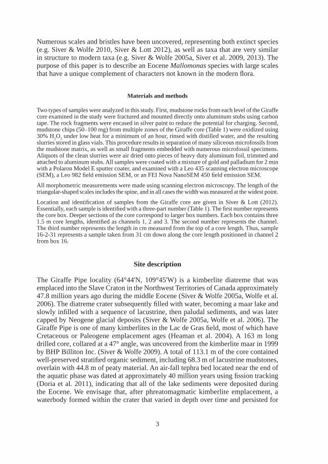
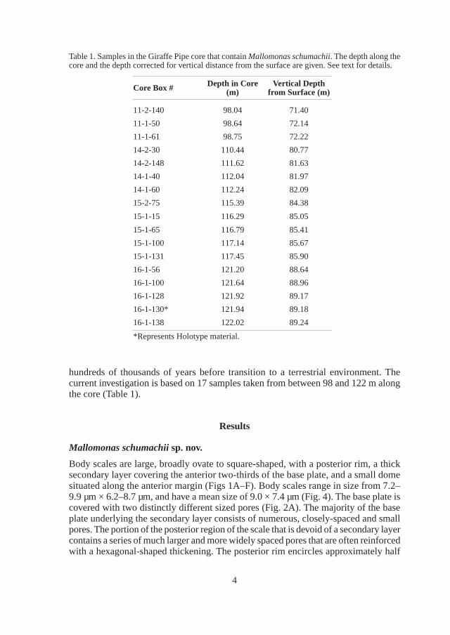
Body scales are large, broadly ovate to square-shaped, with a posterior rim, a thick secondary layer covering the anterior two-thirds of the base plate, and a small dome situated along the anterior margin (Figs 1A–F). Body scales range in size from 7.2– 9.9 µm × 6.2–8.7 µm, and have a mean size of 9.0 × 7.4 µm (Fig. 4). The base plate is covered with two distinctly different sized pores (Fig. 2A). The majority of the base plate underlying the secondary layer consists of numerous, closely-spaced and small pores. The portion of the posterior region of the scale that is devoid of a secondary layer contains a series of much larger and more widely spaced pores that are often reinforced with a hexagonal-shaped thickening. The posterior rim encircles approximately half
Table 1. Samples in the Giraffe Pipe core that contain Mallomonas schumachii. The depth along the core and the depth corrected for vertical distance from the surface are given. See text for details.
Core Box # Depth in Core(m)
Vertical Depth from Surface (m)
11-2-140 98.04 71.40
11-1-50 98.64 72.14
11-1-61 98.75 72.22
14-2-30 110.44 80.77
14-2-148 111.62 81.63
14-1-40 112.04 81.97
14-1-60 112.24 82.09
15-2-75 115.39 84.38
15-1-15 116.29 85.05
15-1-65 116.79 85.41
15-1-100 117.14 85.67
15-1-131 117.45 85.90
16-1-56 121.20 88.64
16-1-100 121.64 88.96
16-1-128 121.92 89.17
16-1-130* 121.94 89.18
16-1-138 122.02 89.24
*Represents Holotype material.
eschweizerbart_xxx
5
of the scale perimeter, is thinner along the posterior margin, and becomes wider along both sides. The secondary layer consists of a honeycomb pattern composed of large hexagonal-shaped chambers. The top surfaces of the chambers can range from being
Fig. 1. Body scales of Mallomonas schumachii. Note the square-shape, shallow and small dome, and the closed (A, C) or open (B, D) nature of the thick secondary layer. E) Scale with partially eroded secondary layer exposing the closely-spaced hexagonal chambers. F) Close-up of the hexagonal chambers forming the secondary layer. Note the base plate pores within each chamber. Scale bars = 4 µm (A), 3 µm (B), 2 µm (C–E) and 500 nm (F).
eschweizerbart_xxx
6
opened (Figs 1B, D–E) to fully closed (Fig. 1A, C). Some body scales have one or two large circular zones within the secondary layer, and generally near the middle of the scale, that lack the secondary honeycomb structure (Figs 1, A, C, E). A few large
Fig. 2. Mallomonas schumachii. A) Undersurface of a body scale denoting both the small and large base plate pores. B–C) Top (B) and bottom (C) views of the large triangular-shaped scales. D–E) Small obovate scales lacking domes and with secondary layers. These scales are believed to cover the posterior portion of the cell. F) The remains of numerous scales and bristles along a fractured rock surface. Scale bars = 2 µm (A–E) and 10 µm (F).
eschweizerbart_xxx
7
and scattered papillae may be present on top of the secondary surface of scales with enclosed chambers (Fig. 1C). A small raised dome with a shallow opening is found along the anterior margin.
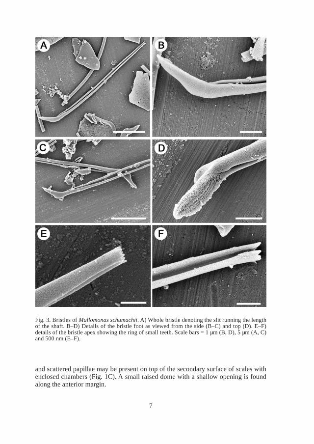
Fig. 3. Bristles of Mallomonas schumachii. A) Whole bristle denoting the slit running the length of the shaft. B–D) Details of the bristle foot as viewed from the side (B–C) and top (D). E–F) details of the bristle apex showing the ring of small teeth. Scale bars = 1 µm (B, D), 5 µm (A, C) and 500 nm (E–F).
eschweizerbart_xxx
8
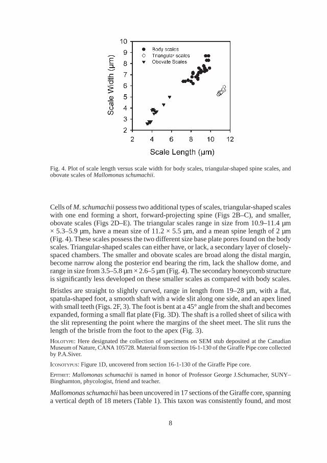
Cells of M. schumachii possess two additional types of scales, triangular-shaped scales with one end forming a short, forward-projecting spine (Figs 2B–C), and smaller, obovate scales (Figs 2D–E). The triangular scales range in size from 10.9–11.4 µm × 5.3–5.9 µm, have a mean size of 11.2 × 5.5 µm, and a mean spine length of 2 µm (Fig. 4). These scales possess the two different size base plate pores found on the body scales. Triangular-shaped scales can either have, or lack, a secondary layer of closely-spaced chambers. The smaller and obovate scales are broad along the distal margin, become narrow along the posterior end bearing the rim, lack the shallow dome, and range in size from 3.5–5.8 µm × 2.6–5 µm (Fig. 4). The secondary honeycomb structure is significantly less developed on these smaller scales as compared with body scales.
Bristles are straight to slightly curved, range in length from 19–28 µm, with a flat, spatula-shaped foot, a smooth shaft with a wide slit along one side, and an apex lined with small teeth (Figs. 2F, 3). The foot is bent at a 45° angle from the shaft and becomes expanded, forming a small flat plate (Fig. 3D). The shaft is a rolled sheet of silica with the slit representing the point where the margins of the sheet meet. The slit runs the length of the bristle from the foot to the apex (Fig. 3).
Holotype: Here designated the collection of specimens on SEM stub deposited at the Canadian Museum of Nature, CANA 105728. Material from section 16-1-130 of the Giraffe Pipe core collected by P.A.Siver.
Iconotypus: Figure 1D, uncovered from section 16-1-130 of the Giraffe Pipe core.
epItHet: Mallomonas schumachii is named in honor of Professor George J.Schumacher, SUNY–Binghamton, phycologist, friend and teacher.
Mallomonas schumachii has been uncovered in 17 sections of the Giraffe core, spanning a vertical depth of 18 meters (Table 1). This taxon was consistently found, and most
Fig. 4. Plot of scale length versus scale width for body scales, triangular-shaped spine scales, and obovate scales of Mallomonas schumachii.
eschweizerbart_xxx
9
abundant, in samples examined from core boxes 14, 15 and 16 that span approximately 8 meters of the core, lacking in boxes 12 and 13, then reappearing in low concentrations in box 11 spanning approximately one meter. Some fractured rock surfaces contained remains of numerous scales and bristles (Fig. 2F). When Mallomonas schumachii is most abundant, it was commonly found with Mallomonas porifera Siver & Wolfe, a taxon similar in structure to Mallomonas lychenensis, and remains of diatoms in the genera Eunotia and Actinella. The testate amoeba Scutyglypha, and a number of different heliozoan taxa were also common, and in several samples large numbers of sponge spicules were also found.
Discussion
There are no known modern species of Mallomonas with a suite of characters equivalent to that of M. schumachii, leading to the conclusion that this Eocene taxon is extinct. Recent works have shown that two major clades exist within the genus, one where species have scales that lack V-ribs, and the other containing species with V-ribs (Jo et al. 2011, 2013). The majority of modern species lacking scales with V-ribs are in the section Planae (Kristiansen 2002, Kristiansen & Preisig 2007). Taxa within this section have bristles of the rolled type that are hollow (Siver, in press), and where a slit is often found along part or the entire length of the shaft. This type of bristle is referred to as craspedodont (Kristiansen 2002). Given the lack of a V-rib on the scale, coupled with the rolled craspedodont bristle structure, M. schumachii likely represents a stem species related to section Planae.
It is likely that the large triangular scales and the smaller obovate scales of M. schu-machii were found on the anterior and posterior ends of the cell, respectively. Within the genus Mallomonas, scales that surround the flagellar pore are typically modified relative to body scales, often asymmetric with a triangular-shape, and with a forward projecting end. Similar shaped scales surrounding the flagellar pore are found on some species within the section Planae, including M. peronoides and M. bangladeshica. However, these types of specialized scales surrounding the flagellar pore are found widely within the genus, and not restricted to taxa in the section Planae. Perhaps the triangular scales of M. schumachii serve a similar function. Likewise, many species of Mallomonas possess smaller scales that in many respects resemble body scales that encircle and cover the posterior end of the cell. Given this fact, it makes sense that the small obovate scales of M. schumachii were likely found on the posterior end of the cell.
Despite an extensive search of rocks from several sections of the Giraffe core that contain numerous scales and bristles of M. schumachii, whole intact cells have not been uncovered. Fractured rock surfaces that contain almost pure remains of siliceous components representing this extinct species (e.g. Fig. 2F) have been closely examined. Based on rough estimates of the numbers of body scales and bristles along these fractured surfaces, it appears that each body scale was associated with one bristle. This would make sense given the small and shallow nature of the dome. There are also only a few triangular-shaped scales relative to numerous body scales indicating that these scales are fewer in number on the cell and as already noted, likely restricted
eschweizerbart_xxx
10
to one end. However, we can’t be certain if they surrounded the flagella or protruded from the posterior end.
The two distinctly different sized base plate pores found on M. schumachii scales are not unique among Mallomonas species (Siver 1991, Kristiansen 2002). Larger pores are more typically found in the posterior region and smaller ones closer to the apical end, especially on scales where a secondary layer covers the apical end as in M. schumachii. For example, in the section Planae, M. matvienkoae and related species have multiple pore sizes (Jo et al. 2013), as does M. bangledeshica (Takahashi & Hayakawa) Wujek & Timpano and M. stellata Cronberg. Differences in base plate pores can serve as another character to aid in distinguishing between closely aligned species (Jo et al. 2013).
The body scales varied greatly in regards to whether the honeycomb chambers were open, partially enclosed, or totally enclosed. Some sections of mudstone contained numerous layers of scales with partially enclosed chambers. Most of these scales did not show any signs of dissolution or breakage, suggesting that they were never totally closed and supporting the idea that scale design is variable in this species. The function of the large depressions lacking secondary chambers is unknown, and this type of structure is not known in the modern flora. Since these depressions are not found on every scale, it is hard to envision a common use or function.
Scales of M. schumachii are large in overall dimensions and total surface area in comparison to the vast majority of modern species (Siver 1991, Kristiansen 2002). The surface area of M. schumachii body scales is estimated to be approximately 62 µm2, almost double the size of the largest of 36 species examined by Siver (1991). Of the remaining Mallomonas species included in the monograph by Kristiansen (2002), the species with the largest body scales are M. bronchartiana and M. leboimei with estimated surface areas of 49 and 54 µm2, respectively. Large scale size has also been reported for other extinct species of Mallomonas, including M. porifera (Siver & Wolfe 2010) and M. media (Siver & Lott 2012), both with surface areas close to 45 µm2. Perhaps large scales posed disadvantageous to the cell and resulted in their extinction. It is reasonable to assume that for cells of the same general size, larger scales, especially ones that are more or less flat like those of M. schumachii, could be more difficult to overlap and form the scale covering while conforming to the curved contours of the cell. Scales and bristles are produced individually within the cell in silica deposition vesicles (SDV) (McGrory & Leadbeater 1981, Mignot & Brugerolle 1982, Kristiansen 2005), and then deployed into position onto the cell covering (Siver & Glew 1990, Leadbeater 1990, Beech et al. 1990). It is also likely that large scales would be more difficult to maneuver within the confines of the cell for precise deployment onto the cell covering, especially if the size of cells were comparable to ones observed today. Unfortunately, we do not know the size of M. schumachii cells. Although there is considerable variability between the mean size of scales and the size of the corresponding cell, Siver (1991) did report a significant relationship between these two variables. Perhaps the large scales of M. schumachii were coupled with large cells, resulting in a slow and cumbersome swimming organism eventually unable to successfully compete. The middle Eocene represented a very warm period in geologic
eschweizerbart_xxx
11
history, and northern latitudes experienced significantly higher temperatures compared to today (Zachos et al. 2008). It is also possible that the lower water densities associated with the elevated temperatures favored smaller cells with correspondingly smaller scales. There is some support for this hypothesis. Siver et al. (2013) found that fossil body scales of Mallomonas insignis from the warm Eocene were significantly smaller than their modern counterparts. Preliminary data also suggests that three additional taxa, Mallomonas lychenensis, M. bangladeshica and M. multiunca var. pocosinensis, also had smaller scales in the Eocene compared to their modern congeners. On the other hand, the conditions that would have supported the evolution of such large scales as found on M. schumachii are not known.
The remains of organisms found in association with M. schumachii suggest that this species thrived in acidic to slightly acidic environments that were high in humic substances. Mudstones containing significant remains of M. schumachii also contain numerous specimens belonging to the acid-loving diatom genera Eunotia and Actinella (Round et al. 1990, Siver & Hamilton 2011). Both of these diatoms are also commonly found in humic stained waterbodies (Sabbe et al. 2001, Melo et al. 2010, Siver & Hamilton 2011). In addition to diatom remains, the samples containing M. schumachii also harbored large concentrations of an organism equivalent to the modern taxon Mallomonas lychenensis, and the extinct M. porifera. Mallomonas lychenensis is commonly reported from humic stained and slightly acidic to acidic waterbodies (Cronberg & Kristiansen 1980, Charles & Smol 1988, Siver 1991), and has been classified as an acidophilic species (Siver 1989). In addition, Eloranta (1989) reported the closely related species Mallomonas allorgei from over 60% of waterbodies sampled from southern and central Finland that were largely acidic and humic stained. The conditions under which M. porifera grew are unknown, but concentrations of this organism are highly correlated with those of M. lychenensis suggesting that this taxon also thrived in acidic localities high in dissolved organic matter. Lastly, samples also contain low concentrations of testate amoebae belonging to Scutiglypha, also suggestive of acidic conditions.
In summary, M. schumachii is a common element of the Giraffe locality that possesses a unique suite of characteristics unknown today. Scales were large with surface areas far greater than the vast majority of modern species. It is possible that the large size of the scales was detrimental to the organism and caused it to go extinct. Mallomonas schumachii is believed to have thrived in shallow lakes that were acidic in nature and high in humic content. Remains of this taxon, along with numerous additional organisms, will eventually aid in reconstructing the history of this Arctic Eocene waterbody.
Acknowledgements
This work was funded with support to PAS from the U.S. National Science Foundation (DEB–0716606 and DEB–1144098), and by an NSF equipment grant (NSF#1126100) to Marie Cantino (University of Connecticut). The work was performed, in part, at the Biosciences Electron Microscopy Facility of the University of Connecticut. The author thanks Anne Lott (Connecticut College) for help with sample preparation, and Alexander Wolfe (University of Alberta) for helpful discussions.
eschweizerbart_xxx
12
References
BEECH, P.L., R. WETHERBEE & J.D. PICKETT HEAPS 1990: Secretion and deployment of bristles in Mallomonas splendens (Synurophyceae). – J. Phycol. 26: 112–122.
CHARLES, D.F. & J.P. SMOL 1988: New methods for using diatoms and chrysophytes to infer past pH of low–alkalinity lakes. – Limnol. Oceanogr. 33: 1451–1462.
CRONBERG, G. & J. KRISTIANSEN 1980: Synuraceae and other Chrysophyceae from central Småland, Sweden. – Bot. Notiser 133: 595–618.
DORIA, G., D.L. ROYER, A.P. WOLFE, A. FOX, J.A. WESTGATE & D.J. BEERLING 2011: Declining atmospheric CO
2 during the late Middle Eocene climate transition. – Am. J. Sci. 311: 63–75.
ELORANTA, P. 1989: Scaled chrysophytes (Chrysophyceae and Synurophyceae) from national park lakes in southern and central Finland. – Nord. J. Bot. 8: 67–81.
GRANT, J., Y.I. TEKLE, O.R. ANDERSEN, D.J. PATTERSON & L.A. KATZ 2009: Multigene evidence for the placement of a heterotrophic amoeboid lineage Leukarachnion sp. among photosynthetic stramenopiles. – Protist 160: 376–38.
HARWOOD, D.M. & R. GERSONDE 1990: Lower Cretaceous diatoms from ODP Leg 113 Site 693 (Weddell Sea). Part 2: Resting spores, chrysophycean cysts, an endoskeletal dinoflagellate, and notes on the origin of diatoms. – Proc. Ocean Drilling Progr., Sci. Results 113: 403–425.
HEAMAN, L.M., B.A. KJARSGAARD & R.A. CREASER 2004: The temporal evolution of North American kimberlites. – Lithos 76: 377–397.
JO, B.Y., W. SHIN, S.M. BOO, H.S. KIM & P.A. SIVER 2011: Studies on ultrastructure and three-gene phylogeny of the genus Mallomonas (Synurophyceae). – J. Phycol. 47: 415–425.
JO, B.Y., W. SHIN, H.S. KIM, P.A. SIVER & R.A. ANDERSEN 2013: Phylogeny of the genus Mallomonas (Synurophyceae) and descriptions of five new species on the basis of morphological evidence. – Phycologia 52: 266–278.
KIM, H.S. & J.H. KIM 2008: Mallomonas koreana sp. nov. (Synurophyceae), a new species from South Korea. – Nova Hedwigia 86: 469–476.
KRISTIANSEN, J. 2002: The genus Mallomonas (Synurophyceae) – A taxonomic survey based on the ultrastructure of silica scales and bristles. – Op. Bot. 139: 1–218.
KRISTIANSEN, J. 2005: Golden algae: A Biology of Chrysophytes. Königstein, Germany, Koeltz Scientific Books, 167 p.
KRISTIANSEN, J. & H.R. PREISIG 2007: Chrysophyte and Haptophyte Algae. – In: BüDEL, B., G. GARTNER, L. KRIENITZ, H.R. PREISIG, M. SCHAGERL (eds.): Süsswasserflora von Mitteleuropa. Volume 2. Berlin, Spektrum Akademischer Verlag. p. 252.
LEADBEATER, B.S.C. 1990: Ultrastructure and assembly of the scale case in Synura (Synurophyceae Andersen). – Br. Phycol. J. 25: 117–132.
MELO, S., L.C. TORGAN & S.V. RUPP 2010: Actinella species (Bacillariophyta) from an Amazon blackwater floodplain lake (Amazonas–Brazil). – Acta Amazonica 40: 269–274.
MCGRORY, C.B. & B.S.C. LEADBEATER 1981: Ultrastructure and deposition of silica in the Chrysophyceae. – In: LEADBEATER B.S.C. & R. RIDING (eds.): Biomineralization in Lower Plants and Animals. Volume 30. Oxford, Clarendon Press, p 315–325.
MIGNOT, J.P. & G. BRUGEROLLE 1982: Scale formation in chrysomonad flagellates. – J. Ultrastr. Res. 81: 13–26.
NĔMCOVÁ, Y., J. KREIDLOVÁ, A. KOSOVÁ & J. NEUSTUPA 2012: Lakes and pools of Aquitaine region (France) – a biodiversity hotspot of Synurales in Europe. – Nova Hedwigia 95: 1–24.
eschweizerbart_xxx
13
NICHOLLS, K.H. & D.E. WUJEK 2003: Chrysophycean algae. – In: WEHR, J.D. & R.G. SHEATH (eds.): Freshwater Algae of North America. New York, Academic Press, 471–509.
ROUND, F.E., R.M. CRAWFORD & D.G. MANN 1990: The Diatoms: Biology and Morphology of the Genera. Cambridge University Press, New York, 747 pp.
SABBE, K., K. VANHOUTTE, R.L. LOWE, E.A. BERGEY, B.J.F. BIGGS, S. FRANCOCEUR, D. HODGSON & W. VYVERMAN 2001: Six new Actinella (Bacillariophyta) species from Papua New Guinea, Australia and New Zealand: further evidence for widespread diatom endemism in the Australasian region. – Eur. J. Phycol. 36: 321–340.
SIVER, P.A. 1989: The distribution of scaled chrysophytes along a pH gradient. – Can. J. Bot. 67: 2120–2130.
SIVER, P.A. 1991: The Biology of Mallomonas: Morphology, Taxonomy and Ecology. – Dordrecht, Kluwer Academic Publishers, 230 p.
SIVER, P.A. 1995: The distribution of chrysophytes along environmental gradients: Their use as biological indicators. – In: SANDGREN, C.D., J.P. SMOL, J. KRISTIANSEN (eds.): Chrysophyte Algae: Ecology, Phylogeny and Development. Cambridge University Press, p. 232–268.
SIVER, P.A. 2003: The Synurophyceae. – In: WEHR, J. & B. SHEATH (eds.): Freshwater Algae of North America. Academic Press, p. 523–558.
SIVER, P.A. (in press): The Synurophyceae. – In: WEHR, J., R. SHEATH & J.P. KOCIOLEK (eds.): Freshwater Algae of North America. Elsevier.
SIVER, P.A. & J.R. GLEW 1990: The arrangement of scales and bristles on Mallomonas (Chrysophyceae) – a proposed mechanism for the formation of the cell covering. – Can. J. Bot. 68: 374–380.
SIVER, P.A. & A.P. WOLFE 2005a: Eocene scaled chrysophytes with pronounced modern affinities. – Int. J. Plant Sci. 166: 533–536.
SIVER, P.A. & A.P. WOLFE 2005b: Scaled chrysophytes in middle Eocene lake sediments from Northwestern Canada, including descriptions of six new species. – Proceedings of the Sixth International Chrysophyte Symposium, pp. 295–308.
SIVER, P.A., A.M. LOTT & A.P. WOLFE 2009: Taxonomic significance of asymmetrical helmet and lance bristles in the genus Mallomonas (Synurophyceae) and their discovery in Eocene lake sediments. – Eur. J. Phycol. 44: 447–460.
SIVER, P.A. & A.P. WOLFE 2009: Tropical ochrophyte algae from the Eocene of Northern Canada: A biogeographic response to past global warming. – Palaios 24: 192–198.
SIVER, P.A. & A.P. WOLFE 2010: A whole–cell reconstruction of Mallomonas porifera Siver and Wolfe from the Eocene: Implications for the evolution of chrysophyte cell architecture. – Nova Hedwigia Beihefte 136: 117–127.
SIVER, P.A. & P.A. HAMILTON 2011: Diatoms of North America: The Freshwater Flora of the Atlantic Coastal Plain. – Icon. Diatomol. 18: 1–920.
SIVER, P.A. & A.M. LOTT 2012: Fossil species of Mallomonas from an Eocene maar lake with recessed dome structures: Early attempts at securing bristles to the cell covering? – Nova Hedwigia 95: 517–529.
SIVER, P.A., A.P. WOLFE, F.J. ROHLF, W. SHIN & B.Y. JO 2013: Combining geometric morphometrics, molecular phylogeny, and micropaleontology to assess evolutionary patterns in Mallomonas (Synurophyceae: Heterokontophyta). – Geobiology 11: 127–138.
ŠKALOUD, P., J. KRISTIANSEN & M. ŠKALOUDOVÁ 2013: Developments in the taxonomy of silica–scaled chrysophytes – from morphological and ultrastructural to molecular approaches. – Nord. J. Bot. 31: 385–402.
eschweizerbart_xxx

14
SMOL, J.P. 1995: Application of chrysophytes to problems in paleoecology. – In: SANDGREN, C.D., J.P. SMOL & J. KRISTIANSEN (eds.): Chrysophyte Algae: Ecology, Phylogeny and Development. Cambridge University Press, p 232–250.
WEE, J.L. 1982: Studies on the Synuraceae (Chrysophyceae) of Iowa. – Bibl. Phycol. 62: 1–183.
WILLIAMS, J.L. 1985: Miocene Chrysophyta cysts from a lacustrine deposit in northern Idaho. – In: SMILEY C.J. (ed.): Late Cenozoic history of the Pacific Northwest. Pacific Division of the American Association for the Advancement of Science, p. 61–66.
WOLFE, A.P., M.B. EDLUND, A.R. SWEET & S. CREIGHTON 2006: A first account of organelle preservation in Eocene nonmarine diatoms: observations and paleobiological implications. – Palaios 21: 298–304.
WOLFE, A.P. & P.A. SIVER 2009: Three extant genera of freshwater thalassiosiroid diatoms from Middle Eocene sediments in northern Canada. – Am. J. Bot. 96: 487–497.
ZACHOS, J.C., G.R. DICKENS & R.E. ZEEBE 2008: An early Cenozoic perspective on greenhouse warming and carbon-cycle dynamics. – Nature 451: 279–283.
Manuscript submitted September 24, 2014; accepted October 28, 2014.
eschweizerbart_xxx