
malformatii_hipofiza.pdf
25
Hypophysis and Hypothalamus Pierre Bessou, Sylviane Hanquinet, and Jean-Franc ¸ois Chateil Contents 1 Introduction .......................................................................... 13 1.1 Embryologic and Anatomical Overview .............................. 13 1.2 Physiology and Function ....................................................... 14 2 Imaging Techniques ............................................................ 14 2.1 MRI versus CT ...................................................................... 14 2.2 Other Techniques................................................................... 16 3 Diseases ................................................................................. 17 3.1 Anterior Pituitary Deficiency ................................................ 17 3.2 Central Diabetes Insipidus .................................................... 21 3.3 Precocious or Delayed Puberty ............................................. 23 3.4 Other Endocrinopathies ......................................................... 23 3.5 Sellar and Suprasellar Tumors .............................................. 24 3.6 Rathke’s Cleft Cysts and Incidentalomas ............................. 34 4 Tips and Tricks for an Easy Diagnosis............................. 34 5 Conclusion ............................................................................ 34 References ...................................................................................... 34 Abstract Pathologies of hypothalamus–hypophysis axis in chil- dren express different clinical presentations, regarding endocrine secretions of numerous hormones. Knowledge of embryology, anatomy, and physiology is mandatory to understand the main features of these diseases. MRI is the best tool to assess the anatomical characteristics of the malformative and acquired pathologies. The main clinical expressions are in relation with isolated GH or combined antehypophysis hormones deficiencies, trou- bles in relation with puberty development, diabetes insipidus; cranial hypertension and visual disturbances may also reveal the disease. Pathologies include devel- opmental disorders, in relation, in most cases with trouble of embryological brain diverticulation, aplasia or hypoplasia of pituitary gland, pituitary stalk inter- ruption. Intra- and suprasellar masses can be a cranio- pharyngioma, a germinoma, and, mainly after 10 years of life, a pituitary adenoma. Systemic and inflammatory diseases include Langerhans cell histiocytosis, and, rarely in children, lymphocytic hypophysitis, sarcoidosis, and tuberculosis. 1 Introduction 1.1 Embryologic and Anatomical Overview The pituitary gland is composed of two portions, the ante- rior adenohypophysis and the posterior neurohypophysis; the development of each is embryologically distinct. At week 4 of embryogenesis, a Rathke’s pouch forms on the top of the stomodeum and before the oropharyngeal mem- brane. The adenohypophysis derives from Rathke’s pouch: this one extends through the sphenoid bone development region until it reaches the sella turcica, then converges with the neuroectoderm of the neurohypophysis. In the path passed by Rathke’s cyst, a solid cell cord forms, resides P. Bessou Á J.-F. Chateil (&) Service d’imagerie anténatale, de l’enfant et de la femme, CHU de Bordeaux, 33000 Bordeaux, France e-mail: [email protected] S. Hanquinet Unit of Pediatric Radiology, Hôpitaux universitaires de Genève, 6 Willy-Donzé, 1205 Geneva, Switzerland J.-F. Chateil RMSB, UMR 5536, Université de Bordeaux, 33000 Bordeaux, France F. Avni (ed.), Imaging Endocrine Diseases in Children, Medical Radiology. Diagnostic Imaging, DOI: 10.1007/174_2012_608, Ó Springer-Verlag Berlin Heidelberg 2012 13
-
Upload
antoniustincel -
Category
Documents
-
view
215 -
download
1
Transcript of malformatii_hipofiza.pdf

Hypophysis and Hypothalamus
Pierre Bessou, Sylviane Hanquinet, and Jean-Francois Chateil
Contents
1 Introduction .......................................................................... 131.1 Embryologic and Anatomical Overview .............................. 131.2 Physiology and Function....................................................... 14
2 Imaging Techniques ............................................................ 142.1 MRI versus CT ...................................................................... 142.2 Other Techniques................................................................... 16
3 Diseases ................................................................................. 173.1 Anterior Pituitary Deficiency................................................ 173.2 Central Diabetes Insipidus .................................................... 213.3 Precocious or Delayed Puberty............................................. 233.4 Other Endocrinopathies ......................................................... 233.5 Sellar and Suprasellar Tumors .............................................. 243.6 Rathke’s Cleft Cysts and Incidentalomas............................. 34
4 Tips and Tricks for an Easy Diagnosis............................. 34
5 Conclusion ............................................................................ 34
References ...................................................................................... 34
Abstract
Pathologies of hypothalamus–hypophysis axis in chil-dren express different clinical presentations, regardingendocrine secretions of numerous hormones. Knowledgeof embryology, anatomy, and physiology is mandatory tounderstand the main features of these diseases. MRI isthe best tool to assess the anatomical characteristics ofthe malformative and acquired pathologies. The mainclinical expressions are in relation with isolated GH orcombined antehypophysis hormones deficiencies, trou-bles in relation with puberty development, diabetesinsipidus; cranial hypertension and visual disturbancesmay also reveal the disease. Pathologies include devel-opmental disorders, in relation, in most cases withtrouble of embryological brain diverticulation, aplasiaor hypoplasia of pituitary gland, pituitary stalk inter-ruption. Intra- and suprasellar masses can be a cranio-pharyngioma, a germinoma, and, mainly after 10 yearsof life, a pituitary adenoma. Systemic and inflammatorydiseases include Langerhans cell histiocytosis, and,rarely in children, lymphocytic hypophysitis, sarcoidosis,and tuberculosis.
1 Introduction
1.1 Embryologic and Anatomical Overview
The pituitary gland is composed of two portions, the ante-rior adenohypophysis and the posterior neurohypophysis;the development of each is embryologically distinct. Atweek 4 of embryogenesis, a Rathke’s pouch forms on thetop of the stomodeum and before the oropharyngeal mem-brane. The adenohypophysis derives from Rathke’s pouch:this one extends through the sphenoid bone developmentregion until it reaches the sella turcica, then converges withthe neuroectoderm of the neurohypophysis. In the pathpassed by Rathke’s cyst, a solid cell cord forms, resides
P. Bessou � J.-F. Chateil (&)Service d’imagerie anténatale, de l’enfant et de la femme,CHU de Bordeaux, 33000 Bordeaux, Francee-mail: [email protected]
S. HanquinetUnit of Pediatric Radiology, Hôpitaux universitaires de Genève,6 Willy-Donzé, 1205 Geneva, Switzerland
J.-F. ChateilRMSB, UMR 5536, Université de Bordeaux,33000 Bordeaux, France
F. Avni (ed.), Imaging Endocrine Diseases in Children, Medical Radiology. Diagnostic Imaging,DOI: 10.1007/174_2012_608, � Springer-Verlag Berlin Heidelberg 2012
13

between two centers of chondrification, which develop intothe sphenoid bone body and wings, and differentiate into acraniopharyngeal canal. The adenohypophysis is made-upof the pars tuberalis, which surrounds the infundibulum, thepars intermedia, the portion of Rathke’s pouch in contactwith the neurohypophysis, and the pars distalis, which is thelargest portion of the anterior lobe. The residual lumenbetween the pars distalis and the pars intermedia decreasesin size, forming Rathke’s cleft, a narrow, non-visible cleftbetween the anterior and posterior lobes.
The hypothalamus develops from the neuroectoderm ofthe floor of the embryonic brain and begins its developmentby days 33–41. There are two major white matter tractsin the hypothalamus: the postcommissural fornix and themamillothalamic tract. The neurohypophysis forms boththe pituitary infundibulum and the posterior lobe proper(Schroeder and Vezina 2011; Yu et al. 2012).
Knowledge concerning genes involved within theformation of these structures becomes larger and larger.Gene defects affecting pituitary transcription factors:HESX1, LHX4, OTX2, or SOX3 are now well-known. Thehomeobox gene HESX1 is expressed in prospective fore-brain tissue, but later becomes restricted to Rathke’s pouch,the primordium of the anterior pituitary gland. Neonateswith HESX1 mutation exhibit abnormalities in the corpuscallosum, the anterior and hippocampal commissures, andthe septum pellucidum (Dattani et al. 1998).
1.2 Physiology and Function
The main function of the hypothalamus is homeostasis.Measurable factors such as blood pressure, body tem-perature, fluid and electrolyte balance, and body weightare maintained at a precise value called the set point.
The hypothalamus does so by regulating three interrelatedfunctions: endocrine secretion, autonomic function andemotions. The hypothalamus controls the release of hor-mones by the pituitary gland. Secretion from the posteriorpituitary gland can occur as a result of direct neuronalstimulation via the infundibulum, whereas secretion fromthe anterior pituitary gland is dependent upon the portalplexus, which carries hypothalamic releasing factors (TRH,CRH, IGF-1 and GH-RH, LH-RH) to the anterior pituitarygland; the precursor of vasopressin is also synthesized in thehypothalamus and then stored in vesicles at the posteriorpituitary (Saleem et al. 2007).
The adenohypophysis produces six established hor-mones: thyroid stimulating hormone (TSH), corticotropin(ACTH), growth hormone (GH), sexual stimulating hor-mones: follicle-stimulating hormone (FSH) and luteinizinghormone (LH), and prolactin (PRL). The first five servetropic functions by stimulating other organs to secretehormonally active substances, whereas PRL serves a tro-phic function on breast tissue. Cells of the anterior lobealso produce propiomelanocortin, which is also made byneurons of the hypothalamus and cells of the intermediatelobe. The posterior lobe or neurohypophysis secretesoxytocin and vasopressin, also called antidiuretic hormone(ADH).
2 Imaging Techniques
2.1 MRI versus CT
MRI is the best tool for imaging hypophysis and hypo-thalamus. CT may be useful in case of emergency, whenMRI is not still available, with clinical signs of acuteintracranial hypertension. Calcifications are also better seen
Fig. 1 Normal hypophysis on T1 sagittal plane: antenatal, neonatal (premature baby), with hypersignal within the entire hypophysis and samebaby at 2 months
14 P. Bessou et al.

Fig. 2 a 3-year-old, normal aspect: T1 sagittal view, isointensity of adenohypohysis and stalk, hypersignal of neurohypophysis. b 3-year-old,normal subject: T1 coronal anterior and posterior views, isointensity of adenohypohysis and stalk, hypersignal of neurohypophysis
Table 1 Height and volume ofantehypophysis regarding the age
Age Height (mm) Pituitary volume (mm3)
\6 weeks 4.5 ± 2
6 weeks–2 years 3.5 ± 1.2 174 ± 118
2 years–5 years 4 ± 0.7 184 to 214 ± 145
5–10 years 4.5 ± 0.6 226 to 277 ± 188
10–20 years (boys) 5 ± 2
10–20 years (girls) 8 ± 2
Fig. 3 14-year-old girl, sagittaland coronal views after contrastinjection: homogeneousenhancement with normalprominent pubertaladenohypophysis
Hypophysis and Hypothalamus 15

classically with CT and may help in cases of craniopha-ryngioma, before and after surgery.
2.1.1 Protocol of MRIMRI of the hypothalamo-pituitary axis includes thin(1–1.5 mm thick) T1-weighted slices focusing on thehypothalamo-pituitary area in the coronal and sagittalplanes. T2-weighted coronal slices are useful to study thehypothalamus, hypophysis and pituitary stalk, chiasm, butalso olfactory bulbs and sulci in cases of isolated gonado-tropin deficiency; axial slices may be useful for assessmentof the neurohypophysis. Constructive interference steadystate (CISS) T2-weighted sequence helps also for studyingthe pituitary stalk. Contrast medium injection is not alwaysmandatory and the use depends on the clinical context andfindings in the absence of contrast injection. A contrastagent is systematically injected if accurate imaging of thepituitary stalk is required, as is the case for children pre-senting hypopituitarism without a spontaneously visiblepituitary stalk and for cases of central diabetes insipidus.Enhanced sequences are useful for assessment of carvenoussinus. The whole brain must be examined because otherabnormalities may be associated with pituitary abnormali-ties. Flair, T2-weighted axial slices may be useful (Gareland Leger 2007). MR angiography is useful to evaluate thesurrounding vessels: internal carotid arteries and branches,cavernous sinus.
2.1.2 Normal AspectsThe fetal pituitary gland consists of the pars distalis (ante-rior lobe), the pars nervosa (posterior lobe) and the parsintermedia. The pars intermedia undergoes involution dur-ing the third trimester of pregnancy. The normal hypophysisis not clearly depicted by antenatal sonography. With MRfetal imaging, the entire pituitary gland is bright on T1sequences in foetuses (Garel and Leger 2007).
In infants under the age of 2 months, the entire pituitarygland is bright on T1 sequences, resulting in very similarsignals for the adenohypophysis and the neurohypophysis(Fig. 1). The brightness of the adenohypophysis may beaccounted for by intense cellular activity in the pituitarygland during this period of development. Moreover, thepituitary gland is bulbous in shape in this period, probablydue to cellular hypertrophy (Garel and Leger 2007). Therelative signal intensity and pituitary height significantlynegatively correlated with postnatal time but not with ges-tational age at birth (Kitamura et al. 2008).
After the neonatal period, signal of the anterior hypo-physis decreases on T1-weighted sequences and reaches theone of the gray matter (Fig. 2). Neurohypophysis remainshyperintense in most of cases and is clearly depicted on thinsagittal and axial slices; this ‘‘bright spot’’ is in relation withthe presence of vesicles of ADH, is seen in most of cases and
is a marker of neurohypophyseal functional integrity; itdepends on patient’s hydration and may be absent in normalindividuals.
The signal of the normal pituitary gland and stalk ismarkedly enhanced by the intravenous injection of contrastmedium. The anterior and posterior lobes differ in theirvascularization: the superior hypophyseal arteries supplythe median eminence. The inferior hypophyseal arteriessupply the neurohypophysis and stalk. The hypophysealportal vessels supply the anterior lobe; so, dynamicenhancement is not the same for all these structures and isseen later within the anterior lobe in comparison to themedian and posterior parts (Garel and Leger 2007).
The pituitary gland gradually increases in size untilpuberty. Table 1 gives some landmarks regarding the age(Argyropoulou et al. 1991; Dietrich et al. 1995; Kato et al.2002). A pituitary gland less than 3 mm high is consideredsmall, but pituitary gland shape and size in this age group ishighly variable. Shape varies from crescent-like to hemi-spherical and near spherical, some are dumbbell-shaped.The posterior pituitary bright spot could be elongated orflattened and extended variably in the anterior direction,often beneath the anterior portion of the gland; someauthors demonstrated that 3D-measurement of pituitaryvolume appears to be more robust, giving new references(Fink et al. 2005). At puberty, the pituitary gland displaysphysiological hypertrophy and may be 8 mm high in boysand 10 mm high in girls. The nearly spherical shape of thepituitary gland in teenage females should be considered anormal developmental feature (Fig. 3). The absence ofvisual symptoms, homogeneous pituitary enlargement onMR images, and a normal endocrine profile exclude apituitary adenoma (Aquilina and Boop 2011).
No data are available concerning the normal dimensionsof the pituitary stalk in children, but it is widely acceptedthat the maximum transverse diameter does not exceed2 mm in children (Dietrich et al. 1995; Garel and Leger2007).
2.2 Other Techniques
Regarding the variety of hormonal secretions, other man-ifestations in relation with hypo or hypersecretion may bevery protean and it is not possible here to give anexhaustive list of imaging explorations. Bilateral simulta-neous inferior petrosal sinus sampling, a very specializedinvestigation, may be useful in Cushing disease. Skull andsella turcica plain X-rays have no more utility. Bone agedetermination is still useful in case of growth abnormality.Sonography of thyroid gland, abdomen and pelvis forgenitals needs to be performed, regarding the clinicalpresentation.
16 P. Bessou et al.

3 Diseases
There are several ways to describe the pathologies that mayinvolve the hypothalamus-hypophysis axis. We choose inthis chapter to categorize the main disease regarding theendocrine dysfunction, with lack or, in the contrary, hy-persecretory states. These pathologies are mainly in relationwith developmental disorders, inflammatory/systemic dis-eases but also due to sellar or suprasellar mass lesions. Onthe other hand, the first clinical signs can be in relation witha neurodevelopmental delay, an intracranial hypertension ora visual disturbance. In this chapter, we will first describepresentations with primitive endocrine dysfunction ofhypothalamus–hypophysis axis, but we have to keep inmind that sellar and suprasellar tumors, described at the endof this chapter, may also be revealed by initial clinical signsin relation with an endocrine dysfunction rather than anoccupying space syndrome.
3.1 Anterior Pituitary Deficiency
Anterior pituitary hormone deficiencies may be isolated forone hormone or expressed by a combined pituitary hormonedeficiency (CPHD). Some of them are related to a knowngenetic abnormality or associated with other malformations;in other cases, hypothalamus–hypophysis axis developmen-tal disorders are demonstrated. Some cases are secondary tosurgery or radiotherapy. Lastly, some cases remain idio-pathic. Isolated GH deficiency (IGHD) is the most frequentone; other isolated pituitary hormone deficiencies may beobserved (Garel and Leger 2007). Hormones deficiencies areconfirmed by static and dynamic blood samples.
Severe congenital GH deficiency of the newborn is a raredisease, which can cause life-threatening hypoglycemiasbeginning in the first week of life. In some cases, the causeis monogenic, including mutations of the GH encodingGH-1. The majority of cases are still idiopathic or associ-ated with a significant malformation of the pituitary glandand multiple pituitary hormone deficiency (Binder et al.2010). In older children, growth retardation with a shortstature is the most frequent presentation (Dutta et al. 2009).
3.1.1 Aplasia and Hypoplasia of Pituitary GlandAplasia of the hypophysis is extremely rare, without pitui-tary fossa within the sphenoid bone (Arrigo et al. 2006).Neurohypophysis can be seen on the floor of hypothalamus.Hypoplasia is defined by a small anterior pituitary gland,regarding the normal values in relation with age, within anormal or a dysplastic pituitary fossa. These cases may beisolated (Fig. 4), or associated with other CNS malforma-tions. The endocrine damage is part of the septo-optic
dysplasia, but it is not constant; also, some children mayhave septal agenesis with an endocrine deficit withoutvisual impairment. Association of a septal agenesis andpituitary stalk interruption syndrome can occur (Belhocineet al. 2005). Other malformations include holoprosence-phaly, optic nerve hypoplasia, Chiari I malformation, all ofthese being part of disorders of diverticulation of theembryonic brain (Fig. 5a, b and c).
Hypoplasia of the pituitary gland must be differentiatedfrom a primitive ‘‘empty sella’’, which is defined by a sellaturcica partially or completely filled with cerebrospinalfluid, with herniation of the sellar diaphragm (Fig. 6). Iso-lated primary empty sella arises in the absence of previouspituitary surgery or radiotherapy and is quite rare in child-hood. The frequency of an empty sella is significantly highin idiopathic intracranial hypertension and nevoid basal cellcarcinoma syndrome, but it can be encountered without anyhypothalamic disorder in normal children (Takanashi et al.2001). Dysplastic enlarged sella can be seen in patient withneurofibromatosis 1 (Fig. 7).
3.1.2 Pituitary Stalk Interruption SyndromePituitary stalk interruption syndrome (PSIS), also known aspituitary dystopia, is characterized by the absence of normalpituitary stalk and an ectopic posterior pituitary lobe, seen onT1-weighted MRI as a bright spot localized between hypo-thalamus floor and pituitary fossa and in some cases withhypoplasia of adenohypophysis (Fig. 8a–d). The stalk maybe very thin, better seen with CISS sequence. Hypopituita-rism can be CPHD or IGHD. Patients with IGHD have amore preserved hypothalamic pituitary region on MRI thanthose with CPHD and therefore, the presence of more thanone hormonal deficiency could be attributed to more severeabnormalities of the pituitary gland, as has been also previ-ously observed (Acharya et al. 2011). Even if the high rate ofextrapituitary birth defects and of familial components sup-ports a role for genetic factors in the pathogenesis, only rarecases have a known genetic cause. HESX1, PROP 1, LHX 3,LHX4, POU1F1 or GLI2 genes mutations accounted for less5 % of cases and were found in consanguineous or familialcases (do Amaral et al. 2007; Franca et al. 2010; Maghnieet al. 2004; Melo et al. 2007; Reynaud et al. 2011; Simonet al. 2006; Zimmermann et al. 2007). Correlations betweeninvolved implicated genes and MRI findings have beengiven (Garel and Leger 2007). Pituitary stalk can be absentor enlarged: pituitary enlargement consisted of a non-enhancing mass lesion interposed between the normallyenhancing anterior lobe and the neurohypophysis. Sponta-neous regression of the mass lesion with normalization of thepituitary stalk position was observed (Voutetakis et al.2006). The initial enlargement of the stalk might bebecause of growth of functioning adenohypophyseal tissuewithin the stalk (Berkowitz et al. 2008).
Hypophysis and Hypothalamus 17

Fig. 4 a Neonate with hypoglycemia: aplasia of adenohypophysis. b Girl, 10-year-old, GH and gonodatrophins deficiencies: hypoplasticadenohypophysis
Fig. 5 a Boy, one-year-old, mildline defect with frontonasal encephalocele, suprasellar arachnoid cyst, hypophysis hypoplasia. b 2-year-old,Kenny Caffey syndrome with Chiari 1 malformation, hypoplastic hypophysis, dysplastic bones with subcutaneous fat hypertrophy
Fig. 6 Boy, 17-year-old,short stature: intra sellararachnoidocele with pseudoempty sella
18 P. Bessou et al.

3.1.3 Other Malformative Abnormalitieswith Anterior Pituitary HormoneDeficiencies
3.1.3.1 Hypogonadotropic Hypogonadism
Hypogonadotropic hypogonadism and congenital olfactorydeficit are common findings in Kallmann’s syndrome,which may display X-linked or autosomal inheritance.Other abnormalities, such as cleft lip or palate, dentalagenesis, renal abnormalities, hearing loss and cerebellardysfunction may be associated. The morphology of thehypothalamo-pituitary axis appears normal on MRI scans,but some cases of pituitary hypoplasia have been reported.In case of olfactory deficit, the olfactory bulbs are absent orhypoplastic. The olfactory sulci may be normal, absent orhypoplastic (Fig. 9). In no instance is an olfactory sulcusabsent when a bulb is present (Garel and Leger 2007).
3.1.3.2 Hypoparathyroidism-Retardation-
Dysmorphism
Hypoparathyroidism-retardation-dysmorphism syndrome(OMIM no. 241410), is an autosomal recessive disorderalmost exclusively reported in children born to consan-guineous parents of Middle Eastern origin. The syndromeconsists of hypoparathyroidism, dysmorphic features,developmental delay, and intrauterine and postnatal growthfailure. The serum IGF-I concentration is low. Neuroim-aging demonstrates reduced white matter mass with delayedmyelination, a hypoplastic anterior pituitary and hypoplasiaof the corpus callosum (Padidela et al. 2009).
3.1.3.3 Prader Willi Syndrome
Prader Willi syndrome is characterized by infantile hypo-tonia, mental retardation, short stature, hypogonadism, earlyonset obesity, hyperphagia, and a characteristic clinicalphenotype. Hyperphagia, hypogonadotropic hypogonadism,growth hormone deficiency are hypothesized to be due to
abnormalities of the hypothalamus and/or pituitary gland.Hypoplastic pituitary gland, a complete absence of theposterior pituitary bright spot can be seen on MRI, but norelationship between these anomalies and the presence ofanterior pituitary hormone deficiencies was found in indi-viduals with Prader Willi syndrome (Fig. 10) (Miller et al.2008). Other neuroradiological alterations could be a ven-tricular enlargement, a thin corpus callosum (Iughetti et al.2008).
3.1.3.4 Other Syndromes
The spectrum of congenital abnormalities affecting also theskull base ranges from the persistence of the craniopha-ryngeal canal, which connects the pituitary fossa andnasopharynx, to large basal cephaloceles with craniofacialdefects. Ectopic hypophysis can be found in associationwith meningo (hypophyso-) encephalocele through thecraniopharyngeal canal (Rabelink et al. 2011).
Ectopic posterior pituitary lobe and cortical dysplasia:the coexistence of ectopic posterior pituitary lobe andperiventricular heterotopia suggests a common underlyinggenetic basis. The presence of a heterozygous HESX1mutation in one case suggests this gene is important in thedevelopment of both ectopic posterior pituitary lobe andperiventricular heterotopia and supports their place in thespectrum of septo-optic dysplasia (Mitchell et al. 2002).
Pituitary abnormalities have been described in patientswith Fanconi anemia. PSIS was associated with hypogo-nadism, thyroid dysfunction, and GH deficiency (Fig. 11).Children with Fanconi anemia tend to have unsuspectedsmall pituitary glands (Sherafat-Kazemzadeh et al. 2007).
3.1.4 Hypogonadism and HemochromatosisPituitary hemochromatosis is an uncommon cause ofhypogonadism in children, except in patients with b-thal-assemia major due to post-transfusional iron overload. MRI
Fig. 7 Girl, 14-year-old,neurofibromatosis type 1,dysplasia of the sphenoid bonewith enlarged sella turcica andhypophysis
Hypophysis and Hypothalamus 19

Fig. 8 a Girl 17-year-old, short stature with GH deficiency but alsobiological combined deficiencies: Pituitary stalk interruption withectopic posterior pituitary lobe; absence of septum lucidum. b Girl9-year-old, short stature, combined pituitary hormone deficiency(thyreotrope and corticotrope) left amblyopia: Pituitary stalk interrup-tion with hypoplasia of the left optic nerve. c Girl, 5-year-old, short
stature: adenohypophysis hypoplasia, interruption of the pituitarystalk, ectopic posterior pituitary lobe. d Girl, 3-year-old; short staturewith GH deficiency: Pituitary stalk interruption with T1 hyperintenseectopic neurohypophysis within hypothalamus. e Boy, 3-year-old,scaphocephaly and short stature: thin pituitary stalk and suprasellarectopic neurohypophysis
20 P. Bessou et al.

is a good technique for detecting pituitary hemochromato-sis, with a markedly decreased signal intensity of the pitu-itary gland on T2 and T2*-weighted images (Sparacia et al.2000).
3.1.5 Anterior Pituitary Deficiencywithout Anatomical Abnormality
A normal pituitary gland on the MRI does not exclude apituitary endocrine deficit, gland and conversely, some chil-dren without biological endocrine abnormalities may have an
abnormality of the hypothalamic-pituitary axis on MRI(Wang et al. 2011).
3.2 Central Diabetes Insipidus
Central diabetes insipidus (CDI) is characterized by theabsence of secretion of ADH. CDI can be in relation withseveral diseases: Langerhans cell histiocytosis, inflamma-tory diseases, intracranial tumor (germinoma, glioma), post
Fig. 9 Boy, 17-year-old,psychomotor development delay,hypogonadism, anosmia,hypertelorism: Kallmann diseasewithout olfactive sulci and tracts,corpus callosum agenesis
Fig. 10 Boy, neonate, hypotonia, Prader Willi syndrome; hypoplasiaof sella turcica
Fig. 11 Boy, 2-year-old, Fanconi disease: hypoplastic hypophysisand pituitary stalk interruption
Hypophysis and Hypothalamus 21

traumatic, autoimmune polyendocrinopathy, familial dis-ease, or idiopathic (Maghnie et al. 2000). The natural his-tory of idiopathic CDI with pituitary stalk thickening isunpredictable, and can be the first manifestation of a ger-minoma. Sampling of HCG in serum will be repeated every3–6 months during the first 3 years after the onset of CDI,and careful MRI evaluation should then be performed onceper year for 2 years and every 2–5 years, thereafterdepending on the size and progression of the lesion (Gareland Leger 2007). On MRI, with sagittal or axial T1-weighted images, the loss of the posterior pituitary brightspot is a sensitive marker for CDI. The pituitary stalk isconsidered enlarged if at least part of the stalk is found tohave a diameter superior to 2.0 mm.
3.2.1 Langerhans Cell HistiocytosisLangerhans cell histiocytosis (LCH) is a clonal proliferativedisorder of cells of the mononuclear phagocytic and den-dritic cell system that often presents in childhood either as asolitary often-curable bone lesion or as widespread, oftenmultisystemic, sometimes lethal disorder (Demaerel andVan Gool 2008). Infundibular and hypothalamic infiltrationoccurs in 10–30 % of the patients with multisystemic LCH(Varan et al. 2008). There is a high degree of suspicion forLangerhans cell histiocytosis as etiology of diabetes in-sipidus, which can be the initial manifestation of the dis-ease. Precocious puberty or hypogonadism, acceleratedgrowth despite growth hormone deficiency, hypothalamicobesity may also occur in LCH (Demaerel and Van Gool2008; Marchand et al. 2011; Priyambada et al. 2011).
Cerebral imaging demonstrates pituitary stalk thicken-ing, moderate (3.0–7 mm) to marked ([7 mm), with huge
enhancement after contrast media injection (Fig. 12). Thecoexistence of osteolytic lesions of the orbit, the sphenoidand petrous bones, the cranial vault with soft-tissueenhancing masses is highly suggestive of LCH, previouslyknown as Hand Schüller Christian disease. The stalk lesioncan extend to the floor of the third ventricle. Small-sizedpituitary gland or atrophic pituitary can be seen. Duringfollow-up, pituitary stalk can increase in volume, decrease,or remaining stable. A normal pituitary stalk can also beseen initially with increase in pituitary stalk volume onfollow-up in 50 % of these cases (Marchand et al. 2011).Regression of the pituitary changes, as visualized on MRimages, is only rarely accompanied by reversal of thesymptomatology and these children are at risk to developfurther deficiencies of anterior pituitary hormones (Dema-erel and Van Gool 2008). Other lesions within the brain canoccur during the evolution: leukoencephalopathy, paren-chymal enhancing lesions, gray matter changes in the cer-ebellar dentate nucleus and in the supratentorial basalganglia, finally cerebral atrophy (Prayer et al. 2004).
3.2.2 Lymphocytic HypophysitisLymphocytic hypophysitis (LYH) is a rare inflammatorydisease of the pituitary gland that usually affects women intheir anterior immediate postpartum period, but can also berarely encountered in children. Symptoms include anteriorand/or posterior pituitary insufficiency of varying degrees.Diagnosis can be based on biopsies or infered from clinicalcharacteristics and typical MRI findings. MRI of the sellarregion revealed an homogeneously enhancing mass lesionin the pituitary stalk and the posterior pituitary gland withlack of the hyperintensity signal of the posterior lobe onunenhanced T1-weighted images. Enlargement of the wholepituitary gland with symmetrical suprasellar expansion canbe observed, with slightly inhomogeneous enhancement.Differential diagnoses lesions include LYH, idiopathic giantcell hypophysitis and granulomatous hypophysitis causedby conditions such as tuberculosis, sarcoidosis, LCH,primitive abscess, and mycotic infections. Follow-up,spontaneously of after steroids treatment, demonstrates aregression in 50 % of cases (Gellner et al. 2008).
3.2.3 SarcoidosisSarcoidosis is a multisystem granulomatous disorder ofunknown cause that most commonly affects young adultsand is exceptional in childhood. Neurosarcoidosis occurs inabout 10 % of affected patients. The disease has a predi-lection for the hypothalamus and pituitary gland but anyportion of the CNS may be affected. CDI and anteriorpituitary failure are the most common feature of neurosar-coidosis. MRI shows granulomatous infiltration of thedura mater or nodular thickening on the infundibular stalkand optic chiasm. The lesion is isointense on T1-weighted
Fig. 12 Boy, 4-year-old, diabetes insipidus: Langerhans cell hystio-cytosis with enlarged pituitary stalk, occipital osteolysis
22 P. Bessou et al.

images and hypointense on T2-weighted images. Aftercontrast media injection, there is a thick enhancing infun-dibulum with intense surrounding meningeal enhancement.Rarely, masslike lesions, particularly in the region of thefloor of the third ventricle and optic chiasm can be found(Saleem et al. 2007).
3.2.4 TuberculosisA previous study demonstrated a 55 % prevalence of absentposterior pituitary bright spot in pediatric patients present-ing with tuberculous meningitis. Those with absent poster-ior pituitary bright spot demonstrated poorer developmentaloutcome at 6 months follow-up (Andronikou et al. 2009).Tuberculosis of sellar region is uncommon despite tuber-culomas being the most common lesion in neurotubercu-losis. Headache, vomiting, visual disturbances, and featuresof hypopituitarism are common. MRI reveals tuberculomasas hypointense on T1-weighted images and iso to hyperin-tense on T2-weighted images with perilesional edema;suprasellar extension of the sellar tuberculoma with thick-ening of pituitary stalk is observed in most of cases (Nayilet al. 2011).
3.2.5 Other Causes of Diabetes Insipidus
3.2.5.1 Familial CDI
Familial CDI is caused by mutations of the gene encoding apreprohormone and involves the progressive postnataldegeneration of ADH producing neurons. This abnormalpreprohormone could not be processed correctly, and theaccumulation of this preprohormone might account for thepersistent posterior pituitary bright spot (Garel and Leger2007).
3.2.5.2 Chronic Neurogenic Hypernatremia
Chronic neurogenic hypernatremia is observed in childrenpresenting midline abnormalities of the brain, such as holo-prosencephaly, callosal agenesis, or septal agenesis. Theunderlying mechanism remains unclear, but there appears tobe a defect in hypothalamic function, leading to the failureof the osmoreceptors, whereas the synthesis and storage ofADH remain intact (Garel and Leger 2007).
3.3 Precocious or Delayed Puberty
3.3.1 Duplication of Pituitary GlandThe duplication of pituitary gland and stalk is a rare mal-formation. Most of the reported cases were associatedwith other anomalies, such as agenesis/hypoplasia of cor-pus callosum, cerebellar hypoplasia, hydrocephalus,absent olfactory bulbs and/or tracts, oropharengeal masses
including teratomas and various orofacial midline defects(Akin et al. 2011). Other cases demonstrate a wide hypo-thalamus, named pseudohamartoma (de Penna et al. 2005).Other association with ‘‘morning glory disk’’ anomaly andMoyamoya disease has been described (Loddenkemperet al. 2008). MRI demonstrates the presence of two para-medial pituitary stalks, coming from hypothalamus andconnected to two separate anterior and posterior pituitaryglands (Fig. 13). Triplication of the pituitary gland isexceptional (Manara et al. 2009).
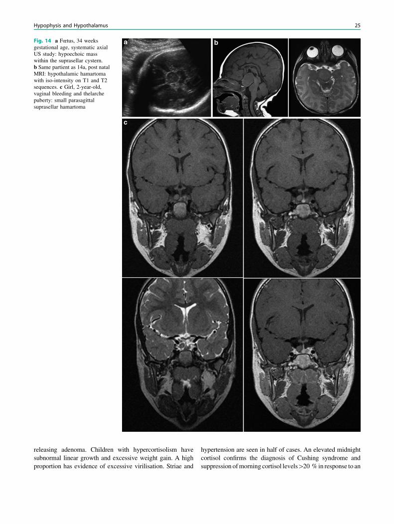
3.3.2 HamartomaHypothalamic hamartomas are developmental malforma-tions consisting of tumorlike masses located in the tubercinereum of the hypothalamus. Most patients present intheir first or second decade of life, with boys being morecommonly affected than girls. These lesions have beendivided into two main clinico-anatomic subsets: parahy-pothalamic and intrahypothalamic hamartomas. Parahy-pothalamic hamartomas are pedunculated masses that areattached to the floor of the hypothalamus by a narrowbase. These lesions seem more likely to be associatedwith precocious puberty. Intrahypothalamic hamartomasare sessile masses with a broad attachment to the hypo-thalamus, often associated with gelastic seizures. MRdemonstrates a well-defined pedunculated or sessile lesionat the tuber cinereum. The mass is isointense or mildlyhypointense on T1-weighted images and iso to hyperin-tense on T2-weighted images, with no contrast enhance-ment or calcification (Fig. 14a–c). The absence of anylong-term change in the size, shape, or signal intensity ofthe lesion strongly supports the diagnosis of hypothalamichamartoma (Saleem et al. 2007). Tuber hamartoma has tobe differentiated from septopreoptic holoprosencephaly,where the midline fusion is restricted to the septal regionor preoptic region of the telencephalon (Hahn et al.2010).
3.4 Other Endocrinopathies
Pathological pituitary hyperplasia may occur in severalcircumstances, including central precocious puberty,ectopic production of hypothalamic-releasing hormonesfrom hypothalamic and nonpituitary tumors, and admin-istration of exogenous oestrogens (Morana et al. 2010).Rapid progression of pituitary hyperplasia may develop incase of peripheral hypothyroidism and evaluation ofthyroid function is needed when a homogeneous pituitarymass is revealed by MR imaging; this hyperplasia dis-appears in a few months after substitutive treatment (Leeet al. 2008).
Hypophysis and Hypothalamus 23

3.5 Sellar and Suprasellar Tumors
3.5.1 Pituitary AdenomasPituitary adenomas are relatively uncommon in childrenand account for less than 3 % of all supratentorial tumors.They are more frequent in adolescents than in younger agegroups. Hormone secreting tumors predominate, whilehormonally inactive adenomas are rare. Prepubertal chil-dren more frequently have ACTH-releasing adenomas,while pubertal and postpubertal patients are most likely tohave prolactinomas (Morana et al. 2010).
3.5.1.1 Prolactinoma
Depending on size, pituitary adenomas are classified intomicroadenomas and macroadenomas. Microadenomas aresmaller than 10 mm in diameter and lie entirely within thepituitary gland. Most common presenting signs of prolactinmicroadenomas are primary amenorrhea, then galactorheain females and gynecomastia and hypogonadism in males.They can also be associated with delayed puberty. Theyappear as small, hypointense lesions on T1-weightedimages (Fig. 15). Some may only become apparent as non-
enhancing spots within the gland on post-contrast images.Their appearance on T2-weighted images is variable.
A giant, solid, invasive prolactinoma in a prepubescentchild is extremely rare (Dinc et al. 2008; Furtado et al.2010). Macroadenomas show intermediate signal in unen-hanced T1-weighted images and enhance after contrastmedium administration (Fig. 16). Invasion of the cavernoussinus is sometimes demonstrated but with normal carotidartery diameter.
Pituitary apoplexy must be considered in case of intenseheadache and worsening visual acuity. MRI showed alarge suprasellar mass with a small sellar component,with heterogeneous hyperintensity on T1-weighted images,suggestive of recent hemorrhage (Fig. 17); sometimes,intralesional-dependent fluid–fluid levels can be detected,mainly on axial slices. T2* sequences may be useful.Heterogeneous enhancement is often present (Satyartheeand Mahapatra 2005).
3.5.1.2 Cushing Disease
Cushing disease (CD) refers only to hypercortisolismsecondary to excess production of ACTH from a ACTH-
Fig. 13 a Girl, 10-year-old, precocious puberty: bone age: 14-year-old. b Same patient: duplication of pituitary stalk and hypophysis
24 P. Bessou et al.
releasing adenoma. Children with hypercortisolism havesubnormal linear growth and excessive weight gain. A highproportion has evidence of excessive virilisation. Striae and
hypertension are seen in half of cases. An elevated midnightcortisol confirms the diagnosis of Cushing syndrome andsuppression of morning cortisol levels[20 % in response to an
Fig. 14 a Fœtus, 34 weeksgestational age, systematic axialUS study: hypoechoic masswithin the suprasellar cystern.b Same partient as 14a, post natalMRI: hypothalamic hamartomawith iso-intensity on T1 and T2sequences. c Girl, 2-year-old,vaginal bleeding and thelarchepuberty: small parasagittalsuprasellar hamartoma
Hypophysis and Hypothalamus 25

overnight, high-dosage dexamethasone test excludes allpatients with adrenal tumors and identifies almost all patientswith CD.
ACTH-releasing adenomas are frequently small anddifficult to localize. In previous studies, nearly half ofchildren with CD (confirmed histologically) had an
identifiable adenoma of the pituitary gland by imaging(Batista et al. 2007; Morana et al. 2010). Most of the cor-ticotrophin adenomas are small (\4 mm), and have similarintensity to those of normal pituitary tissue (Oliveira et al.2010). A half dose of contrast media with 3T dynamicresonance imaging study seems to increase the sensitivity
Fig. 15 Girl, 8-year-old, precocious puberty, small lesion with the right part of the adenohypophysis with T2 hyperintensity and lack ofenhancement after injection: microadenoma? No surgical confirmation
Fig. 16 a Girl, 10-year-old precociuous puberty, obesity: antehypo-physis adenoma with suprasellar extension. b Girl, 6-year-old,growth acceleration: prolactinoma without enhancement after contrastmedia injection. c Girl, 14-year-old, secondary amenorrhea, hyper
prolactinemia: prolactinoma: heterogeneous enhancement with exten-sion to the right cavernous sinus. d Girl, 15-year-old, headaches:invasive prolactinoma with extension through the floor of sella turcica
26 P. Bessou et al.

(Portocarrero-Ortiz et al. 2010). ACTH-producing macro-adenomas are rare (Min et al. 2007). Pituitary imagingperformed in all the patients showed poor concordance withfindings at surgery. In contrast, bilateral simultaneousinferior petrosal sinus sampling, performed in selectedcenters, demonstrates a good correlation with surgicalfindings, but the sensitivity regarding the lateralization ofthe microadenoma is variable (Dias et al. 2010).
In the other hand, CD-like with ectopic ACTH secretioncan be in relation with neuro-endocrine tumors: bronchialcarcinoid tumor, pancreatic neuro-endocrine tumor, but alsoEwing’s sarcoma, stromal epithelial tumors of the liver,ganglioneuroblastoma, Wilm’s tumor, pancreatoblastoma(More et al. 2011).
3.5.1.3 Others Pituitary Adenomas
McCune–Albright syndrome is characterized by a triad ofpoly/monostotic fibrous dysplasia, cafe au lait macules andhyperfunctioning endocrinopathies including growth hor-mone excess. GH secreting pituitary macroadenoma can beresponsible of gigantism; treatment of patients with suchmacroadenoma is difficult because of thickened calvariumand dysplastic skull bone (Bhansali et al. 2003; Subbiahet al. 2011). Hyperthyroidism due to TSH-secretingpituitary adenomas is a very rare disorder in childhood(Nakayama et al. 2010).
3.5.2 CraniopharyngiomaCraniopharyngiomas are benign epithelial tumors accoun-ting for 5–13 % of all intracranial neoplasms in the pedi-atric age group. These tumors arise from remnants of thecraniopharyngeal duct: they may arise anywhere alongthe infundibular stalk from the floor of the third ventricle tothe pituitary gland. They may be intrasellar (25 % of cases),suprasellar, or a combination of both (Morana et al. 2010).Most cases occur between 4 and 12-year-old. The clinical
picture at the time of diagnosis is often characterized bynonendocrine manifestations, such as headache (60 %)and visual disturbances (46 %). However, up to 80 %have evidence of endocrine dysfunction at diagnosis (shortstature, inappropriate secretion of ADH, diabetes insipidus,delayed or precocious puberty, even CD. (Sosa et al. 2005;Tomita and Bowman 2005).
3.5.2.1 Imaging
A suprasellar enhancing lesion with a cystic component andcalcifications is characteristic of a craniopharyngioma. Themost common pattern is represented by a cystic lesion thatis hyperintense on both T1-weighted and T2-weightedimages due to high protein concentration and/or to thepresence of methemoglobin, with enhancing walls andsubtle peripheral calcifications. Solid tumor components,often located in the intra or parasellar region, are oftenheavily calcified and appear isohypointense in T1-weightedimages with variable, often low signal intensity on T2-weighted images; these components typically enhancefollowing gadolinium administration (Fig. 18a–f). CT issuperior to MRI in the identification of calcifications: cal-cifications may appear as shell-like deposits along the cystwalls, or may form fine punctuations or lumps within thesubstance of the lesion. Proton MR spectroscopy may showa prominent lipid peak or doublet of lactate (Morana et al.2010). The preoperative MR images can classify the tumoraccording to the degree of hypothalamic involvement asfollows: Grade 0, no hypothalamic involvement; Grade 1,the tumor abuting or displacing the hypothalamus; andGrade 2, hypothalamic involvement (Puget et al. 2007).Intrasellar craniopharyngioma are classified into two typesaccording to possible origin regions: the first one, origi-nating in the sella turcica and developing downward to thesphenoidal sinus, and the second one, originating from theresidual embryo craniopharyngeal canal (Yu et al. 2012).
Fig. 17 Girl, 14-year-old,galactorhea and secondaryamenorrhea, headache: prolactinadenoma with spontaneoushemorrhage
Hypophysis and Hypothalamus 27

3.5.2.2 Follow-up Evaluation
The extent of tumor resection after surgery influenced therecurrence-free survival, and patients with total resectionhave a high-rate survival. Postoperative CT and MRI have tosearch for small residual tumor or calcifications. In case ofsubtotal resection or residual tumor, radiotherapy is used
(Tomita and Bowman 2005). After radiotherapy, a highincidence of vascular abnormalities is seen in children withcraniopharyngioma (temporal cavernomas, moyamoya syn-drome, aneurysm or decreases in the caliber of the internalcarotid artery. Intracystic bleomycin infusion may contributeto radiation-related vasculopathy (Liu et al. 2009).
Fig. 18 a Girl, 6-year-old, short stature, reduction of the visual field:craniopharyngioma with predominant cystic component, thin wallcalcifications seen on CT, spontaneous hypersignal on T1 weightedimages in relation with high cholesterol concentration within the cyst.b Boy, 5-year-old: short stature, headache and vomiting; craniopha-ryngioma with hydrocephalus, ‘‘egg-shell’’ calcification on CT,spontaneous T1 hyperintense cystic part on MRI. c Girl 9-year old,visual disturbance since several months, papillary edema on fundo-scopy: craniopharyngioma with predominantly cystic component,
peripheral rim enhancement after contrast injection. d Girl, 4-year-old, anorexia with slimming: craniopharyngioma with multicysticcomponent, hypointense ‘‘pop-corn’’ calcification within the solidenhanced part. e Boy, 4-year-old, vomits since 2 weeks, palsy of theright 6th nerve: craniopharyngioma with T1 isointense cystic compo-nent, intra sellar solid component. f Boy, 4-year-old, same as Fig. 18e,localized MR spectroscopy within the cyst demonstrates a doubletlactate peak
28 P. Bessou et al.

Fig. 18 (continued)
Fig. 18 (continued)
Hypophysis and Hypothalamus 29

3.5.3 GerminomaIntracranial germinoma is a rare malignant tumor, onlyconstituting 0.5–2.0 % of all primary intracranial tumorsbut constitutes 50–60 % of central nervous system germcell tumor. Age at diagnosis ranged from 3 to 21 years(mean 12.5 years) with a peak between 10 and 18 years.Almost 60 % of intracranial germinoma are located inpineal region, 30 % in suprasellar region, and 10 % in basalganglia region. Synchronous lesions in pineal and supra-sellar region are also possible. With regard to suprasellarregion germinoma, endocrinic syndromes including centraldiabetes insipidus, abnormality of sexual development(precocious puberty or delayed sexual development) andgrowth hormone deficiency (Gottschling et al. 2006). Visual
symptoms or headache in relation with intracranial hyper-tension may be the first signs.
MRI demonstrates a ill-defined margin tumor withirregular shape. It often has necrosis, cysts, and hemorrhageinside the tumor, but has no calcification. The lesiondemonstrated hypointense to isointense signal on T1-weightedimages and isointense to hyperintense on T2-weightedimages with markedly heterogeneous enhancement(Fig. 19). Diffusion-weighted MR imaging shows restricteddiffusion (Wang et al. 2010). Craniospinal metastases haveto be searched with entire head and spine MR evaluation.As written before, in children suffering from diabetes in-sipidus showing absence of visualization of the posterior‘bright spot’, a small germinoma could not yet be visible on
Fig. 19 a Girl, 11-year-old with diabetes insipidus: suprasellargerminoma with cysts. b Boy, 14-year-old, diabetes insipidus since6 months: intra and suprasellar germinoma with heterogeneous solidmass. c Boy, 11-year-old, oculomotor palsy: suprasellar malignant
germinoma with heterogeneous enhancement. d Girl, 9-year-old,headaches and visual disturbances: Synchronous lesions in pineal andsuprasellar regions
30 P. Bessou et al.

the initial MR images (Morana et al. 2010). A close follow-up with repeated imaging studies should therefore be car-ried out in these patients; MRI evidence of an increase in
the size of the anterior pituitary with thickening of the stalkis strongly associated with the presence of a germinoma,whereas a decrease of normal gland parenchyma can
Fig. 20 a Girl, 14-year-old,secondary amenorrhea, thenacute headache: enlargement ofadenohypophysis, with fluid–fluid level: macroadenoma withpituitary apoplexy was suspected,without surgical confirmation.b Same girl as Fig. 20a, sixmonths later, hyperprolactinemia:heterogeneous enhanced masswithin the andenohypophysis,biopsy: primitive neuroectodermal tumor
Fig. 21 Girl, 8-year-old,headaches and left visualimpairement: suprasellar and prepontine epidermoid cyst
Hypophysis and Hypothalamus 31

suggest an inflammatory or autoimmune process such aslymphocytic infundibulo-hypophysitis (Edouard et al. 2009;Maghnie et al. 2000).
Confirmation of the diagnosis requires measurementof serum and CSF tumor markers (a-fetoprotein and/orb-human chorionic gonadotropin) and/or biopsy. Germi-noma are highly sensitive to radiotherapy or specificchemotherapy.
3.5.4 Other TumorsExtremely rare tumors, such as pituitary astrocytomas,granular cell tumors or primitive neuro ectodermal tumors(Fig. 20), may arise within the sella turcica (Huang andCastillo 2005). Pituitary carcinoma is defined as a primaryadeno-hypophyseal neoplasm with documented craniospi-nal and/or systemic metastases. They are exceptional inchildhood, are hormonally active, and they can havemetastases in all parts of the central nervous system (Guzelet al. 2008).
The pituitary stalk is the most common site for thedevelopment of supratentorial hemangioblastomas in VonHippel Lindau disease. Patients with pituitary stalk
hemangioblastomas often remain asymptomatic and do notrequire treatment (Lonser et al. 2009).
Trilateral retinoblastoma is a rare combination of unila-teral or bilateral retinoblastoma with a midline malignantneuroectodermal tumor (3 % incidence). There are onlythree published cases of histologically confirmed trilateralretinoblastoma involving suprasellar tumors (Dai et al.2008).
3.5.5 Dermoid and Epidermoid Cystsof the Suprasellar Cistern
Dermoid and epidermoid cysts are rare benign maldeve-lopmental lesions that arise from epithelial inclusionsoccurring during neural tube closure. Dermoid and epider-moid cysts consist of a capsule composed of epidermalelements, with dermoid cysts containing dermal derivatives(fat, sebaceous glands, hair). Suprasellar lesions can causevisual abnormalities and endocrinologic disturbances(Saleem et al. 2007). CT and MRI can demonstrate thepresence of fat (with characteristic hypodensity with CT,or lack of signal with fat-saturation sequences on MRI;diffusion imaging can also help to characterize the cystic
Fig. 22 Boy, 11-year-old, MRIfor advanced puberty: incidentallipoma demonstrated, withspontaneous T1 hypersignalposterior to the pituitary stalk
Fig. 23 Girl, 1-year-old, MRIperformed for psychomotordevelopment delay: incidentalRathke’s cleft cyst
32 P. Bessou et al.

Ta
ble
2S
umm
ary
ofcl
inic
alpr
esen
tati
ons
and
path
olog
ies
HP
axis
deve
lopm
enta
ldi
sord
ers
Intr
aor
supr
asel
lar
mas
ses
Infl
amm
ator
yan
dsy
stem
icdi
seas
es
Dis
orde
rsof
dive
rtic
ulat
ion
Pit
uita
rygl
and
apla
sia/
hypo
plas
ia
Pit
uita
ryst
alk
inte
rrup
tion
synd
rom
e
Dup
lica
tion
ofpi
tuit
ary
glan
dan
dst
alk
Hyp
otha
lam
icha
mar
tom
aP
itui
tary
aden
oma
Cra
niop
hary
ngio
ma
Ger
min
oma
Lan
gerh
ans
cell
hist
iocy
tosi
s
Lym
phoc
ytic
hypo
phys
itis
/S
arco
idos
is/
Tub
ercu
losi
s
Gro
wth
dist
urba
nce
rr
r–
–r
rr
––
Com
bine
dan
tehy
poph
ysis
defi
cien
cy
rr
r–
––
r–
–r
Pre
coci
ous
pube
rty
––
–r
r(+
gela
stic
seiz
ures
)r
rr
r–
Del
ayed
pube
rty
-hy
pogo
nadi
sm
r–
r–
–r
rr
r–
Cen
tral
diab
etes
insi
pidu
s–
––
––
rr
rr
r
Intr
acra
nial
hype
rten
sion
––
––
–r
rr
rr
Vis
ual
dist
urba
nce
r–
––
–r
rr
–r
Hypophysis and Hypothalamus 33

component by showing a restricted diffusion of water withinthe hypo-T1 cyst (Fig. 21). Benign lipomas can also befound on the midline (Fig. 22).
3.5.6 Optic Chiasm and Third Ventricule’s FloorGlioma
Endocrinic syndromes, including precocious puberty andgrowth hormone deficiency, may reveal gliomas arisingfrom the optic chiasm or the floor of the third ventricle.Children who suffer neurofibromatosis type 1, but alsoNoonan syndrome should always be carefully examined forclinical signs of precocious puberty (Chateil et al. 2001).
3.6 Rathke’s Cleft Cysts and Incidentalomas
Rathke’s cleft cyst (RCC) is a benign cystic lesion that isconsidered to be derived from remnants of Rathke’s pouch.The majority seem to remain asymptomatic and only a partof the cyst becomes symptomatic throughout its wholelifetime. The common symptoms in symptomatic RCC areheadache (32.1–80 %), endocrine disturbance (30–69.4 %)and visual impairment (14.3–55.8 %) (Wen et al. 2010).
A study evaluating MR imaging studies in a group of 341patients aged less than 15 years revealed only four pituitarycystic lesions (Takanashi et al. 2005). On MRI, they appearas rounded cysts with variable signal behavior both on T1and T2-weighted images. On T1-weighted images, abouttwo-thirds are hyperintense to brain and one-third showslow signal intensity, similar to CSF. On T2-weighted ima-ges, about 50 % are hyperintense, 25 % isointense, and25 % hypointense; presence of a hypointense spot within ahyperintense cyst is said to be a characteristic finding.Contrast enhancement is absent (Fig. 23). Regarding dif-ferentiation from pituitary adenomas, location is animportant factor in that RCC typically lies centrally in thepars intermedia, between the anterior and posterior pituitarylobes, whereas pituitary adenomas are often eccentric andtypically located within the adenohypophysis. On diffusion-weighted imaging, RCC is hypointense relative to normalbrain parenchyma. It has recently been demonstrated thatADC values of RCC are significantly higher than those ofthe cystic components of craniopharyngiomas and hemor-rhagic components of pituitary adenomas in the subacutephase thus, providing useful information in the differentialdiagnosis of RCC from other sellar cystic lesions (Kuniiet al. 2007).
Incidentalomas may create management difficulties.Incidental identification of a small cyst in the pituitary glandof a child should be considered an incidental finding in theabsence of signs or symptoms referable to pituitary dys-function (Morana et al. 2010; Takanashi et al. 2005).
4 Tips and Tricks for an Easy Diagnosis
Table 2 gives the different diagnosis regarding the clinicalpresentation and the pathophysiology.
5 Conclusion
Diseases of hypothalomo-hypohysis axis may express awide variety of symptoms, including endocrine dysfunc-tions with lack or hypersecretion of one or several hor-mones, and also in relation with a mass effect. Brain MRI,completed with localized multiplanar thin slices, is themandatory tool to define the anatomic abnormalities. Anormal examination is some presentations does not permitto exclude a lesion, mainly in diabetes insipidus, and has tobe repeated in such cases.
References
Acharya SV, Gopal RA, Lila A, Sanghvi DS, Menon PS, Bandgar TR,Shah NS (2011) Phenotype and radiological correlation in patientswith growth hormone deficiency. Indian J Pediatr 78:49–54. doi:10.1007/s12098-010-0211-1
Akin L, Kendirci M, Doganay S, Kurtoglu S, Tucer B, Coskun A(2011) Pituitary duplication: a rare cause of precocious puberty.Childs Nerv Syst 27:1157–1160. doi:10.1007/s00381-011-1443-8
Andronikou S, van Toorn R, Boerhout E (2009) MR imaging of theposterior hypophysis in children with tuberculous meningitis. EurRadiol 19:2249–2254. doi:10.1007/s00330-009-1408-4
Aquilina K, Boop FA (2011) Nonneoplastic enlargement of thepituitary gland in children. J Neurosurg Pediatr 7:510–515. doi:10.3171/2011.2.peds10509
Argyropoulou M, Perignon F, Brunelle F, Brauner R, Rappaport R(1991) Height of normal pituitary gland as a function of ageevaluated by magnetic resonance imaging in children. PediatrRadiol 21:247–249
Arrigo T, Wasniewska M, De Luca F, Valenzise M, Lombardo F,Vivenza D, Vaccaro T, Coradi E, Biason-Lauber A (2006)Congenital adenohypophysis aplasia: clinical features and analysisof the transcriptional factors for embryonic pituitary development.J Endocrinol Invest 29:208–213
Batista DL, Riar J, Keil M, Stratakis CA (2007) Diagnostic tests forchildren who are referred for the investigation of Cushing syndrome.Pediatrics 120:e575–e586. doi:10.1542/peds.2006-2402
Belhocine O, Andre C, Kalifa G, Adamsbaum C (2005) Doesasymptomatic septal agenesis exist? A review of 34 cases. PediatrRadiol 35:410–418. doi:10.1007/s00247-004-1378-2
Berkowitz F, Lee PJ, Martin AL, Martin MM (2008) Enlargement ofthe proximal pituitary stalk associated with spontaneous recoveryfrom multiple pituitary hormone deficiencies. AJNR Am JNeuroradiol 29:1601–1602. doi:10.3174/ajnr.A1117
Bhansali A, Sharma BS, Sreenivasulu P, Singh P, Vashisth RK, DashRJ (2003) Acromegaly with fibrous dysplasia: McCune-AlbrightSyndrome—clinical studies in 3 cases and brief review ofliterature. Endocr J 50:793–799
Binder G, Weidenkeller M, Blumenstock G, Langkamp M, Weber K,Franz AR (2010) Rational approach to the diagnosis of severe
34 P. Bessou et al.

growth hormone deficiency in the newborn. J Clin EndocrinolMetab 95:2219–2226. doi:10.1210/jc.2009-2692
Chateil JF, Soussotte C, Pédespan JM, Brun M, Le Manh C, Diard F(2001) MR imaging and clinical difference between optic pathwaytumours in children with and without neurofibromatosis. Brit JRadiol 74:24–31
Dai S, Dimaras H, Heon E, Budning A, Doyle J, Halliday W, Drake J,Gallie BL, Chan HS (2008) Trilateral retinoblastoma with pitui-tary-hypothalamic dysfunction. Ophthalmic Genet 29:120–125.doi:10.1080/13816810802043678
Dattani MT, Martinez-Barbera JP, Thomas PQ, Brickman JM, GuptaR, Martensson IL, Toresson H, Fox M, Wales JK, Hindmarsh PC,Krauss S, Beddington RS, Robinson IC (1998) Mutations inthe homeobox gene HESX1/Hesx1 associated with septo-opticdysplasia in human and mouse. Nat Genet 19:125–133. doi:10.1038/477
de Penna GC, Pimenta MP, Drummond JB, Sarquis M, Martins JC, deCampos RC, Dias EP (2005) Duplication of the hypophysisassociated with precocious puberty: presentation of two cases andreview of pituitary embryogenesis. Arq Bras Endocrinol Metabol49:323–327. doi:/S0004-27302005000200023
Demaerel P, Van Gool S (2008) Paediatric neuroradiological aspectsof Langerhans cell histiocytosis. Neuroradiology 50:85–92. doi:10.1007/s00234-007-0323-0
Dias RP, Kumaran A, Chan LF, Martin L, Afshar F, Matson M,Plowman PN, Monson JP, Besser GM, Grossman AB, Savage MO,Storr HL (2010) Diagnosis, management and therapeutic outcomein prepubertal Cushing’s disease. Eur J Endocrinol 162:603–609.doi:10.1530/eje-09-0509
Dietrich RB, Lis LE, Greensite FS, Pitt D (1995) Normal MRappearance of the pituitary gland in the first 2 years of life. AJNRAm J Neuroradiol 16:1413–1419
Dinc C, Bikmaz K, Iplikcioglu AC, Kosdere S, Latifaci I (2008) Cysticgiant prolactinoma in childhood. J Clin Neurosci 15:76–79. doi:10.1016/j.jocn.2006.09.010
do Amaral LL, Ferreira RM, Ferreira NP, Mendonca RA, Marussi VH,da Cunha JL, Macaranduba BR, Medeiros JD (2007) Combinedpituitary hormone deficiency and PROP-1 mutation in twosiblings: a distinct MR imaging pattern of pituitary enlargement.AJNR Am J Neuroradiol 28:1369–1370. doi: 10.3174/ajnr.A0545
Dutta P, Bhansali A, Singh P, Rajput R, Khandelwal N, Bhadada S(2009) Congenital hypopituitarism: clinico-radiological correla-tion. J Pediatr Endocrinol Metab 22:921–928
Edouard T, Stafford DE, Oliver I, Jesuran M, Bertozzi AI, Cances C,Boetto S, Guilbeau-Frugier C, Delisle B, Tauber M (2009) Isolatedlymphocytic infiltration of pituitary stalk preceding the diagnosisof germinoma in 2 prepubertal children treated with growthhormone. Horm Res 72:57–62. doi:10.1159/000224342
Fink AM, Vidmar S, Kumbla S, Pedreira CC, Kanumakala S, WilliamsC, Carlin JB, Cameron FJ (2005) Age-related pituitary volumes inprepubertal children with normal endocrine function: volumetricmagnetic resonance data. J Clin Endocrinol Metab 90:3274–3278.doi:10.1210/jc.2004-1558
Franca MM, Jorge AA, Carvalho LR, Costalonga EF, Vasques GA,Leite CC, Mendonca BB, Arnhold IJ (2010) Novel heterozygousnonsense GLI2 mutations in patients with hypopituitarism andectopic posterior pituitary lobe without holoprosencephaly. J ClinEndocrinol Metab 95:E384–E391. doi:10.1210/jc.2010-1050
Furtado SV, Saikiran NA, Ghosal N, Hegde AS (2010) Giant, solid,invasive prolactinoma in a prepubescent boy with gynecomastia.Pediatr Neurol 42:72–74. doi:10.1016/j.pediatrneurol.2009.08.005
Garel C, Leger J (2007) Contribution of magnetic resonance imagingin non-tumoral hypopituitarism in children. Horm Res 67:194–202.doi:10.1159/000097755
Gellner V, Kurschel S, Scarpatetti M, Mokry M (2008) Lymphocytichypophysitis in the pediatric population. Childs Nerv Syst24:785–792. doi:10.1007/s00381-007-0577-1
Gottschling S, Graf N, Meyer S, Reinhard H, Krenn T, Rohrer T (2006)Intracranial germinoma: a rare but important differential diagnosis inchildren with growth retardation. Acta Paediatr 95:302–305. doi:10.1080/08035250500430262
Guzel A, Tatli M, Senturk S, Guzel E, Cayli SR, Sav A (2008) Pituitarycarcinoma presenting with multiple metastases: case report. J ChildNeurol 23:1467–1471. doi:10.1177/0883073808319078
Hahn JS, Barnes PD, Clegg NJ, Stashinko EE (2010) Septopreopticholoprosencephaly: a mild subtype associated with midlinecraniofacial anomalies. AJNR Am J Neuroradiol 31:1596–1601.doi:10.3174/ajnr.A2123
Huang BY, Castillo M (2005) Nonadenomatous tumors of the pituitaryand sella turcica. Topics in magnetic resonance imaging: TMRI16:289–299. doi:10.1097/01.rmr.0000224685.83629.18
Iughetti L, Bosio L, Corrias A, Gargantini L, Ragusa L, Livieri C, PredieriB, Bruzzi P, Caselli G, Grugni G (2008) Pituitary height andneuroradiological alterations in patients with Prader-Labhart-Willisyndrome. Eur J Pediatr 167:701–702. doi:10.1007/s00431-007-0555-3
Kato K, Saeki N, Yamaura A (2002) Morphological changes on MRimaging of the normal pituitary gland related to age and sex: mainemphasis on pubescent females. J Clin Neurosci 9:53–56. doi:10.1054/jocn.2001.0973
Kitamura E, Miki Y, Kawai M, Itoh H, Yura S, Mori N, Sugimura K,Togashi K (2008) T1 signal intensity and height of the anteriorpituitary in neonates: correlation with postnatal time. AJNR Am JNeuroradiol 29:1257–1260. doi:10.3174/ajnr.A1094
Kunii N, Abe T, Kawamo M, Tanioka D, Izumiyama H, Moritani T(2007) Rathke’s cleft cysts: differentiation from other cystic lesionsin the pituitary fossa by use of single-shot fast spin-echo diffusion-weighted MR imaging. Acta Neurochir (Wien) 149: 759–769;discussion 769. doi: 10.1007/s00701-007-1234-x
Lee CY, Hsu HH, Lai HY, Lee ST (2008) Rapid progression ofhypothyroidism-related pituitary hyperplasia. J Neurosurg Pediatr2:212–214. doi:10.3171/ped/2008/2/9/212
Liu AK, Bagrosky B, Fenton LZ, Gaspar LE, Handler MH, McNattSA, Foreman NK (2009) Vascular abnormalities in pediatriccraniopharyngioma patients treated with radiation therapy. PediatrBlood Cancer 52:227–230. doi:10.1002/pbc.21787
Loddenkemper T, Friedman NR, Ruggieri PM, Marcotty A, Sears J,Traboulsi EI (2008) Pituitary stalk duplication in association withmoya moya disease and bilateral morning glory disc anomaly-broadening the clinical spectrum of midline defects. J Neurol255:885–890. doi:10.1007/s00415-008-0799-5
Lonser RR, Butman JA, Kiringoda R, Song D, Oldfield EH (2009)Pituitary stalk hemangioblastomas in von Hippel-Lindau disease.J Neurosurg 110:350–353. doi:10.3171/2008.4.17532
Maghnie M, Cosi G, Genovese E, Manca-Bitti ML, Cohen A, Zecca S,Tinelli C, Gallucci M, Bernasconi S, Boscherini B, Severi F, AricoM (2000) Central diabetes insipidus in children and young adults.N Engl J Med 343:998–1007. doi:10.1056/nejm200010053431403
Maghnie M, Ghirardello S, Genovese E (2004) Magnetic resonanceimaging of the hypothalamus-pituitary unit in children suspected ofhypopituitarism: who, how and when to investigate. J EndocrinolInvest 27:496–509
Manara R, Citton V, Rossetto M, Padoan A, D’Avella D (2009)Hypophyseal triplication: case report and embryologic consider-ations. AJNR Am J Neuroradiol 30:1328–1329. doi:10.3174/ajnr.A1520
Marchand I, Barkaoui MA, Garel C, Polak M, Donadieu J (2011) Centraldiabetes insipidus as the inaugural manifestation of Langerhans cellhistiocytosis: natural history and medical evaluation of 26 children
Hypophysis and Hypothalamus 35

and adolescents. J Clin Endocrinol Metab 96:E1352–E1360. doi:10.1210/jc.2011-0513
Melo ME, Marui S, Carvalho LR, Arnhold IJ, Leite CC, MendoncaBB, Knoepfelmacher M (2007) Hormonal, pituitary magneticresonance, LHX4 and HESX1 evaluation in patients with hypopi-tuitarism and ectopic posterior pituitary lobe. Clin Endocrinol(Oxf) 66:95–102. doi:10.1111/j.1365-2265.2006.02692.x
Miller JL, Goldstone AP, Couch JA, Shuster J, He G, Driscoll DJ, LiuY, Schmalfuss IM (2008) Pituitary abnormalities in Prader-Willisyndrome and early onset morbid obesity. Am J Med Genet A146A:570–577. doi:10.1002/ajmg.a.31677
Min HS, Lee SJ, Kim SK, Park SH (2007) Pituitary adenoma with richfolliculo-stellate cells and mucin-producing epithelia arising in a2-year-old girl. Pathol Int 57:600–605. doi:10.1111/j.1440-1827.2007.02145.x
Mitchell LA, Thomas PQ, Zacharin MR, Scheffer IE (2002) Ectopicposterior pituitary lobe and periventricular heterotopia: cerebralmalformations with the same underlying mechanism? AJNR Am JNeuroradiol 23:1475–1481
Morana G, Maghnie M, Rossi A (2010) Pituitary tumors: advances inneuroimaging. Endocrine development 17:160–174. doi:10.1159/000262537
More J, Young J, Reznik Y, Raverot G, Borson-Chazot F, Rohmer V,Baudin E, Coutant R, Tabarin A (2011) Ectopic ACTH syndromein children and adolescents. J Clin Endocrinol Metab96:1213–1222. doi:10.1210/jc.2010-2276
Nakayama Y, Jinguji S, Kumakura SI, Nagasaki K, Natsumeda M,Yoneoka Y, Saito T, Fujii Y (2010) Thyroid-stimulating hormone(thyrotropin)-secretion pituitary adenoma in an 8-year-old boy:case report. Pituitary. doi: 10.1007/s11102-010-0275-y
Nayil K, Singh S, Makhdoomi R, Ramzan A, Wani A (2011) Sellar-suprasellar tuberculomas in children: 2 cases and literature review.Pediatr Neurol 44:463–466. doi:10.1016/j.pediatrneurol.2011.01.020
Oliveira RS, Castro M, Antonini SR, Martinelli CE Jr, Moreira AC,Machado HR (2010) Surgical management of pediatric Cushing’sdisease: an analysis of 15 consecutive cases at a specializedneurosurgical center. Arq Bras Endocrinol Metabol 54:17–23
Padidela R, Kelberman D, Press M, Al-Khawari M, Hindmarsh PC,Dattani MT (2009) Mutation in the TBCE gene is associated withhypoparathyroidism-retardation-dysmorphism syndrome featuringpituitary hormone deficiencies and hypoplasia of the anteriorpituitary and the corpus callosum. J Clin Endocrinol Metab94:2686–2691. doi:10.1210/jc.2008-2788
Portocarrero-Ortiz L, Bonifacio-Delgadillo D, Sotomayor-Gonzalez A,Garcia-Marquez A, Lopez-Serna R (2010) A modified protocolusing half-dose gadolinium in dynamic 3-Tesla magnetic reso-nance imaging for detection of ACTH-secreting pituitary tumors.Pituitary 13:230–235. doi:10.1007/s11102-010-0222-y
Prayer D, Grois N, Prosch H, Gadner H, Barkovich AJ (2004) MRimaging presentation of intracranial disease associated withLangerhans cell histiocytosis. AJNR Am J Neuroradiol 25:880–891
Priyambada L, Bhatia V, Krishnani N, Agarwal V, Bhattacharyya A, JainS, Mishra SK, Marwaha RK (2011) Primary hypothyroidism, preco-cious puberty and hypothalamic obesity in Langerhans cell histiocy-tosis. Indian J Pediatr 78:351–353. doi:10.1007/s12098-010-0271-2
Puget S, Garnett M, Wray A, Grill J, Habrand JL, Bodaert N, Zerah M,Bezerra M, Renier D, Pierre-Kahn A, Sainte-Rose C (2007)Pediatric craniopharyngiomas: classification and treatment accord-ing to the degree of hypothalamic involvement. J Neurosurg106:3–12. doi:10.3171/ped.2007.106.1.3
Rabelink NM, Lips P, Castelijns JA (2011) Be careful…. She has apituitary gland in her nose. Pituitary. doi: 10.1007/s11102-011-0320-5
Reynaud R, Albarel F, Saveanu A, Kaffel N, Castinetti F, Lecomte P,Brauner R, Simonin G, Gaudart J, Carmona E, Enjalbert A, Barlier A,Brue T (2011) Pituitary stalk interruption syndrome in 83 patients:
novel HESX1 mutation and severe hormonal prognosis in malforma-tive forms. Eur J Endocrinol 164:457–465. doi:10.1530/eje-10-0892
Saleem SN, Said AH, Lee DH (2007) Lesions of the hypothalamus:MR imaging diagnostic features. Radiographics 27:1087–1108.doi:10.1148/rg.274065123
Satyarthee GD, Mahapatra AK (2005) Pituitary apoplexy in a childpresenting with massive subarachnoid and intraventricular hemor-rhage. J Clin Neurosci 12:94–96. doi:10.1016/j.jocn.2003.10.030
Schroeder JW, Vezina LG (2011). Pediatric sellar and suprasellarlesions. Pediatr Radiol 41: 287–298; quiz 404-285. doi: 10.1007/s00247-010-1968-0
Sherafat-Kazemzadeh R, Mehta SN, Care MM, Kim MO, WilliamsDA, Rose SR (2007) Small pituitary size in children with Fanconianemia. Pediatr Blood Cancer 49:166–170. doi:10.1002/pbc.21148
Simon D, Hadjiathanasiou C, Garel C, Czernichow P, Leger J (2006)Phenotypic variability in children with growth hormone deficiencyassociated with posterior pituitary ectopia. Clin Endocrinol (Oxf)64:416–422. doi:10.1111/j.1365-2265.2006.02484.x
Sosa IJ, Krieger MD, McComb JG (2005) Craniopharyngiomas ofchildhood: the CHLA experience. Childs Nerv Syst 21:785–789.doi:10.1007/s00381-005-1225-2
Sparacia G, Iaia A, Banco A, D’Angelo P, Lagalla R (2000)Transfusional hemochromatosis: quantitative relation of MRimaging pituitary signal intensity reduction to hypogonadotropichypogonadism. Radiology 215:818–823
Subbiah S, Palikhe G, Bhadada SK, Mukherjee KK, Bhansali A (2011)Acrogigantism and facial asymmetry: McCune-Albright syndrome.J Pediatr Endocrinol Metab 24:835–837
Takanashi J, Suzuki H, Nagasawa K, Kobayashi K, Saeki N, Kohno Y(2001) Empty sella in children as a key for diagnosis. Brain Dev23:422–423
Takanashi J, Tada H, Barkovich AJ, Saeki N, Kohno Y (2005)Pituitary cysts in childhood evaluated by MR imaging. AJNR Am JNeuroradiol 26:2144–2147
Tomita T, Bowman RM (2005) Craniopharyngiomas in children:surgical experience at Children’s Memorial Hospital. Childs NervSyst 21:729–746. doi:10.1007/s00381-005-1202-9
Varan A, Cila A, Akyuz C, Kale G, Kutluk T, Buyukpamukcu M(2008) Radiological evaluation of patients with pituitary langer-hans cell histiocytosis at diagnosis and at follow-up. PediatrHematol Oncol 25:567–574. doi:10.1080/08880010802237112
Voutetakis A, Sertedaki A, Livadas S, Xekouki P, Bossis I, Dacou-Voutetakis C, Argyropoulou MI (2006) Pituitary size fluctuation inlong-term MR studies of PROP1 deficient patients: A persistentpathophysiological mechanism? J Endocrinol Invest 29:462–466
Wang CY, Chung HW, Cho NY, Liu HS, Chou MC, Kao HW, JuanCJ, Lee MS, Huang GS, Chen CY (2011) Idiopathic growthhormone deficiency in the morphologically normal pituitary glandis associated with perfusion delay. Radiology 258:213–221. doi:10.1148/radiol.10100504
Wang Y, Zou L, Gao B (2010) Intracranial germinoma: clinical andMRI findings in 56 patients. Child’s nervous system : ChNS :official journal of the International Society for Pediatric Neuro-surgery 26:1773–1777. doi:10.1007/s00381-010-1247-2
Wen L, Hu LB, Feng XY, Desai G, Zou LG, Wang WX, Zhang D(2010) Rathke’s cleft cyst: clinicopathological and MRI findings in22 patients. Clin Radiol 65:47–55. doi:10.1016/j.crad.2009.09.010
Yu X, Liu R, Wang Y, Wang H, Zhao H, Wu Z (2012) Infrasellarcraniopharyngioma. Clin Neurol Neurosurg 114:112–119. doi:10.1016/j.clineuro.2011.09.010
Zimmermann A, Schenk JP, Grigorescu Sido P, Pfaffle R, Lazea C,Zimmermann T, Heinrich U, Weber MM, Bettendorf M (2007)MRI findings and genotype analysis in patients with childhoodonset growth hormone deficiency–correlation with severity ofhypopituitarism. J Pediatr Endocrinol Metab 20:587–596
36 P. Bessou et al.

http://www.springer.com/978-3-642-20702-0


