male cone development and microsporogenesis in pinus radiata
23
285 MALE CONE DEVELOPMENT AND MICROSPOROGENESIS IN PINUS RADIATA D. Y. WANG*, Institute of Molecular Biosciences, Massey University, P. O. Box 11122, Palmerston North, New Zealand D. R. SMITH, Metagenetics, 93 State Highway 30, R.D. 4, Rotorua, New Zealand H. A. OUTRED, R. E. ROWLAND, and D. W. FOUNTAIN! Institute of Molecular Biosciences, Massey University, P.O. Box 11122, Palmerston North, New Zealand (Received for publication 16 June 1999; revision 13 June 2000) ABSTRACT The developmental timing and cytological detail of male cone development and microsporogenesis in Pinus radiata D.Don (radiata or Monterey pine) growing in the central North Island of New Zealand were studied. Potential male cone primordia were formed by mid-January, and microsporophylls were initiated in late February, the late New Zealand summer. Unlike northern temperate pine species, development of microsporophylls and differentiation of the microsporogenous tissue progressed continuously from late February to early July without a break for dormancy. Meiosis, as inferred by nuclear changes prior to tetrad formation, occurred in late May and was completed by the beginning of July. Pollen was shed in July. Electron microscopy showed major ultrastructural changes during microsporogenesis. Early in development, well-developed plasmodesmata connected all the sporogenous cells and tapetal cells which contained vacuoles, plastids, mitochondria, dictyosomes, an extensive rough endoplasmic reticulum (RER) network, and an abundance of ribosomes. As the pollen mother cells differentiated, the number ofplastids, mitochondria, and ribosomes remained abundant but, in contrast, the plasmodesmata between these adjoining cells became blocked and putative autophagic vacuoles appeared. The tapetal cells became radially flattened, their nuclei and cytoplasm were intensely basophilic, and the RER system became highly dilated to form wide channels containing fibrillar material. As development progressed further, a thick callosic wall isolated the pollen mother cells from the tapetal cells. Invagination of the pollen mother cell plasmalemma became prominent along with a reduction in the numbers of plastids and mitochondria. Keywords: male cone development; microsporogenesis; Pinus radiata. * Present address: Department of Biology, University of Miami, Coral Gables, Florida 33124, United States t Corresponding author New Zealand Journal of Forestry Science 30(3): 285-307 (2000)
Transcript of male cone development and microsporogenesis in pinus radiata
285
MALE CONE DEVELOPMENT AND MICROSPOROGENESIS IN PINUS RADIATA
D. Y. WANG*,
Institute of Molecular Biosciences, Massey University, P. O. Box 11122, Palmerston North, New Zealand
D. R. SMITH,
Metagenetics, 93 State Highway 30, R.D. 4, Rotorua, New Zealand
H. A. OUTRED, R. E. ROWLAND, and D. W. FOUNTAIN!
Institute of Molecular Biosciences, Massey University, P.O. Box 11122, Palmerston North, New Zealand
(Received for publication 16 June 1999; revision 13 June 2000)
ABSTRACT The developmental timing and cytological detail of male cone development and
microsporogenesis in Pinus radiata D.Don (radiata or Monterey pine) growing in the central North Island of New Zealand were studied. Potential male cone primordia were formed by mid-January, and microsporophylls were initiated in late February, the late New Zealand summer. Unlike northern temperate pine species, development of microsporophylls and differentiation of the microsporogenous tissue progressed continuously from late February to early July without a break for dormancy. Meiosis, as inferred by nuclear changes prior to tetrad formation, occurred in late May and was completed by the beginning of July. Pollen was shed in July. Electron microscopy showed major ultrastructural changes during microsporogenesis. Early in development, well-developed plasmodesmata connected all the sporogenous cells and tapetal cells which contained vacuoles, plastids, mitochondria, dictyosomes, an extensive rough endoplasmic reticulum (RER) network, and an abundance of ribosomes. As the pollen mother cells differentiated, the number of plastids, mitochondria, and ribosomes remained abundant but, in contrast, the plasmodesmata between these adjoining cells became blocked and putative autophagic vacuoles appeared. The tapetal cells became radially flattened, their nuclei and cytoplasm were intensely basophilic, and the RER system became highly dilated to form wide channels containing fibrillar material. As development progressed further, a thick callosic wall isolated the pollen mother cells from the tapetal cells. Invagination of the pollen mother cell plasmalemma became prominent along with a reduction in the numbers of plastids and mitochondria.
Keywords: male cone development; microsporogenesis; Pinus radiata.
* Present address: Department of Biology, University of Miami, Coral Gables, Florida 33124, United States
t Corresponding author
New Zealand Journal of Forestry Science 30(3): 285-307 (2000)

286 New Zealand Journal of Forestry Science 30(3)
INTRODUCTION In reviewing studies of Pinus growing in northern temperate zones, Owens (1986) noted
that reproductive buds of pines underwent early development before winter dormancy and over-wintered at various stages. Pollination occurred in the spring or early summer of the second year; pollen tubes and ovules partially developed but then stopped, usually in midsummer. Development resumed the following spring; fertilisation occurred, and seeds were mature in autumn. Seeds were usually shed in the year they matured. The reproductive cycle generally encompassed 3 calendar years, with commonly about 27 months from reproductive bud initiation to seed maturity. The time of reproductive bud initiation might vary from one species to another, as might the sites of cone buds in the crown and on the shoot.
Long-shoot buds consisted of a series of scale leaves (cataphylls) initiated throughout the growing season. Pollen-cone-bearing shoots were initiated as buds in the axils of the cataphylls (as are dwarf shoots or lateral long shoots). The time when an axillary bud differentiated was determined by its position in the long-shoot bud — the proximal buds which were initiated first, developed before the more distal axillary buds. These generalisations were drawn from studies of the evolution of foliar types, dwarf shoots, and cone scales of Pinus by Doak (1935a,b), the structure and seasonal activity of the shoot apices of P. lambertiana Douglas and P. ponderosa P.Lawson et Lawson (Sacher 1954), the time scale of morphogenesis at the stem apex of P. resinosa Aiton (Duff & Nolan 1958), shoot apex studies in eastern white pine (P. stricheckbus) (Owston 1969), bud development in lodgepole pine (P. contorta Loudon^ (Van den Berg & Lanner 1971), the timing and rate of bud formation in P. resinosa (Sucoff 1971), the developmental anatomy of long-branch terminal buds of P. banksiana Lamb. (Curtis & Popham 1972), studies of vegetative buds and shoots of lodgepole pine (P. contorta) (Lanner & Van den Berg 1975), and of the development of long-shoot terminal buds of P. contorta and P. monticola D.Don (Owens & Molder 1975, 1977).
Within the genus Pinus, growth of long-shoot buds can be monocyclic consisting of one complete sequence, or polycyclic consisting of two or more sequences (Owens 1986). In general, complex polycyclic growth is often characteristic of warm temperature or tropical climates, as observed in young Caribbean pine (P. caribaea var. hondurensis Morelet) (Chudnoff& Geary 1973) and P. radiata, growing in New Zealand (Bollman& Sweet 1976).
Pinus radiata growing in New Zealand differs in its growth and development patterns from other pine species growing in the northern temperate zone. The broad picture of the growth pattern in P. radiata, the timing of long-shoot initiation of leader and branch shoots, and the morphology of long-shoot development and its polycyclic activity have been described (Bollmann & Sweet 1976, 1979; Bollmann 1983; Sweet & Bollmann 1976). Initiation of the components of the annual shoot begins between mid-September and mid-October and finishes during August in Rotorua (central North Island of New Zealand). The appearance of sterile cataphylls represents the start of a growing cycle of the long shoot, and a cluster of branches and/or seed cones represents the end of a growing cycle. Generally, five clusters 6f branches develop each year, the first three of which may bear seed cones. These are initiated in December, at the end of January, and during March. After a number of cataphylls are laid down by the apical meristem of the leading and subordinate branch shoots, axillary meristem primordia develop in the axils of the cataphylls below the fourth sterile cataphyll from the apex.
Wang et al.—Male cone development and microsporogenesis 287
Soon after initiation, axillary primordia form cataphylls behind the apex. Axillary primordia can be short-shoot or long-shoot primordia; short-shoot primordia occur within a cycle, and long-shoot primordia occur at the end of a cycle. Short-shoot primordia, normally developing into needle fascicles, might be modified to form pollen cones; long-shoot primordia might develop into branches or be modified to form seed cones (Bollmann & Sweet 1976, 1979; Bollmann 1983).
A detailed study of the seed cone developmental process from seed cone initiation to seed maturity was completed to meet the needs of a large breeding programme for P. radiata in New Zealand (Lill 1975; Bollmann & Sweet 1976). There is, however, little published information on male cone developmental processes from cone initiation to pollen maturity in P. radiata. The morphological nature of pollen-bearing shoots and a framework of the developmental pattern have been described by Burdon (1977) who noted that the timing of determination of pollen cone initials was unknown.
This paper presents the results of a cytological study which was designed to determine the timing of the initiation of male cones and subsequent development of the male floral structures on P. radiata clonal trees. Tissue and cellular changes from the initiation of the male cone to the formation of mature pollen are described. Selected stages within this development process were explored further at the cellular and sub-cellular levels, using electron microscopy to describe further the nature and the timing of developmental events. In particular, the nature of the connection and disconnection among pollen mother cells and tapetal cells, the fate of tapetal cells, and the reorganisation of the cellular organelles of pollen mother cells and tapetal cells during the presumptive meiotic processes were examined.
DSB ER LSTB
PCB
PMC RER TEM
dwarf shoot buds endoplasmic reticulum long-shoot terminal buds
pollen cone buds
pollen mother cells rough endoplasmic reticulum transmission electron microscopy
MATERIALS AND METHODS Light Microscopy
Pollen-cone-bearing shoot terminal buds were collected from a 30-year-old P. radiata clonal tree (880-606) growing in the New Zealand Forest Research Institute nursery in the central North Island, Rotorua, New Zealand (latitude 38°24', altitude 544 m). The annual mean average daily temperature is 11°C; mean winter temperature is 7.46°C and mean summer temperature is 16.57°C (Bollmann & Sweet 1976). Collections were made weekly or fortnightly from early November 1991 to late July 1992. On each collection date, branches which bore pollen cones were sampled from the middle of the crown. Before mid-April, the male long-shoot terminal buds were fixed for light microscopy study. After mid-April, only

288 New Zealand Journal of Forestry Science 30(3)
the male cone buds collected from the middle region of the pollen-cone-bearing shoot were fixed. They were fixed in FAA (formalimacetic acid:alcohol, in volume ratio 5:5:90 of 70% ethanol), dehydrated in a graded tert-butyl alcohol series (Johansen 1940), and embedded in Paraplast+™. Specimens were longitudinally sectioned with a rotary microtome set at 8 jum. Bright field photomicrographs were made from serial median or near-median sections mounted on glass microscope slides using 10% PVA Glue and stained with Heidenhain's haematoxylin and safranin (Johansen 1940).
Transmission Electron Microscopy Microsporophylls removed from the middle region of male long-shoot terminal buds
collected on 19/4,20/5,27/5,3/6, and 16/6 1992 were fixed in modified Karnovsky fixation fluid (Karnovsky 1965) and then dehydrated through an acetone series (20%, 40%, 60%, 80%) allowing a minimum of 20 minutes in each, and three changes in 100% acetone, each time allowing 5—10 minutes. After three periods of infiltration with two volumes of acetone (absolute) to one of resin (Vinylcyclohexene dioxide VCD-DER 736) (Spun* 1969), one part of acetone to two of resin, and finally, pure resin (each period allowing 12 hours), specimens were embedded in fresh embedding resin and polymerised at 60°C overnight. Blocks were sectioned with a Diamond knife on an ultracut microtome (Reichert-Jung) at a thickness of 0.1 jum. The sections were picked up with 200- or 400-mesh unsupported grids and stained with aqueous uranyl acetate and lead nitrate following the methods of Roland & Vian (1991). Sample grids were examined under a Phillips 20IC transmission electron microscope, with an accelerating voltage of 60 KV.
RESULTS Light Microscopy
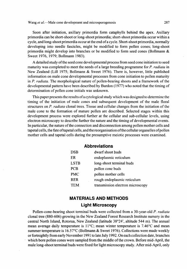
The youngest long-shoot terminal buds (LSTB) for this study were collected in early December 1991, when the LSTB apical meristem was encased in a series of broad, sclerified, achlorophyllous sterile cataphylls, formed from pockets of localised mitotic activity within the peripheral zone of the LSTB apical meristem. Random division of hypodermal cells in the peripheral zone around the apical meristem caused buttresses to develop from the apical flank. Epidermal cells divided periclinally and the primordium grew outwards. Gradually a meristem appeared along the primordium margin, and a broad flattened sterile cataphy 11 was formed (Fig. 1). A proximal to basal developmental gradient was apparent. The forming of sterile cataphylls signalled the beginning of a single growing cycle of the LSTB. After four to five sterile cataphylls were formed, the fertile cataphylls started to develop in the same manner, except that each bore an axillary meristem (Fig. 1). The axillary apices developed from pockets of mitotically active cells in the axil of the cataphyll. They remained relatively small and took on a faintly stained apical zonation. These axillary apices were the earliest-formed short-shoot initials. It was these predetermined buds which eventually developed into pollen cone buds. This determination is based on their location on the shoot apex, but it must be emphasised that the appearance of these axillary primordia at this stage does not necessarily mean that they are male cone primordia; vegetative dwarf shoot buds (DSB) which will develop into needle fascicles have a similar appearance at this stage.
The axillary apices became increasingly larger, as cell division continued, and by 15 January bullet-shaped apices had been formed below the three to four earlier-formed
Wang et al.—Male cone development and microsporogenesis 289
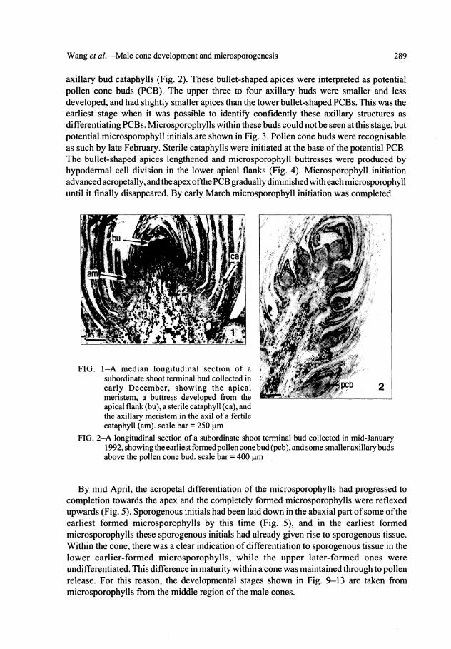
axillary bud cataphylls (Fig. 2). These bullet-shaped apices were interpreted as potential pollen cone buds (PCB). The upper three to four axillary buds were smaller and less developed, and had slightly smaller apices than the lower bullet-shaped PCBs. This was the earliest stage when it was possible to identify confidently these axillary structures as differentiating PCBs. Microsporophylls within these buds could not be seen at this stage, but potential microsporophyll initials are shown in Fig. 3. Pollen cone buds were recognisable as such by late February. Sterile cataphylls were initiated at the base of the potential PCB. The bullet-shaped apices lengthened and microsporophyll buttresses were produced by hypodermal cell division in the lower apical flanks (Fig. 4). Microsporophyll initiation advanced acropetally, and the apex of the PCB gradually diminished with each microsporophyll until it finally disappeared. By early March microsporophyll initiation was completed.
FIG. 2—A longitudinal section of a subordinate shoot terminal bud collected in mid-January 1992, showing the earliest formed pollen cone bud (pcb), and some smaller axillary buds above the pollen cone bud. scale bar = 400 \xm
By mid April, the acropetal differentiation of the microsporophylls had progressed to completion towards the apex and the completely formed microsporophylls were reflexed upwards (Fig. 5). Sporogenous initials had been laid down in the abaxial part of some of the earliest formed microsporophylls by this time (Fig. 5), and in the earliest formed microsporophylls these sporogenous initials had already given rise to sporogenous tissue. Within the cone, there was a clear indication of differentiation to sporogenous tissue in the lower earlier-formed microsporophylls, while the upper later-formed ones were undifferentiated. This difference in maturity within a cone was maintained through to pollen release. For this reason, the developmental stages shown in Fig. 9-13 are taken from microsporophylls from the middle region of the male cones.

290 New Zealand Journal of Forestry Science 30(3)
FIG. 3-A higher magnification of the tissue seen in Fig. 2, showing the relatively larger and bullet-shaped apex (ax), the initiation of the pollen cone bud sterile cataphyll (mi), and the central pith cells, scale bar = 50 \xm
FIG. 4-A near-median longitudinal section of a developing pollen cone bud collected in late February, showing the acropetally initiated microsporophyll (dm) beneath the apex, and the darkly stained pith cells, scale bar = 50 |im
FIG. 5—A near median longitudinal section of a pollen cone bud, after the completion of microsporophyll initiation in late April, showing asymmetrical growth resulting in an upward turning of the microsporophylls. The sporogenous initials have been laid down in the abaxial part of some of the oldest microsporophylls (spi). scale bar = 250 \xm.
FIG. 6-A higher magnification of the tissue seen in Fig. 5, showing the upper epidermis (adaxial surface) (ue) cells filled with a darkly-stained material and the dehiscent zone (dz) in the normal abaxial surface cells with absence of stain. The closely packed sporogenous cells (sc) are surrounded within the microsporangium by a wall of four to five cell layers (mw). scale bar = 20 [im
The upper epidermis (adaxial surface) of the microsporophylls at this stage (see Fig. 6) consisted of broadly elongated cells which underwent frequent anticlinal divisions. Their outer walls were cutinised and most of them were filled with a uniformly darkly-stained material which persisted to the time of pollen shedding. Only the cells at the abaxial surface, marking the line of dehiscence, were free from staining. The newly formed microsporangium with a wall of four to five cell layers and sporogenous cells inside the wall were well-defined at this stage. The tapetal layer could not be distinguished from other wall layers. The sporogenous cells were larger in diameter, had a denser cytoplasm, and stained more darkly

Wang et al—Male cone development and microsporogenesis 291
than the surrounding cells. They formed a compact mass with no apparent intercellular spaces among them (Fig. 6). At the morphological level, there were two well-defined microsporangia formed on the underside of each microsporophyll.
By late May, the sporogenbus cells had divided in several planes to form a mass of pollen mother cells, each with dense cytoplasm and a large granular nucleus. They were also much larger than the wall layer cells (Fig. 7). Division of pollen mother cells (PMC) was noted. These PMCs were surrounded by a microsporangial wall layer two to three cells thick. The innermost layer that completely surrounded the PMCs was presumed to be the tapetum. The tapetal cells were smaller than the pollen mother cells, but their cytoplasm was strongly stained (Fig. 7).
Closer examination of some of these PMCs under high power showed they had probably entered the prophase stage of meiosis. The nucleus was larger than the previous stage, the chromosomes appeared as fine strands, and the nucleolus remained large and distinct (Fig. 8). PMCs seemed to be separated from each other by inter-cellular spaces. These, however, may be an artifact of fixation. By 27 May the chromosomes had become compact, indicating the onset of meiosis in the PMCs (Fig. 9). Walls between PMCs were also noticeably thicker. The nature of the wall was further investigated by transmission electron microscopy and is discussed later.
pollen mother cells within the microsporangium surrounded by the tapetal layer (ta) and the outside wall layer cells. Note compact chromatin (ch) of a pollen mother cell entering meiosis, scale bar = 20 urn.
FIG. 8—A higher magnification of sporogenous tissue, showing a larger nucleus (N), at an early prophase stage of meiosis in a pollen mother cell. Chromosomes appear as fine strands (ch), and the nucleolus (nu) remains large and distinct, scale bar = 10 jum
By mid-June, pollen mother cells had reached the late pachytene stage of prophase I. Paired chromosomes had contracted as thick fuzzy strands, clumped in a tangled mass and covering the nucleolus (Fig. 10). At this stage, PMCs had also accumulated a number of colourless granules which had the appearance of starch grains (Fig. 10). Subsequent development from this stage was rapid. Sixteen days later, on 2 July, pollen cones collected from the same lateral shoot at different positions showed both microspore tetrads and mature pollen grains. Pollen cones collected from the upper part of the shoot tips, within 20 mm from
292 New Zealand Journal of Forestry Science 30(3)
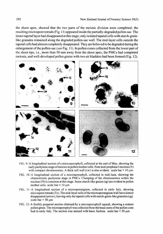
the shoot apex, showed that the two parts of the meiotic division were completed; the resulting microspore tetrads (Fig. 11) appeared inside the partially-degraded pollen-sac. The intact tapetal layer had disappeared at this stage; only isolated tapetal cells with starch-grainlike granules remained along the degraded pollen-sac wall. The mid-layer cells outside the tapetal cells had almost completely disappeared. They are believed to be degraded during the enlargement of the pollen-sac (see Fig. 11). In pollen cones collected from the lower part of the shoot tips, i.e., more than 50 mm away from the shoot apex, the PMCs had completed meiosis, and well-developed pollen grains with two air bladders had been formed (Fig. 12).
FIG. 9-A longitudinal section of a microsporophyll, collected at the end of May, showing the early pachytene stage of meiosis in pollen mother cells. Note mid-prophase I nucleus (N) with compact chromosomes. A thick cell wall (cw) is also evident, scale bar = 10 fam.
FIG. 10-A longitudinal section of a microsporophyll, collected in mid-June, showing the characteristic pachytene stage in PMCs. Clumping of the chromosomes within the nucleus (N) is common at this stage. Some starch-like grains (sg) are evident in pollen mother cells, scale bar = 10 jum
FIG. 11-A longitudinal section of a microsporangium, collected in early July, showing microspore tetrads (Te). The mid-layer cells of the microsporangium wall have almost disappeared (arrow), leaving only the tapetal cells with starch-grain-like granules (sg). scale bar = 50 jum
FIG. 12-A freshly prepared section obtained by a microsporophyll squash, showing a mature pollen grain. The microsporophyll was collected from the basal zone of the pollen cone bud in early July. The section was stained with basic fuchsin. scale bar = 50 jam

Wang et al.—Male cone development and microsporogenesis 293
Transmission Electron Microscopy Ultrastructural changes during microsporogenesis (from microsporangium formation
(29 April 1992), until late pachytene stage of meiosis for pollen mother cells (16 June 1992)) were studied by TEM.
In sections prepared from microsporangium tissue collected on 29 April, showing the larger central sporogenous cells and the surrounding smaller tapetal cells, several nucleoli were present inside the nucleus of both cell types (Fig. 13). Vacuoles, plastids, and mitochondria were seen in both sporogenous cells and tapetal cells (Fig. 14, 15). Well-developed plasmodesmatal connections more frequently occurred between tapetum cells than among the sporogenous cells (Fig. 15). A clear plasmodesmatal connection between two sporogenous cells, apparently showing contact between rough endoplasmic reticulum (RER), is seen in Fig. 14. Cell membranes were well-defined, as was the nuclear envelope. Free ribosomes were abundant (Fig. 14,15) and the RER, although not abundant, was evenly dispersed throughout the cytoplasm in both tissues. Dictyosomes were also seen in both tissues.
FIG. 13—Electron micrograph of part of a microsporangium from a microsporophyll collected in late April. Note the central sporogenous cells (sc) surrounded by tapetal cells (ta). Multiple nucleoli (nu) were present inside the nucleus of both the sporogenous cells and the tapetal cells. A number of plastids (pi) can be seen in both sporogenous cells and tapetal cells, scale bar = 5 |iim
FIG. 14-A higher magnification of the tissue seen in Fig. 13, showing part of three sporogenous cells each containing numbers of mitochondria (m) and free ribosomes (r). A plasmodesmatal strand (p) is evident along with rough endoplasmic reticulum (RER). A dictyosome (d) is also present, scale bar = 1 urn
By 20 May, after the sporogenous cells had developed into pollen mother cells, the general shape of the PMCs was distinctly different from that of the tapetal cells. Pollen mother cells were hexagonal-pentagonal while the tapetal cells were rectangular-trapezoidal (Fig. 16). The number of plastids and mitochondria remained abundant, as did the free ribosomes (Fig. 17, 18). The nuclear envelope of the PMCs was well-defined and contact of the RER to the nuclear envelope was evident (Fig. 17). Cell membranes of the PMCs were still visible,

294 New Zealand Journal of Forestry Science 30(3)
FIG. 15-A higher magnification of the electron micrograph of Fig. 13, showing part of two tapetal cells with frequent plasmodesmatal connections between them (p) Manv nbosomes (r) and some dictyosomes (d) are evident. Mitochondria (m) are also present, scale bar = 1 jam
FIG. 1 ̂ Electron micrograph of part of a microsporangium from a microsporophyll collected in late May, showing pollen mother cells (pmc) and narrow tapetal cells (ta). scale bar = 3.3 (im
FIG. 17-A higher magnification of tissue seen in Fig. 16, showing part of two pollen mother cells. The contact of RER to the well-defined nuclear envelope is evident (arrow) Abundant mitochondria (m) are also evident. Autophagic-like vacuoles (v) are also apparent, scale bar = 1 um
FIG. 18-A higher magnification of tissue seen in Fig. 16, showing parts of three pollen mother cells. Unstained callosic wall material (cw) has apparently blocked the intercellular connections among the cells. Autophagic-like vacuoles (v) are evident. Plastids and mitochondria are abundant, scale bar = 1 urn
but an unstained wall of a probable callosic nature had blocked the plasmodesmata
connectionsamongthemandtheirconnectionswiththetapetalcells(Fig.l8).Plasmodesmata

Wang et al.—Male cone development and microsporogenesis 295
were rarely seen at this stage, and vacuolated structures similar to autophagic vacuoles appeared (Fig. 17, 18).
By 27 May, when pollen mother cells started their pachytene stage, more and larger autophagic vacuoles appearirj; and previously well-defined cell walls appeared thinner and less distinctive. A number of mitochondria gave the appearance of being engulfed by the autophagic vacuoles (Fig. 19). Most plastids examined were encircled by the dilated RER (Fig. 19,20) and there was more dilated RER and more plastids and dictyosomes present in PMCs than seen in the previous stage more than 30 days previously. There was little change in the number of mitochondria or ribosomes. The appearance of the cell wall was different from earlier stages and showed a suggestion of reduced rigidity. It appeared to be callosic in nature, as judged by staining properties and uneven thickening. There were signs that the plasmodesmata connections among the PMCs were blocked by this callosic wall (Fig. 20).
FIG. 20-Electron micrograph of part of a pollen mother cell from a microsporophyll collected at the end of May. The plastids are encircled by the dilated RER and a plasmodesmatal connection appears to be blocked by the callosic wall (arrow), scale bar = 1 jum
By 3 June, when the PMCs were at about mid-pachytene stage, both tapetal cells and PMCs had apparently undergone a modification to a hypersecretory appearance (Fig. 2 1 -23). The tapetal cells became radially flattened, nuclei and cytoplasm became intensely basophilic, and dilation of the RER system was observed to be extreme throughout the cytoplasm (Fig. 21). Osmiophilic granules and globules appeared on the radial surface of the tapetal cells. Channels formed by extreme dilation of ER occurred adjacent to the nucleus and near the cell surface (Fig. 22). These channels contained fibrillar material and appeared to open to extensive spaces in the cytoplasm. It is possible that these areas are evidence of cytoplasmic degradation. Cell membranes were evident on the anticlinal surfaces of the tapetal cells but, from examination of many sections, no cytoplasmic connections between tapetal cells were seen. Dilated portions of the RER/ER containing fibrillar flocculant

296 New Zealand Journal of Forestry Science 30(3)
FIG. 21—Electron micrograph of part of a microsporangium from a microsporophyll collected in early June, showing a hypersecretory tapetal cell with an extremely dilated RER system, the appearance of osmiophilic granules (og), and degraded cells of the middle wall layer (arrow ). scale bar = 3.3 jam
FIG. 22-Higher magnification of tissue seen in Fig. 21, showing part of a tapetal cell. Note dilation of ER and degraded appearance of the cytoplasm. Free ribosomes are arranged into groups, scale bar = 1 ̂ m
FIG. 23-Higher magnification of Fig. 21, showing part of a tapetal cell. Channels formed by extreme dilation of ER/RER are seen adjacent to the cell surface. Polyribosomes (pr) are evident. The callosic wall (cw) appears to block any possible intercellular connections, scale bar = 1 jum
FIG. 24-Electron micrograph of part of a microsporangium from a microsporophyll collected in early June, showing parts of two tapetal cells (right) and one pollen mother cell (left). Broad channels formed by extremely dilated ER and RER are evident. Polyribosomes (pr) are also evident. A thick callosic wall (cw) has apparently isolated the pollen mother cells from the tapetal cells. Arrow shows dilated channel, scale bar = 1 fim

Wang et al.—Male cone development and microsporogenesis 297
material were confluent with envelopes of the autophagic vesicles or the broad and channellike loculi (Fig. 23). Polyribosomes were observed (Fig. 23). There was a reduction in the relative density of plastids, mitochondria, and dictyosomes. The callosic wall appeared to have blocked any possible intercellular connections between the tapetal cells. At the same time, PMCs also showed some hypersecretory features; broad channels formed by extremely dilated RER were evident (arrow in Fig. 24). The dilated portions of the RER containing fibrillar flocculant material were also seen confluent with envelopes of the autophagic vesicles. The density of ribosomes decreased. The free ribosomes were observed to be arranged into polyribosomes in a similar way to the tapetal cells. There was also a reduction in the relative densities of plastids, mitochondria, and dictyosomes. A thick callosic wall had apparently isolated the PMCs from the tapetal cells (Fig. 24). In comparison to tapetal cells, the cytoplasm of PMCs was less basophilic. This may indicate that the tapetal cells are more hypersecretory than PMCs.
By 16 June, when the PMCs entered the end of the pachytene stage of prophase I, prominent invagination occurred along the cytoplasmic membrane of the PMCs beneath the cell wall (Fig. 25). Osmiophilic granules and globules were seen along these invaginations (Fig. 26). Increased surface area to facilitate absorbing of nutrients secreted by the tapetal cells seemed to be a conceivable function of this invagination. The callosic wall of the PMCs was evident (Fig. 26). The density of ribosomes was reduced again compared to the earlier stage. The numbers of plastids and mitochondria were also reduced significantly (Fig. 25, 26). These ultrastructural modifications may indicate preparation for the switch from sporophytic development to gametophyte development, which started with the two continuous reduction divisions around late June.
FIG. 25-Electron micrograph of part of a microsporangium collected in mid-June 1992, showing a pollen mother cell with an invaginated plasmamembrane (pm) coated with a thick callose wall (cw). scale bar = 3.3 jam
FIG. 26-Partof two pollen mother cells from amicrosporophyll collected in mid-June, showing invagination of the plasma membrane. Some osmiophilic granules (og) and globules are seen along these invaginations. Fewer ribosomes, plastids, and mitochondria are seen at this stage. The callosic wall (cw) of these two pollen mother cells is evident, scale bar = 1 urn

298 New Zealand Journal of Forestry Science 30(3)
DISCUSSION
Timing of Developmental Events During Male Cone Development in Pinus radiata, and its Relationship with Environmental Factors
Male cones of P. radiata are initiated by the terminal buds of the subordinate shoots (at least second-order shoots). These subordinate shoots exhibit a pre-determined growth pattern similar to the leading shoots, but differ in their structural components. The components of the new season's subordinate shoots are initiated in the previous growing season in a bud form. Pollen cones of P. radiata growing in New Zealand are initiated in spring when the first short shoots are formed (Bollmann & Sweet 1976). For P. radiata growing at a site near Canberra, Australia, the cataphylls that were due to bear the male cones initiated between October and December. The short-shoot initials in the axils of these cataphylls became visible in January, and their differentiation, i.e., their commitment to either foliage or male cone production, occurred in early February (Cremer 1992).
The present study examined the earliest appearance of the axillary apices which will later form the first short shoots (including pollen cones) of the subordinate branches. These were in shoots collected in early summer (5 December), under the apparently favourable growing conditions at Rotorua. Development into pollen cone primordia does not occur until three or four axillary apices are formed above them on the axis of the subordinate shoots. In this study, the differentiation appeared to be occurring at the collection date of mid January. Before this date, classification of axillary buds as male cone buds or vegetative dwarf shoot buds could not be made with confidence. Position of the male LSTB on the crown, exposure to maximal light direction, and intra-specific genetic factors may all modify the timing of cone development. Clonal differences are also indicated as male strobilus differentiation has been observed as early as December (K. Horgan, pers. comm.). The clone selected for the present study was judged to be representative of the species generally.
Mid-January to February is in the middle of the New Zealand summer, which is the warmest and often the driest time of the year (Bollmann & Sweet 1979). The warm temperature provides favourable conditions for the development of pollen cones. By late February and early March, the initiation of microsporophylls was completed. Sporogenous tissue then developed continuously until the shedding of the mature pollen. The whole process took only about 5 months. Once sporogenous cells had developed into PMCs and entered the meiosis process, it took 6 weeks for PMCs to complete the developmental process resulting in the formation of pollen grains. In the first 4 weeks, PMCs were at the prophase stage. It appears to take a relatively long period of time for PMCs to prepare for the reduction division; thereafter it is completed within just 2 weeks.
In northern temperate pines, by contrast, all axillary buds necessary for the development of reproductive buds are initiated in summer and autumn inside the LSTB before the LSTB becomes dormant through winter (Owens 1986). Development is resumed in the next spring. However, depending on the species, reproductive bud differentiation and development may pause at various climate-dependent points. For example, in P. contorta, PCBs were differentiated and developed to near completion before winter (Owens & Molder 1975). In P. taeda L. (Greenwood 1980), P. resinosa (Duff & Nolan 1958), and P. ponderosa (Gifford & Mirov 1960), PCBs differentiated and underwent considerable development before

Wang et al.—Male cone development and microsporogenesis 299
winter. Pollen cones in slash pine (P. elliottii Engelm.) passed winter in the pollen mother cell stage and meiosis occurred between the end of winter and the beginning of next spring (Mergen & Koerting 1957).
In other gymnosperm species, a developmental break is also apparent. In some species of Larix, PCBs started meiosis during autumn and stopped, passing winter at the diplotene stage. Meiosis was completed in spring (Ekberg et al. 1968). However, in P. monticola (Owens & Molder 1977) PCBs differentiated but showed little development before winter. All microsporophylls were initiated but sporogenous tissue did not form before PCBs became dormant in winter. Environmental or climatic conditions appear to be the major factor controlling the developmental timing of events leading to pollen production. The higher latitudes of the Northern Hemisphere impose a cessation of development and break pollen cone development into two quite separate parts by a period of up to 6 months' dormancy (Luomajoki 1977, 1982).
Pollen cone development in the milder climatic conditions of New Zealand is quite different from the developmental timing of northern temperate pines. The organogenic sequence is the same but the "rate" or developmental timing of pollen cone differentiation and development, especially of the meiosis event, is strongly influenced by climate or other environmental cues such as daylength. Evidence reviewed here suggests that temperature is of greatest importance.
In north-west British Columbia, Canada, pollen cones of P. monticola only develop into the microsporophyll stage with no initiation of the sporogenous tissue before winter, and pollen shedding does not occur until late the following June (Owens & Molder 1977). On the other hand, in Baker County, Florida, a warmer and lower latitude area, pollen mother cell development in P. elliottii occurs before a winter dormancy period, and pollen shedding will occur as early as the end of the following January (Mergen & Koerting 1957). Pinus radiata growing in its natural environment on the Pacific coast of California sheds pollen in March after winter dormancy (Mirov 1967). In New Zealand, pollen cones of P. radiata develop through winter and pollen shedding is in the middle of the winter (early July) according to this study, or around August-September according to Bollmann & Sweet (1976). Fountain & Cornford (1991) over a 3-year study period found the starting date for pollen release varied from early July to early August. A dependence of development on temperature is suggested by this variation.
In reviewing environmental impact on pollen cone development, Mirov (1967) stated that in the temperate zone, pines shed their pollen during the season of the year designated as spring. In north-west British Columbia this is in June; in coastal California it is in March, and in temperate Southern Hemisphere countries such as New Zealand it is in July to September. Mirov also pointed out that the closer to the Equator, the earlier pine pollen is shed. However, he regarded changing photoperiod as unlikely to affect the "flowering" of pines, suggesting rather that the involvement of thermoperiodicity was a major factor (Mirov 1967).
Studies on P. sylvestris L. (Sarvas 1962) and P. palustris Miller (Boyer & Woods 1973) yielded a similar conclusion: higher temperatures will cause earlier pollen shedding.
Pollen shedding in P. radiata has, however, also been linked to photoperiod. In plants disentrained from normal seasonal rhythm, a powerful inhibition of pollen shedding during autumn was suggested to be best explained by decreasing photoperiod. Certainly some
300 New Zealand Journal of Forestry Science 30(3)
external factor was responsible for delaying shedding until the normal late winter period (Burdon 1977).
Abnormal pollen cone development in Larix growing in Sweden also illustrates another possible locus of developmental control by temperature. If the pollen cone buds were not exposed to low temperatures, frequently no wall formation was seen after the completion of meiosis of the PMCs. Eight microspores were formed rather than four, and were regarded as non-functional (Ekberg et al. 1968; Eriksson 1968). No reports, however, have been found allowing comment on whether or not this occurs in pines. Low temperatures are also known to cause disturbances to meiosis leading to permanent damage in microsporogenesis (Luomajoki 1977).
The winter dormancy period appears to be essential for meiotic completion in these conifer species. Once pollen cones have completed winter dormancy, it takes only a short period of time to complete the rest of their development and still allow pollination to occur in the coming spring. For example, after the winter dormancy at the diffuse diplotene stage, the remaining part of meiosis in Larix finished within 4 days in the next spring (Ekberg et al 1968). In P. elliottii, after the winter dormancy at the PMC stage, meiosis of the PCBs occurred during the middle of January; subsequent development from this stage on took only 3 days (Mergen & Koerting 1957). The same kind of rapid meiosis after winter dormancy was also recorded by Ferguson (1904) in P. austriaca L., P. strobus L., and P. rigida Hook. & Arn.
The first appearance of potential pollen cone primordia of P. radiata growing in New Zealand is in the early part of the summer. Bullet-shaped primordia and microsporophylls appear by late February. Primordia can be confidently identified as PCBs at this time. In our study the initiation and differentiation processes of PCBs started at early summer and finished in late summer. There was no apparent pause, indicating that the generally favourable temperatures in New Zealand could be a major factor responsible for this continuous development. Some slowing of later stages of pollen cone maturation may occur in the New Zealand environment, when PMCs entered the prophase stage of meiosis around late autumn to late May. Temperatures become lower and the subsequent meiosis process from this stage on took almost 6 weeks to complete before the formation of morphologically mature pollen. This appears to contrast with the rapid development of the meiosis process in some Northern Hemisphere pine species, after their winter dormancy. This comparison may again emphasise the significant impact of the temperature factor upon "floral" development. The accelerated development of the meiosis process of the Northern Hemisphere pine species is an apparent result of the rapid increase of the temperature in the spring season (Mirov 1967). In New Zealand, meiosis of PMCs appears to start in late autumn, and to finish in mid-winter. When temperatures become increasingly reduced during autumn, a slower development at the earlier stages of meiosis seems inevitable.
Morphological Aspects of Male Cone Development in Pinus radiata Light microscopy not only determined the timing of some important stages of male cone
development, but also revealed morphological changes that occurred during this process. When differentiation of the male cone was completed, PCBs consisted of an axis with spirally arranged microsporophylls each bearing two microsporangia on the underside.

Wang et ah—Male cone development and microsporogenesis 301
Microsporangia became proportionally increasingly larger, the mature microsporophyll consisting of upper epidermis (adaxial surface) and lower epidermis (abaxial surface) separated by the microsporangium. The outer walls of the upper epidermal cells were cutinised and the majority were filled with uniformly stained (apparently suberised) material. The lower epidermal cells, however, wereunsuberised. This difference in the upper and lower surface tissue is most likely associated with release of the mature pollen grains caused by a dehiscence zone line along the lower epidermis. Similar events have been reported in P. wallichiana A.B.Jackson (Konar 1960).
Pollen cones mature at somewhat different rates, depending on their positions on the male-cone-bearing shoots. In P. caribaea growing in Northern Queensland, Australia, each cluster of pollen cones commonly released pollen at two different times; the proximal pollen cones shed first, followed 2 weeks later by the distal pollen cones (Harrison & Slee 1992). Ho & Owens (1974) also found that on a shoot of P. contorta, proximal male cones had PMCs at more advanced stages than did the more distal cones. The size and appearance of the male cone also varied considerably during early stages of meiosis, and there appeared to be little correlation between male cone size and the stage of meiosis (Ho & Owens 1974). Similar results were seen in the present study. Pollen cone size at different locations on the shoot axis from three developmental stages revealed that the size of pollen cones located at the basal region was not significantly different from that of male cones located at the distal region. Pollen cones collected from the basal region of the pollen-cone-bearing shoot in early July, however, contained well-developed pollen grains while pollen cones collected from the distal region of the same shoot showed only microspore tetrads. It appears that the maturation rate of male cones relies more on their location on the shoot axis than on their size.
Maturation rate of microsporophylls is also dependent on position within the male cone. Chamberlain (1935) found that in P. banksiaria, when microsporangia in the lower section were in the early sporogenous stages, sporogenous cells had not been differentiated in the apical part of the cone. Ho & Owens (1974) also reported that, within a male cone, PMCs in the proximal microsporophylls were generally at a more advanced stage of meiosis than those in the distal part. The longitudinal section of the male cone bud collected from mid-April in this study showed a similar result. Microsporangia in basal microsporophylls were at a much more advanced stage of development than the microsporangia in the distal microsporophylls. Sporogenous tissue had been formed in the lower section, but sporogenous tissue cells were not obvious in the upper section of the microsporophylls (Fig. 7). Such a variation of male cone maturation within the shoot and within the cone itself no doubt spreads pollen release over a longer period of time, allowing a longer pollination time.
Structural and Ultrastructural Changes During Male Cone Development and Microsporogenesis in Pinus radiata
Changes during the development of PMCs and tapetal cells were examined in this study. In the earlier stages of the male cone, the sporogenous cells were initially compact, forming a mass of cells with no apparent intercellular spaces. Intercellular spaces became more obvious with the development of PMCs prior to the meiotic process. Once PMCs entered the prophase stage of meiosis, pairing and contraction of the chromosomes occurred in pollen mother cells. Similar events have also been described in other pine species. Ferguson (1904) reported that during the period preceding reduction division, the nucleus of the PMCs
302 New Zealand Journal of Forestry Science 30(3)
gradually condensed, the chromosomes became thicker, and the meshes of this nuclear reticulum became smaller. Contractions of chromosomes continued until the network formed a compact mass at one side of the nucleus. These changes occurred when PMCs started reduction division. Owens & Molder (1971) found prolonged pachytene and diffuse diplotene stages in meiosis in several northern conifer species. In Douglas-fir {Pseudotsuga menziesii (Mirb.) Franco) characteristics of the pachytene stage of the PMCs were identified. The chromosomes appeared as thick fuzzy strands and they were usually clumped in a tangled mass filling only a portion of the nucleus. Comparison of chromosome behaviour occurring in Pinus radiata with that of Douglas-fir at pachytene and late pachytene stages suggests the pachytene stage of PMCs in P. radiata started between late-May and mid-June.
Initially, tapetal cells appeared as an intact layer surrounding the earlier-formed PMCs. After the PMCs had completed meiosis and had entered the tetrad stage, only a few isolated tapetal cells along the degraded pollen-sac wall remained. The cells of the middle layer of the microsporangium outside the tapetum almost completely disappeared at this stage.
The nature of the intercellular connections between tapetal cells and PMCs and changes occurring in the cell wall of PMCs during their maturation as well as developmental changes in tapetal cells and PMCs, were examined by TEM. In the early stages of development of the microsporangium, the central sporogenous cells were slightly larger than the surrounding tapetal cells. The nuclei of cells of both tissues were found to contain two to three nucleoli. Similar observations have been reported in other Pinus species (Ferguson 1904; Singh 1978). Tapetal and sporogenous cells were seen to have similar numbers of small vacuoles, and dictyosomes. Both tissues were rich in free ribosomes, mitochondria, and plastids. They both had well-defined cytoplasmic membranes, and the RER from both tissues was similar in appearance. These features are consistent with those found in cells of an actively dividing meristematic tissue.
Dickinson & Bell (1976a,b) also reported that early in their development the tapetal cells and sporogenous cells in P. banksiana were ultrastructurally alike with rich cytoplasm and having a multinucleate appearance. Some intercellular connections via plasmodesmata were evident at this stage both between cells of the sporogenous tissue, as well as between the tapetal cells and also between the two cell types. These features indicate the syncytium character of cells of both tissues, especially between the tapetal cells. This syncytium character has been described previously in P. banksiana by Dickinson & Bell (1976a,b). Cytoplasmic organelles as large as mitochondria were observed in passage through channels formed by the plasmodesmata between two tapetal cells. This remarkable observation suggests that the plasmodesmata have an extremely high size exclusion limit, much greater than is generally thought to be present between cells. The syncytium character of tapetal cells and early sporogenous cells is consistent with the probability of the two tissues developing simultaneously, and also displaying similar structural features. This similarity remained until at least the sampling time of the following month (end of May), when callose walls around the PMCs formed (Fig. 20). Willemse (1971) found that in P. sylvestris callose wall formation started at the diplotene stage of prophase I, appearing first between the plasma membrane and the cell wall. A fine electron-dense fibrillar material accumulated against the cell wall and the plasma membrane. This fibrillar network gradually changed into a highly electron-transparent line between the cell wall and the flat plasma membrane. Such electron-transparent layers of cell walls seen at these developmental stages are interpreted here and

Wang et al.—Male cone development and microsporogenesis 303
elsewhere as callose. The callose wall enveloped the whole PMC and grew in thickness until the tetrad stage. The formation of a callose wall around PMCs signals the start of meiosis (Willemse 1971). At this time PMCs are isolated from their surrounding tapetal cells, and significant ultrastructural modifications occur in both cell types. Taken together these observations are in contrast to those for angiosperm species such as Lilium henryi where the tapetal and sporogenous cells are distinct well before the formation of the callose wall (Dickinson & Heslop-Harrison 1970).
Once PMCs were further developed towards meiosis, tapetal cells underwent some significant structural changes. Their cell wall appeared to be thinner and vacuoles, interpreted from their appearance as autophagic vacuoles, appeared alongside extremely dilated RER. The cytoplasm was intensely basophilic because of the density in ribosomes which seemed to be arranged into polyribosome groups. The numbers of plastids and mitochondria were reduced, possibly the result of degradation by the prominent autophagic vacuoles. The apparent hypersecretory features of the tapetum cells may allow them to secrete nutrients supplied by the tapetal cells and the mid-layer cells into the locule of the microsporangium. Subsequently these breakdown products would be expected to be taken up by the developing PMCs or later on by the microspores.
The intercellular connections via plasmodesmata between tapetal cells and PMCs and among the tapetal cells themselves were blocked by the appearance of the callosic wall between them, suggesting that PMCs and tapetal cells then develop independently along different routes: one group differentiating towards meiosis, one group towards hypersecretory tissue. A block of the now-incompatible genetic messages between cells of these two tissues would be expected to be necessary.
Osmiophilic granules and globules were seen on the radial surfaces of the tapetal cells and subsequently throughout the microsporangial loculus in this study. These are interpreted as sporopollenin or its precursors, of sporophytic origin, associated with sporopollenin deposition on to the pollen grain exine. Detailed studies in P. banksiaria (Dickinson & Bell 1976a,b) and P. sylvestris (Rowley & Walles 1985) have established such relationships. The nature of these osmiophilic granules was first investigated by gas chromatography (Brooks 1971). Brooks found numerous carotene residues in the osmiophilic granules in sporopollenin, suggesting the lipid nature of the osmiophilic granules. Dickinson & Bell (1976a,b) found a clear correlation between the fall in the number of these lipid-like granules in the cytoplasm of the tapetal cell and the development of these external granules in the loculus in P. banksiaria, and they pointed out that these granules originated in the tapetal protoplasts. They also reported that the granules were prominent in the tapetal cytoplasm at the beginning of sporopollenin production, but gradually declined as increasing amounts of sporopollenin were deposited on the pollen wall, suggesting that these lipid-like granules were possibly metabolised in the cisternae of the tapetal cell and used ultimately to form the sporopollenin on the pollen wall (Dickinson & Bell 1972). In an angiosperm genus, Raphanus, it has been reported that pollen grains might be coated to a depth of some 5 urn with the lipid-rich sporopollenin, and it has been demonstrated that this layer plays a part in the self-incompatibility system in this plant (Dickinson & Lewis 1973).
Mid-May was inferred as the time when meiosis begins in PMCs. This determination was based on both the observations of the condensation of chromosomes by light microscopy and the occurrence of callose walls among pollen mother cells as seen by electron microscopy.

304 New Zealand Journal of Forestry Science 30(3)
The TEM study of material from the same sampling period revealed that PMCs underwent some substantial structural modifications. The cell walls appeared to be thinner, and were well-coated by a thick callose wall. The cytoplasmic membrane at a later stage (mid-June) showed shrinking to some degree and formed a number of invaginations inside the callose wall apparently increasing the absorbing surface at the later pachytene stage, just before the reduction division. Rough endoplasmic reticulum became very dilated, more autophagic vacuoles appeared, and the density of the ribosomes, plastids, and mitochondria was reduced compared to the earlier stages. The formation of autophagic vesicles has been traced in differentiating cells of barley {Hordeum sativum Linn.) by Buvat & Robert (1979) from dictyosome vesicles and tubules that concentrate enzymes. They reported that some of these vesicles and tubules became autophagic vacuoles which degraded the imprisoned cytoplasmic fraction by the process of autophagy. Similar findings in P. sylvestris were reported by Walles & Rowley (1982) and by Willemse (1971). The fully developed autophagic vacuoles seen here at late pachytene just before the reduction division seem likely to be responsible for the reduction of some of the free ribosomes, plastids, and mitochondria noted.
In reviewing angiosperm pollen development, Dickinson (1987) stated that plastids and mitochondria de-differentiated to an almost unrecognisable state during the meiotic prophase stage in most plant species examined. This de-differentiation preceded a very active period of DNA synthesis and was followed by division and de-differentiation of most of the organelles. A significant proportion of both mitochondria and plastids may be degraded. In Cosmos bipinnatus Cav.Ic, for example, this degeneration may account for up to 20% of the total mitochondria population during prophase (Dickinson 1987). Sheffield & Bell (1979) pointed out that meiosis in both angiosperms and gymnosperms shared some common features, including the partial de-differentiation of the mitochondria and plastids and the appearance of vacuoles within the nuclei. The undoubted degradation of a major proportion of ribosomes during prophase in Lilium had been reported by Mackenzie et al. (1967). More recently, similar events have also been found in the gymnosperm species Taxus (Pennel & Bell 1986). These events have been interpreted as crucial in the reorganisation of a diploid cell (sporogenous cell) into one whose genome is haploid (microspore), and in which expression of that part of the genome which is concerned specifically with sporophytic growth must be replaced by that part concerned specifically with gametophyte development (Dickinson & Heslop-Harrison 1977).
ACKNOWLEDGMENTS
Based on a PhD thesis by Daniel Yunqiu Wang, Massey University, New Zealand. This research was supported by research grants from New Zealand Forest Research Institute Ltd and the Massey University Graduate Research Fund. Dr J. Owens and colleagues at University of Victoria, British Columbia, Canada, are thanked for valuable comments. Technical assistance with electron microscopy by D. Hopcroft and R. Bennett of the Horticulture and Food Research Institute Ltd, Palmerston North, New Zealand, is gratefully acknowledged.
REFERENCES BOLLMANN, M.P. 1983: Morphology of long-shoot development in Pinus radiata. New Zealand
Journal of Forestry Science 13(3): 275-290.

Wang et al.—Male cone development and microsporogenesis 305
BOLLMAN, M.P.; SWEET, G.B. 1976: Bud morphogenesis of Pinus radiata in New Zealand. I: The initiation and extension of the leading shoot of one clone at two sites. New Zealand Journal of Forestry Science 6: 376-392. 1979: Bud morphogenesis of Pinus radiata in New Zealand. II: The seasonal shoot growth pattern of seven clones at four sites. New Zealand Journal of Forestry Science 9: 153—165.
BOYER, W.D.; WOODS, F. W. 1973: Date of pollen shedding by longleaf-pine advanced by increased temperatures at strobili. Forest Science 19: 315—318.
BROOKS, J. 1971: "Sporopollenin". Academic Press, London and New York. BURDON, R.D. 1977: Photoperiodic effect on pollen shedding in Pinus radiata. New Zealand Journal
of Forestry Science 7: 214-215. BUVAT, R.; ROBERT, G. 1979: Vacuole formation in the actively growing root meristem of barley
(Hordeum sativum). American Journal of Botany 66: 121 9-1 237. CHAMBERLAIN, CS. 1935: "Gymnosperms, Structure and Evolution". University of Chicago
Press. 484 p. CHUDNOFF, M.; GEARY, T.F. 1973: Terminal shoot elongation and cambial growth rhythms in
Pinus caribaea. Commonwealth Forestry Review 52: 317—324. CREMER, K.W. 1992: Relations between reproductive growth and vegetative growth of Pinus
radiata. Forest Ecology and Management 52: 179-199. CURTIS, J.D.; POPHAM, R.A. 1972: The developmental anatomy of long-branch terminal buds of
Pinus banksiana. American Journal of Botany 59: 194-202. DICKINSON, H.G. 1987: The physiology and biochemistry of meiosis in the anther. International
Review of Cytology 107: 79-109. DICKINSON, H.G.; BELL, P.R. 1972: The role of the tapetum in the formation of sporopollenin
containing structures during microsporogenesis in Pinus banksiana. Planta 107: 205-215. 1976a: The changes in the tapetum of Pinus banksiana accompanying formation and maturation of the pollen. Annals of Botany 40: 1101-1109. 1976b: Development of the tapetum in Pinus banksiana preceding sporogenesis. Annals of Botany 40: 103-113.
DICKINSON, H.G.; HESLOP-HARRISON, J. 1970: The ribosome cycle, nucleoli and cytoplasmic nucleoloids in the meiocytes of Lilium. Protoplasma 69: 187-200. 1977: Ribosomes, membranes and organelles during meiosis in angiosperms. Philosophical transactions of the Royal Society of London B. 277: 327-342.
DICKINSON, H.G.; LEWIS, D. 1973: The formation of the tryphine coating the pollen grains of Raphanus and its properties relating to the self-ineompatibility system. Proceedings of the Royal Society of London. Series B. 184: 149-165.
DO AK, C.C. 1935a: Evolution of foliar types, dwarf shoots, and cone scales of Pinus. University of Illinois Bulletin 32. 106 p. 1935b: Evolution of foliar types, dwarf shoots, and cone scales of Pinus. Illinois Biological Monographs 13 (3): 1-106.
DUFF, G.H.; NOLAN, N.J. 1958: Growth and morphogenesis in the Canadian forest species. Canadian Journal of Botany 36: 687—706.
EKBERG, I.; ERIKSSON, G.S.; SULIKOVA, Z. 1968: Meiosis and pollen formation in Larix. Hereditas 59(24): 427-438.
ERIKSSON, G.S. 1968: Temperature response of pollen mother cells in Larix and its importance for pollen formation. Studia Forestalia Suecica 63: 131.
FERGUSON, M. 1904: Contributions to the life history of pines with special reference to sporogenesis, the development of gametophytes and fertilization. Proceedings of the Washington Academy of Sciences 6: 1—202.
FOUNTAIN, D.W.; CORNFORD, CA. 1991: Aerobiology and allergenicity of Pinus radiata pollen in New Zealand. Grana 30: 71-75.

306 New Zealand Journal of Forestry Science 30(3)
GIFFORD, E.M.; MIROV, N.T. 1960: Initiation and ontogeny of the ovulate strobilus in ponderosa pine. Forest Science 6: 19-25.
GREENWOOD, M.S. 1980: Reproductive development in loblolly pine. I. The early development of male and female strobili in relation to the long shoot growth behaviour. American Journal of Botany 67: 1414-1422.
HARRISON, D.L.S.; SLEE, M.U. 1992: Long shoot terminal bud development and the differentiation of pollen- and seed-cone buds in Pinus caribaea var. hondurensis. Canadian Journal of Forest Research 22: 1656-1668.
HO, R.; OWENS, J.N. 1974: Microsporogenesis and pollen formation in lodgepole pine. Canadian Journal of Botany 52: 1669-1674.
JOHANSEN, D.A. 1940: "Plant Microtechnique". McGraw-Hill, New York. KARNOVSKY, M.J. 1965: A formaldehyde-glutaraldehyde fixative of high osmolarity for use in
electron microscopy. Journal of Cell Biology 2 7: 137-13 8 KONAR, R.N. 1960: The morphology and embryology of Pinus roxburghii Sarg. with a comparison
with Pinus wallichiana Jack. Phytomorphology 10: 305—319. LANNER, R.M.; VAN DEN BERG, D.A. 1975: The vegetative buds and shoots of lodgepole pine.
Management of lodgepole pine ecosystems. Vol. {.Cooperative Extension Service Washington State University 1: 68-85.
LILL, B.S.I 975: Ovule and seed development inPinus radiata: postmeiotic development, fertilization,
and embryogeny. Canadian Journal of Botany 54: 2141-2154.
LUOMAJOKI, A. 1977: Effects of temperature on spermatophute male meiosis. Hereditas 85:33-48.
1982: Temperature and dates of male meiosis in trees. Hereditas 97: 167-178. MACKENZIE, A.; HESLOP-HARRISON, J.; DICKINSON, H.G. 1967: Elimination of ribosomes
during meiotic prophase. Nature 215: 997-999. MERGEN, F.; KOERTING, L.E. 1957: Initiation and development of flower primordia in slash pine.
Forest Science 3(2): 145-155. MIROV, N.T. 1967: "The Genus Pinus ". Ronald Press, New York. OWENS, J.N. 1986: Cone and seed biology. Pp. 14-31 in Proceedings of "Conifer Tree Seed in the
Inland Mountain West" Symposium. USDA Forest Service, Intermountain Research Station General Technical Report No. INT-302.
OWENS, J.N.; MOLDER, M. 1971: Meiosis in conifers: prolonged pachytene and diffuse diplotene stages. Canadian Journal of Botany 49: 2061-2064.
1975: Development of long-shoot terminal buds of Pinus contorta. Pp. 86-104 in Proceedings of "Management of Lodgepole Pine Ecosystems", 1973, Washington State University Cooperative Extension Service.
1977: Development of long-shoot terminal buds of western white pine (Pinus monticola). Canadian Journal of Botany 55: 1308-1321.
OWSTON, P.W. 1969: The shoot apex in eastern white pine: its structure, seasonal development and variation within the crown. Canadian Journal of Botany 47: 1181—1188.
PENNELL, R.I.; BELL, P.R. 1986: Microsporogenesis in Taxus baccata L. : The formation of the tetrad and development of the microspores. Annals of Botany 57: 545-555.
ROLAND, J.C.; VIAN, B. 1991: General preparation and staining of thin sections. Pp. 2-64 in Hall, J.L.; Hawes, C. (Ed.) "Electron Microscopy of Plant Cells". Academic Press, London.
ROWLEY, J.R.; WALLES, B. 1985: Cell differentiation in microsporangia of Pinus sylvestris. II. Early pachytene. Nordic Journal of Botany 5: 241-254.
SACHER, J.A. 1954: Structure and seasonal activity of the shoot apices of Pinus lambertiana and Pinus ponderosa. American Journal of Botany 41: 749-759.
SARVAS, R. 1962: Investigation on the flowering and seed apices ofPinus sylvestris. Communications, Instituti Forestalls Fenniae 53: 1-198.

Wang et al.—Male cone development and microsporogenesis 307
SHEFFIELD, E.; BELL, P.R. 1979: Ultrastructural aspects of sporogenesis in a fern, Pteridium aquilinum (L.) Kuhn. Annals of Botany 44: 393-^405.
SINGH, H. 1978: "Embryology of Gymnosperms". Gerbrder, Borntraeger, Berlin. SPURR, A.R. 1969: A low viscosity epoxy resin embedding medium for electron microscopy. Journal
of Ultrastructure Research 26: 31-43. SUCOFF, E. 1971: Timing and rate of bud formation in Pinus resinosa. Canadian Journal of Botany
49: 1821-1832. SWEET, G.B.; BOLLMANN, M.P. 1976: The terminology of pine shoot growth. New Zealand
Journal of Forestry Science 6: 393—396. VAN DEN BERG, D. A.; LANNER, R.M. 1971: Bud development in lodgepole pine. Forest Science
17: 479-486. WALLES, B.; ROWLEY, J.R. 1982: Cell differentiation in microsporangia of Pinus sylvestris with
special attention to the tapetum. I. The pre-and early-meiotic periods. Nordic Journal of Botany 2: 53-70.
WILLEMSE, M.T.M. 1971: Morphological and quantitative changes in the population of cell organelles during microsporogenesis of Pinus sylvestris L. I. Morphological changes from zygotene until prometaphase I. Acta Botanica Neerlandica 20(3): 261—274.