Energy Dispersive X-ray Spectrometry and X-ray Microanalysis.

The BV Pulsera mobile C-arm solution provides the power and superb visualizations to support a variety of challenging interventions. From open to minimally invasive, from Abdominal Aortic Aneurysms (AAA) to pacemaker implants. From spinal to hip surgery.
This counter-balanced system can be positioned quickly and easily. It provides the extra power to image obese patients or technically diffi cult projections.
Key benefi ts
• Powerful system with choice of 9" or 12" image intensifi er to perform a wide range of routine and challenging applications
• Ortho Plus – applies extra X-ray power to visualize challenging regions of interest with normal or obese patients
• Single user vascular workfl ow - tap the footswitch once to perform Subtraction, Trace, or Roadmap
• Pulsed exposure mode visualizes fast moving anatomy with outstanding image quality for a virtually unlimited amount of time
• Compact Mobile View Station with exceptional viewing fl exibility
Making the di� erence with Live Image GuidanceBV Pulsera mobile C-arm system specifi cations
BV Pulsera
Surgery
The print quality of this copy is not an accurate representation of the original.

Contents
1 System overview 4
2 Image detection 6
2.1 Image intensifier 6
3 X-ray generation 7
3.1 X-ray generator 7
3.2 X-ray tube 8
3.3 X-ray collimation 9
4 Workflow 10
4.1 C-arm stand 10
4.2 Mobile View Station 10
4.3 Connectivity 10
4.4 Handheld remote control 11
4.5 Touchscreen Examination monitor 11
4.6 DICOM and IHE 11
4.7 Stand monitor 11
5 Imaging 12
5.1 SmartVision 12
5.2 DoseWise 12
5.3 Automatic Programmed Fluoroscopy 13 programs
5.4 Real-time processing functions 13
5.5 Post processing functions 13
5.6 Mobile View Station monitors 13
6 Options 14
7 Geometry 15
7.1 C-arm stand 15
7.2 Mobile View Station 15
8 Service 16
9 Dimensions 18
9.1 C-arm stand 18
9.2 Mobile View Station 19
2 The print quality of this copy is not an accurate representation of the original.

3The print quality of this copy is not an accurate representation of the original.

1 System overviewThe BV Pulsera is a counterbalanced mobile C-arm system. Its
C-arm stand has a compact foot and rear-wheel steering for easy
maneuverability and positioning. The intelligent design of the Mobile
View Station supports easy transport and set-up, ergonomic viewing
and full connectivity capabilities.
4
2
1
3
4 The print quality of this copy is not an accurate representation of the original.

1 Easy workfl ow• Automatic Programmed
Fluoroscopy (APF) programs are fi ne-tuned for each clinical application to enable high quality visualizations
2 Image intensifi er• 1K2 high resolution imaging chain• 9" or 12" triple-mode image
intensifi er• 9/7/5" (23/17/13 cm)• 12/9/7" (31/23/17 cm)
3 High power X-ray tank• 15 kW rotating anode power for
demanding procedures• Excellent heat management
for lengthy interventional procedures
• Monoblock architecture delivers sharp defi ned pulses
4 Monitors• C-arm stand:
12” stand monitor (optional)• Mobile View Station:
19” color LCD• Contrast ratio >500:1
(optional: HiBri >700:1)• Convenient touchscreen user
interface (optional)• Flexible monitor positioning:
stepless height adjustment and 180° rotation
• Foldable for easy transport and storage
5 Archiving and documentation• Fully integrated DICOM solution
(optional)• Medical DVD recorder (optional)• Printer (optional)• USB image storage
6 Connectivity• Video-in to display external
video signals such as ultrasound or endoscopy displayed on reference monitor
• Digital video out (optional) and analog video out (standard) to display BV Pulsera images on separate monitors (such as ceiling suspended monitors)
4
5
6
5The print quality of this copy is not an accurate representation of the original.

2 Image detectionDesigned to handle a variety of routine and complex procedures,
this surgical imaging system is compact, flexible and easy to move.
Feature Specification
Image intensifier type Triple-mode 9" HRC / Triple-mode 12"
Nominal II formats 31, 23, and 17 cm (12", 9", and 7")23, 17, and 13 cm (9", 7", and 5")
Entrance screen Cesium Iodine
Grid type Circular, carbon fiber; 60 lines/cm Ratio = 1:10SID = 100 cm
TV camera type CCD; high-resolution 1k2
Image rotation Digital, live and on LIH
Image reversal Yes
Mirror up/down Digital, live and on LIH
Mirror left/right Digital, live and on LIH
Automatic anatomical measuring field Yes with ‘BodySmart’
2.1 Image intensifier The BV Pulsera comes with a 9" or 12" image intensifierand can go wherever you need it - surgery, intensive care and the emergency room.
Choose either a 9" or 12" triple-mode image intensifier, to match your applicational requirements.
6 The print quality of this copy is not an accurate representation of the original.

3 X-ray generationThe rotating anode technology and 15 kW generator of the BV Pulsera
provide the power to see through virtually any normal or obese patient
and visualize fi ne details in steep projections.
Feature Specifi cation
X-ray generator type Monoblock 80 kHz high-frequency generator microprocessor controlled
Max. generator output 15 kW
Max. X-ray tube voltage 120 kV
Max. X-ray tube current 125 mA
3.1 X-ray generatorBV Pulsera uses a Monoblock concept with the high-tension transformer in the X-ray tank. This means pulses do not have to be transmitted over high voltage cables, which can result in ramping up and trailing
down eff ects, due to the electrical impedance of the cables. The BV Pulsera Monoblock generator produces immediate pulse rise so all radiation is in the useful spectrum. The result is excellent quality images with less soft radiation and long fl uoro times.
mA
ramping imaging pulse trailingimaging pulse
mA
time time
The BV Pulsera Monoblock concept decreases blurring and soft radiation
Monoblock Without monoblock
7The print quality of this copy is not an accurate representation of the original.

3.2 X-ray tubeBV Pulsera has a rotating anode and high power generator with excellent heat management to perform demanding interventional procedures. An integrated beam fi lter helps to reduce patient skin dose1. Automatic Programmed Fluoroscopy (APF) provides provide consistent image quality for every examination.
Feature Specifi cation
Tube type Rotating anode X-ray tube
Nominal X-ray tube voltage 120 kV
Nominal focal spot values 0.3 IEC and 0.6 IEC
Maximum anode heat content 311 kHU
Maximum heat dissipation 73 kHU/min
Cooling method Active oil-circulation cooling
Inherent fi ltration 1.0 mm Al eq.
Additional fi ltration 3.0 mm Al eq. + 0.1 mm Cu
Operating values Continuous Fluoroscopy
kV range 40 - 120 kV
mA range for Low Dose Fluoroscopy mode 0.10 - 8.33 mA (up to 10 mA during Auto High Penetration)
mA range for High Defi nition Fluoroscopy mode 0.24 - 20 mA
Operating values Half Dose Fluoroscopy
kV range 40 - 120 kV
mA range 0.4 - 12 mA
Pulse width 11 - 37 ms
Pulse rate 12.5 pps
Operating values Quarter Dose Fluoroscopy
kV range 40 - 120 kV
mA range 0.4 - 12 mA
Pulse width 11 - 37 ms
Pulse rate 6.25 pps
Rating values Pulsed Exposure
kV range 40 - 110 kV
mA range 2.0 - 60 mA
Pulse width 8.0, 9.5 and 11.1 ms
Pulse rate 3, 5, 8 pps (optional 15, 30 pps)
Operating values Digital Exposure
kV range 40 - 110 kV
mA range 1.5 - 75 mA
Pulse width 120 - 440 ms
1. Compared to conventional fi ltration of 3 millimeters aluminium as required by IEC 60601-2-43, 2010.8 The print quality of this copy is not an accurate representation of the original.

Iris collimation
Additional beam fi lter
Near focus collimation
Anode
Independently movable lead shutters
3.3 X-ray collimationCollimation reduces exposure dose by reducing the radiated surface and enhances image quality. BV Pulsera makes collimation easy. Its full lead (assymetric) shutters can be rotated and moved independently, and the unique Philips Automatic Shutter Positioning (ASP) feature automatically positions shutters for high image quality at the touch of a button. You can position shutters or adjust the iris on the last X-ray image (Last Image Hold), eliminating additional X-ray dose during collimation.
Iris collimation
Feature Specifi cation
Shutters Two independent lead shutters with steel wedge: shutters can be coupled for rotation and translation, or moved individually for asymmetric collimation
Automatic Shutter Postioning Automatic shutter placement based on image content
Shutter material 3 mm Pb
Wedge material 0.2 to 2.5 mm stainless steel
Adjustment Stepless
Rotation 360°
Minimal iris diameter < 50 mm on II entrance
Position indication On screen and also on LIH without radiation
At a touch of a button, Automatic Shutter Positioning (ASP) places shutters in the optimal position
9The print quality of this copy is not an accurate representation of the original.

Enter patient demographics andconnect to PACS/RIS/HIS convenientlyusing the handy touchscreen.
For ergonomic viewing, the LCD monitors can be rotated 180° and adjusted in height (23 cm/9").
For easy transport and storage,simply fold the monitors and movethem to their lowest position.
4 Workfl owThe BV Pulsera combines intelligent, ergonomic design with
workfl ow-friendly features. From transport to setup to operation,
the BV Pulsera is a system built for the way you want to work.
4.1 C-arm standSmooth, form fi tting handles and responsive movements make the C-arm easy to transport and position. Its easy steering and lightweight design allow it to be quickly moved through crowded hallways and into position in the OR.
The intuitive user interface is designed so that trained staff can easily operate the BV Pulsera. Controls are laid out in a logical fashion with a minimum of functions and menus to streamline operations. For more procedure control at table side, the remote control and footswitch can be added as an option.
4.2 Mobile View StationThe compact Mobile View Station (MVS) fi ts perfectly in the surgical workfl ow. Its intelligent design supports easy system set up and fast transportation. To provide ergonomic viewing for diff erent medical staff , the MVS can be placed close to the operating table. The height and angles of the monitors can be adjusted to comfortable viewing positions. The smooth, sealed controls simplify cleaning.
4.3 Connectivity• Analog video out (left Examination monitor).• Digital video out (optional) displays left and right
images on separate monitors (such as ceiling suspended monitors).
• Video in allows you to conveniently display external video signals such as endoscopy or ultrasound on the right Reference monitor, so all the information you need is in one place.
• USB storage provides a convenient way to store images for use in reports or presentations.
Feature Specifi cation
Analog video out 1 BNC connector left monitor
Digital Video out (optional)
2 DVI connectors left and right monitor
Video in S-Video
USB storage bmp format
10 The print quality of this copy is not an accurate representation of the original.

4.4 Handheld remote controlThe remote control unit is a handheld infrared keypadused to control the main image handling functions.For sterile operation, it can be used in a transparentsterile plastic cover. The functions include:• Run loop• Overview run/exam• Retrieve previous image/run• Retrieve next image/run• Park image on Reference monitor• Retrieve image from Reference monitor• Protect image/release image• SmartMask• Fluoroscopy mode selection• II-format selection• Subtraction on/off • Image grab
4.5 Examination monitorThe touchscreen on the Examination monitor of the Mobile View Station makes it easy to access the system’s graphical user interface. It’s all at your fi ngertips. You can intuitively set up an exam, post-process images, or export a case to PACS. Because Philips uses advanced infra-red technology in the touchscreen, it delivers the same image quality as non-touchscreen monitors.
4.6 DICOM and IHEBV Pulsera can be equipped with the Philips IntegratedDICOM solution, which transfers images from the BV Pulsera onto the hospital network in a Seconary Capture DICOM SC or a DICOM XA format. The Basic DICOM package supports DICOM Print and DICOM Store.
The advanced DICOM/IHE package (optional) supports:• Modality Worklist Management• Modality Performed Procedure Step• Storage Commit• Full compliance to the IHE Scheduled Workfl ow
integration profi le as an Acquisition Modality Actor• The BV Pulsera supports DICOM Structured Dose
Reporting• Query/Retrieve (ViewForum option)
4.7 Stand monitorIn a busy operating room, monitors are positionedfor optimal viewing by the surgeon. BV Pulsera gives visual control to the operator by providing an extra 12” LCD monitor on the C-arm. The extra stand monitor can be rotated and tilted to the angle needed for the operator to follow the procedure, all without compromising the view of the surgeon. The extra monitor displays the live image, enabling accurate positioning of the C-arm.
Optional
You can quickly display previous images on the fl y during a procedure using the remote control.
Stand monitor can be rotated and tilted to improve
the viewing position
11The print quality of this copy is not an accurate representation of the original.
without
with
+ 0.1mm Cu
energy keV
inte
nsi
ty
without
with
+ 0.1mm Cu
energy keV
inte
nsi
ty
5.1 SmartVisionUnique BodySmart software allows free positioningof the anatomy, even at the edge of the imagedetector. BodySmart detects the anatomy and adjuststhe technique and image processing to producehigh quality images.• Automatic Shutter Positioning (ASP) positions
shutters around the anatomy of interest for excellent image quality at the touch of a button.
• Users can enhance the contrast and brightness automatically in real time, or adjust them manually for the preferred Image quality.
• Unique dynamic movement detection reduces motion artifacts. Millions of calculations are made every second to apply the appropriate level of noise reduction to every pixel in the image. Less noise reduction is applied to dynamic structures to reduce motion artifacts. More integration is applied to static structures to produce high quality images that are virtually free of noise.
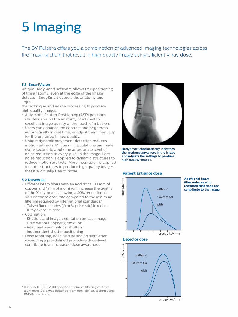
5.2 DoseWise• Efficient beam filters with an additional 0.1 mm of
copper and 1 mm of aluminum increase the quality of the X-ray beam, allowing a 40% reduction in skin entrance dose rate compared to the minimum filtering required by international standards.*
- Pulsed fluoro modes (1/2 or 1/4 pulse rate) to reduce X-ray exposure dose.
• Collimation: - Shutters and image orientation on Last Image
Hold without applying radiation - Real lead asymmetrical shutters - Independent shutter positioning• Dose reporting, dose display and an alert when
exceeding a pre-defined procedure dose-level contribute to an increased dose awareness
BodySmart automatically identifies the anatomy anywhere in the image and adjusts the settings to produce high quality images.
Additional beam filter reduces soft radiation that does not contribute to the image
Patient Entrance dose
Detector dose
5 Imaging
The BV Pulsera offers you a combination of advanced imaging technologies across
the imaging chain that result in high quality image using efficient X-ray dose.
* IEC 60601-2-43: 2010 specifies minimum filtering of 3 mm aluminum. Data was obtained from non-clinical testing using PMMA phantoms.
12 The print quality of this copy is not an accurate representation of the original.

5.3 Automatic Programmed Fluoroscopy
programsThe Automatic Programmed Fluoroscopy (APF) programs apply pre-set application-specific fluoroscopy settings to obtain a high quality image for the anatomy of interest.
Within each program there are different X-ray modes available (depending on region):• Low Dose Fluoroscopy• Fluoroscopy• High Definition Fluoroscopy• Pulsed exposure mode to produce high quality
images of virtually every patient• Digital exposure, for extra-sharp, single
snapshot images
5.4 Real time processing functions
Feature
360˚ digital rotation, mirror left/right and up/down without radiation
(Automatic) contrast and brightness
Dynamic noise reduction (Adaptive temporal recursive noise reduction)
Adaptive 2D edge enhancement
White compression
Image disk storage: 2,000/10,000/20,000 images
5.5 Post processing functions
Feature
360° digital rotation, mirror left/right and up/down
Contrast and brightness
Annotation (for a single image or all images in an examination)
Video invert
Zoom and roam (factor 2x real-time magnification, freely movable to any section of an image)
Measurement (to quantify lengths and angles in images)
Electronic shutters
Digital zoom that can be easily moved over the image
5.6 Mobile View Station monitorsTwo 19" Color LCD monitors for diagnostic imagequality display.
Feature Specification
Resolution 1280 x 1024 pixels
Contrast ratio >500:1>700:1 (optional 19" High Brightness monitor)
Viewing angle 170˚ in horizontal and vertical direction
Touchscreen (optional for left monitor)
Offers easy access to post-processing of acquired images, patient demographics as well as PACS
13The print quality of this copy is not an accurate representation of the original.

6 Options
Feature Specification
Tank laser aiming device Laser projects a crosshair towards the image intensifier, indicating the center of the X-ray beam and enabling alignment of the C-arm without X-ray
II laser aiming device Positioning device for use at the image intensifier side
Medical DVD Recorder Recording of static and dynamic live fluoroscopy on a DVD (up to 2 hours)
Video Paper printer Thermal printer to print video images from the Examination (left) monitor onto paper during or after examinations.
Video Paper/transparency printer Thermal printer to print video images from the Examination (left) monitor onto paper or transparencies during or after examinations.
Multi modality workstation(ViewForum option)
Intuitive multi-purpose platform for retrieving and handling images from different modalities. Allows comparison of pre-operative images side-by-side with live fluoroscopy images.
ViewForum options • MIP/MPR – maximum intensity projection singles out high intensity areas for optimized 2D projection of a 3D volume
• DVD DICOM Store – record DICOM images onto a DVD
Vascular package • Subtracted fluoroscopy mode• Trace-mode shows opacification of the vessels in real time• Roadmap images support catheter guidance• Remask lets you reselect the best image in a run as a new mask image• SmartMask uses previously acquired mask images for roadmapping to
support efficient use of X-ray dose and contrast medium• Landmarking provides a non-subtracted background for anatomical
reference• Real-time pixel shift compensates for movement artifacts• Subtraction on/off simplifies the orientation for subtracted images during
roadmap procedures• View Trace creates a trace image in post-processing• CO2 mode for subtraction, trace white and roadmap with SmartMask
Advanced Vascular package • Pulsed exposure at a maximum pulse rate of 15 pps, with max. 60 mA• All vascular package features
Ortho plus • Extra examination type to obtain low noise images in dense patients• 12.5 pps, with max. 60mA
Cardiac package • Three dedicated APF sets for cardiac procedures, advanced pacemaker placements, and electrophysiology
• Pulsed exposure at 8, 15 and 30 pps, with max. 60 mA
Stand monitor 12" LCD monitor on C-arm stand
14 The print quality of this copy is not an accurate representation of the original.

7 Geometry
The BV Pulsera consists of a mobile C-arm stand with monitor for image
acquisition and a Mobile View Station with two LCD monitors for image
processing, review, archiving and display.
BV Pulsera and MVS movements
* depending on configuration
7.1 C-arm stand
Feature Specification
Longitudinal movement 20 cm (7.9")
Panning movement (swivel) ± 10°
Vertical movement Motorized 49 cm (+43 cm / -6 cm) (19.3", +16.9", -2.4")
Rotation ± 180°, with safety stop at ± 135°
Angulation +90°, -25°
Extended angulation (optional) +90°, -45° for increased projection flexibility
Source to Image Distance 98 cm (38.7")
Free space within C-arm 77 cm (30.3")
C-arm depth 61 cm (24.0")
Lowest lateral working position 102 cm (40.2")
Brakes for all movements Yes, manual
Steering Rear wheel
Parallel movement Dedicated parallel movement via rear wheel control
Cable deflectors Yes
C-arm stand weight 9": 310 kg (683 lbs) – 12": 305 kg (672 lbs)
C-arm stand length 196 cm (77.2")
C-arm stand width 81 cm (31.9")
C-arm stand height 9": 173 cm (68.2") – 12": 182 cm (71.7")
7.2 Mobile View Station
Feature Specification
Mobile view station depth 70 cm (27.6")
Mobile view station width 94 cm (37.1")70 cm (27.6") monitors folded
Mobile view station height Max height is 185 cm or higher (72.8”)*
Weight (including options) 195 kg or lower (313 lbs)*
Monitor rotation 180°
Monitor height movement 23 cm (9")
15The print quality of this copy is not an accurate representation of the original.

8 Service
Services – a full lifecycle solutionThe success of your organization depends on people.Philips Services are designed with that in mind—creating healing environments, developing your staff , improving your organization’s performance, and increasing patient satisfaction. Rely on us. The resources, training, and support we off er, enable you to focus on what’s most important - your patients. Philips provides a full lifecycle solution designed around your patients, your people, and your organization. We help you succeed in every phase of system ownership, from planning to start-up, through peak usage and renewal.
PlanningUnderstand how and when the right equipment andservices contribute to enhanced patient care andeconomics.
Start-upMake the most of your system as quickly as possible.
Peak UsageExtract maximum utility out of your system day to day.
RenewalWe’ll help you make smart decisions on upgrading ortransitioning to a new system.
First-rate carePhilips global service network, our highly qualifi edservice engineers, the individual attention of our service technicians, and the international availability of spare parts combine to provide our seamless service support.
Count on us as your patients count on you
Staying on top of today’s complex and ever changing healthcare environment is
challenging enough. The last thing you need to be concerned with is keeping your care
systems up and running smoothly.
At Philips, we work as one with your teams. We share their dedication to solve issues
before they start and their drive to keep going day and night until the job is done. With
us taking care of your systems you can focus on what really matters – delivering better
care, to more people, at lower cost. Together we can create a healthier future.
16 The print quality of this copy is not an accurate representation of the original.

17The print quality of this copy is not an accurate representation of the original.

532
(20.9"
)
752
- 124
2 (2
9.6"
- 48
.9")
45° (option)
25°
90° 45
(17.
5")
770
(30.
3")
9":
1732
- 22
22 (6
8.2"
- 87
.5")
12":
1822
- 23
12 (7
1.7"
- 91
.0")
1016
(40.
0")
430
(16.
9")
672 (26.4")
607 (23.9")
1226 - 1426 (48.3" - 56.1")
803
(31.
6")
60 (2
.4")
118
(4.
6")
389 (15.3")
9": 297 (11.7")
176
(6.9
")
200 (7.9")
12": 385 (15.2")
1020
- 15
10 (4
0.2"
- 59
.4")2"
: 385
(15.
2")
8.1 C-arm stand
9 Dimensions
All dimensions are in millimeters (inches)
18 The print quality of this copy is not an accurate representation of the original.

Max
18
50 m
m (
72.8
") o
r h
igh
er*
594 mm (23.4")
943 mm or lower (37.1")*
701
mm
(27
.6")
max 281 mm (11.1") or lower*
max 702 mm (27.6) or lower*
min
16
50 m
m o
r lo
we
r (6
4.9
")*
104
8 m
m (
41.
3")
180°
90° 90°
8.2 Mobile View Station
* depending on configuration
19The print quality of this copy is not an accurate representation of the original.

© 2017 Koninklijke Philips N.V. All rights reserved. Specifications are subject to change without notice. Trademarks are the property of Koninklijke Philips N.V. (Royal Philips) or their respective owners.
4522 991 19161 * JAN 2017
How to reach usPlease visit www.philips.com/healthcarewww.philips.com/[email protected]
The print quality of this copy is not an accurate representation of the original.