Majd Michael, MD Richard W. McCallum, MD CASE …€¦ · Majd Michael, MD Richard W. McCallum, MD...
3
Volume 40 Number 2 June 2017 22 El Paso Physician Majd Michael, MD Richard W. McCallum, MD Continued on page 23 Abstract Chilaiditi syndrome is a rare entity in which inter position of bowel between the liver and right hemidiaphragm manifests with abdominal pain, nausea, vomiting, anorexia, and consti- pation that can be easily misdiagnosed as bowel perforationor intestinal obstruction. Most patients with radiologic evidence of hepatodiaphragmatic interposition of bowel remain a symp- tomatic (i.e., Chilaiditi sign). We report the case of a patient with Chilaiditi syndrome who suffered from acute on chronic abdominal pain accompanied by nausea and vomiting, and un- derwent exploratory laparoscopy based on a working diagnosis of small bowel obstruction. Case 50-year old Hispanic male presented to our hospital with severe 8/10 abdominal pain, localized to the right upper quadrant, and associated with nausea, vomiting and anorexia. His medical history included chronic kidney disease, rheumatoid arthritis, hypertension, and previous hospitalizations for unexplained abdominal pain. Initial evaluation included an abdominal ultra- sound which revealed cholelithiasis without evidence of chole- cystitis. This was followed by a CT of the abdomen without contrast that revealed dilated loops of small bowel, mostly of the jejunum and ileum, concerning for a small bowel obstruc- tion. Subsequently, the patient underwent an exploratory lap- aroscopy with a preoperative diagnosis of small bowel obstruc- tion. Laparoscopic exploration of the bowel did not reveal any obstruction or ischemia. However, the surgeons described seg- ments of dilated small bowel and evidence of increased peri- stalsis. Symptoms persisted after the exploratory laparoscopy and a new CT of the abdomen, and pelvis was obtained on post-operative day 5 that showed diffusely dilated colon without any evidence of mechanical obstruction suggesting colonic ileus or Ogilvie syndrome. In addition, a colonic loop at the hepatic flexure was interposed anterior to the liver and between the superior margin of the liver capsule and the diaphragm [figures 1-2]. This dis- placed colonic bowel loop is consistent with Chilaiditi sign, and explains the patient’s symptoms. Our patient was stable during ongoing observation post-operatively, but continued to have significant abdominal pain and food intolerance that recovered gradually and was able to be successfully discharged Discussion Chilaiditi sign is a rare radiologic finding in which hepatodia- phragmatic interposition of the colon or small bowel occurs. This sign was first described in 1910 by Demetrius Chilaiditi, a Greek radiologist who practiced in Vienna, Austria. He de- scribed three cases of patients with bowel interposition between the liver and diaphragm. 1 Chilaiditi sign is a radiographic term, as most patients with this anomaly will remain asymptomatic throughout their lives. Those who become symptomatic will develop Chilaiditi syndrome, which manifests with intermittent abdominal pain, distention, vomiting, anorexia, and constipa- tion that may require surgical intervention. 2 The ability to recognize Chilaiditi sign is crucial, as it is com- monly misinterpreted as free air under the diaphragm, pneu- moperitoneum, which is an indication for immediate surgical exploration based on a working diagnosis of a perforated vis- cus; e.g. peptic ulcer or colonic diverticulum. The prevalence of Chilaiditi sign is around 0.025 to 0.28% of general popula- tion, more prevalent in males than females, and the incidence increases with age. The bowel segments, most commonly found interposed between the liver and diaphragm or abdominal wall are the colonic hepatic flexure and transverse colon, although interposition of the small bowel has been reported. 3 Recog- nition of this condition becomes especially important when CASE REPORT
Transcript of Majd Michael, MD Richard W. McCallum, MD CASE …€¦ · Majd Michael, MD Richard W. McCallum, MD...

Volume 40 Number 2 � June 201722 El Paso Physician
Majd Michael, MD
Richard W. McCallum, MD
Continued on page 23
AbstractChilaiditi syndrome is a rare entity in which inter position of bowel between the liver and right hemidiaphragm manifests with abdominal pain, nausea, vomiting, anorexia, and consti-pation that can be easily misdiagnosed as bowel perforationor intestinal obstruction. Most patients with radiologic evidence of hepatodiaphragmatic interposition of bowel remain a symp-tomatic (i.e., Chilaiditi sign). We report the case of a patient with Chilaiditi syndrome who suffered from acute on chronic abdominal pain accompanied by nausea and vomiting, and un-derwent exploratory laparoscopy based on a working diagnosis of small bowel obstruction.
Case50-year old Hispanic male presented to our hospital with severe 8/10 abdominal pain, localized to the right upper quadrant, and associated with nausea, vomiting and anorexia. His medical history included chronic kidney disease, rheumatoid arthritis, hypertension, and previous hospitalizations for unexplained abdominal pain. Initial evaluation included an abdominal ultra-sound which revealed cholelithiasis without evidence of chole-cystitis. This was followed by a CT of the abdomen without contrast that revealed dilated loops of small bowel, mostly of the jejunum and ileum, concerning for a small bowel obstruc-tion. Subsequently, the patient underwent an exploratory lap-aroscopy with a preoperative diagnosis of small bowel obstruc-tion. Laparoscopic exploration of the bowel did not reveal any obstruction or ischemia. However, the surgeons described seg-ments of dilated small bowel and evidence of increased peri-stalsis.
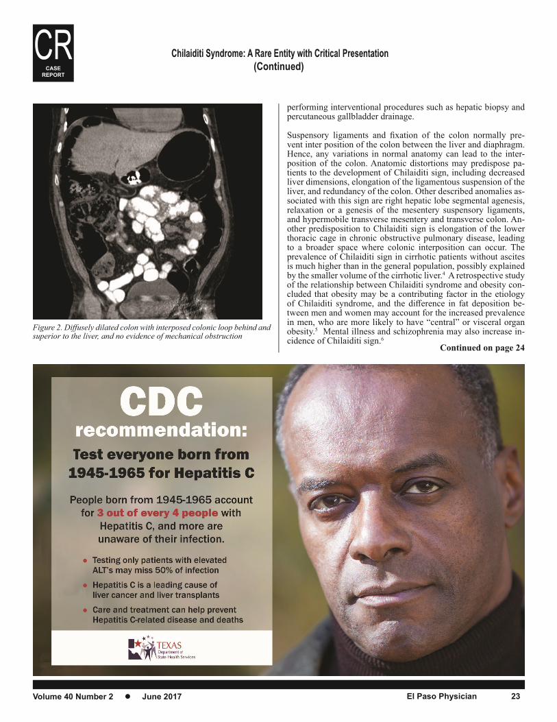
Symptoms persisted after the exploratory laparoscopy and a new CT of the abdomen, and pelvis was obtained on post-operative day 5 that showed diffusely dilated colon without any evidence of mechanical obstruction suggesting colonic ileus or Ogilvie syndrome. In addition, a colonic loop at the hepatic flexure was interposed anterior to the liver and between the superior margin of the liver capsule and the diaphragm [figures 1-2]. This dis-placed colonic bowel loop is consistent with Chilaiditi sign, and explains the patient’s symptoms. Our patient was stable during ongoing observation post-operatively, but continued to have significant abdominal pain and food intolerance that recovered gradually and was able to be successfully discharged
DiscussionChilaiditi sign is a rare radiologic finding in which hepatodia-phragmatic interposition of the colon or small bowel occurs.
This sign was first described in 1910 by Demetrius Chilaiditi, a Greek radiologist who practiced in Vienna, Austria. He de-scribed three cases of patients with bowel interposition between the liver and diaphragm.1 Chilaiditi sign is a radiographic term, as most patients with this anomaly will remain asymptomatic throughout their lives. Those who become symptomatic will develop Chilaiditi syndrome, which manifests with intermittent abdominal pain, distention, vomiting, anorexia, and constipa-tion that may require surgical intervention.2
The ability to recognize Chilaiditi sign is crucial, as it is com-monly misinterpreted as free air under the diaphragm, pneu-moperitoneum, which is an indication for immediate surgical exploration based on a working diagnosis of a perforated vis-cus; e.g. peptic ulcer or colonic diverticulum. The prevalence of Chilaiditi sign is around 0.025 to 0.28% of general popula-tion, more prevalent in males than females, and the incidence increases with age. The bowel segments, most commonly found interposed between the liver and diaphragm or abdominal wall are the colonic hepatic flexure and transverse colon, although interposition of the small bowel has been reported.3 Recog-nition of this condition becomes especially important when
CASE REPORT
Volume 40 Number 2 � June 2017 El Paso Physician 23
Continued on page 24
(Continued)
performing interventional procedures such as hepatic biopsy and percutaneous gallbladder drainage.
Suspensory ligaments and fixation of the colon normally pre-vent inter position of the colon between the liver and diaphragm. Hence, any variations in normal anatomy can lead to the inter-position of the colon. Anatomic distortions may predispose pa-tients to the development of Chilaiditi sign, including decreased liver dimensions, elongation of the ligamentous suspension of the liver, and redundancy of the colon. Other described anomalies as-sociated with this sign are right hepatic lobe segmental agenesis, relaxation or a genesis of the mesentery suspensory ligaments, and hypermobile transverse mesentery and transverse colon. An-other predisposition to Chilaiditi sign is elongation of the lower thoracic cage in chronic obstructive pulmonary disease, leading to a broader space where colonic interposition can occur. The prevalence of Chilaiditi sign in cirrhotic patients without ascites is much higher than in the general population, possibly explained by the smaller volume of the cirrhotic liver.4 A retrospective study of the relationship between Chilaiditi syndrome and obesity con-cluded that obesity may be a contributing factor in the etiology of Chilaiditi syndrome, and the difference in fat deposition be-tween men and women may account for the increased prevalence in men, who are more likely to have “central” or visceral organ obesity.5 Mental illness and schizophrenia may also increase in-cidence of Chilaiditi sign.6
Figure 2. Diffusely dilated colon with interposed colonic loop behind and superior to the liver, and no evidence of mechanical obstruction
CASE
REPORT
24 El Paso Physician
Chilaiditi sign has been associated with a variety of functional disorders, such as chronic constipation (colonic elongation and redundancy), aerophagia, cirrhosis, diaphragmatic paralysis, chronic lung disease, obesity, and multiple pregnancies.
Patients with Chilaiditi syndrome usually present with gastroin-testinal symptoms followed by respiratory distress, and less fre-quently angina-like chest pain. Gastrointestinal symptoms include anorexia, nausea, emesis, abdominal pain, distension, potentially with serious complications including volvulus and bowel perfora-tion.7 Our patient presented with these symptoms requiring hos-pitalization in the past for recurrent vomiting and abdominal pain episodes, which had no explanation. A working diagnosis of cy-clic vomiting syndrome was being considered. Intestinal pseudo-obstruction which can overlap with Oglivie syndrome, has also been described in a patient with Chilaiditi syndrome.8
Diagnosis of Chilaiditi syndrome is based upon clinical findings and signs observed on plain radiographs and CT scans. CT scans of the abdomen enables clinicians to exclude diaphragmatic her-nia and differentiate subphrenic fluid, true pneumoperitoneum, and air within the bowel lumen. This differentiation is of a criti-cal importance because perforation can also complicate Chilaiditi syndrome when the involved bowel segment strangulates, and eventually perforates. The radiologic differential diagnosis is es-tablished by observing an elevation of the right hemi-diaphragm due to caudal displacement of the liver, haustral markings between the liver and diaphragmatic surface, and the absence of image dis-placement with changes in the patient’s position. Pneumoperito-neum and subdiaphragmatic fluid collections have different char-acteristics being more mobile on lateral decubitus radiographs, and can be accompanied by pulmonary findings such as ipsilateral pleural effusion and basilar atelectasis.7,9
In most cases of Chilaiditi syndrome, management is conserva-tive and consists of bowel decompression, bowel rest, and ag-gressive fluid rehydration. Patients who fail conservative therapy should undergo an exploratory procedure. Colonoscopy should be performed with great caution considering the risk of gas being trapped in the acutely angulated and interposed bowel, potentially leading to perforation. Administration of carbon dioxide as the in-sufflating agent for colonoscopy is appropriate for decreasing this risk because it is nonexplosive, rapidly absorbed, and increases colon blood flow.10
REFERENCES1. Chilaiditi D. Zur frage der hepatoptose und ptose im allgemeinen im anschluss an drei fälle von temporärer, partieller leberverlagerung. . Fortschritte auf dem Gebiete der Röntgenstrahlen 1910;16:173-208.
2. Plorde JJ, Raker EJ. Transverse colon volvulus and associated Chi-laiditi’s syndrome: Case report and literature review. American Journal of Gastroenterology 1996;91:2613-6.
3. Mateo de Acosta Andino DA, Aberle CM, Ragauskaite L, et al. Chi-laiditi syndrome complicated by a closed-loop small bowel obstruction. Gastroenterol Hepatol (N Y) 2012;8:274-6.
4. Nakagawa H, Toda N, Taniguchi M, Ibukuro K, Tagawa K. Prevalence and Sonographic Detection of Chilaiditi’s Sign in Cirrhotic Patients Without Ascites. American Journal of Roentgenology 2006;187:w589-w93.
5. Murphy JM, Maibaum A, Alexander G, Dixon AK. Chilaiditi’s syn-drome and obesity. Clinical Anatomy 2000;13:181-4.
6. Lekkas CN, Lentino W. Symptom-producing interposition of the colon. Clinical syndrome in mentally deficient adults. JAMA 1978;240:747-50.
7. Moaven O, Hodin RA. Chilaiditi syndrome: a rare entity with impor-tant differential diagnoses. Gastroenterol Hepatol (N Y) 2012;8:276-8.
8. Suarez-Grau JM, Chaves CR, Bernal FL, Ciuro FP. [Colonic pseudo-colonic obstruction (Ogilvie syndrome) in a patient with Chilaiditi syn-drome]. Cir Esp 2011;89:e2.
9. Saber AA, Boros MJ. Chilaiditi’s syndrome: what should every surgeon know? . Am Surg 2005;71:261-3.
10. Gurvits GE, Lau N, Gualtieri N, Robilotti JG. Air under the right dia-phragm: colonoscopy in the setting of Chilaiditi syndrome. Gastrointest Endosc 2009;69:758-9; discussion 9.
(Continued)CASE
REPORT