Maintenance of host DNA integrity in field-preserved mosquito ...
11
RESEARCH Open Access Maintenance of host DNA integrity in field-preserved mosquito (Diptera: Culicidae) blood meals for identification by DNA barcoding Lawrence E. Reeves 1* , Chris J. Holderman 2 , Jennifer L. Gillett-Kaufman 1 , Akito Y. Kawahara 3 and Phillip E. Kaufman 1 Abstract Background: Determination of the interactions between hematophagous arthropods and their hosts is a necessary component to understanding the transmission dynamics of arthropod-vectored pathogens. Current molecular methods to identify hosts of blood-fed arthropods require the preservation of host DNA to serve as an amplification template. During transportation to the laboratory and storage prior to molecular analysis, genetic samples need to be protected from nucleases, and the degradation effects of hydrolysis, oxidation and radiation. Preservation of host DNA contained in field-collected blood-fed specimens has an additional caveat: suspension of the degradative effects of arthropod digestion on host DNA. Unless effective preservation methods are implemented promptly after blood-fed specimens are collected, host DNA will continue to degrade. Preservation methods vary in their efficacy, and need to be selected based on the logistical constraints of the research program. Methods: We compared four preservation methods (cold storage at -20 °C, desiccation, ethanol storage of intact mosquito specimens and crushed specimens on filter paper) for field storage of host DNA from blood-fed mosquitoes across a range of storage and post-feeding time periods. The efficacy of these techniques in maintaining host DNA integrity was evaluated using a polymerase chain reaction (PCR) to detect the presence of a sufficient concentration of intact host DNA templates for blood meal analysis. We applied a logistic regression model to assess the effects of preservation method, storage time and post-feeding time on the binomial response variable, amplification success. Results: Preservation method, storage time and post-feeding time all significantly impacted PCR amplification success. Filter papers and, to a lesser extent, 95 % ethanol, were the most effective methods for the maintenance of host DNA templates. Amplification success of host DNA preserved in cold storage at -20 °C and desiccation was poor. Conclusions: Our data suggest that, of the methods tested, host DNA template integrity was most stable when blood meals were preserved using filter papers. Filter paper preservation is effective over short- and long-term storage, while ethanol preservation is only suitable for short-term storage. Cold storage at -20 °C, and desiccation of blood meal specimens, even for short time periods, should be avoided. Keywords: Aedes aegypti, Blood meal preservation, Blood meal analysis, Blood meal identification, COI barcoding, Degraded DNA, DNA preservation, Host preference, Vector, Vector ecology * Correspondence: [email protected] 1 Entomology and Nematology Department, University of Florida, PO Box 110620, 1881 Natural Area Drive, Gainesville, FL 32611, USA Full list of author information is available at the end of the article © 2016 The Author(s). Open Access This article is distributed under the terms of the Creative Commons Attribution 4.0 International License (http://creativecommons.org/licenses/by/4.0/), which permits unrestricted use, distribution, and reproduction in any medium, provided you give appropriate credit to the original author(s) and the source, provide a link to the Creative Commons license, and indicate if changes were made. The Creative Commons Public Domain Dedication waiver (http://creativecommons.org/publicdomain/zero/1.0/) applies to the data made available in this article, unless otherwise stated. Reeves et al. Parasites & Vectors (2016) 9:503 DOI 10.1186/s13071-016-1791-z
Transcript of Maintenance of host DNA integrity in field-preserved mosquito ...

Reeves et al. Parasites & Vectors (2016) 9:503 DOI 10.1186/s13071-016-1791-z
RESEARCH Open Access
Maintenance of host DNA integrity infield-preserved mosquito (Diptera:Culicidae) blood meals for identificationby DNA barcoding
Lawrence E. Reeves1*, Chris J. Holderman2, Jennifer L. Gillett-Kaufman1, Akito Y. Kawahara3 and Phillip E. Kaufman1Abstract
Background: Determination of the interactions between hematophagous arthropods and their hosts is a necessarycomponent to understanding the transmission dynamics of arthropod-vectored pathogens. Current molecularmethods to identify hosts of blood-fed arthropods require the preservation of host DNA to serve as anamplification template. During transportation to the laboratory and storage prior to molecular analysis, geneticsamples need to be protected from nucleases, and the degradation effects of hydrolysis, oxidation and radiation.Preservation of host DNA contained in field-collected blood-fed specimens has an additional caveat: suspension ofthe degradative effects of arthropod digestion on host DNA. Unless effective preservation methods areimplemented promptly after blood-fed specimens are collected, host DNA will continue to degrade. Preservationmethods vary in their efficacy, and need to be selected based on the logistical constraints of the research program.
Methods: We compared four preservation methods (cold storage at -20 °C, desiccation, ethanol storage of intactmosquito specimens and crushed specimens on filter paper) for field storage of host DNA from blood-fedmosquitoes across a range of storage and post-feeding time periods. The efficacy of these techniques inmaintaining host DNA integrity was evaluated using a polymerase chain reaction (PCR) to detect the presence of asufficient concentration of intact host DNA templates for blood meal analysis. We applied a logistic regressionmodel to assess the effects of preservation method, storage time and post-feeding time on the binomial responsevariable, amplification success.
Results: Preservation method, storage time and post-feeding time all significantly impacted PCR amplificationsuccess. Filter papers and, to a lesser extent, 95 % ethanol, were the most effective methods for the maintenance ofhost DNA templates. Amplification success of host DNA preserved in cold storage at -20 °C and desiccation waspoor.
Conclusions: Our data suggest that, of the methods tested, host DNA template integrity was most stable whenblood meals were preserved using filter papers. Filter paper preservation is effective over short- and long-termstorage, while ethanol preservation is only suitable for short-term storage. Cold storage at -20 °C, and desiccation ofblood meal specimens, even for short time periods, should be avoided.
Keywords: Aedes aegypti, Blood meal preservation, Blood meal analysis, Blood meal identification, COI barcoding,Degraded DNA, DNA preservation, Host preference, Vector, Vector ecology
* Correspondence: [email protected] and Nematology Department, University of Florida, PO Box110620, 1881 Natural Area Drive, Gainesville, FL 32611, USAFull list of author information is available at the end of the article
© 2016 The Author(s). Open Access This article is distributed under the terms of the Creative Commons Attribution 4.0International License (http://creativecommons.org/licenses/by/4.0/), which permits unrestricted use, distribution, andreproduction in any medium, provided you give appropriate credit to the original author(s) and the source, provide a link tothe Creative Commons license, and indicate if changes were made. The Creative Commons Public Domain Dedication waiver(http://creativecommons.org/publicdomain/zero/1.0/) applies to the data made available in this article, unless otherwise stated.
Reeves et al. Parasites & Vectors (2016) 9:503 Page 2 of 11
BackgroundThe need to understand transmission system dynamicsin insect-vectored pathogens has generated substantialinterest in the taxonomic identification of vector bloodmeals [1]. Analysis of the blood meals of arthropods pro-vides data necessary to understanding the epidemiologyof arthropod-borne diseases. Techniques for blood mealidentification have existed since the early twentieth cen-tury [2]. Some of the first methods for assigning taxo-nomic identities to blood meals were serological (e.g.enzyme-linked immunosorbent assays, precipitin tests)[3, 4]. Serological and early DNA-based methods werelabor intensive, requiring the collection of serologic orgenetic reference samples from each potential host taxonfor laboratory comparison against blood meal specimenscollected from arthropods in the field [1]. Because ofthis, it is difficult to screen blood meal samples againstthe entire range of potential hosts using these methods,and in some cases, host identifications are limited tohigher taxonomic levels.The use of mitochondrial DNA (mtDNA) and riboso-
mal DNA (rDNA) sequences to determine host speciesin contemporary blood meal identification methods im-proves the ability to identify a broader range of hosts tolower taxonomic levels [1]. Comparisons to referencesamples remain necessary for taxonomic determinationsof hosts. The rapid growth of DNA barcoding initiatives,such as Barcode of Life Data Systems (BOLD) [5], allowsreference sample comparisons to be outsourced to pub-licly accessible sequence databases, without the need toindependently collect and compare them to blood mealsin the laboratory. In general, current methods arestraightforward, and involve the extraction of host DNAfrom an arthropod, a PCR to amplify a diagnostic hostDNA fragment, Sanger sequencing of the PCR product,and a database search for referenced sequences identicalor similar to the blood meal-derived sequence. Severalgenetic markers have been used to identify hosts, butmtDNA is well suited to analyses of degraded DNA.Mitochondrial DNA is present in a high number of cop-ies in many cells, is well conserved across animal taxa,and evolves rapidly, allowing even closely related taxa tobe distinguished from one another. Regions of the cyto-chrome c oxidase subunit I (cox1) and cytochrome b(cyt-b) genes often are used because of their broad taxo-nomic coverage in databases.DNA-based blood meal analyses require non-degraded
fragments of vertebrate host DNA to serve as a templatefor amplification. Two major issues affect host DNA in-tegrity and the subsequent success of PCR amplificationof host DNA from blood meals: the extent of digestion [6]and DNA degradation during the preservation periodprior to extraction [7]. Shortly after ingestion, imbibedblood meals are gradually digested and host DNA is
degraded over time. Field samples of blood-fed arthropodsconsist of individuals containing blood meals at variousstages of digestion [8]. Rates of DNA degradation due todigestion vary among hematophagous taxa. In mosquitoes,digestion proceeds quickly, with host DNA undetectableafter 36–72 h [9, 10]. A similar rate of host DNA degrad-ation is known from Chrysomya blowflies, with mamma-lian host DNA reliably detected up to 24 h after feeding,with detection dropping rapidly 48–96 h post-feeding[11]. In contrast, host DNA persists in leeches substan-tially longer, and can be amplified up to four months post-feeding [12]. Regardless of taxon, as post-feeding timeincreases, host DNA degrades and amplification becomesincreasingly difficult. Unless effective preservationmethods are implemented promptly after specimens arecollected, downstream amplification of template DNA canbe rendered impossible [13].When blood-fed arthropods are field collected, subse-
quent preservation, transport, and storage can adverselyaffect the integrity of template DNA molecules, in partthrough the action of endogenous nucleases [14]. Preser-vation methods that are appropriate to the logistical con-straints of a particular study program are required tooptimize the ability to determine the taxonomic identityof host DNA. A variety of blood meal preservationmethods have been used including chilling live arthropodsduring transport and prompt DNA extraction [9, 15], coldstorage or cryopreservation until DNA extraction [16–19],desiccation of whole arthropod specimens [20, 21], blot-ting blood meals on filter paper for transport and storage[22, 23] and preservation of whole specimens in ethanol[24, 25]. When logistical constraints allow, the optimalmethod of preserving arthropod DNA is cryopreservationat -80 °C [26]. However, field logistics, particularly thoseof remote field sites or locations with limited access tofreezers, can restrict the ability to utilize this method.Therefore, it is beneficial to identify preservation and stor-age methods appropriate to conducting blood meal identi-fication research far from the laboratory or in the absenceof access to ultra-low freezers.In this study, we assessed the ability of four mosquito
blood meal preservation methods to maintain templatehost DNA for subsequent analysis using the rapid andinexpensive HotSHOT DNA extraction method [27] inconjunction with a semi-nested primer set that select-ively amplifies a 758 bp fragment of the cox1 gene of awide range of vertebrate classes (Mammalia, Aves, Rep-tilia, Amphibia) [28]. Although primer sets are availablethat target shorter fragments of bovine or mammalianmtDNA [21], we selected these primers for their versa-tility in targeting a wide range of potential host classes.This primer selection was made under the assumptionthat these primers are a logical choice for host identifica-tion from field-collected samples. The success of PCR
Reeves et al. Parasites & Vectors (2016) 9:503 Page 3 of 11
amplification was used to assess the presence of suffi-cient concentrations of non-degraded template DNA formolecular analyses. The potential effects of storage timeand post-feeding time on amplification success were in-vestigated in our assessment of the performance of eachpreservation method.
MethodsWe experimentally compared the efficacy of four preser-vation methods (cold storage at -20 °C, silica desiccation,95 % ethanol and filter papers) on the integrity of tem-plate host DNA in preserved mosquito blood meals.Maintenance of host DNA integrity during preservationwas assessed by PCR amplification success. To evaluatethe effects of storage and post-feeding time we preservedblood meal specimens at controlled intervals of post-feeding time points (every 6 h, from 6 to 54 h post-feeding), and held preserved specimens for a range ofstorage times (7, 30, 90 and 180 days). Our experimentalprocedures were replicated four times. Additionally, toconfirm that the PCR worked properly with mosquitoblood meals, and to estimate baseline PCR amplificationsuccess at the zero-hour post-feeding and 0 days of stor-age time point, we killed 16 blood-fed mosquitoesimmediately following the two-hour feeding period. Fourspecimens were preserved using each preservationmethod, and DNA was extracted six hours after thefeeding period ended.
MosquitoesIn each replication, approximately 700 laboratory-rearedadult female Aedes aegypti (L.) (Diptera: Culicidae) (UFStrain, [29]) were removed from the colony maintained atthe University of Florida, Veterinary Entomology Labora-tory and held in a 30 × 30 × 30 cm wire-mesh cage, provi-sioned with one 14.8 ml vial of water with a paper wick,and one 7.4 ml vial of 10 % sugar solution with a cottonwick. All mosquitoes had eclosed approximately 7 daysprior to removal from the colony, and had not taken ablood meal. Thereafter, two cotton pads (11 × 7 cm) weresoaked in bovine blood until saturated and placed on topof the holding cage allowing mosquitoes to feed. After 2 h,individual mosquitoes were visually inspected for the pres-ence of a blood meal. Approximately 200 blood-fed femalemosquitoes were removed by aspiration, transferred to anidentical cage and stored at 25 °C until preservation.
Blood meal preservationWithin each replication, we preserved a sample of 144blood-fed mosquitoes. Mosquitoes were preserved acrossnine serial time points, separated by six-hour intervals,from 6 to 54 h post-feeding. At each post-feeding timepoint, 16 mosquitoes were removed from the cage, andkilled by exposure to ethyl acetate-soaked plaster in a
473 ml glass jar for 10 min. Of these, four individualswere preserved per preservation method describedbelow, and placed into storage. From these four (perpost-feeding time point), DNA was extracted from onemosquito blood meal at 7, 30, 90 and 180 days of stor-age. Altogether, this yielded a total sample of 576 mos-quito blood meals (four preservation methods × 9 post-feeding time points × 4 storage times × 4 replications).
DesiccationPrior to preservation, 1.5 ml graduated microcentrifugetubes were prepared by filling to the 1 ml mark with sil-ica beads (3.5 mm in diameter) (Consolidated Chemicaland Solvents LLC, Quakertown, USA). A 3.5 × 3.5 cmsquare of crumpled Kimwipe® tissue paper was insertedabove the silica beads to prevent the mosquito specimenfrom directly contacting the silica. Specimens were pre-served individually by placing the whole specimen ontop of the tissue with sterile forceps. Tubes containingspecimens were sealed for storage.
Cold storageWhole mosquito specimens were placed individuallyinto 1.5 ml microcentrifuge tubes. Within 10 min afterdeath, specimens were transferred to, and stored in a-20 °C non-frost-free freezer until DNA extraction.
EthanolWhole mosquito specimens were transferred to 1.5 mlmicrocentrifuge tubes using sterile forceps. One ml of95 % ethanol was added to each tube, and the tubes weresealed.
Filter paperUsing sterile forceps, mosquito specimens were individu-ally transferred to the sampling area of a four-sampleFlinders Technology Associates (FTA) card (Whatman®,Maidstone, United Kingdom). One mosquito was placedin each sampling area. The blood meal was released ontothe sampling area by applying pressure with a sterile,plastic pestle. The pestle was then used to spread theblood meal around the sampling area until the cardabsorbed all viscous droplets. The card was air dried for5 min before storage.
StorageBlood meal specimens preserved by desiccation, ethanoland filter paper were placed inside a cardboard box andstored inside an incubator at 30 °C and 80 % relative hu-midity. These settings were selected to simulate the con-ditions of field sites located in tropical or subtropicalregions during the warmer months, under the assump-tion that preservation of specimens for molecular studiesis most challenging under similar field conditions.
Reeves et al. Parasites & Vectors (2016) 9:503 Page 4 of 11
DNA extractionDNA was extracted from the blood meal specimens fol-lowing the HotSHOT protocol [27]. Whole mosquitospecimens preserved by desiccation, cold storage andethanol were removed from storage and transferred tonew 1.5 ml microcentrifuge tubes using sterile forceps.Seventy-five μl of lysis solution consisting of 25 mMNaOH and 0.2 mM EDTA were added to each tube. Theabdomen of each specimen was individually maceratedusing a sterile pestle, releasing the blood meal into thesolution. To avoid the release of endonucleases con-tained in the eyes, maceration of the thorax and headwas avoided, and both were removed after the release ofthe blood meal. The lysis solution, containing homoge-nized blood meals, was transferred to 0.2 ml eight-wellPCR strips.Specimens preserved on filter paper were removed
from storage and a 1 mm hole punch was used to re-move 2 × 1 mm punch samples from the area containingthe specimen residue. Using sterile forceps, the 2 punchsamples per specimen were transferred to a 0.2 ml tube,and 75 μl of lysis solution was added to each well.The eight-well strips, containing lysis solution and
blood meals from all preservation methods, were incu-bated in a DNA Engine thermocycler (BioRad®, Hercules,USA) at 95 °C for 30 min followed by 4 °C for 5 min.Seventy-five μl of neutralization buffer consisting of 40mM tris-HCL was added to each tube. The tubes werebriefly vortexed and immediately frozen at -20 °C untilPCR amplification. Polymerase chain reactions were per-formed within ten days of DNA extraction, and eachsample underwent one freeze-thaw cycle (samples werefrozen immediately after DNA extraction, and thawedprior to PCR).
PCRThe presence of a sufficient concentration of non-degraded template DNA for PCR amplification was de-termined through a semi-nested PCR using primer pairsdesigned by Alcaide et al. [28]. The expected product ofthis reaction is a 758 bp fragment of the vertebrate cox1gene. The degenerated primers M13BCV-FW (5′-TGTAAA ACG ACG GCC AGT HAA YCA YAA RGA YATYGG-3′) and BCV-RV1 (5′-GCY CAN ACY ATN CCYATR TA-3′) were used in the first reaction. BCV-FW1contains a M13 tail at the 5′ end to facilitate the second,semi-nested amplification reaction and sequencing. TheM13 primer (5′-GTA AAA CGA CGG CCA CTG-3′)and the reverse primer BCV-RV2 (5′-ACY ATN CCYATR TAN CCR AAN GG-3′) were used in the semi-nested reaction. Although primer pairs targeting shorteramplicons are preferred in blood meal analyses, the Alcaide[28] sets were used because they putatively amplify DNAtemplates from a wide range of vertebrate groups.
A BioRad® DNA Engine thermocycler was used in allreactions. Our semi-nested reactions followed a protocolslightly modified from Alcaide et al. [28]. The first reac-tion was performed in a final volume of 15 μl that con-tained 1 U of Taq polymerase (Sigma-Aldrich, St. Louis,USA), 1 × PCR buffer (Sigma-Aldrich), 2.5 mM MgCl2,0.25 mM of each dNTP, 5 % DMSO, 10 μg of bovineserum albumin, 0.13 μM of each primer (M13BCV-FW1, BCV-RV1), and 1 μl of DNA extract. The firstamplification reaction consisted of an initial denaturationstep of 94 °C for 4 min, followed by 35 cycles of 45 °C for40 s, 72 °C for 1 min and 94 °C for 4 min, with a final ex-tension step of 72 °C for 7 min. The second reaction wasperformed in a final volume of 15 μl that contained 1 unitof Taq polymerase (Sigma-Aldrich), 1× PCR buffer, 1.7mM MgCl2, 0.25 mM of each dNTP, 5 % DMSO, 5 μg bo-vine serum albumin, 0.16 μM of each primer (M13, BCV-RV2), and 0.5 μl of PCR product from the previous reac-tion. The second reaction consisted of an initial denatur-ation step of 94 °C for 3 min, followed by 16 cycles of atouchdown program of 60 °C to 45 °C for 40 s, 72 °C for 1min, and 94 °C for 40 s. During these 16 cycles, the anneal-ing temperature began at 60 °C and was decreased by 1 °Cper cycle to 45 °C on the 16th cycle. The touchdown cycleswere followed by 24 cycles of 94 °C for 40 s, 45 °C for 40 sand 72 °C for 40 s, with a final extension step of 72 °C for7 min. Negative controls containing no DNA were used inevery set of reactions to monitor for contamination.The products of the semi-nested reaction were electro-
phoresed and visualized on an ethidium bromide-stained1.5 % agarose gel. The presence of a band at the ex-pected amplicon size was interpreted as a positive result,indicating sufficient host DNA preservation for bloodmeal identification. We verified amplification of the cor-rect vertebrate template using Sanger sequencing on anABI 3130® automated sequencer at the University ofFlorida Interdisciplinary Center for Biotechnology Re-search (ICBR). A subset of PCR products from success-ful amplification reactions were sequenced, and theresulting sequences were searched in the National Cen-ter for Biotechnology Information (NCBI) sequencedatabase using the Standard Nucleotide Basic LocalAlignment Search Tool.
Statistical analysisData were analyzed in the statistical program R® Version3.2.0 using the stats, lsmeans, and multcompView pack-ages [30]. We modeled the effects of the independentvariables, preservation method, storage time and post-feeding time, on the dependent variable, amplificationsuccess, using a multivariate analysis, binomial logisticregression. Because direct comparisons of independentvariable coefficients can be misleading in logistic regres-sion models [31], we used equality tests of predicted

Reeves et al. Parasites & Vectors (2016) 9:503 Page 5 of 11
probabilities to identify differences in amplification suc-cess [32]. We applied this approach to our data to testfor amplification success differences among the fourpreservation methods with the continuous variables heldat a range of constant values.An initial model included all independent variables
and their interactions. Using a backward stepwise elim-ination process, we selected the terms of the final modelthat best explained data variability based on the Akaikeinformation criterion [33]. Model fit was evaluated withthe Hosmer-Lemeshow goodness-of-fit test, and the like-lihood ratio test. We evaluated the significance of theoverall effect of each variable coefficient in the modelusing the Wald Chi-squared statistic. Least squaresmeans (LS means) were used to conduct pairwise com-parisons of the predicted probability of amplification foreach preservation method. Because amplification successvaries with the levels of the continuous variables, pair-wise comparisons were made with storage time andpost-feeding time held at constant values. For this ana-lysis, we selected 7, 77 and 180 days, and 6, 30 and 54 has constant values of storage time and post-feeding time,
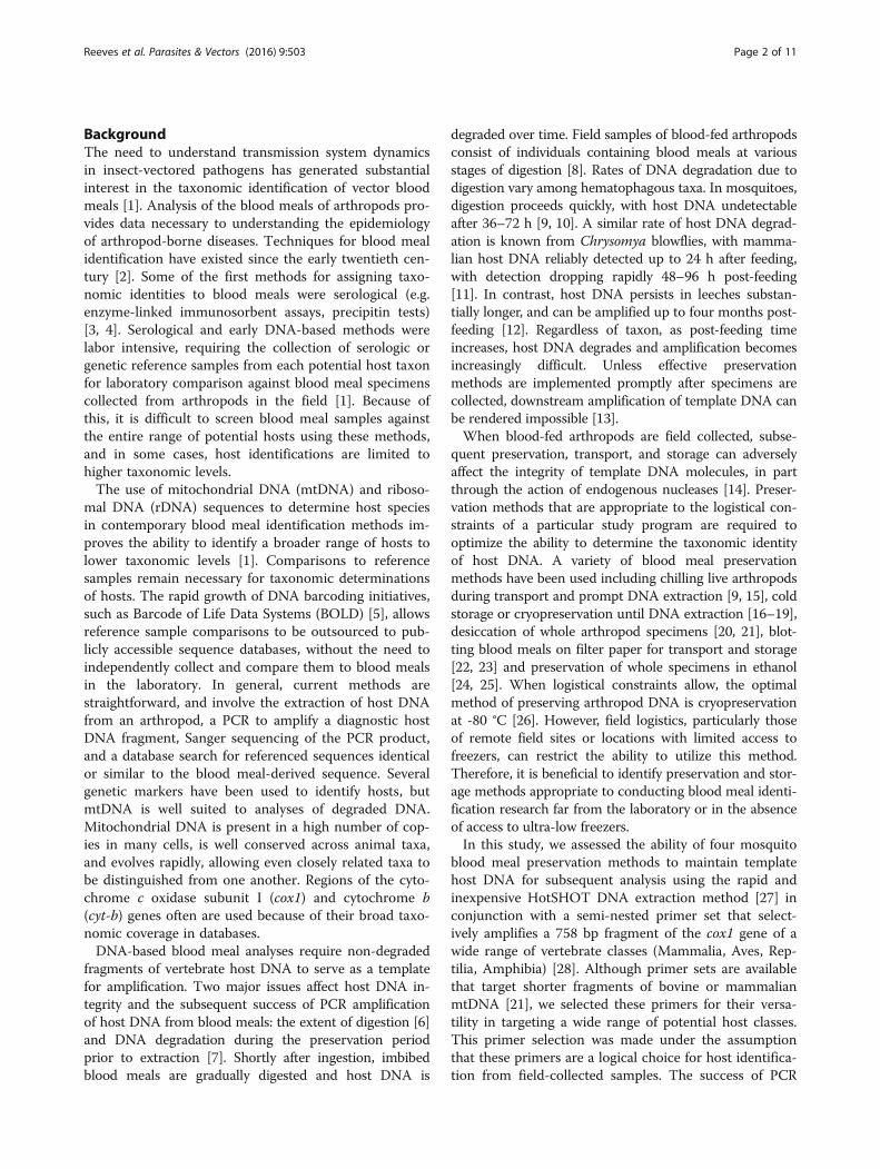
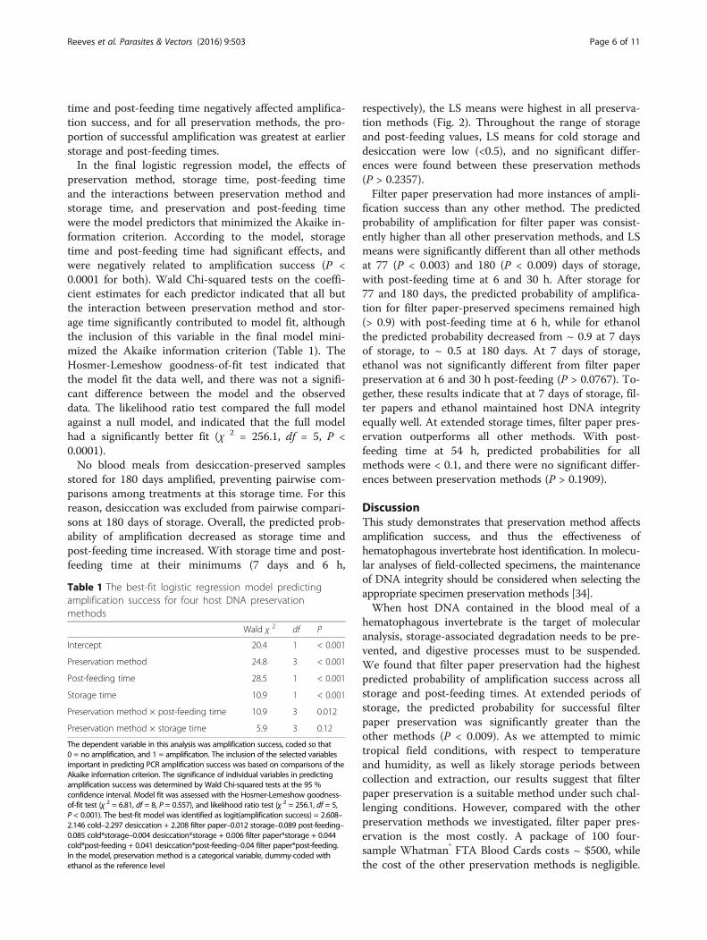
Fig. 1 The observed proportion of amplification success by preservation metspecimens were stored for seven days (a), 30 days (b), 90 days (c) and 180 datreatment combination, a total of four blood meal specimens were tested. Thfor each treatment across storage times (n = 16 for each preservation methodobserved proportion of amplification success for each treatment, with blue in
respectively, to represent brief, intermediate and ex-tended values of each variable. Pairwise comparisonswere made on the LS means of the predicted probabilityat all combinations of these values, and all comparisonswere made at the 95 % confidence level.
ResultsWe verified that the PCR procedure amplified the cor-rect DNA template, and amplification success was 100 %for blood meal specimens preserved at and stored for 0h post-feeding. All sequenced PCR products containedDNA fragments that matched the cox1 sequence of theexpected host species, with > 99 % similarity to data-based Bos taurus cox1 sequences.Host DNA templates were amplified from specimens
preserved by each of the 4 preservation methods.Altogether, 159 of the 576 blood meal specimens con-tained host DNA that could be detected by our PCR.Amplification success was greatest in specimens pre-served on filter papers, and in ethanol (Fig. 1). Amplifi-cation success was poor for specimens preservedthrough cold storage, and desiccation. Overall, storage
hod at each storage time (a–e), and post-feeding time. Blood mealys (d). For each preservation method, storage time and post feeding timee bottom sub-figure (e) presents the proportion of amplification successand post-feeding time combination). Cell shading corresponds with the
dicating a proportion of 1.0, and white indicating zero
Reeves et al. Parasites & Vectors (2016) 9:503 Page 6 of 11
time and post-feeding time negatively affected amplifica-tion success, and for all preservation methods, the pro-portion of successful amplification was greatest at earlierstorage and post-feeding times.In the final logistic regression model, the effects of
preservation method, storage time, post-feeding timeand the interactions between preservation method andstorage time, and preservation and post-feeding timewere the model predictors that minimized the Akaike in-formation criterion. According to the model, storagetime and post-feeding time had significant effects, andwere negatively related to amplification success (P <0.0001 for both). Wald Chi-squared tests on the coeffi-cient estimates for each predictor indicated that all butthe interaction between preservation method and stor-age time significantly contributed to model fit, althoughthe inclusion of this variable in the final model mini-mized the Akaike information criterion (Table 1). TheHosmer-Lemeshow goodness-of-fit test indicated thatthe model fit the data well, and there was not a signifi-cant difference between the model and the observeddata. The likelihood ratio test compared the full modelagainst a null model, and indicated that the full modelhad a significantly better fit (χ 2 = 256.1, df = 5, P <0.0001).No blood meals from desiccation-preserved samples
stored for 180 days amplified, preventing pairwise com-parisons among treatments at this storage time. For thisreason, desiccation was excluded from pairwise compari-sons at 180 days of storage. Overall, the predicted prob-ability of amplification decreased as storage time andpost-feeding time increased. With storage time and post-feeding time at their minimums (7 days and 6 h,
Table 1 The best-fit logistic regression model predictingamplification success for four host DNA preservationmethods
Wald χ 2 df P
Intercept 20.4 1 < 0.001
Preservation method 24.8 3 < 0.001
Post-feeding time 28.5 1 < 0.001
Storage time 10.9 1 < 0.001
Preservation method × post-feeding time 10.9 3 0.012
Preservation method × storage time 5.9 3 0.12
The dependent variable in this analysis was amplification success, coded so that0 = no amplification, and 1 = amplification. The inclusion of the selected variablesimportant in predicting PCR amplification success was based on comparisons of theAkaike information criterion. The significance of individual variables in predictingamplification success was determined by Wald Chi-squared tests at the 95 %confidence interval. Model fit was assessed with the Hosmer-Lemeshow goodness-of-fit test (χ 2 = 6.81, df = 8, P = 0.557), and likelihood ratio test (χ 2 = 256.1, df = 5,P < 0.001). The best-fit model was identified as logit(amplification success) = 2.608–2.146 cold–2.297 desiccation + 2.208 filter paper–0.012 storage–0.089 post-feeding–0.085 cold*storage–0.004 desiccation*storage + 0.006 filter paper*storage + 0.044cold*post-feeding + 0.041 desiccation*post-feeding–0.04 filter paper*post-feeding.In the model, preservation method is a categorical variable, dummy-coded withethanol as the reference level
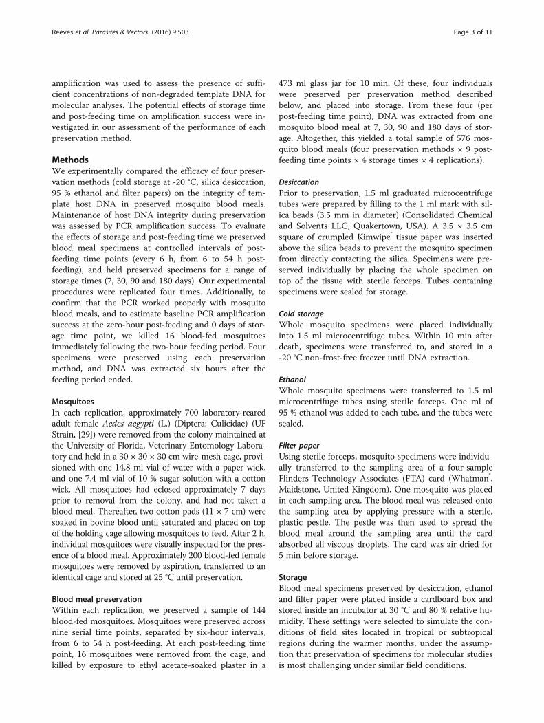
respectively), the LS means were highest in all preserva-tion methods (Fig. 2). Throughout the range of storageand post-feeding values, LS means for cold storage anddesiccation were low (<0.5), and no significant differ-ences were found between these preservation methods(P > 0.2357).Filter paper preservation had more instances of ampli-
fication success than any other method. The predictedprobability of amplification for filter paper was consist-ently higher than all other preservation methods, and LSmeans were significantly different than all other methodsat 77 (P < 0.003) and 180 (P < 0.009) days of storage,with post-feeding time at 6 and 30 h. After storage for77 and 180 days, the predicted probability of amplifica-tion for filter paper-preserved specimens remained high(> 0.9) with post-feeding time at 6 h, while for ethanolthe predicted probability decreased from ~ 0.9 at 7 daysof storage, to ~ 0.5 at 180 days. At 7 days of storage,ethanol was not significantly different from filter paperpreservation at 6 and 30 h post-feeding (P > 0.0767). To-gether, these results indicate that at 7 days of storage, fil-ter papers and ethanol maintained host DNA integrityequally well. At extended storage times, filter paper pres-ervation outperforms all other methods. With post-feeding time at 54 h, predicted probabilities for allmethods were < 0.1, and there were no significant differ-ences between preservation methods (P > 0.1909).
DiscussionThis study demonstrates that preservation method affectsamplification success, and thus the effectiveness ofhematophagous invertebrate host identification. In molecu-lar analyses of field-collected specimens, the maintenanceof DNA integrity should be considered when selecting theappropriate specimen preservation methods [34].When host DNA contained in the blood meal of a
hematophagous invertebrate is the target of molecularanalysis, storage-associated degradation needs to be pre-vented, and digestive processes must to be suspended.We found that filter paper preservation had the highestpredicted probability of amplification success across allstorage and post-feeding times. At extended periods ofstorage, the predicted probability for successful filterpaper preservation was significantly greater than theother methods (P < 0.009). As we attempted to mimictropical field conditions, with respect to temperatureand humidity, as well as likely storage periods betweencollection and extraction, our results suggest that filterpaper preservation is a suitable method under such chal-lenging conditions. However, compared with the otherpreservation methods we investigated, filter paper pres-ervation is the most costly. A package of 100 four-sample Whatman® FTA Blood Cards costs ~ $500, whilethe cost of the other preservation methods is negligible.
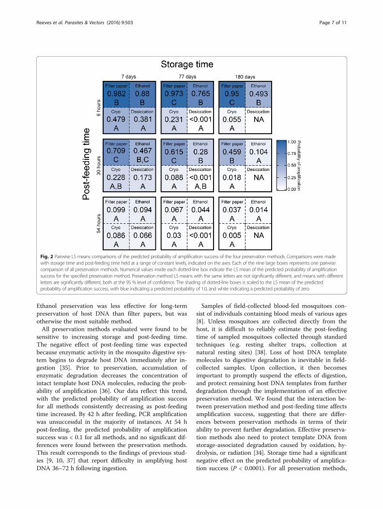
Fig. 2 Pairwise LS means comparisons of the predicted probability of amplification success of the four preservation methods. Comparisons were madewith storage time and post-feeding time held at a range of constant levels, indicated on the axes. Each of the nine large boxes represents one pairwisecomparison of all preservation methods. Numerical values inside each dotted-line box indicate the LS mean of the predicted probability of amplificationsuccess for the specified preservation method. Preservation method LS means with the same letters are not significantly different, and means with differentletters are significantly different, both at the 95 % level of confidence. The shading of dotted-line boxes is scaled to the LS mean of the predictedprobability of amplification success, with blue indicating a predicted probability of 1.0, and white indicating a predicted probability of zero
Reeves et al. Parasites & Vectors (2016) 9:503 Page 7 of 11
Ethanol preservation was less effective for long-termpreservation of host DNA than filter papers, but wasotherwise the most suitable method.All preservation methods evaluated were found to be
sensitive to increasing storage and post-feeding time.The negative effect of post-feeding time was expectedbecause enzymatic activity in the mosquito digestive sys-tem begins to degrade host DNA immediately after in-gestion [35]. Prior to preservation, accumulation ofenzymatic degradation decreases the concentration ofintact template host DNA molecules, reducing the prob-ability of amplification [36]. Our data reflect this trend,with the predicted probability of amplification successfor all methods consistently decreasing as post-feedingtime increased. By 42 h after feeding, PCR amplificationwas unsuccessful in the majority of instances. At 54 hpost-feeding, the predicted probability of amplificationsuccess was < 0.1 for all methods, and no significant dif-ferences were found between the preservation methods.This result corresponds to the findings of previous stud-ies [9, 10, 37] that report difficulty in amplifying hostDNA 36–72 h following ingestion.
Samples of field-collected blood-fed mosquitoes con-sist of individuals containing blood meals of various ages[8]. Unless mosquitoes are collected directly from thehost, it is difficult to reliably estimate the post-feedingtime of sampled mosquitoes collected through standardtechniques (e.g. resting shelter traps, collection atnatural resting sites) [38]. Loss of host DNA templatemolecules to digestive degradation is inevitable in field-collected samples. Upon collection, it then becomesimportant to promptly suspend the effects of digestion,and protect remaining host DNA templates from furtherdegradation through the implementation of an effectivepreservation method. We found that the interaction be-tween preservation method and post-feeding time affectsamplification success, suggesting that there are differ-ences between preservation methods in terms of theirability to prevent further degradation. Effective preserva-tion methods also need to protect template DNA fromstorage-associated degradation caused by oxidation, hy-drolysis, or radiation [34]. Storage time had a significantnegative effect on the predicted probability of amplifica-tion success (P < 0.0001). For all preservation methods,
Reeves et al. Parasites & Vectors (2016) 9:503 Page 8 of 11
predicted probability of amplification decreased with in-creasing storage time, suggesting that none of themethods entirely protected host DNA integrity.Investigations of the host-use of mosquitoes and other
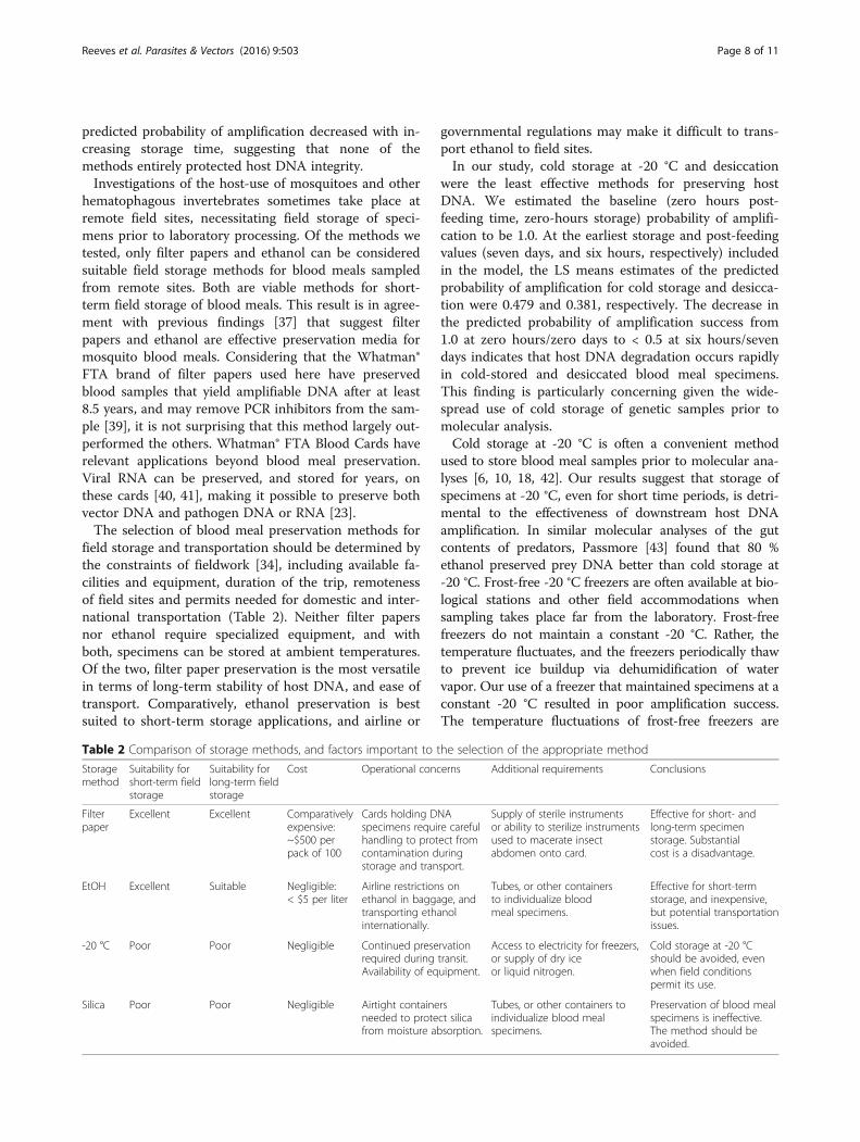
hematophagous invertebrates sometimes take place atremote field sites, necessitating field storage of speci-mens prior to laboratory processing. Of the methods wetested, only filter papers and ethanol can be consideredsuitable field storage methods for blood meals sampledfrom remote sites. Both are viable methods for short-term field storage of blood meals. This result is in agree-ment with previous findings [37] that suggest filterpapers and ethanol are effective preservation media formosquito blood meals. Considering that the Whatman®FTA brand of filter papers used here have preservedblood samples that yield amplifiable DNA after at least8.5 years, and may remove PCR inhibitors from the sam-ple [39], it is not surprising that this method largely out-performed the others. Whatman® FTA Blood Cards haverelevant applications beyond blood meal preservation.Viral RNA can be preserved, and stored for years, onthese cards [40, 41], making it possible to preserve bothvector DNA and pathogen DNA or RNA [23].The selection of blood meal preservation methods for
field storage and transportation should be determined bythe constraints of fieldwork [34], including available fa-cilities and equipment, duration of the trip, remotenessof field sites and permits needed for domestic and inter-national transportation (Table 2). Neither filter papersnor ethanol require specialized equipment, and withboth, specimens can be stored at ambient temperatures.Of the two, filter paper preservation is the most versatilein terms of long-term stability of host DNA, and ease oftransport. Comparatively, ethanol preservation is bestsuited to short-term storage applications, and airline or
Table 2 Comparison of storage methods, and factors important to t
Storagemethod
Suitability forshort-term fieldstorage
Suitability forlong-term fieldstorage
Cost Operational conc
Filterpaper
Excellent Excellent Comparativelyexpensive:~$500 perpack of 100
Cards holding Dspecimens requihandling to protcontamination dstorage and tran
EtOH Excellent Suitable Negligible:< $5 per liter
Airline restrictionethanol in baggatransporting ethinternationally.
-20 °C Poor Poor Negligible Continued preserequired duringAvailability of eq
Silica Poor Poor Negligible Airtight containeneeded to protefrom moisture ab
governmental regulations may make it difficult to trans-port ethanol to field sites.In our study, cold storage at -20 °C and desiccation
were the least effective methods for preserving hostDNA. We estimated the baseline (zero hours post-feeding time, zero-hours storage) probability of amplifi-cation to be 1.0. At the earliest storage and post-feedingvalues (seven days, and six hours, respectively) includedin the model, the LS means estimates of the predictedprobability of amplification for cold storage and desicca-tion were 0.479 and 0.381, respectively. The decrease inthe predicted probability of amplification success from1.0 at zero hours/zero days to < 0.5 at six hours/sevendays indicates that host DNA degradation occurs rapidlyin cold-stored and desiccated blood meal specimens.This finding is particularly concerning given the wide-spread use of cold storage of genetic samples prior tomolecular analysis.Cold storage at -20 °C is often a convenient method
used to store blood meal samples prior to molecular ana-lyses [6, 10, 18, 42]. Our results suggest that storage ofspecimens at -20 °C, even for short time periods, is detri-mental to the effectiveness of downstream host DNAamplification. In similar molecular analyses of the gutcontents of predators, Passmore [43] found that 80 %ethanol preserved prey DNA better than cold storage at-20 °C. Frost-free -20 °C freezers are often available at bio-logical stations and other field accommodations whensampling takes place far from the laboratory. Frost-freefreezers do not maintain a constant -20 °C. Rather, thetemperature fluctuates, and the freezers periodically thawto prevent ice buildup via dehumidification of watervapor. Our use of a freezer that maintained specimens at aconstant -20 °C resulted in poor amplification success.The temperature fluctuations of frost-free freezers are
he selection of the appropriate method
erns Additional requirements Conclusions
NAre carefulect fromuringsport.
Supply of sterile instrumentsor ability to sterilize instrumentsused to macerate insectabdomen onto card.
Effective for short- andlong-term specimenstorage. Substantialcost is a disadvantage.
s onge, andanol
Tubes, or other containersto individualize bloodmeal specimens.
Effective for short-termstorage, and inexpensive,but potential transportationissues.
rvationtransit.uipment.
Access to electricity for freezers,or supply of dry iceor liquid nitrogen.
Cold storage at -20 °Cshould be avoided, evenwhen field conditionspermit its use.
rsct silicasorption.
Tubes, or other containers toindividualize blood mealspecimens.
Preservation of blood mealspecimens is ineffective.The method should beavoided.
Reeves et al. Parasites & Vectors (2016) 9:503 Page 9 of 11
believed to accelerate DNA degradation [44]. When avail-able, the use of -20 °C freezers at field sites presents fur-ther challenges in that the temperature needs to bemaintained during transport back to the laboratory. Al-though blood meal specimens are typically held at -70 or-80 °C in the laboratory, such equipment is often unavail-able in remote field sites, leaving researchers in search ofmethods that effectively preserve DNA at ambienttemperatures.Desiccation of blood meal specimens does not require
cold temperatures, but our results suggest preservation ofmosquito blood meals through desiccation can be problem-atic. This result aligns with a previous study that reportedPCR amplification to be rare in silica-preserved mosquitoblood meal specimens [20]. However, Kent and Norris [21],and Logue et al. [45] successfully used silica to preservemosquito blood meals collected from remote field sites.Kent and Norris [21] used a species-specific multiplexedPCR that amplifies a 132–680 bp fragment, depending onhost species, of the mammalian cyt-b gene. Logue et al. [45]used high throughput (Illumina) sequencing of a 140 bpfragment of the mammalian 16S ribosomal RNA gene.High throughput sequencing and primer sets targetingshorter amplicons are well suited for analyses of degradedhost DNA. The use of such molecular methods that arebetter suited to degraded DNA templates may be compat-ible with desiccation-preserved blood meals. However, theresults presented here suggest that preservation of bloodmeals by desiccation should be avoided. Sanger sequencingand primers targeting longer template fragments are used.In addition, desiccation by drying insects with silica is suc-cessfully used to preserve insect DNA for molecular ana-lyses [46, 47]. We suspect that when host DNA, rather thaninsect DNA, is the target of molecular analysis, desiccationof blood-fed mosquito specimens with silica gel is not ac-complished quickly enough to adequately block the actionof digestive enzymes.In addition to the variables we tested, amplification
success of host DNA can be affected by the method usedto kill invertebrates [48], the method used to extractDNA [26, 34] and the length of the amplicon [22, 49].Martinez-de la Puente et al. [36] found amplificationsuccess improved by 17 % when DNA was extractedusing commercial Qiagen® DNA extraction kits as com-pared with the HotSHOT DNA extraction method [27].However, the cost per sample of using commercial DNAextraction kits is substantial in comparison to the Hot-SHOT method. Amplicon length also can affect theprobability of amplification success.Molecular analyses that require the amplification of
highly degraded DNA (e.g. environmental DNA, ancientDNA, DNA from old museum specimens) often targetshort amplicons 80–250 bp in length [50]. The numberof strand breaks in individual DNA molecules increases
with time, resulting in DNA degradation, thereby de-creasing the number of copies of intact DNA moleculesavailable to serve as templates. As a result, intact copiesof 100 bp DNA templates are likely to persist longerthan 1,000 bp DNA templates, and amplification is morelikely to be successful with shorter amplicons [51]. Cur-rently, no barcoding primer sets have been published forshorter amplicons in the ~100 bp size range that target adiverse set of vertebrate classes, while excluding amplifi-cation of insect templates.Universal barcoding primers are available to amplify
shorter fragments, however these co-amplify inverte-brate and host DNA. Such an approach requires highthroughput sequencing, coupled with a bioinformaticsplatform to parse amplified invertebrate DNA fragmentsfrom those of hosts. Host identification through highthroughput sequencing can be advantageous because theuse of universal primer pairs targeting shorter templatesmakes the method more sensitive to degraded DNA,and by making identification of mixed blood meals, de-rived from more than one host, tractable. These resultsshould be equally applicable to research that uses a highthroughput sequencing approach.
ConclusionsIn this study we demonstrate that preservation methodaffects the success of PCR amplification of host DNA con-tained in mosquito blood meals. When sampling mos-quito populations that are located in close proximity tothe laboratory, preservation of blood meal specimens isrelatively straightforward. When blood-fed invertebratesare collected from remote locations, maintenance of hostDNA integrity during field storage can be problematic.Here, we show that all the preservation methods we testedwere sensitive to storage time and post-feeding time. It isimpossible to correct DNA degradation due to digestionthrough a preservative. However, because host DNA is in-herently degraded when it is placed in storage, it is im-portant that the preservation method block continuedenzymatic digestion, as well as degradation due to oxida-tion, hydrolysis and radiation. Our results strongly suggestthat filter paper preservation is the most reliable and ver-satile of the methods we tested. At extended periods ofstorage, filter paper preservation outperformed all othermethods. Ethanol preservation was not significantly differ-ent from filter paper preservation over short-term periodsof storage. In general, cold storage at -20 °C and desicca-tion performed poorly and neither method is a viabletechnique for preserving mosquito blood meals collectedat remote field sites. Cold storage is a widespread methodfor preserving DNA integrity, and -80 °C is thought to bean acceptable method for storing host DNA. Future workshould examine these variables further to determine thethreshold at which cold storage is ineffective.
Reeves et al. Parasites & Vectors (2016) 9:503 Page 10 of 11
AcknowledgementsWe thank Shari Linn for editing the initial draft of the manuscript, JamesColee for statistical consulting, and the University of Florida, Entomology andNematology Department faculty and staff.
FundingThis material is based upon work supported by the National ScienceFoundation Graduate Research Fellowship under Grant No. 00107251 to LER.
Availability of data and materialsNot applicable.
Authors’ contributionsCJH, JLG-K, AYK, PEK and LER conceptualized the study. CJH and LER preservedmosquitoes and performed all molecular work in PEK’s laboratory, usingmosquitoes maintained in a colony by PEK. LER analyzed the data and wrotethe first draft of the manuscript. Initial drafts were reviewed by PEK, JLG-K, andCJH. All authors read and approved the final manuscript.
Competing interestsThe authors declare they have no competing interests.
Consent for publicationNot applicable.
Ethics approval and consent to participateNot applicable.
Author details1Entomology and Nematology Department, University of Florida, PO Box110620, 1881 Natural Area Drive, Gainesville, FL 32611, USA. 2BiosecurityResearch Institute, Kansas State University, 1041 Pat Roberts Hall, Manhattan,KS 66506, USA. 3McGuire Center for Lepidoptera and Biodiversity, FloridaMuseum of Natural History, University of Florida, 3215 Hull Road, Gainesville,FL 32611, USA.
Received: 27 June 2016 Accepted: 2 September 2016
References1. Kent RJ. Molecular methods for arthropod bloodmeal identification and
applications to ecological and vector-borne disease studies. Mol EcolResour. 2009;9:4–18.
2. Washino RK, Templis CH. Mosquito host blood meal identification:methodology and data analysis. Annu Rev Entomol. 1983;28:179–201.
3. Weitz B. Identification of blood meals of blood-sucking arthropods. BullWorld Health Organ. 1956;15:473–90.
4. Beier JC, Perkins PV, Wirtz RA, Koros J, Diggs D, Gargan TP, Koech DK. Bloodmealidentification by direct enzyme-linked immunosorbent assay (ELISA), tested onAnopheles (Diptera: Culicidae) in Kenya. J Med Entomol. 1988;25:9–16.
5. Ratnasingham S, Hebert PDN. BOLD: The barcode of life data system (http://www.barcodinglife.org). Mol Ecol Notes. 2007;7:355–64.
6. Mukabana WR, Takken W, Seda P, Killeen GF, Hawley WA, Knols BGJ. Extent ofdigestion affects the success of amplifying human DNA from blood meals ofAnopheles gambiae (Diptera: Culicidae). Bull Entomol Res. 2002;92:233–9.
7. King RA, Read DS, Traugott M, Symondson WOC. Molecular analysis of predation:a review of best practice for DNA-based approaches. Mol Ecol. 2008;17:947–63.
8. Garcia-Rejon JE, Lorono-Pino MA, Farfan-Ale JA, Flores-Flores LF, Lopez-Uribe MP, Najera-Vasquez MR, et al. Mosquito infestation and Dengue virusinfection in Aedes aegypti females in schools in Merida, Mexico. Am J TropMed Hyg. 2011;84:489–96.
9. Ngo KA, Kramer LD. Identification of mosquito bloodmeals usingpolymerase chain reaction (PCR) with order-specific primers. J Med Entomol.2003;40:215–22.
10. Oshaghi MA, Chavshin AR, Vatandoost H, Yaaghoobi F, Mohtarami F, NoorjahN. Effects of post-ingestion and physical conditions on PCR amplification ofhost blood meal DNA in mosquitoes. Exp Parasitol. 2006;112:232–6.
11. Lee PS, Sing KW, Wilson JJ. Reading mammal diversity from flies: thepersistence period of amplifiable DNA mammal mtDNA in blowfly guts(Chrysomya megacephala) and a new DNA mini-barcode target. PLoS One.2015;10, e0123871.
12. Schnell IB, Thomsen PF, Wilkinson P, Rasmussen M, Jensen LRD, Wilerslev E,et al. Screening mammal biodiversity using DNA from leeches. Curr Biol.2012;22:262–3.
13. Seutin G, White BN, Boag PT. Preservation of avian blood and tissuesamples for DNA analyses. Can J Zoolog. 1991;69:82–90.
14. Mukabana WR, Takken W, Knols BGJ. Analysis of arthropod blood mealsusing molecular genetic markers. Trends Parasitol. 2002;18:505–9.
15. Molaei G, Andreadis TG, Armstrong PM, Diuk-Wasser M. Host-feedingpatterns of potential mosquito vectors in Connecticut, USA: molecularanalysis of bloodmeals from 23 species of Aedes, Anopheles, Culex,Coquillettidia, Psorophora, and Uranotaenia. J Med Entomol. 2008;45:1143–51.
16. Watts SL, Fitzpatrick DM, Maruniak JE. Blood meal identification from Floridamosquitoes (Diptera: Culicidae). Fla Entomol. 2009;92:619–22.
17. Unlu I, Kramer WL, Roy AF, Foil LD. Detection of West Nile virus RNA inmosquitoes and identification of mosquito blood meals collected atalligator farms in Louisiana. J Med Entomol. 2010;47:625–33.
18. Muñoz J, Eritja R, Alcaide M, Montalvo T, Soriguer RC, Figuerola J. Host-feeding patterns of native Culex pipiens and invasive Aedes albopictusmosquitoes (Diptera: Culicidae) in urban zones from Barcelona, Spain. J MedEntomol. 2011;48:956–60.
19. Calvignac-Spencer S, Merkel K, Kutzner N, Kuhl H, Boesch C, Kappeler P,et al. Carrion fly-derived DNA as a tool for comprehensive and cost-effectiveassessment of mammalian biodiversity. Mol Ecol. 2013;23:299–304.
20. Coulson RM, Curtis CF, Ready PD, Hill N, Smith DF. Amplification and analysis ofhuman DNA present in mosquito bloodmeals. Med Vet Entomol. 1990;4:357–66.
21. Kent RJ, Norris DE. Identification of mammalian blood meals in mosquitoesby a multiplexed polymerase chain reaction targeting cytochrome B. Am JTrop Med Hyg. 2005;73:336–42.
22. Chow-Scheffer E, Sina B, Hawley WA, De Benedictis J, Scott TW. Laboratoryand field evaluation of polymerase chain reaction-based forensic DNAprofiling for use in identification of human blood meal sources of Aedesaegypti (Diptera: Culicidae). J Med Entomol. 2000;37:492–502.
23. Sant’Anna MR, Jones NG, Hindley JA, Mendes-Sousa AF, Dillon RJ,Cavalcante RR, et al. Blood meal identification and parasite detection inlaboratory-fed and field-captured Lutzomyia longipalpis by PCR using FTAdatabasing paper. Acta Trop. 2008;107:230–7.
24. Townzen JS, Brower AVZ, Judd DD. Identification of mosquito blood mealsusing mitochondrial cytochrome oxidase subunit I and cytochrome b genesequences. Med Vet Entomol. 2006;22:386–93.
25. Mota J, Chacon JC, Gutiérrez-Cabrera AE, Sánchez-Cordero V, Wirtz RA,Ordoñez R, et al. Identification of blood meal source and infection withTrypanosoma cruzi of Chagas disease vectors using a multiplex cytochromeb polymerase chain reaction assay. Vector-Borne Zoonot. 2007;7:617–28.
26. Mtambo J, Van Bortel W, Madder M, Roelants P, Backeljau T. Comparison ofpreservation methods of Rhipicephalus appendiculatus (Acari: Ixodidae) forreliable DNA amplification by PCR. Exp Appl Acarol. 2006;38:189–99.
27. Truett GE, Heeger P, Mynatt RL, Truett AA, Walker JA, Warman ML.Preparation of PCR quality mouse genomic DNA with sodium hydroxideand Tris (HotSHOT). Biotechniques. 2000;29:52–4.
28. Alcaide M, Rico C, Ruiz S, Soriguer R, Munoz J, Figuerola J. Disentanglingvector-borne transmission networks: a universal DNA barcoding method toidentify vertebrate hosts from arthropod bloodmeals. PLoS One. 2009;4, e7092.
29. Kaufman PE, Mann RS, Butler JF. Evaluation of semiochemical toxicity toAedes aegypti, Ae. albopictus and Anopheles quadrimaculatus (Diptera:Culicidae). Pest Manag Sci. 2010;66:497–504.
30. R Development Core Team. R: A language and environment for statisticalcomputing. 2142nd ed. Vienna: R Foundation for Statistical Computing; 2012.
31. Allison P. Comparing logit and probit coefficients across groups. Socio MethRes. 1999;28:186–208.
32. Mood C. Logistic regression: why we cannot do what we think we can do,and what we can do about it. Eur Sociol Rev. 2010;26:67–82.
33. Akaike H. A new look at the statistical model identification. IEEE T AutomatContr. 1974;19:716–23.
34. Prendini L, Hanner R, DeSalle R. Obtaining, storing and archiving specimensand tissue samples for use in molecular studies. In: Desalle R, Giribet G,Wheeler W, editors. Techniques in Molecular Systematics and Evolution.Basel: Birkhauser; 2002. p. 176–248.
35. Billingsly PF, Hecker H. Blood digestion in the mosquito, Anopheles stephensiListon (Diptera: Culicidae): activity and distribution of trypsin,aminopeptidase and alpha-glucosidase in the midgut. J Med Entomol.1991;28:865–71.

Reeves et al. Parasites & Vectors (2016) 9:503 Page 11 of 11
36. Martinez-de la Puente J, Ruiz M, Soriguer R, Figuerola J. Effect of blood mealdigestion on the success of blood meal identification in the malaria vectorAnopheles atroparvus. Malaria J. 2013;12:109.
37. Srisawat R, Sungvornyothin S, Jacquet M, Komalamisra N, Apiwathnasorn C,Dujardin JP, et al. Preserving blood-fed Aedes albopictus from field tolaboratory for blood source determination. Joint Int Trop Med Meet 2013.2013;3:31–9.
38. Burkett-Cadena ND, Eubanks MD, Unnasch TR. Preference of mosquitoes fornatural and artificial resting sites. J Am Mosq Contr. 2008;24:228–35.
39. Mullen MP, Howard DJ, Powell R, Hanrahan JP. A note on the use of FTA™technology for storage of blood samples for DNA analysis and removal ofPCR inhibitors. Irish J Agr Food Res. 2009;48:109–13.
40. Li CC, Beck IA, Seidel KD, Frenkel LM. Persistence of humanimmunodeficiency virus type 1 subtype B DNA in dried-blood samples onFTA filter paper. J Clin Microbiol. 2004;42:3847–9.
41. Moscoso H, Raybon EO, Thayer SG, Hofacre CL. Molecular detection andserotyping of infectious bronchitis virus from FTA® filter paper. Avian Dis.2005;49:24–9.
42. Gariepy TD, Lindsay R, Ogden N, Gregory TR. Identifying the last supper:utility of the DNA barcode library for bloodmeal identification in ticks. MolEcol Resour. 2012;12:646–52.
43. Passmore AJ, Jarman SN, Swadling KM, Kawaguchi S, McMinn A, Nicol S.DNA as a dietary biomarker in Antarctic krill, Euphausia superba. MarBiotechnol. 2006;8:686–96.
44. Harvey MG, Bonter DN, Stenzler LM, Lovette IJ. A comparison of pluckedfeathers versus blood samples as DNA sources for molecular sexing. J FieldOrnithol. 2006;77:136–40.
45. Logue K, Keven JB, Cannon MV, Reimer L, Siba P, Walker ED, et al. Unbiasedcharacterization of Anopheles mosquito blood meals by targeted high-throughput sequencing. PLoS Negl Trop Dis. 2016;10, e0004512.
46. Dillon N, Austin AD, Bartowsky E. Comparison of DNA preservationtechniques for DNA extraction from hymenopterous insects. Insect Mol Bio.1996;5:21–4.
47. Cooper RD. Preservation of anopheline mosquitoes for DNA probe analysis.J Am Mosq Contr. 1998;14:58–60.
48. Dean MD, Ballad JWO. Factors affecting mitochondrial DNA quality frommuseum specimens of Drosophila simulans. Entomol Exp Appl. 2001;98:279–83.
49. Wiegand P, Kleiber M. Less is more - length reduction of STR ampliconsusing redesigned primers. Int J Med. 2001;114:285–7.
50. Epp LS, Boessenkool S, Bellemain EP, Haile J, Esposito A, Riaz T, et al. Newenvironmental metabarcodes for analyzing soil DNA: potential for studyingpast and present ecosystems. Mol Ecol. 2012;21:1821–33.
51. Pääbo S, Poinar H, Serre D, Jaenicke-Despres V, Hebler J, Rohland N, et al.Genetic analyses from ancient DNA. Annu Rev Genet. 2004;38:645–79.
• We accept pre-submission inquiries
• Our selector tool helps you to find the most relevant journal
• We provide round the clock customer support
• Convenient online submission
• Thorough peer review
• Inclusion in PubMed and all major indexing services
• Maximum visibility for your research
Submit your manuscript atwww.biomedcentral.com/submit
Submit your next manuscript to BioMed Central and we will help you at every step: