Magnetic Resonance Imaging in...
16
Journal of Cardiovascular Magnetic Resonance”, 2( I), 67-82 (2000) INVITED PAPER Magnetic Resonance Imaging in Cardiomyopathies Matthias G. Friedrich Franz-Volhard-Klinik, Charite, Humboldt University, Berlin, Germany INTRODUCTION Cardiomyopathies are chronic progressive myocardial diseases with distinct morphological, functional, and/or electrophysiological characteristics. On clinical, morpho- logical, and histological grounds, they have been classi- fied into four categories: dilated cardiomyopathy (DCM), hypertrophic cardiomyopathy (HCM), restrictive cardio- myopathy (RCM), and arrhythmogenic right ventricular cardiomyopathy (ARVC) (1). Although originally understood as “primary” or “id- iopathic,” several etiological factors leading to the phe- notype have been identified for each cardiomyopathy. A genetic predisposition with a possible additional effect of inflammatory or toxic injuries of the myocardium may contribute to the development of DCM (2) and ARVC (3). Genetic defects are made responsible for HCM (4). RCM may be idiopathic but also may be due to infiltra- tive systemic diseases such as amyloidosis, which is also often inherited. The diagnosis of cardiomyopathies must be estab- lished by exclusion of other cardiovascular etiologies, and the specific type of cardiomyopathy must be con- firmed. Therapy is guided by the individual stage and hemodynamic relevance of the disease, which is fre- quently a long-term management problem. Thus, im- aging techniques are of paramount importance for both diagnosis and therapy. The modalities frequently used in these diseases are echocardiography, conventional an- Received December 8, 1998; Accepted September 8, 1999 Address correspondence to M. G. Friedrich. giography, radionuclide ventriculography, and magnetic resonance imaging (MRI). ROLE OF OTHER DIAGNOSTIC MODALITIES IN CARDIOMYOPATHIES Echocardiography and conventional left ventricular (LV) angiography are the tools routinely used for func- tion analysis. Transthoracic echocardiography serves as the standard technique to assess LV parameters, includ- ing M mode, two-dimensional (2D), and Doppler meth- ods. The technique is widely available, noninvasive, fast, and easily performed in most patients. However, obtained results feature substantial interstudy and interobserver variability, which is a limiting factor in their use (5-8). Because of the poor ultrasound transmission of adjacent tissues such as pulmonary air and sternal or costal bones, the echocardiographic fields of view may be restricted. Consequently, echocardiography is susceptible to angular errors of the ultrasonic plane and diameters, resulting in underestimation of (especially systolic) intraventricular dimensions and subsequent overestimation of the ejec- tion fraction (9). The use of newer techniques such as acoustic quantification ( 1 O), automated border detection (1 I), or three-dimensional postprocessing (9) may im- prove this state of affairs to a minor degree. However, their value in clinical cardiology is not yet established. Transesophageal echocardiography ( 12) is semiinvasive, 67 Copyright 0 2000 by Marcel Dekker, Inc. www .dekker.com
Transcript of Magnetic Resonance Imaging in...

Journal of Cardiovascular Magnetic Resonance”, 2( I ) , 67-82 (2000)
INVITED PAPER
Magnetic Resonance Imaging in Cardiomyopathies
Matthias G. Friedrich Franz- Volhard-Klinik, Charite, Humboldt University, Berlin, Germany
INTRODUCTION
Cardiomyopathies are chronic progressive myocardial diseases with distinct morphological, functional, and/or electrophysiological characteristics. On clinical, morpho- logical, and histological grounds, they have been classi- fied into four categories: dilated cardiomyopathy (DCM), hypertrophic cardiomyopathy (HCM), restrictive cardio- myopathy (RCM), and arrhythmogenic right ventricular cardiomyopathy (ARVC) (1).
Although originally understood as “primary” or “id- iopathic,” several etiological factors leading to the phe- notype have been identified for each cardiomyopathy. A genetic predisposition with a possible additional effect of inflammatory or toxic injuries of the myocardium may contribute to the development of DCM (2) and ARVC (3). Genetic defects are made responsible for HCM (4). RCM may be idiopathic but also may be due to infiltra- tive systemic diseases such as amyloidosis, which is also often inherited.
The diagnosis of cardiomyopathies must be estab- lished by exclusion of other cardiovascular etiologies, and the specific type of cardiomyopathy must be con- firmed. Therapy is guided by the individual stage and hemodynamic relevance of the disease, which is fre- quently a long-term management problem. Thus, im- aging techniques are of paramount importance for both diagnosis and therapy. The modalities frequently used in these diseases are echocardiography, conventional an-
Received December 8, 1998; Accepted September 8, 1999 Address correspondence to M. G. Friedrich.
giography, radionuclide ventriculography, and magnetic resonance imaging (MRI).
ROLE OF OTHER DIAGNOSTIC MODALITIES IN CARDIOMYOPATHIES
Echocardiography and conventional left ventricular (LV) angiography are the tools routinely used for func- tion analysis. Transthoracic echocardiography serves as the standard technique to assess LV parameters, includ- ing M mode, two-dimensional (2D), and Doppler meth- ods. The technique is widely available, noninvasive, fast, and easily performed in most patients. However, obtained results feature substantial interstudy and interobserver variability, which is a limiting factor in their use (5-8). Because of the poor ultrasound transmission of adjacent tissues such as pulmonary air and sternal or costal bones, the echocardiographic fields of view may be restricted. Consequently, echocardiography is susceptible to angular errors of the ultrasonic plane and diameters, resulting in underestimation of (especially systolic) intraventricular dimensions and subsequent overestimation of the ejec- tion fraction (9). The use of newer techniques such as acoustic quantification ( 1 O ) , automated border detection (1 I ) , or three-dimensional postprocessing (9) may im- prove this state of affairs to a minor degree. However, their value in clinical cardiology is not yet established. Transesophageal echocardiography ( 12) is semiinvasive,
67
Copyright 0 2000 by Marcel Dekker, Inc. www .dekker.com

68 Friedrich
uncomfortable, and not free of risks for cardiovascular patients ( 13).
One of the most important limitations of echocardio- graphic (as well as other “ray” modalities) is the lack of techniques to analyze tissue pathology itself. Despite the initial hope to identify specific pathology-related changes of echogenicity (14), results to date have gener- ally been disappointing.
LV angiography after intraventricular injection of contrast media reveals an accurate projection of the con- tracting LV. Biplane data acquisition is possible with a high temporal and high in-plane spatial resolution. The technique provides reliable information on cardiac vol- ume and ventricular function (15). The investigation of coronary vessels in the same session is important to ex- clude coronary artery disease. As a strong limitation of the method, invasive angiography poses substantial risks to the patient, including vessel injury, plaque disruption, volume overload, and arrhythmia, leading to an overall mortality of 0.14% (16). Further limitations include radi- ation and adrenergic stimulation of the patient’s cardio- vascular system (17).
MRI IN CARDIOMYOPATHIES: GENERAL ASPECTS
MRI noninvasively visualizes LV and right ventricu- lar (RV) morphology and function with a very high de- gree of accuracy and reproducibility (18,19). MRI is superior to 2D echocardiography in determination of ven- tricular mass (5,20) and volumes (21). In the last few years, the upsurge of cardiac MRI as the in vivo gold standard for identifying the phenotype of cardiomyopath- ies in the diagnosis and follow-up of these patients is increasingly apparent.
The power of MRI to obtain visual information on the pathological processes of the myocardium and to perform tissue analysis in cardiomyopathies has not yet been fully exploited. This task will be easier in the future because of faster and stronger gradient systems currently avail- able and a wider spectrum of sequences. The MRI tec- nique will most likely overcome the limitations of density projections, such as x-ray or analysis of reflected ultra- sound. Because proton relaxivity depends on the chemi- cal environment, pathological processes with a more or less destinct local chemistry may allow a specific identi- fication of diseased tissue. This principle was recognized rather early on and will incresingly be exploited (22, 23).
MRI is contraindicated when metallic objects may harm the patient during exposure to the Bo field. This factor must be considered in the case of implanted pace- maker electrodes or projectiles due to trauma, whereas surgical implants (exceptions include metallic devices with magnetic properties rarely used in vessel surgery), valve prostheses, and state-of-the-art coronary stents are harmless and are nor a contraindication to MRI. Claustro- phobia occurs in about 5% of the patients but can be man- aged by careful sedation before moving the patient into the device. Other limitations are the lack of wide avail- ability of scanners, costs, and limited knowledge and per- sonal experience of cardiologists with this technique.
MRI APPROACH TO THE PATIENT WITH CARDIOMYOPATHY
Morphology and Function
Cardiomyopathies are characterized by specific alter- ations of ventricular and myocardial geometry and/or function. To assess volumes and mass, generally white blood gradient echo sequences are applied with 10 to 30 phases per heartbeat (Fig. I). Breathholding techniques with acquisition times of 15 to 20 sec reduce blurring of the endocardium-blood border and are preferred, although a small shift of the heart’s position between the breathhold studies may occur. Developments of breathhold multislice techniques covering the entire ven- tricles are under way. To cover the whole diastolic phase, techniques exist with continuous data acquisition and ret- rospective gating (24). Whereas under routine clinical circumstances a biplanar approach (long-axis and short- axis view) may be sufficient (25), entire coverage of the left ventricle with short-axis views from the mitral plane to the apex is preferable for accurate measurements of volume and mass (26).
Total coverage is especially important in the follow- up after therapeutic interventions. Reliable angulation of the images by a series of at least three angulated scouts is crucial, because the anatomical axis of the heart is not perpendicular to any of the orthogonal planes of the mag- netic field. Slice thickness should not exceed 10 mm; in case of circumscribed or subtle global changes it should be reduced adequately. It is important to notice a substan- tial shortening of the ventricular long axis (27) leading to a smaller number of slices covering the heart in systole as compared with diastole (28). Small doses of gadolin- ium enhance image quality in patients with arrhythmia or other causes of a low contrast-to-noise ratio (29). In

MRI in Cardiomyopathies 69
Figure 1. Dilated cardiomyopathy. Gradient-echo images (TE 4.6 msec) in a long-axis view (left) showing extensive global dilata- tion of the left ventricle. The end-diastolic diameter is 88 mm. Diastolic (middle) and systolic (right) gradient-echo image in a short- axis view revealing global hypokinesie with septa1 akinesia.
the future, automated edge detection may facilitate the evaluation process in clinical routine (30).
A frequent finding in patients with cardiomyopathies is mild to moderate mitral regurgitation. Promoting fac- tors are dilatation of the mitral valve ring (DCM, infiltra- tive cardiomyopathies) and papillary muscle dysfunction due to infiltration (sarcoidosis, amyloidosis, hemochro- matosis, or tumor). Because mitral valve competence is of prognostic value, it should be assessed using gradient- echo sequences (with adjusted TE) or phase-contrast techniques. If quantification of mitral regurgitation is re- quired for therapeutic decision-making, an established technique using flow analysis should be performed (3 I ,32). “Eyeball” quantification of the regurgitant jet, as used in ventriculography or evaluation of the jet size performed in echocardiography (33), may be misleading and should be used with great caution.
Tissue
For delineation of cardiac anatomy, “black-blood’’ T 1 -weighted spin-echo techniques are preferable, be- cause of the excellent contrast between the myocardium and adjacent structures such as epicardial fat and intra- cavital blood. Slice orientation depends on the question being posed to the MR study; however, the orientation should include views orthogonal to the anatomical axis of the heart. Gadolinium administration followed by a repeat TI study may be helpful in infiltrative and inflammatory myocardial disease. TZweighted image quality has markedly improved by short T1 inversion re- covery techniques, and fluid accumulation such as edema and effusion in inflammatory or malignant diseases may
be sensitively visualized. Cardiomyopathic tissue trans- formation can be identified, including granulomatous infiltrates in sarcoidosis or iron deposits in hemochro- matosis. However, MRI still lacks reliable techniques to detect intramyocardial fibrosis as a frequent type of pa- thology with prognostic relevance. Preliminary studies with contrast-enhanced MRI suggest that the increase of interstitial space in fibrotic tissue may be reflected by gadolinium accumulation (34); however, clinical and controlled data are not available.
Metabolism
MR spectroscopy (MRS) has generally relied on ‘H and 31 P and has been applied in several studies of cardio- myopathy. Changes of high-energy phosphates as studied by 3’P-MRS in cardiomyopathy were reported for DCM (35,36) and HCM (37). However, MRS remains an ex- perimental approach for several reasons. ‘H-MRS is lim- ited by a strong signal from water-bound protons and dif- ficulties in spectral interpretation. 3’P-MRS is limited by the weakness of the phophorous signal. Thus, voxels must be sufficiently large to cover circumscribed myocar- dial regions, and spectra are often altered by blood or adjacent tissue (e.g., skeletal muscle). Newer techniques feature irregularly shaped voxels and a significantly lower degree of spectral contamination (38). This ap- proach may allow reproducible acquisition of reliable and highly informative myocardial spectra and even identify local pathology. However, MRS techniques require ex- tensive experience, strong support from an MR physicist, highly sophisticated hardware and software, and a close collaboration with the manufacturer. Thus, the number

70 Friedrich
of centers with access to this promising tool is currently limited.
DILATED CARDIOMYOPATHY
DCM is characterized by a progressive dilatation of the heart with loss of contractile function. Its etiology is not clarified in about half of the cases (39); however, the typical pattern may be the final result of a disease process initiated by various insults, including myocardial in- flammation, toxic agents like alcohol or anthracyclines, or genetic disorders (2). The histological hallmark of DCM is a progressive interstitial fibrosis with a numeri- cal decrease of contractile myocytes. In advanced stages, DCM is also associated with at least relative wall third- ning.
Main targets of MRI studies in DCM are LV morphol- ogy and function, and gradient-echo sequences are suit- able. MRI has been proven to have a low interobserver and intraobserver variability of LV mass and volume measurements (18,40), with a good correlation to results obtained with radionuclide ventriculography (41). It is superior to echocardiography (20,42). MRI was also used to analyze wall thickening in DCM (43), visualize im- paired fiber shortening (44), and calculate end-systolic wall stress that may be a very sensitive parameter for changes of LV function (45). The right ventricle is also frequently affected in DCM, and its morphology and function is accurately assessed by MRI (46,47). Using three-dimensional MRI, atrial volumes and function were approached (48), and in patients with DCM, enlargement and reduction of the atrial ejection fraction were found (49). Cardiac MR may be the method of choice for a longitudinal follow-up in patients with DCM under phar- macological interventions (50) or after cardiomyoplasty (51). MRI-derived parameters also serve for reliable end point definitions in clinical studies of DCM (52). In a retrospective analysis of a study in patients with DCM, the estimated sample size needed to detect LV parameter changes in a clinical trial on pharmacological interven- tions was by far lower when MRI was chosen instead of 2D echocardiography (21). Thus, costs could be reduced markedly and time could be saved in clinical research.
However, the application of MRI as a method with a higher sensitivity to subtle changes should not be con- fined to scientific considerations. The extremely high ac- curacy enables the physician to adequately adjust the therapy. Moreover, inconsistency in the results is much less likely to occur. Both improvements in therapy and reduction in hospital admissions for repeat studies are
likely to overcome the additional costs of an MRI study. However, an analysis of cost effectiveness has not yet been prospectively conducted.
Spectroscopic studies have shown that high-energy phosphate metabolism is altered in dilated cardiomyopa- thy (53). A low ratio of phosphocreatine to ATP as as- sessed by MRS was shown to be of prognostic value in DCM (36). A study with a similar technique related these changes to a reduction of creatine kinase activity (54). Future studies will shed more light on these exciting field of research, and further clinical studies are warranted.
Investigating the early onset of DCM is an important undertaking. The incidence of inflammation-induced forms of DCM is unclear (2); however, a substantial pro- portion of DCM patients may in fact suffer from viral myocarditis (55). In a series of endomyocardial biopsies in patients with clinically suspected hypertrophic and di- lated cardiomyopathy , the proportion of inflammatory changes as detected in biopsy material was as high as 25% (56). Inflammation may serve as a trigger to initiate myocardial tissue transformation. Autoimmunological mechanisms and persistence of active viruses are cur- rently under active investigation. In these cases, an ongo- ing inflammatory process is likely to be present. In a re- cent study, contrast media-enhanced T 1-weighted MR images visualized reversible myocardial signal changes in acute myocarditis (57). The increase of gadolinium ac- cumulation and subsequent signal increase were presum- ably caused by a combination of increased inflow (in- flammatory hyperemia), slow interstitial wash-idwash- out kinetics (capillary leakage and edema), and diffusion into cells (necrosis). Long-term follow-up revealed per- sisting changes in patients with clinical and functional evidence for ongoing inflammation (58). Signal enhance- ment was also found to be increased in patients with Cha- gas myocarditis (59).
There is evidence for similar changes in chronic DCM (60). Associated edema during acute inflammation may be detected by conventional and breathhold TZweighted MRI (61,62). Contrast-enhanced MRI could also increase the sensitivity of endomyocardial biopsy by visualization of inflammatory areas and preinterventional definition of the biopsy site (63). Regional changes of contrast media may also occur when the myocardium is involved in sys- temic vasculitis (Fig. 2). Thus, MRI may be a very help- ful tool for the diagnosis and noninvasive follow-up of patients with inflammatory myocardial disease present- ing as DCM. However, further studies are needed to en- hance the specificity of contrast-enhanced MRI by addi- tional or improved imaging techniques and to assess the prognostic value of these changes. Moreover, similar ap-
MRI in Cardiomyopathies 71
Figure 2. Myocardial involvement in systemic lupus erythe- matodes in a patient with known disease and new onset of an- gina, tachycardia, ST segment changes, arrhythmia, and small pericardial effusion. TI -weighted spin-echo image after appli- cation of Gd-DTPA. Subendocardial contrast media accumula- tion (arrow) and a suspected septa1 focus.
proaches are warranted in toxic injuries to the myocar- dium.
Progressive fibrotic replacement of the myocardium characterizes advanced stages of the disease. In a pilot study, the attempt was made to visualize myocardial fi- brosis by contrast-enhanced MRI (34), but further results have to be awaited before firm conclusions can be drawn.
HYPERTROPHIC CARDIOMYOPATHY
HCM features inappropriate myocardial hypertrophy with loss of diastolic function and a narrowing of the LV outflow tract (LVOT). The condition is a common cause of sudden death in young people (64). Histologically, ar- eas of hypertrophy reveal a pattern of myofibrillar disar- ray and patchy areas of necrotic tissue due to relative coronary insufficiency.
Echocardiographic findings include wall thickening and accelerated flow in the LVOT in obstructive HCM. For the clinical follow-up, the pressure gradient is esti- mated from measurements of velocity and according to a modified Bernoulli formula. In a small series of pa- tients, the correlation with direct catheter measurements was adequate (65). However, there is an unacceptably high intraindividual variation of results, probably due to the susceptibility of flow velocities to the hemodynamic
status (66). Moreover, the pressure gradient may be over- estimated easily (67). Thus, although frequently used, echocardiography has substantial limitation in defining the morphology and hemodynamics in individual HCM.
MRI studies have been applied to morphology, mass, function, tissue characterization, and hemodynamic rele- vance of obstruction. Because of its high sensitivity of detecting regional morphological changes and its nonin- vasive character, MRI may be of special importance in screening families of index patients.
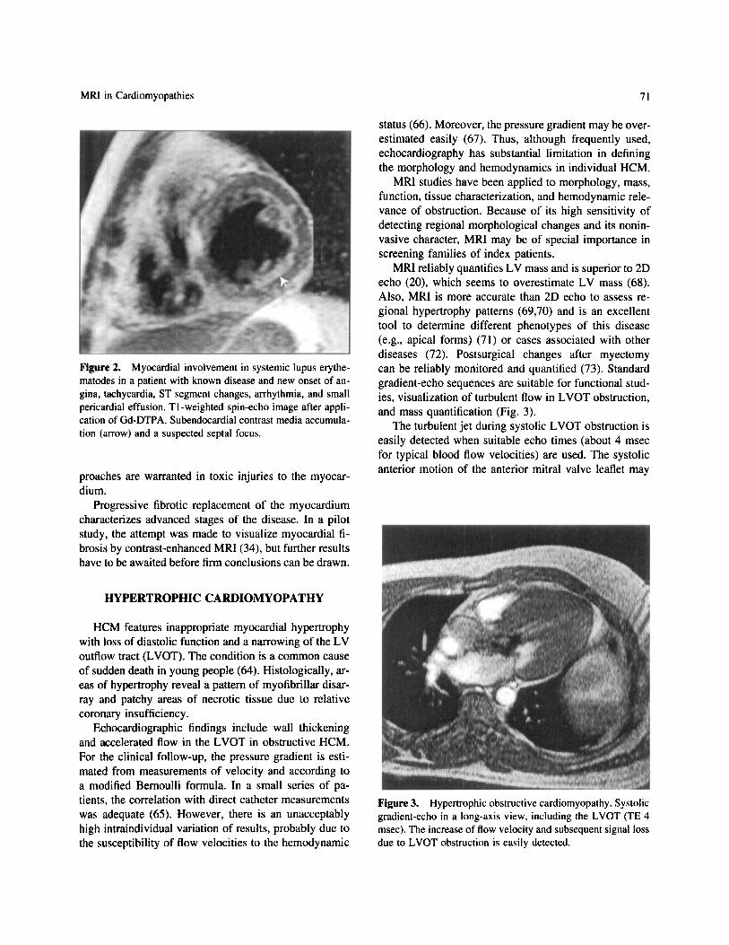
MRI reliably quantifies LV mass and is superior to 2D echo (20), which seems to overestimate LV mass (68). Also, MRI is more accurate than 2D echo to assess re- gional hypertrophy patterns (69,70) and is an excellent tool to determine different phenotypes of this disease (e.g., apical forms) (71) or cases associated with other diseases (72). Postsurgical changes after myectomy can be reliably monitored and quantified (73). Standard gradient-echo sequences are suitable for functional stud- ies, visualization of turbulent flow in LVOT obstruction, and mass quantification (Fig. 3).
The turbulent jet during systolic LVOT obstruction is easily detected when suitable echo times (about 4 msec for typical blood flow velocities) are used. The systolic anterior motion of the anterior mitral valve leaflet may
Figure 3. Hypertrophic obstructive cardiomyopathy. Systolic gradient-echo in a long-axis view, including the LVOT (TE 4 msec). The increase of flow velocity and subsequent signal loss due to LVOT obstruction is easily detected.

72 Friedric h
contribute significantly to the LVOT obstruction and is a typical feature of obstructive HCM. The phenomenon is detectable by MRI (74) and best visualized in the four- chamber view or a short-axis view through the valvular plane. Mitral valve regurgitation, probably due to a pathological change of leaflet geometry, is frequent and should be included in an MRI workup.
MRI may be helpful in the exclusion or verification of hypertrophy due to extracardiac causes such as amy- loidosis (75). Preliminary studies indicate that hypertro- phic tissue may reveal native signal heterogeneities (76). The tissue signal of HCM-associated hypertrophy may also differ from normal myocardium in contrast- enhanced MRI studies (77).
MR techniques have been established to investi- gate phosphate metabolism in HCM. Myocardial phosphocreatine/ATP ratio and the signal of phospho- monoesters reveal changes in patients with HCM (37,78), and the decrease of myocardial energy reserve related to high-energy phophate metabolism correlates to diastolic dysfunction (79). Phosphorous metabolism was also found to be altered in skeletal muscles of HCM (80).
Blood flow analysis in the coronary sinus using MR is feasible, and a preliminary study suggested alterations of coronary flow reserve in patients with HCM (81). A promising approach to assess the hemodynamic rele- vance of LVOT obstruction may be the noninvasive mea- surement of the effective outflow tract area by MRI pla- nimetry of transplanar flow in the LVOT during systole
(82). This method may overcome the problem of pressure recovery with subsequent overestimation of the pressure gradient (67) and the high interstudy variability of pres- sure gradient measurements by echocardiography (66), which is presumably also a feature of invasive studies.
Diastolic function (or dysfunction) is a powerful clini- cal and prognostic factor in hypertrophy but is not part of the routine MRI. Preliminary clinical results suggest that the analysis of the early untwisting motion of the myocardium may be a helpful tool to assess early dia- stolic function in hypertrophic heart diseases (83). Other functional changes detected by the use of myocardial tag- ging are a reduction of posterior rotation, a reduced radial displacement of the inferior septal myocardium (84), het- erogeneities of regional function (85). and reduced 3- dimensional myocardial shortening (86). These findings may be more sensitive in the detection and quantification of functional impairment than conventional parameters such as mitral valve inflow patterns in echocardiography. Future studies will have to show the feasibility and practi- cability of these approaches in clinical routine.
MRI may also be very important in the follow-up of patients after surgical (74) or pharmacological interven- tions. A long-term follow-up is sensitive to morphologi- cal changes during the natural course of the disease (87). The acute and chronic morphological and functional changes due to interventional ablation of a septal artery in obstructive HCM are easily detected by TI-weighted images (Fig. 4).
Figure 4. Effect of septal artery ablation in severely symptomatic hypertrophic cardiomyopathy. TI-weighted spin-echo images (TE 25 msec, non-breathhold, acquisition time = 6 min) in axial orientation after administration of Gd-DTPA. Left: 3 days after the intervention there is contrast media accumulation in the thickened septum. Right: 6 months later the septum shows a significant involution with subsequent reduction of LVOT obstruction (images not shown).
MRI in Cardiomyopathies 73
ARRHYTHMOGENIC RV CARDIOMYOPATHY
ARVC is characterized by a progressive degeneration of the RV and to a lesser extent the LV myocardium with localized disturbance of myocardial function. Morpho- logical features include fibrous and/or fatty replacement of myocardial tissue, extensive wall thinning, and atypi- cal arrangement of trabecular muscles. The associated morphological spectrum ranges from subtle changes to extensive fibrofatty dysplasia of the right ventricle (88,89).The clinical course is generally determined by ventricular arrhythmias with a substantial risk of sudden cardiac death (90). The morphological substrate to ven- tricular arrhythmias probably are fibromuscular bundles isolated from each other by fatty tissue, leading to reentry phenomena (91).
Echocardiography is able to show regional or global changes of myocardial contractility (92) and thus may be very helpful in detecting the disease during a routine study. However, visibility of the apex and the RV outflow tract (RVOT) are limited, areas of wall thinning may be very difficult to detect, and fat does not provide any spe- cific signal to diffentiate it from surrounding tissue or fluid. Studies have shown a limited accuracy compared with electrocardiographic and angiographic criteria (93). So, echocardiography is a useful tool to detect findings leading to a further diagnostic workup but lacks the power to rule out or confirm the diagnosis of ARVC.
MRI noninvasively visualizes ventricular cavities and
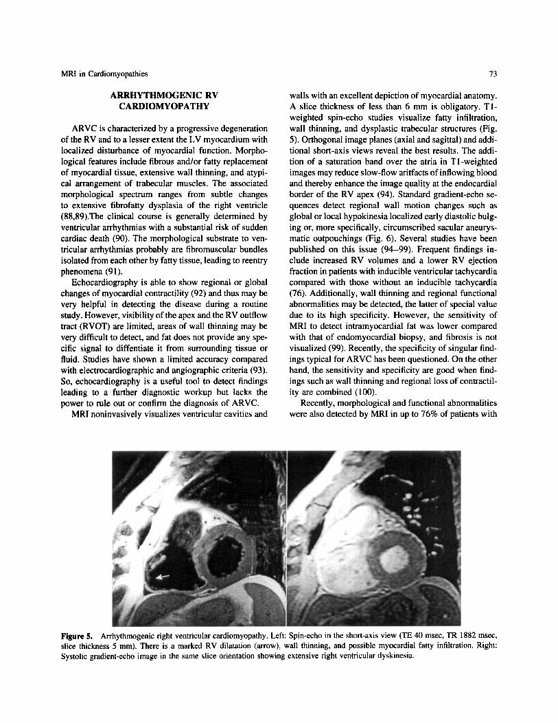
walls with an excellent depiction of myocardial anatomy. A slice thickness of less than 6 mm is obligatory. T l - weighted spin-echo studies visualize fatty infiltration, wall thinning, and dysplastic trabecular structures (Fig. 5) . Orthogonal image planes (axial and sagittal) and addi- tional short-axis views reveal the best results. The addi- tion of a saturation band over the atria in T1-weighted images may reduce slow-flow aritfacts of inflowing blood and thereby enhance the image quality at the endocardia1 border of the RV apex (94). Standard gradient-echo se- quences detect regional wall motion changes such as global or local hypokinesia localized early diastolic bulg- ing or, more specifically, circumscribed sacular aneurys- matic outpouchings (Fig. 6). Several studies have been published on this issue (94-99). Frequent findings in- clude increased RV volumes and a lower RV ejection fraction in patients with inducible ventricular tachycardia compared with those without an inducible tachycardia (76). Additionally, wall thinning and regional functional abnormalities may be detected, the latter of special value due to its high specificity. However, the sensitivity of MRI to detect intramyocardial fat was lower compared with that of endomyocardial biopsy, and fibrosis is not visualized (99). Recently, the specificity of singular find- ings typical for ARVC has been questioned. On the other hand, the sensitivity and specificity are good when find- ings such as wall thinning and regional loss of contractil- ity are combined (100).
Recently, morphological and functional abnormalities were also detected by MRI in up to 76% of patients with
Figure 5. Arrhythmogenic right ventricular cardiomyopathy. Left: Spin-echo in the short-axis view (TE 40 msec, TR 1882 msec, slice thickness 5 mm). There is a marked RV dilatation (arrow), wall thinning, and possible myocardial fatty infiltration. Right: Systolic gradient-echo image in the same slice orientation showing extensive right ventricular dyskinesia.

74 Friedric h
Figure 6. Arrhythmogenic right ventricular cardiomyopathy. Systolic gradient-echo image in a sagittal view. There are sev- eral small dyskinetic areas of the diaphragmal right ventricular wall (arrows).
idiopathic RVOT tachycardia (101- 104). The results concerning the incidence of regional wall motion abnor- malities are conflicting; however, such abnormalities were found in up to 97% of patients (104). The etiological and pathophysiological relation of idiopathic RVOT tachycardia with its favorable prognosis to ARVC is un- der discussion. MRI will play an important role as the tool of choice for follow-up studies in ARVC.
RESTRICTIVE CARDIOMYOPATHY
Primary infiltration of the myocardium by fibrosis or other tissues leads to the rare entity of RCM. The condi- tion is characterized by a severe diastolic dysfunction, biatrial dilatation, preserved LV size, and usually normal systolic function. Atrial thrombi occur. The main differ- ential diagnostic consideration is constrictive pericarditis, which must be excluded in a patient with suspected RCM.
MRI studies in RCM should focus on myocardial mor- phology and function and on the exclusion of constrictive pericardial disease. LV size and wall thickness are quan- tified in gradient-echo image sets. Long-axis views are obligatory. Biatrial dilatation is easily visualized by
means of a four-chamber view. MR volumetry of the en- larged atria may be recommended (105). Although MRI might be at least as powerful as echocardiography to as- sess atrial thrombi (106), slow-flowing blood in the atria may lead to false-positive results when spin-echo tech- niques are used (107). The choice of a longer TE, satura- tion of inflowing blood, and combination with gradient- echo sequences may be helpful. Concomitant mitral re- gurgitation should be visualized. The exclusion of rele- vant pericardial thickening rules out constrictive pericar- ditis and may prevent unnecessary operative exploration of the pericardium (108). This exclusion is possible by T1 weighted spin echo-techniques (109.1 lo).
SARCOIDOSIS
The incidence of myocardial involvement in systemic sarcoidosis is about 20-30% (1 I 1). although the heart was involved in about half of the cases in a large Japanese autopsy series (1 12). Up to 50% of deaths in sarcoidosis may be related to cardiac involvement (1 13), primarily due to sudden death and congestive heart failure. Histo- logical findings are granulomas that are often transmural. Sarcoid lesions may lead to different signal intensity be- haviors, possibly because of different stages of disease activity. Muscular sarcoidosis was reported as high signal intensity areas in T2-weighted MRI (114). In another study of skeletal muscle sarcoidosis, the granulomatous nodules exhibited a central region with low signal inten- sity in T1- and T2-weighted imaging but were sur- rounded by a high signal ring ( I 15). Gadolinium gado- pentetate dimeglumine (Gd-DTPA) seems to accumulate in sarcoid lesions of the brain (1 16). This behavior may be compatible with fibrotic nonactive granulomatous nodules with inflammatory response of the surrounding tissue. Similar findings were reported in several cases of sarcoid infiltration of the heart (117-120). Thus, T2- weighted followed by T1-weighted spin-echo techniques in short and long axis before and after Gd-DTPA may be useful to detect or exclude suspected granulomas (Fig. 7). Occasionally, the MRI findings could be used to “guide” endomyocardial biopsy (1 18).
Because follow-up is very important to guide therapy, MRI may be very helpful in these patients. However, suf- ficient data sets and standardized protocols must still be generated.
AMYLOIDOSIS
Infiltration of the heart by amyloid deposits is found in almost all cases of primary amyloidosis and in about

MRI in Cardiomyopathies 75
Figure 7. Cardiac involvement in sarcoidosis in a patient with clinical signs of heart failure, nonspecific electrocardiographic changes, and invasive exclusion of coronary artery disease. Diastolic TI-weighted short-axis view before (left) and after (rightl) administration of Gd-DTPA (TE 23 msec, TR 635 msec). There is a circumscribed area of increased gadolinium uptake in the lateral segment, including the papillary muscle (arrow). The arrowhead marks the central region of an extensive posterolateral hypokinesia.
one fourth of familial amyloidosis (121), leading to a loss of atrial (122) or LV function and congestive LV failure (123). There are only a few reports on MRI in cardiac amyloidosis (75,124). Similar to sarcoidosis, the MRI ap- proach is directed toward the detection of signal changes after Gd-DTPA administration. At present, it is unclear
how to differentiate between different infiltrative dis- eases. Because of the rather low incidence of these dis- eases, the difficulties to enroll a sufficient number of patients are obvious. Furthermore, the underlying mecha- nisms of a change in magnetization and relaxation have to be clarified and standard protocols have to be developed.
Figure 8. Cardiac involvement in hemochromatosis. Left: T2-weighted short TI inversion recovery, TE 68 msec, TR 1552 msec). Right: TI-weighted fast spin-echo (TE 23 msec, TR 768 msec). Both techniques show a low intensity appearence of the liver and an area with signal reduction in the anterolateral myocardium (arrows)

76 Friedrich
HEMOCHROMATOSIS
Hemochromatosis of the myocardium is characterized by sometimes extensive iron deposits, leading to wall thickening, ventricular dilatation, progressive loss of function with congestive heart failure, and subsequent death. Because of a predominantly subepicardial deposi- tion of iron, endomyocardial biopsy may fail to confirm the diagnosis (125).
The MRI approach is directed toward the detection of iron deposits as the specific marker for the disease. Iron has very strong paramagnetic properties, and myocardial deposits imply an extensive signal loss in native T1-
weighted, but also T2- and T2*-weighted, imaging of dif- ferent regions of the body (126,127). Similar findings were also reported for cardiac hemochromatosis (128) (Fig. 8). The pattern of focal signal loss in a dysfunctional myocardium combined with a “dark” liver may be suf- ficient to confirm the diagnosis of cardiac hemochro- matosis by MRI alone. LV function should be carefully assessed.
Because an intensified therapy may improve LV func- tion (129), MRI is an ideal tool to follow-up infiltrations and LV parameters in these patients. The highly specific detection of typical tissue changes are especially an ex- ample of the diagnostic power of MRI.
Figure 9. Endomyocardial fibrosis in a patients with a variant of Churg-Strauss vasculitis. (a) T1-weighted spin-echo in an axial orientation (TE 25 msec, TR 821 msec) crossing the apex. There is an apical wall thickening of intermediate signal intensity with a subsequent reduction of left ventricular stroke volume. (b) Left: Gradient-echo images in a long-axis view of the left ventricle (LV) and the left atrium (LA). Right: Right ventricle (RV) and right atrium (RA). Ventricles are small and both atria are dilated due to the restrictive ventricular physiology.
MRI in Cardiomyopathies
ENDOMYOCARDIAL FIBROSIS
Endomyocardial fibrosis is related to two forms, one occuring in the tropics and the other in temperate climate, termed Loffler’s endocarditis. Both conditions lead to primarily posterobasal concentric wall thickening fol- lowed by extensive subendocardial fibrosis and frequent apical thrombus formation. Both ventricles may be af- fected. The course is determined by progressive diastolic dysfunction and decreased stroke volume.
The morphological and functional features can be vis- ualized and quantified by MRI (130,131) (Fig. 9). Fibro- sis or calcifications may be visible as a dark rim in bright bloodprepared gradient-echo sequences but may also re- veal an intermediate signal intensity (132). Thus, there is a certain lack of accuracy to assess fibrosis (low sensi- tivity and specificity) and calcifications (low specificity). Differentiation from apical infarction is easy by visualiza- tion of the preserved (or increased) wall thickness and the v-shaped outer form of the apex in the long-axis views.
SUMMARY
The emerging role of MRI for the understanding and treatment of cardiomyopathies cannot be overestimated. Establishing the diagnosis is generally possible by a sin- gle noninvasive MRI study. The follow-up examination is sensitive to even small changes. MRI data on ventricu- lar morphology, volumes, and function are very reliable, and the use of MRI-derived end points in clinical studies may lead to a substantial decrease in subjects needed to test a given hypothesis. In addition to established ap- proaches, MRI analysis of myocardial tissues should be the focus in future studies. If MR coronary angiography will be available in a routine clinical setting, a single MR study could provide a complete and comprehensive diag- nostic procedure
1. Richardson
in patients with cardiomyopathies.
REFERENCES
P, McKenna W, Bristow M, Maisch B, Mautner B, O’Connell J, Olsen E. Thiene G, Goodwin J, Gyarfas I, Martin I and Nordet P. Report of the 1995 World Health Organization/Intemationnl Society and Federation of Cardiology Task Force on the Defini- tion and Classification of Cardiomyopathies [see com- ments]. Circulation, 1996; 93:841-842.
2. Bender JR. Idiopathic dilated cardiomyopathy. An im- munologic, genetic, or infectious disease, or all of the
3.
4.
5 .
6.
7.
8.
9.
10.
11 .
12.
13.
77
above [editorial; comment]? Circulation, 1991 ; 83:
Burke AP, Farb A, Tashko G and Virmani R. Arrhyth- mogenic right ventricular cardiomyopathy and fatty re- placement of the right ventricular myocardium: are they different diseases [see comments]? Circulation, 1998;
Spinto P, Seidman CE, McKenna WJ and Maron BJ. The management of hypertrophic cardiomyopathy [see comments]. N Engl J Med, 1997; 336:775-785. Germain P, Roul G, Kastler B, Mossard JM, Bareiss P and Sacrez A. Inter-study variability in left ventricular mass measurement. Comparison between M-mode echography and MRI. Eur Heart J, 1992; 13:lOll- 1019. Gottdiener JS, Livengood SV, Meyer PS and Chase GA. Should echocardiography be performed to assess effects of antihypertensive therapy? Test-retest reliability of echocardiography for measurement of left ventricular mass and function. J Am Coll Cardiol, 1995; 25:424- 430. van Royen N, Jaffe CC, Krumholz HM, Johnson KM. Lynch PJ, Natale D, Atkinson P, Deman P and Wackers FJ. Comparison and reproducibility of visual echocar- diographic and quantitative radionuclide left ventricular ejection fractions. Am J Cardiol, 1996; 775343-850. Jensen Urstad K, Bouvier F, Hojer J, Ruiz H, Hulting J, Samad B, Thorstrand C and Jensen Urstad M. Com- parison of different echocardiographic methods with ra- dionuclide imaging for measuring left ventricular ejec- tion fraction during acute myocardial infarction treated by thrombolytic therapy. Am J Cardiol, 1998; 81538- 544. Kupferwasser I, Mohr Kahaly S. Stahr P, Rupprecht HJ, Nixdorff U, Fenster M, Voigtlander T, Erbel R and Meyer J. Transthoracic three-dimensional echocardio- graphic volumetry of distorted left ventricles using rota- tional scanning. JAm Soc Echocardiogr, 1997; 10:840- 852. Chandra S, Bahl VK, Reddy SC, Bhargava B, Malhotra A and Wasir HS. Comparison of echocardiographic acoustic quantification system and radionuclide ventric- ulography for estimating left ventricular ejection frac- tion: validation in patients without regional wall motion abnormalities. Am Heart J, 1997; 133:359-363. Lucariello RJ, Sun Y, Doganay G and Chiarainida SA. Sensitivity and specificity of left ventricular ejection fraction by echocardiographic automated border detec- tion: comparison with radionuclide ventriculography. Clin Cardiol, 1997; 20:943-948. Nessly ML, Bashein G, Detmer PR, Graham MM, Kao R and Martin RW. Left ventricular ejection fraction: single-plane and multiplanar transesophageal echocar- diography versus equilibrium gated-pool scintigraphy. J Cardiothorac Vasc Anesth, 1991; 5:40-45. Tam JW. Burwash IG, Ascah KJ, Baird MG and Chan
704-706.
97:1571-1580.

78 Friedrich
14.
15.
16.
17.
18.
19.
20.
21.
22.
23.
24.
-
KL. Feasibility and complications of single-plane and biplane versus multiplane transesophageal imaging: a review of 2947 consecutive studies. Can J Curdiol,
Skorton DJ, Collins SM. Clinical potential of ultrasound tissue characterization in cardiomyopathies. J Am Soc Echocurdiogr 1988; 1:69-77. Rogers WJ, Smith LR, Hood WP Jr, Mantle JA, Rackley CE and Russell RO Jr. Effect of filming projection and interobserver variability on angiographic biplane left ventricular volume determination. Circulation, 1979;
American College of CardiologylAmerican Heart Asso- ciation Ad Hoc Task Force on Cardiac Catheterization. ACC/AHA guidelines for cardiac catheterization and cardiac catheterization laboratories. J Am Cull Cardiol,
Takenaka A, lwase M, Sobue T and Yokota M. The dis- crepancy between echocardiography, cineventriculogra- phy and thennodilution. Evaluation of left ventricular volume and ejection fraction. Int J Curd Imaging, 1995;
Benjelloun H. Cranney GB, Kirk KA, Blackwell GG, Lotan CS and Pohost GM. Interstudy reproducibility of biplane cine nuclear magnetic resonance measurements of left ventricular function. Am J Curdiol, 1991; 67: 141 3- 1420. Pattynama PM, Lamb HJ. Van der Velde EA, Van der Geest RJ, Van der Wall EE and De Roos A. Reproduc- ibility of MRI-derived measurements of right ventricu- lar volumes and myocardial mass. Magn Reson Im- aging, 1995; 1353-63. Bottini PB, Carr AA, Prisant LM, Flickinger FW, Alli- son JD and Gottdiener JS. Magnetic resonance imaging compared to echocardiography to assess left ventricular mass in the hypertensive patient. Am J Hypertens, 1995;
Strohm 0, Schulz-Menger J, Schuler J, Pilz B, Osterziel KJ and Dietz R. The impact of the imaging modality on the sample size of clinical studies of left ventricular morphology and function-comparison of two-dimen- sional echocardiography and magnetic resonance im- aging in dilated cardiomyopathy. submitted. Higgins CB, Lanzer P, Stark D, Botvinick E, Schiller NB, Lipton MJ, Crooks LE and Kaufman L. Assessment of cardiac anatomy using nuclear magnetic resonance imaging. J Am Coll Curdiol, 1985; 5:77s-81s. Lund G, Morin RL, Olivari MT and Ring WS. Serial myocardial T2 relaxation time measurements i n normal subjects and heart transplant recipients. J Hrcirr Truri.7- plant, 1988; 7:274-279. Feinstein JA, Epstein FH, Arai AE, Foo TK, Hartley MR, Balaban RS and Wolff SD. Using cardiac phase to order reconstruction (CAPTOR): a method to improve diastolic images. J Mugn Reson Imaging, 1997; 7:794- 798.
1997; 13:81-84.
59~96-104.
1991; 18:ll49-1l82.
I 1 :255-262.
81221-228.
25.
26.
27.
28.
29.
30.
31.
32.
33.
34.
35.
Lawson MA, Blackwell GG, Davis ND, Roney M, Dell'Italia LJ and Pohost GM. Accuracy of biplane long-axis left ventricular volume determined by cine magnetic resonance imaging in patients with regional and global dysfunction. Am J Curdiol, 1996; 77: 1098- 1104. Sinha S, Mather R, Sinha U, Goldin J, Fonarow G and Yoon HC. Estimation of the left ventricular ejection fraction using a novel multiphase, dark-blood, breath- hold MR imaging technique. AJR Am J Roentgenol,
Rogers WJ Jr, Shapiro EP, Weiss JL. Buchalter MB, Rademakers FE, Weisfeldt ML and Zerhouni EA. Quan- tification of and correction for left ventricular systolic long-axis shortening by magnetic resonance tissue tag- ging and slice isolation. Circulation, 199 I ; 84:721-73 1. Marcus JT, Gotte MJW, DeWaal LK, Stam MR, Van der Geest RJ and Heethaar RM. The influence of through-plane motion on left ventricular volumes mea- sured by magnetic resonance imaging: implications for image acquisition and analysis. J Curdiovusc Mugn Res.
Pennell DJ, Underwood SR and Longmore DB. Im- proved cine MR imaging of left ventricular wall motion with gadopentetate dimeglumine. J Mugn Resun Im- aging, 1993; 3: 13- 19. Singleton HR and Pohost GM. Automatic cardiac MR image segmentation using edge detection by tissue clas- sification in pixel neighborhoods. Mugn Reson Med,
Sechtem U, Pflugfelder PW, Cassidy MM, White RD, Cheitlin MD, Schiller NB and Higgins CB. Mitral or aortic regurgitation: quantification of regurgitant vol- umes with cine MR imaging. Radiology, 1988; 167:
Fujita N, Chazouilleres AF, Hartiala JJ, O'Sullivan M. Heidenreich P, Kaplan JD, Sakuma H, Foster E, Capulo GR and Higgins CB. Quantification of mitral regurgita- tion by velocity-encoded cine nuclear magnetic reso- nance imaging [see comments]. J Am Coll Curdiol,
McCully RB, Enriquez Sarano M, Tajik AJ and Seward JB. Overestimation of severity of ischemic/functional mitral regurgitation by color Doppler jet area. Am J Curdiol, 1994; 74:790-793. Aso H, Takeda K, Ito T, Shiraishi T, Matsumura K and Nakagawa T. Assessment of myocardial fibrosis in cardiomyopathic hamsters with gadolinium-DTPA en-
1997; 169:101-112.
1998; 1:1-6.
1997; 37:418-424.
425-430.
1994; 23:951-958.
hanced magnetic resonancc imaging. / n v m Rrrcliol, 1998; 33:22-32. Neubauer S, KraheT, Schindler R. Horn M, Hillenbrand H, Entzeroth C, Mader H, Kromer EP, Riegger GA, Lackner K, and Ertl. 31P magnetic resonance spectros- copy in dilated cardiomyopathy and coronary artery dis- ease. Allercd cardiac high-energy phosphate metaho- lism in heart failure. Circulation, 1992; 86: I X 10- 18 18.
MRI in Cardiomyopathies 79
36.
37.
38.
39.
40.
41.
42.
43.
44.
45.
46.
47.
Neubauer S, Horn M, Cramer M, Harre K, Newell JB, Peters W, Pabst T, Ertl G, Hahn D, Ingwall JS and Kochsiek K. Myocardial phosphocreatine-to-ATP ratio is a predictor of mortality in patients with dilated cardio- myopathy. Circulation, 1997; 96:2190-2196. Jung WI, Sieverding L, Breuer J, Hoess T, Widmaier S, Schmidt 0, Bunse M, van Erckelens F, Apitz J, Lutz 0 and Dietze GJ. 3 I P NMR spectroscopy detects meta- bolic abnormalities in asymptomatic patients with hypertrophic cardiomyopathy. Circulation, 1998; 97:
Loffler R, Sauter R, Kolem H, Haase A and von Kienlin M. Localized spectroscopy from anatomically matched compartments: improved sensitivity and localization for cardiac 3 1 P MRS in humans. J Magn Reson, 1998; 134:
Kasper EK, Agema WR, Hutchins GM, Deckers JW, Hare JM and Baughman KL. The causes of dilated car- diomyopathy: a clinicopathologic review of 673 consec- utive patients [see comments]. JAm Coll Cardiol, 1994;
Semelka RC, Tomei E, Wagner S, Mayo J, Caputo G, O’Sullivan M, Parmley WW, Chatterjee K, Wolfe C and Higgins CB. Interstudy reproducibility of dimensional and functional measurements between cine magnetic resonance studies in the morphologically abnormal left ventricle. Am Heart J , 1990; 1 19: 1367- 1373. Gaudio C, Tanzilli G, Mazzarotto P, Motolese M, Ro- meo F, Marino B and Reale A. Comparison of left ven- tricular ejection fraction by magnetic resonance imaging and radionuclide ventriculography in idiopathic dilated cardiomyopathy. Am J Cardiol, 1991; 67:411-415. Friedrich MG, Strohm 0, Osterziel KJ and Dietz R. Growth hormone therapy in dilated cardiomyopathy monitored with MRI. MAGMA, 1998; 6:152-154. Buser PT, Auffermann W, Holt WW, Wagner S, Kircher B, Wolfe C and Higgins CB. Noninvasive eval- uation of global left ventricular function with use of cine nuclear magnetic resonance. J Am Coll Cardiol, 1989; 13:1294-13oo. MacGowan GA, Shapiro EP, Azhari H, Siu CO, Hees PS, Hutchins GM, Weiss JL and Rademakers FE. Non- invasive measurement of shortening in the fiber and cross-fiber directions in the normal human left ventricle and in idiopathic dilated cardiomyopathy. Circulation,
Fujita N, Duerinekx AJ and Higgins CB. Variation in left ventricular regional wall stress with cine magnetic resonance imaging: normal subjects versus dilated car- diomyopathy. Am Heart J , 1993; 1251337-1345. Sechtem U, Pflugfelder PW, Could RG, Cassidy MM and Higgins CB. Measurement of right and left ventric- ular volumes in healthy individuals with cine MR im- aging. Radiology. 1987; I63:697-702. Globits S, Pacher R, Frank H, Pacher B, Mayr H, Neu- hold A and Glogar D. Comparative assessment of right
2536-2542.
287-299.
23586-590.
1997; 96535-541.
48.
49.
50.
51.
52.
53.
54.
55.
56.
57.
58.
ventricular volumes and ejection fraction by thermodi- lution and magnetic resonance imaging in dilated car- diomyopathy. Cardiology, 1995; 86:67-72. Jarvinen VM, Kupari MM, Hekali PE and Poutanen VP. Right atrial MR imaging studies of cadaveric atrial casts and comparison with right and left atrial volumes and function in healthy subjects. Radiology, 1994; 191 : 137- 142. Jarvinen VM, Kupari MM, Poutanen VP and Hekali PE. Right and left atrial phasic volumetric function in mildly symptomatic dilated and hypertrophic cardiomyopathy: cine MR imaging assessment, Radiology, 1996; 198:
Doherty NEd, Seelos KC, Suzuki J , Caputo GR, O’Sul- livan M, Sobol SM, Cavero P, Chatterjee K, Parmley WW and Higgins CB. Application of cine nuclear mag- netic resonance imaging for sequential evaluation of response to angiotensin-converting enzyme inhibitor therapy in dilated cardiomyopathy. J Am Coll Cardiol,
Kalil Filho R, Bocchi E, Weiss RG, Rosemberg L, Bacal F, Moreira LF, Stolf NA, Magalhaes AA, Bellotti G, Jatene A, et al. Magnetic resonance imaging evaluation of chronic changes in latissimus dorsi cardiomyoplasty. Circulation, 1994; 90: 102-106. Osterziel KJ, Strohm 0, Schuler J, Friedrich M, Hanlein D, Willenbrock R, Anker SD, Poole Wilson PA, Ranke MB and Dietz R. Randomised, double-blind, placebo- controlled trial of human recombinant growth hormone in patients with chronic heart failure due to dilated car- diomyopathy. Lancet, 1998; 35 1 : 1233- 1237. Hardy CJ, Weiss RG, Bottomley PA and Gerstenblith G. Altered myocardial high-energy phosphate metabo- lites in patients with dilated cardiomyopathy. Am Heart
Liao R, Nascimben L, Friedrich J, Gwathmey JK and Ingwall JS. Decreased energy reserve in an animal model of dilated cardiomyopathy. Relationship to con- tractile performance. Circ Res, 1996; 782393-902. Kawai C. From myocarditis to cardiomyopathy: mecha- nisms of inflammation and cell death: learning from the past for the future. Circulation, 1999; 99:1091- 1 loo. Leatherbury L, Chandra RS, Shapiro SR and Perry LW. Value of endomyocardial biopsy in infants, children and adolescents with dilated or hypertrophic cardiomyo- pathy and myocarditis. J Am CON Cardiol, 1988; 12:
Friedrich MG, Strohm 0, Schulz Menger J, Marciniak H, Luft FC and Dietz R. Contrast media-enhanced mag- netic resonance imaging visualizes myocardial changes in the course of viral myocarditis. Circulation, 1998;
Friedrich MG, Strohm 0, Schulz-Menger JE, Marciniak H and Dietz R. Noninvasive diagnosis of acute myocar- ditis by contrast-enhanced magnetic resonance imaging.
487-495.
1992; 19:1294-1302.
J , 1991; 122:795-801.
1547-1554.
97: 1802- 1809.

80 Friedric h
59.
60.
61.
62.
63.
64.
65.
66.
67.
68.
69.
70.
Proceedings of the ISMRM 6th Annual Meeting, Syd- ney, 1998, p. 912. Kalil R, Bocchi EA, Ferreira BM, de Lourdes Higuchi M, Lopes NH, Magalhaes AC, Mady C, Pereira Barretto AC, Albuquerque CP, Bellotti G, et al. Magnetic reso- nance imaging in chronic Chagas cardiopathy. Correla- tion with endomyocardial biopsy findings. Arq Bras Cardiol, 1995; 6541 3-41 6. Koito H, Suzuki J, Ohkubo N, lshiguro Y, Iwasaka T and Inada M. Gadolinium-diethylenetriamine penta- acetic acid enhanced magnetic resonance imaging of di- lated cardiomyopathy: clinical significance of abnor- mally high signal intensity of left ventricular myocardium. J Cardiol, 1996; 28:41-49. Gagliardi MG, Polletta B and Di Renzi P. MRI for the diagnosis and follow-up of myocarditis [letter]. Circula- tion, 1999; 99:458. Friedrich MG. Strohm 0, Schulz Menger J, Marciniak H, Luft FC and Dietz R. Response to the author. Circu- lation, 1999; 99:459. Bellotti G, Bocchi EA, de Moraes AV, Higuchi ML, Barber0 Marcia1 M, Sosa E, Esteves Filho A, Kalil R, Weiss R, Jatene A and Pileggi F. In vivo detection of Trypanosoma cruzi antigens in hearts of patients with chronic Chagas’ heart disease. Am Heart J, 1996; 131:
Drory Y, Turetz Y, Hiss Y, Lev B, Fisman EZ, Pines A and Kramer MR. Sudden unexpected death in persons less than 40 years of age. Am J Cardiol. 199 I ; 68: 1388- 1392. Sasson Z, Yock PG, Hatle LK, Alderman EL, Popp RL. Doppler echocardiographic determination of the pressure gradient in hypertrophic cardiomyopathy. J Am Coll Cardiol, 1988; 11:752-756. Kizilbash AM, Heinle SK and Grayburn PA. Spontane- ous variability of left ventricular outflow tract gradient in hypertrophic obstructive cardiomyopathy. Circula- tion, 1998; 97:461-466. Levine RA, Jimoh A, Cape EG, McMillan S, Yogana- than AP and Weyman AE. Pressure recovery distal to a stenosis: potential cause of gradient “overestimation” by Doppler echocardiography. JAm CON Cardiol, 1989;
Missouris CG, Forbat SM, Singer DR, Markandu ND, Underwood R and MacGregor GA. Echocardiography overestimates left ventricular mass: a comparative study with magnetic resonance imaging in patients with hy- pertension. J Hypertens, 1996; 14: 1005-1010. Posma JL, Blanksma PK, van der Wall EE, Hamer HP, Mooyaart EL and Lie KI. Assessment of quantitative hypertrophy scores in hypertrophic cardiomyopathy: magnetic resonance imaging versus echocardiography. Am Heart J , 1996; 132:1020-1027. Pons Llado G, Cameras F, Borras X, Palmer J, Llauger J and Bayes de Luna A. Comparison of morphologic assessment of hypertrophic cardiomyopathy by mag-
301 -307.
1 3 :706-7 1 5.
71.
72.
73.
74.
75.
76.
77.
78.
79.
80.
81.
netic resonance versus echocardiographic imaging. Am J Cardiol, 1997; 79: 165 1 - 1656. Soler R, Rodriguez E, Rodriguez JA, Perez ML and Penas M. Magnetic resonance imaging of apical hyper- trophic cardiomyopathy. J Thoruc Imaging, 1997; 12:
Pongratz G, Friedrich M. Unverdorben M, Kunkel B and Bachmann K. Hypertrophic obstructive cardiomy- opathy as a manifestation of a cardiocutaneous syn- drome (Noonan syndrome). Klin Wochenschr, 1991 ; 69:
Franke A, Schondube FA, Kuhl HP, Klues HG. Erena C, Messmer BJ, Flachskampf FA and Hanrath P. Quan- titative assessment of the operative results after ex- tended myectomy and surgical reconstruction of the subvalvular mitral apparatus in hypertrophic obstruc- tive cardiomyopathy using dynamic three-dimensional transesophageal echocardiography. J Am Coll Cardiol,
White RD, Obuchowski NA, Gunawardena S, Lipchik EO. Lever HM, Van Dyke CW and Lytle BW. Left ven- tricular outflow tract obstruction in hypertrophic cardio- myopathy: presurgical and postsurgical evaluation by computed tomography magnetic resonance imaging. Am J Card Imaging, 1996; 10:1-13. Fattori R, Rocchi G, Celletti F, Bertaccini P, Rapezzi C and Gavelli G. Contribution of magnetic resonance imaging in the differential diagnosis of cardiac amy- loidosis and symmetric hypertrophic cardiomyopathy. Am Heart J, 1998; 136324-830. Zahler R, Chelmow D, Gore J, Wilkens K, Pope C, Sost- man HD. Meese R, Negro Vilar R, Herfkens RJ, Wack- ers FJ, et al. Heterogeneous signal intensity in magnetic resonance images of hypertrophied left ventricular myo- cardium. Magn Reson Imaging, 1989; 7517-528. Tsukihashi H, Ishibashi Y, Shimada T, Hatano J, Tanabe K. Ooyake N, Morioka S and Moriyama K. Changes in gadolinium-DTPA enhanced magnetic reso- nance signal intensity ratio in hypertrophic cardiomyop- athy. J Cardiol, 1994; 24: 185- I9 1. de Roos A, Doornbos J, Luyten PR, Oosterwaal U, van der Wall EE and den Hollander JA. Cardiac metabolism in patients with dilated and hypertrophic cardiomyopa- thy: assessment with proton-decoupled P-3 1 MR spec- troscopy. J Magn Reson Imaging, 1992; 2:711-719. Spindler M, Saupe KW, Christe ME, Sweeney HL. Seidman CE, Seidman JG and lngwall JS. Diastolic dys- function and altered energetics in the alphaMHC403/ + mouse model of familial hypertrophic cardiomyopathy. J Clin Invest, 1998; 101:1775-1783. Thompson CH, Kemp GJ. Taylor DJ, Conway M, Raja- gopalan B, O’Donoghue A, Styles P, McKenna WJ and Radda GK. Abnormal skeletal muscle bioenergetics in familial hypertrophic cardiomyopathy. Heart, 1997; 78:
Kawada N, Sakuma H, Yamakado T, Takeda K, Isaka
22 1-225.
9 3 2 - 9 3 6.
1998; 31:1641-1649.
177- 181.
MRI in Cardiomyopathies 81
82.
83.
84.
85.
86.
87.
88.
89.
90.
N, Nakano T and Higgins CB. Hypertrophic cardiomy- opathy: MR measurement of coronary blood flow and vasodilator flow reserve in patients and healthy subjects. Radiology, 1999; 2 1 1 : 129- 135. Schulz-Menger JE, Strohm 0, Waigand J, Marciniak H, Uhlich F, Dietz R and Friedrich MG. Magnetic reso- nance imaging accurately detects delayed improve- ment in left ventricular outflow tract following septa1 embolisation in hypertrophic obstructive cardiomyopa- thy [abstract]. Circulation, 1997; 96: 1054. Stuber M, Scheidegger M, Fischer S, Nagel E, Steine- mann F, Hess 0 and Boesiger T. Alterations in the local myocardial motion pattern in patients suffering from pressure overload due to aortic stenosis. Circulation,
Maier SE, Fischer SE, McKinnon GC, Hess OM, Kray- enbuehl HP and Boesiger P. Evaluation of left ventricu- lar segmental wall motion in hypertrophic cardiomyopa- thy with myocardial tagging. Circulation, 1992; 86:
Kramer CM, Reichek N, Ferrari VA, Theobald T, Daw- son J and Axel L. Regional heterogeneity of function in hypertrophic cardiomyopathy. Circulation, 1994; 90:
Young AA. Kramer CM, Ferrari VA, Axel L and Rei- chek N. Three-dimensional left ventricular deformation in hypertrophic cardiomyopathy. Circulation, 1994; 90:
Suzuki J, Shimatnoto R, Nishikawa J, Yamazaki T, Tsuji T, Nakamura F, Shin WS, Nakajima T, Toyo Oka T and Ohotomo K. Morphological onset and early di- agnosis in apical hypertrophic cardiomyopathy : a long term analysis with nuclear magnetic resonance imaging. JAm Coll Cardiol, 1999; 33:146-151. McKenna WJ, Thiene G, Nava A, Fontaliran F, Bloms- trom Lundqvist C, Fontaine G and Camerini F. Diag- nosis of arrhythmogenic right ventricular dysplasiakar- diomyopathy. Task Force of the Working Group Myocardial and Pericardial Disease of the European So- ciety of Cardiology and of the Scientific Council on Car- diomyopathies of the International Society and Federa- tion of Cardiology. Br Heart J , 1994; 71:215-218. Corrado D, Basso C, Thiene G, McKenna WJ, Davies MJ, Fontaliran F, Nava A, Silvestri F, Blomstrom Lundqvist C, Wlodarska EK, Fontaine G and Camerini F. Spectrum of clinicopathologic manifestations of ar- rhythmogenic right ventricular cardiomyopathy/dyspla- sia: a multicenter study. J Am Coll Cardiol, 1997; 30:
Committees UI-US. Survivors of out-of-hospital cardiac arrest with apparently normal heart. Need for definition and standardized clinical evaluation. Consensus State- ment of the Joint Steering Committees of the Unex- plained Cardiac Arrest Registry of Eumpe and of the Idiopathic Ventricular Fibrillation Registry of the United States. Circulation, 1997; 95:265-272.
1999; 100:361-368.
1919- 1928.
186- 194.
854-867.
1512-1520.
91.
92.
93 *
94.
95.
96.
97.
98.
99.
100.
101.
102.
103.
Fontaliran F, Arkwright S, Vilde F and Fontaine G. Ar- rhythmogenic right ventricular dysplasia and cardiomy- opathy. Clinical and anatomic-pathologic aspects, noso- logic approach. Arch Anat Cytol Pathol, 1998; 46: 17 1 - 177. Blomstrom Lundqvist C, Beckman Suurkula M, Wal- lentin l, Jonsson R and Olsson SB. Ventricular dimen- sions and wall motion assessed by echocardiography in patients with arrhythmogenic right ventricular dyspla- sia. Eur Heart J , 1988; 9:1291-1302. Kisslo J. Two-dimensional echocardiography in ar- rhythmogenic right ventricular dysplasia. Eur Hean J,
Blake LM, Scheinman MM and Higgins CB. MR fea- tures of arrhythmogenic right ventricular dysplasia. AJR Am J Roentgenol, 1994; 162:809-812. Casolo GC, Poggesi L, Boddi M, Fazi A, Bartolozzi C, Lizzadro G and Dabizzi RP. ECG-gated magnetic reso- nance imaging in right ventricular dysplasia. Am Heorr
Frank H, Rimpfl T, Weber H, Globits S, Neuhold A, Sochor H, Mayr H and Glogar D. Diagnosis of arrhyth- mogenic right ventricular disease using magnetic reso- nance tomography. Z Kardiol, 1991 ; 80569-573. Ricci C, Longo R, Pagnan L, Dalla Palma L, Pinamonti B, Camerini F, Bussani R and Silvestri F. Magnetic res- onance imaging in right ventricular dysplasia. Am J Cardiol, 1992; 70:1589-1595. Auffermann W, Wichter T, Breithardt G, Joachimsen K and Peters PE. Arrhythmogenic right ventricular dis- ease: MR imaging vs angiography. AJR Am J Roent- genol, 1993; 161549-555. Menghetti L, Basso C, Nava A, Angelini A and Thiene G. Spin-echo nuclear magnetic resonance for tissue characterisation in arrhythmogenic right ventricular car- diomyopathy. Heart, 1996; 76:467-470. Friedrich MG, Strohm 0, Schulz-Menger J, Thierfelder L, Schirdewan A, Schroeder G and Dietz R. Evidence for a low specificity of right ventricular morphological changes related to arrhythmogenic right ventricular car- diomyopathy as assessed by magnetic resonance im- aging [abstract]. Circulation, 1998; 982357. Carlson MD, White RD, Trohman RG, Adler LP, Biblo LA, Merkatz KA and Waldo AL. Right ventricular out- flow tract ventricular tachycardia: detection of previ- ously unrecognized anatomic abnormalities using cine magnetic resonance imaging. J Am Coll Cardiol, 1994;
Globits S, Kreiner G, Frank H, Heinz G, Klaar U, Frey B and Gossinger H. Significance of morphological ab- normalities detected by MRI in patients undergoing suc- cessful ablation of right ventricular outflow tract tachy- cardia. Circulation, 1997; 96:2633-2640. Proclemer A, Basadonna PT, Slavich GA, Miani D, Fresco C and Fioretti PM. Cardiac magnetic resonance imaging findings in patients with right ventricular out-
1989; 10122-26.
J, 1987; 113:1245-1248.
24~720-727.

82 Friedrich
104.
105.
106.
107.
108.
109.
110.
1 1 1 .
112.
113.
114.
115.
116.
117.
118.
flow tract premature contractions [see comments]. Eur Heart J , 1997; 18:2002-2010. White RD, Trohman RG. Flamm SD, VanDyke CW, Optican RJ, Sterba R, Obuchowski NA, Carlson MD and Tchou PJ. Right ventricular arrhythmia in the absence of arrhythmogenic dysplasia: MR imaging of myocardial abnormalities. Radiology, 1998; 207:743- 751. Jarvinen VM, Kupari MM, Poutanen VP and Hekali PE. A simplified method for the determination of left atrial size and function using cine magnetic resonance im- aging. Mugri Reson Imaging, 1996; 14:215-226. Johnson DE, Vacek J, Gollub SB. Wilson DB and Dunn M. Comparison of gated cardiac magnetic resonance imaging and two-dimensional echocardiography for the evaluation of right ventricular thrombi: a case report with autopsy correlation. Cuthet Cardiovasc Diugn,
Raggi P, Daniels M, Shanoudy H and Jarmukli NF. MRI misinterpretation of spontaneous echo-contrast as a large left atrial thrombus. Inr J Curd Imaging, 1996; 12:
Restrictive cardiomyopathy or constrictive pericarditis [editorial]? Lancet, 1987; 2:372-374. Sechtem U, Higgins CB, Sommerhoff BA. Lipton MJ and Huycke EC. Magnetic resonance imaging of restric- tive cardiomyopathy. Am J Curdiol, 1987; 59:480-482. Masui T, Finck S and Higgins CB. Constrictive peri- carditis and restrictive cardiomyopathy: evaluation with MR imaging. Radiology, 1992; 182:369-373. Flora GS and Sharma OP. Myocardial sarcoidosis: a re- view. Surcoidosis, 1989; 6:97- 106. Sugie T, Hashimoto N and lwai K. Clinical and autopsy studies on prognosis of sarcoidosis. Nippon Rinsho,
Perry A and Vuitch F. Causes of death in patients with sarcoidosis. A morphologic study of 38 autopsies with clinicopathologic correlations. Arch Puthol Lab Med,
Kurashima K, Shimizu H, Ogawa H, Ohka T, Nobata K. Ueno K, Rikimaru S, Fujimura M and Matsuda T. MR and CT in the evaluation of sarcoid myopathy. J Comput Assist Tomogr, 1991; 15:1004-1007. Otake S, Banno T, Ohba S, Noda M and Yamamoto M. Muscular sarcoidosis: findings at MR imaging. Rudiol-
Seltzer S, Mark AS and Atlas SW. CNS sarcoidosis: evaluation with contrast-enhanced MR imaging. AJNR Am J Neurorudiol, 1991; 12:1227-1233. Riedy K, Fisher MR, Belic N and Koenigsberg DI. MR imaging of myocardial sarcoidosis. AJR Am J Roent- genol, 1988; 151:915-916. Dupuis JM, Victor J, Furber A, Pezard P, Lejeune LL and Tadei A. Value of magnetic resonance imaging in cardiac sarcoidosis. Apropos of a case. Arch Mu1 Coeur Vuiss, 1994; 87:105-110.
1988; 14:266-268.
85-88.
1994; 52:1567-1570.
1995; I19:167-172.
O ~ V , 1990: 1761145-148.
119.
120.
121.
122.
123.
124.
125.
126.
127.
128.
129.
130.
131.
132.
Chandra M, Silverman ME, Oshinski J and Pettigrew R. Diagnosis of cardiac sarcoidosis aided by MRI. Chest, 1996; I10:562-565. Doherty MJ, Kumar SK, Nicholson AA and McGivern DV. Cardiac sarcoidosis: the value of magnetic reso- nance imaging in diagnosis and assessment of response to treatment. Respir Med, 1998; 92:697-699. Gertz MA, Kyle RA and Thibodeau SN. Familial amy- loidosis: a study of 52 North American-born patients examined during a 30-year period. Muyo Clin Proc,
Plehn JF, Southworth J and Cornwell GGd. Brief report: atrial systolic failure in primary amyloidosis [see com- ments]. N Engl J Med, 1992; 327:1570-1573. Kyle RA, Spittell PC, Gertz MA, Li CY, Edwards WD, Olson LJ and Thibodeau SN. The premortem recog- nition of systemic senile amyloidosis with cardiac involvement. Am J Med, 1996; I0 I :395-400. Matsuoka H, Hamada M, Honda T, Kawakami H, Okay- ama H, Abe M, Shigematsu Y, Sumimoto T and Hiwada K. Precise assessment of myocardial damage associated with secondary cardiomyopathies by use of Gd-DTPA- enhanced magnetic resonance imaging. Angiology,
Olson LJ, Edwards WD, McCall JT, Ilstrup DM and Gersh BJ. Cardiac iron deposition in idiopathic hemoch- romatosis: histologic and analytic assessment of 14 hearts from autopsy. J Am Coll Curdiol, 1987; 10:
Siegelman ES, Mitchell DG and Semelka RC. Abdomi- nal iron deposition: metabolism, MR findings, and clini- cal importance. Radiology, 1996; 199: 13-22. Jager HJ, Mehring U, Gotz GF, Neise M, Erlemann R, Kapp HJ and Mathias KD. Radiological features of the visceral and skeletal involvement of hemochromatosis. Eur Radiol, 1997; 7: 1199-1206. Waxman S, Eustace S and Hartnell GG. Myocardial involvement in primary hemochromatosis demonstrated by magnetic resonance imaging. Am Heart J , 1994; 128:
Rahko PS. Salerni R and Uretsky BF. Successful rever- sal by chelation therapy of congestive cardiomyopathy due to iron overload. J Am Coll Curdiol, 1986; 8:436- 440. D’Silva SA, Kohli A, Dalvi BV and Kale PA. MRI in right ventricular endomyocardial fibrosis. Am Heart J ,
Celletti F. Fattori R, Napoli G , Leone 0, Rocchi C , Re- ggiani LB and Gavelli G . Assessment of restrictive car- diomyopathy of amyloid or idiopathic etiology by mag- netic resonance imaging. Am J Curdiol, 1999; 83:798- 801. Huong DL, Wechsler B, Pap0 T, de Zuttere D. Bletry 0, Hernigou A, Delcourt A, Godeau P and Piette JC. Endomyocardial fibrosis in Behcet’s disease. Ann Rheum Dis, 1997; 56:205-208.
1992; 67:428-440.
1993; 44:945-50.
1239- 1243.
1047-1049.
1992; 123:1390-1392.