Functional magnetic resonance imaging outcomes from a comprehensive magnetic resonance
University of Wollongong University of Wollongong
Research Online Research Online
Faculty of Engineering and Information Sciences - Papers: Part B
Faculty of Engineering and Information Sciences
2016
Magnetic resonance imaging in lung: a review of its potential for Magnetic resonance imaging in lung: a review of its potential for
radiotherapy radiotherapy
Shivani Kumar Cancer Therapy Centre Liverpool Hospital
Gary P. Liney University of Wollongong, [email protected]
Robba Rai Cancer Therapy Centre Liverpool Hospital
Lois C. Holloway University of Wollongong, [email protected]
Daniel Moses University of New South Wales
See next page for additional authors
Follow this and additional works at: https://ro.uow.edu.au/eispapers1
Part of the Engineering Commons, and the Science and Technology Studies Commons
Recommended Citation Recommended Citation Kumar, Shivani; Liney, Gary P.; Rai, Robba; Holloway, Lois C.; Moses, Daniel; and Vinod, Shalini K., "Magnetic resonance imaging in lung: a review of its potential for radiotherapy" (2016). Faculty of Engineering and Information Sciences - Papers: Part B. 249. https://ro.uow.edu.au/eispapers1/249
Research Online is the open access institutional repository for the University of Wollongong. For further information contact the UOW Library: [email protected]

Magnetic resonance imaging in lung: a review of its potential for radiotherapy Magnetic resonance imaging in lung: a review of its potential for radiotherapy
Abstract Abstract MRI has superior soft-tissue definition compared with existing imaging modalities in radiation oncology; this has the added benefit of functional as well as anatomical imaging. This review aimed to evaluate the current use of MRI for lung cancer and identify the potential of a MRI protocol for lung radiotherapy (RT). 30 relevant studies were identified. Improvements in MRI technology have overcome some of the initial limitations of utilizing MRI for lung imaging. A number of commercially available and novel sequences have shown image quality to be adequate for the detection of pulmonary nodules with the potential for tumour delineation. Quantifying tumour motion is also feasible and may be more representative than that seen on four-dimensional CT. Functional MRI sequences have shown correlation with flu-deoxy-glucose positron emission tomography (FDG-PET) in identifying malignant involvement and treatment response. MRI can also be used as a measure of pulmonary function. While there are some limitations for the adoption of MRI in RT-planning process for lung cancer, MRI has shown the potential to compete with both CT and PET for tumour delineation and motion definition, with the added benefit of functional information. MRI is well placed to become a significant imaging modality in RT for lung cancer.
Disciplines Disciplines Engineering | Science and Technology Studies
Publication Details Publication Details Kumar, S., Liney, G., Rai, R., Holloway, L., Moses, D. & Vinod, S. K. (2016). Magnetic resonance imaging in lung: a review of its potential for radiotherapy. British Journal of Radiology, 89 (1060), 1-14.
Authors Authors Shivani Kumar, Gary P. Liney, Robba Rai, Lois C. Holloway, Daniel Moses, and Shalini K. Vinod
This journal article is available at Research Online: https://ro.uow.edu.au/eispapers1/249

BJR © 2016 The Authors. Published by the British Institute of Radiology
Received:27 May 2015
Revised:21 January 2016
Accepted:1 February 2016
doi: 10.1259/bjr.20150431
Cite this article as:Kumar S, Liney G, Rai R, Holloway L, Moses D, Vinod SK. Magnetic resonance imaging in lung: a review of its potential for radiotherapy. Br JRadiol 2016; 89: 20150431.
REVIEW ARTICLE
Magnetic resonance imaging in lung: a review of itspotential for radiotherapy
1,2,3SHIVANI KUMAR, MPH, B App Sc (MRS), 1,2,3,4GARY LINEY, PhD, 2,3ROBBA RAI, MHlthSc (MRI),1,2,3,4,5LOIS HOLLOWAY, PhD, 1,6,7DANIEL MOSES, MEngSc, FRANZCR and 1,2,7SHALINI K VINOD, MD, FRANZCR
1South Western Clinical School, School of Medicine, University of New South Wales, Liverpool, NSW, Australia2Liverpool and Macarthur Cancer Therapy Centres, Liverpool Hospital, Liverpool, NSW, Australia3Ingham Institute of Applied Medical Research, Liverpool, NSW, Australia4Centre for Medical Radiation Physics, University of Wollongong, Liverpool, NSW, Australia5Institute of Medical Physics, School of Physics, University of Sydney, Sydney, NSW, Australia6Department of Medical Imaging, Northern Hospital Network, Sydney, NSW, Australia7Western Sydney University, Penrith, NSW, Australia
Address correspondence to: Mrs Shivani KumarE-mail: [email protected]
ABSTRACT
MRI has superior soft-tissue definition compared with existing imaging modalities in radiation oncology; this has the
added benefit of functional as well as anatomical imaging. This review aimed to evaluate the current use of MRI for lung
cancer and identify the potential of a MRI protocol for lung radiotherapy (RT). 30 relevant studies were identified.
Improvements in MRI technology have overcome some of the initial limitations of utilizing MRI for lung imaging. A number
of commercially available and novel sequences have shown image quality to be adequate for the detection of pulmonary
nodules with the potential for tumour delineation. Quantifying tumour motion is also feasible and may be more
representative than that seen on four-dimensional CT. Functional MRI sequences have shown correlation with flu-deoxy-
glucose positron emission tomography (FDG-PET) in identifying malignant involvement and treatment response. MRI can
also be used as a measure of pulmonary function. While there are some limitations for the adoption of MRI in RT-planning
process for lung cancer, MRI has shown the potential to compete with both CT and PET for tumour delineation and
motion definition, with the added benefit of functional information. MRI is well placed to become a significant imaging
modality in RT for lung cancer.
INTRODUCTIONRadiotherapy (RT) plays a significant role in the treatmentof lung cancer1 and relies on accurate imaging for precisetreatment delivery. Improvements in imaging and the useof multimodality imaging have improved tumour de-lineation for RT planning and treatment of lung cancer.2,3
CT is the standard imaging modality in RTwith a relativelyhigh spatial resolution, but limited specificity. Tumourdefinition on CT can be obscured in the presence of ad-jacent lung collapse or consolidation. The incorporation ofpositron emission tomography (PET) with glucose ana-logue flu-deoxy-glucose (FDG) tracer has significantlyimproved the discrimination between benign and malignanttissue.4 The use of FDG-PET has reduced gross tumourvolumes (GTV) owing to the improved differentiation be-tween benign and malignant tissue5–7 and reduced in-terobserver variability in the delineation of GTV.4–13 WhileFDG-PET with CT is currently the standard of care for
tumour delineation, there are limitations. The spatial reso-lution of PET is poor, ranging between 5 and 7mm com-pared with 2mm for CT.14 This low spatial resolution resultsin blurred edges, and tumours ,4mm may be falsely neg-ative on FDG-PETscans. There is also a lack of consensus onthe FDG-PET visualization method within RT, with a num-ber of different methods reported in the literature.6–9,13,15
Use of ionizing radiation to acquire images for PET and CTcan be a limiting factor in repeated examinations.
The mobility of tumour and normal anatomy duringrespiration can lead to a large degree of uncertainty intumour position. In order to visualize and quantify tu-mour motion, a number of options have been utilizedincluding fluoroscopy, slow CT scans and breath-holddevices.16 However, these have largely been supersededby respiratory-correlated CT or four-dimensional CT(4DCT).17 4DCT image acquisition is based on acquiringCT images with an external respiratory trace while the
patient is free breathing. During post processing, the acquiredimages are correlated with an external respiratory signalusually either in the form of a reflective marker and camerasystem or a pressure-sensing belt around the abdomen. Theadvent of 4DCT in RT planning has overcome some of theproblems associated with imaging the thorax18 and hasallowed for the definition of patient-specific margins for tu-mour motion.19–21 However, accurate definition of motion on4DCT is reliant on a consistent respiratory cycle.22
MRI is a well-established diagnostic tool in oncology.23 UnlikeCT where tissue contrast primarily depends on electron density,MRI contrast can be varied extensively by imaging other in-trinsic properties of the tissue (e.g. spin lattice and spin–spinrelaxation time, proton density, diffusion etc.). Typically, a MRIexamination will consist of multiple series of scans in severalimaging planes using different pulse sequences which exploitthese properties.24 This allows flexibility in facilitating optimaltumour visualization and evaluation.23 The benefit of MRI indelineating soft tissues has been demonstrated for a number ofdisease sites in RT23,25,26 and is being incorporated into thetreatment-delivery process with the development of MRI linearaccelerators,27–29 making it a significant imaging modality infuture. The use of MRI in the lung has been complicated byfirstly by respiratory motion and also low proton density of thelung tissue, which can reduce the signal-to-noise ratio and in-crease magnetic susceptibility effects.30
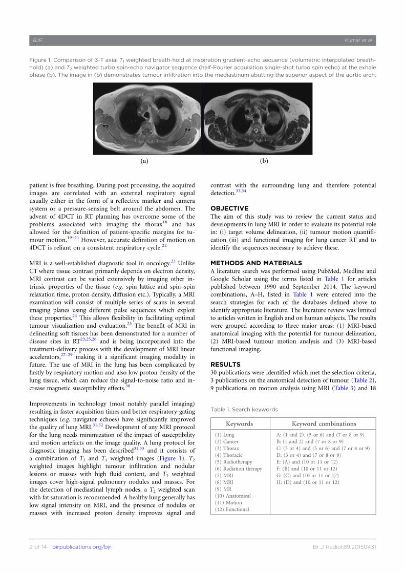
Improvements in technology (most notably parallel imaging)resulting in faster acquisition times and better respiratory-gatingtechniques (e.g. navigator echoes) have significantly improvedthe quality of lung MRI.31,32 Development of any MRI protocolfor the lung needs minimization of the impact of susceptibilityand motion artefacts on the image quality. A lung protocol fordiagnostic imaging has been described31,33 and it consists ofa combination of T2 and T1 weighted images (Figure 1). T2weighted images highlight tumour infiltration and nodularlesions or masses with high fluid content, and T1 weightedimages cover high-signal pulmonary nodules and masses. Forthe detection of mediastinal lymph nodes, a T2 weighted scanwith fat saturation is recommended. A healthy lung generally haslow signal intensity on MRI, and the presence of nodules ormasses with increased proton density improves signal and
contrast with the surrounding lung and therefore potentialdetection.33,34
OBJECTIVEThe aim of this study was to review the current status anddevelopments in lung MRI in order to evaluate its potential rolein: (i) target volume delineation, (ii) tumour motion quantifi-cation (iii) and functional imaging for lung cancer RT and toidentify the sequences necessary to achieve these.
METHODS AND MATERIALSA literature search was performed using PubMed, Medline andGoogle Scholar using the terms listed in Table 1 for articlespublished between 1990 and September 2014. The keywordcombinations, A–H, listed in Table 1 were entered into thesearch strategies for each of the databases defined above toidentify appropriate literature. The literature review was limitedto articles written in English and on human subjects. The resultswere grouped according to three major areas: (1) MRI-basedanatomical imaging with the potential for tumour delineation,(2) MRI-based tumour motion analysis and (3) MRI-basedfunctional imaging.
RESULTS30 publications were identified which met the selection criteria,3 publications on the anatomical detection of tumour (Table 2),9 publications on motion analysis using MRI (Table 3) and 18
Figure 1. Comparison of 3-T axial T1 weighted breath-hold at inspiration gradient-echo sequence (volumetric interpolated breath-
hold) (a) and T2 weighted turbo spin-echo navigator sequence (half-Fourier acquisition single-shot turbo spin echo) at the exhale
phase (b). The image in (b) demonstrates tumour infiltration into the mediastinum abutting the superior aspect of the aortic arch.
Table 1. Search keywords
Keywords Keyword combinations
(1) Lung(2) Cancer(3) Thorax(4) Thoracic(5) Radiotherapy(6) Radiation therapy(7) MRI(8) MRI(9) MR(10) Anatomical(11) Motion(12) Functional
A: (1 and 2), (5 or 6) and (7 or 8 or 9)B: (1 and 2) and (7 or 8 or 9)C: (3 or 4) and (5 or 6) and (7 or 8 or 9)D: (3 or 4) and (7 or 8 or 9)E: (A) and (10 or 11 or 12)F: (B) and (10 or 11 or 12)G: (C) and (10 or 11 or 12)H: (D) and (10 or 11 or 12)
BJR Kumar et al
2 of 14 birpublications.org/bjr Br J Radiol;89:20150431

publications on functional MRI for lung tumour and pulmonarynodules including functional imaging of healthy lung (Table 4).The majority of the studies were conducted on 1.5-T scannersusing a combination of breath-hold and free-breathing scanswith and without respiratory and/or cardiac gating. Most pro-tocols used fast-imaging-sequence variants of both the gradient-echo (GRE) or turbo-spin echo techniques. In most cases,
parallel imaging—using radiofrequency coil encoding—wasapplied to further reduce the scan time and limit the breath-holdduration required. Table 5 highlights the basic sequences usedand their applications and limitations in lung imaging. Whilethere was evidence available for lung cancer imaging from a di-agnostic perspective, there was limited evidence in its applica-tion in radiation oncology imaging.
Table 2. Literature summary of scan protocols for studies evaluating MRI-based anatomical detection of lung cancer
Reference Scanner ProtocolAcquisition
planeBreathing manoeuvre
Physiologyassessed
Biedereret al35
1.5T(Siemens)
3D GRE VIBE Coronal Breath-hold 20 s Vessels and airways
Bruegelet al36
1.5T(Siemens)
T2 HASTE,T2 IR-HASTE,T2 TSE,STIR,VIBE
Axial
Breath-hold 14–19 s at endinspiration Pulmonary lesions
STIR Respiratory and pulse triggered
Chin et al37 3T (Phillips)T2 triple-inversionblack-blood TSE,T1 3D TSE
Axial Breath-hold 16 s Pulmonary nodules
3D, three-dimensional; GRE, gradient-recalled echo sequence; HASTE, half-Fourier acquisition single-shot turbo spin echo; IR-HASTE, inversionrecovery HASTE; STIR, short-tau inversion recovery; TSE, turbo spin echo; VIBE, volumetric interpolated breath-hold.GE; Milwaukee, WI; Phillips, Amsterdam, Netherlands; Siemens, Erlangen, Germany.
Table 3. Literature summary of studies evaluating MRI-based tumour motion
Reference Scanner Protocol Acquisition plane Breathing manoeuvre Physiology assessed
Biedereret al38
1.5T(Siemens)
3D GRE Coronal Phantom studyPorcine heart and lungcollocated into a chestphantom
Cai et al391.5T(Siemens)
TrueFISP Sagittal Quiet breathing Tumour and lung motion
Cai et al401.5T(Siemens)
TrueFISP SagittalNormal breathing cycle—300-s continuous scan
Tumour and healthy lung
Koch et al41 1.5T (GE)FSEfGRE
Sagittal coronal and axial NA Phantom
Liu et al42 1.5T (GE)fGRE—modified
Axial, sagittal and coronal Free breathing Pulmonary vessels
Plathowet al43
1.5T(Siemens)
TrueFISPLung motion—coronal;tumour motion—sagittal,coronal
Quiet tidal breathing followedby maximum inspiration andexpiration
Tumour volume,lung volume
Plathowet al44
1.5T(Siemens)
TrueFISPLung motion—coronal;tumour motion—sagittal,coronal and axial
Quiet tidal breathing followedby maximum inspiration andexpiration
Lung and tumour volume
Blackallet al45
1.5T(Phillips)
SSFPCoronal
Breath-hold 15 s at tidalinhalation and exhalation Lung and tumour motion
FFE Free breathing
Koch et al46 1.5T (GE)fGRE—modified
Axial,sagittal and coronal
Free breathing Pulmonary vessels
3D, three-dimensional; FFE, fast field echo; fGRE, fast gradient echo; FSE, fast spin echo; GRE, gradient echo; NA, not applicable; SSFP, steady-statefree precession; TrueFISP, true fast imaging with steady-state precession.GE; Milwaukee, WI; Phillips, Amsterdam, Netherlands; Siemens, Erlangen, Germany.
Review article: MRI in lung radiotherapy BJR
3 of 14 birpublications.org/bjr Br J Radiol;89:20150431

Table
4.MRI-base
dfunctionalim
aging
Author
Scanner
MRIprotocol
b-value
(smm
22)
ADC
parameter
Acquisition
plane
Breathing
manoeuvre
Imagereference
Outcom
e
Lichy
etal47
1.5T
(Siemens)
DWI(EPIwith
SE),
respiratorygated
0,400,
1000
NS
Transverse
Free
breathing
FDG-PET(uptake
value,NS)
MetastaticdiseaseDWIat
b51000
smm
22correspo
nded
tothat
onFD
G-PETim
ages;
ADCmapsdidnot
improve
nod
aldetection
Mori
etal48
1.5T
(Phillips)
DWI(EPI
withSE
)1000
ADCmin
Transverse
Shallow
breathing
FDG-PET
(SUVmax,SU
V-C
R)
Inversecorrelationbetween
ADCminandSU
V-C
R;DWI
had
higher
sensitivity
inthe
presence
ofinflam
mation
Pauls
etal49
1.5T
(Siemens)
DWI(EPI
withSS)
0,400,
800
NA
Transverse
NS
FDG-PET(uptake—
NS)
T1DCEim
ages
were
equivalentto
DWIin
nod
aldetectionwhen
compared
withPET;DCEandDWI
understagednod
aldisease
when
comparedwithPET
DCE(T
12D
GE1FL
ASH
)NA
NA
Transverse
NS
Abd
elRazek
etal50
1.5T
(Siemens)
DWI(EPI
withSE
)0,
300,
600
ADCmean
Transverse
NS
Histology
MeanADCvalues
for
malignantdiseasesignificantly
lower
than
benignnod
es
Xuet
al51
1.5T
(Phillips)
DWI(EPI
withSE
)0,
1000
ADCmean
Transverse
Free
breathing
Histology
Visual
detectionof
malignant
nod
eswas
higher
than
that
ofbenignnod
es;subsequently,
ADCvalues
weresignificantly
lower
formalignantnod
eswhen
comparedwith
benignnod
es
Wang
etal52
1.5T
(Siemens)
IVIM
—DWI(EPI
withSS)
respiratorygated
0,5,
10,15,20,
25,50,
80,150,
300,
500,
800
NA
Transverse
Free
breathing
FDG-PEThistology
IVIM
parameterswerelower
andDCEparameterswere
higher
inthepresence
ofdiseasewhen
comparedwith
consolid
ation;how
ever,there
was
poor
correlationbetween
IVIM
andDCEparameters
T13D
VIBE
NA
NA
Transverse
Breath-hold
Yang
etal53
1.5T
(GE)
DWI(EPIwith
SSSE
1ASSET)
500
ADCmean
Transverse
Breath-hold
FDG-PET
DWIim
ages
wereequivalent
toPE
Tin
thedifferentiation
betweentumourandatelectasis;
T2im
ages
allowed
differentiationbetweentumour
andatelectasisthen
T1;SC
LChadhigher
ADCvalues
than
SCCandAC
T1SE
;respiratory
and
cardiacgated
NA
NA
Transverse
Free
breathing
T2FR
FSE
respiratory
andcardiacgated
NA
NA
Transverse
Free
breathing
(Continued)
BJR Kumar et al
4 of 14 birpublications.org/bjr Br J Radiol;89:20150431

Table
4.(C
ontinued)
Author
Scanner
MRIprotocol
b-value
(smm
22)
ADC
parameter
Acquisition
plane
Breathing
manoeuvre
Imagereference
Outcom
e
Ohno
etal54
3T(G
E)
DWI(EPI
withST
IR)
1,1000
ADCmean
Transverse
Free
breathing
FDG-PET(SUVmax)
DWI(ADC)may
have
better
potentialthan
FDG-PET
(SUVmax)forthepredictio
nof
tumou
rrespon
sedu
ring
chem
oradiotherapyforNSC
LC
Chang
etal55
3T(G
E)
DWI(EPI
withASSET)
600
ADCmean
Transverse
Breath-hold
NA
Patientswho
respon
dedto
treatm
entdemon
strated
increasedADCvalues;those
who
hadno
treatm
entrespon
sehadaslight
decrease
inADC
Yabu
uchi
etal56
1.5T
(Phillips)
DWI(EPI)
respiratory
andpu
lse-gated
0,1000
ADCmean
Transverse
Free
breathing
NA
Correlation
betweenearly
ADCchange
and
post-treatmenttumou
rsize;
better
progression-free
survival
inpatientswith
increasedADC
DCEwithSE
Breath-hold
Iizuka
etal57
1.5T
(Siemens)
DWI(EPI)
respiratorygated
0,500,
1000
ADCmedian
Transverse
Free
breathing
FDG-PET(SUVmax)
Nocorrelationnoted
between
SUVmax
andADCmedian;
how
ever,patientswithlow
ADCvalueandhighSU
Vmax
demon
strateddisease
progression
Chen
etal58
3T (Siemens)
DWI(EPI
withSE
)50,1000
ADCmean,
ADCmedian
andADCmin
Transverse
Free
breathing
Histology
Significantinverse
relation
ship
betweentumou
rcellu
larity
andADCmeanand
minim
um
values
Hunter
etal59
1.5T
(GE)
T1DCEwithEPI
T1SE
NA
NA
Transverse
Quietbreathing
FDG-PET(M
RGI c)
Tumou
rvascular
physiology
asmeasuredon
DCE
correlated
withglucose
metabolism;changesin
vascularph
ysiology
noted
duringandaftertreatm
ent
Regier
etal60
1.5T
(Phillips)
EPIwithSE
;respiratory
triggered
0,500
ADCminand
ADCmean
Transverse
Free
breathing
FDG-PET(SUVmax
and
SUVmean)
Inversecorrelationbetween
ADCminandSU
Vmax
T13D
GRE
NA
NA
Breath-hold
(,20
s)
Pauls
etal61
1.5T
(Siemens)
T1sG
RE
NA
NA
Transverse
Maxim
um
end
inspiration
breath-hold
Histology
DCEparametersallowed
differentiationbetween
tumou
rsubtypes (C
ontinued)
Review article: MRI in lung radiotherapy BJR
5 of 14 birpublications.org/bjr Br J Radiol;89:20150431
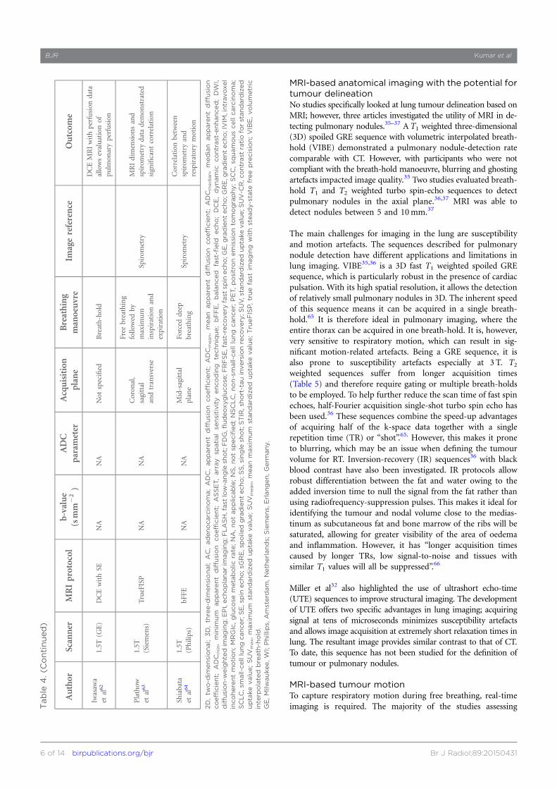
MRI-based anatomical imaging with the potential fortumour delineationNo studies specifically looked at lung tumour delineation based onMRI; however, three articles investigated the utility of MRI in de-tecting pulmonary nodules.35–37 A T1 weighted three-dimensional(3D) spoiled GRE sequence with volumetric interpolated breath-hold (VIBE) demonstrated a pulmonary nodule-detection ratecomparable with CT. However, with participants who were notcompliant with the breath-hold manoeuvre, blurring and ghostingartefacts impacted image quality.35 Two studies evaluated breath-hold T1 and T2 weighted turbo spin-echo sequences to detectpulmonary nodules in the axial plane.36,37 MRI was able todetect nodules between 5 and 10mm.37
The main challenges for imaging in the lung are susceptibilityand motion artefacts. The sequences described for pulmonarynodule detection have different applications and limitations inlung imaging. VIBE35,36 is a 3D fast T1 weighted spoiled GREsequence, which is particularly robust in the presence of cardiacpulsation. With its high spatial resolution, it allows the detectionof relatively small pulmonary nodules in 3D. The inherent speedof this sequence means it can be acquired in a single breath-hold.65 It is therefore ideal in pulmonary imaging, where theentire thorax can be acquired in one breath-hold. It is, however,very sensitive to respiratory motion, which can result in sig-nificant motion-related artefacts. Being a GRE sequence, it isalso prone to susceptibility artefacts especially at 3 T. T2weighted sequences suffer from longer acquisition times(Table 5) and therefore require gating or multiple breath-holdsto be employed. To help further reduce the scan time of fast spinechoes, half-Fourier acquisition single-shot turbo spin echo hasbeen used.36 These sequences combine the speed-up advantagesof acquiring half of the k-space data together with a singlerepetition time (TR) or “shot”.65. However, this makes it proneto blurring, which may be an issue when defining the tumourvolume for RT. Inversion-recovery (IR) sequences36 with blackblood contrast have also been investigated. IR protocols allowrobust differentiation between the fat and water owing to theadded inversion time to null the signal from the fat rather thanusing radiofrequency-suppression pulses. This makes it ideal foridentifying the tumour and nodal volume close to the medias-tinum as subcutaneous fat and bone marrow of the ribs will besaturated, allowing for greater visibility of the area of oedemaand inflammation. However, it has “longer acquisition timescaused by longer TRs, low signal-to-noise and tissues withsimilar T1 values will all be suppressed”.
66
Miller et al32 also highlighted the use of ultrashort echo-time(UTE) sequences to improve structural imaging. The developmentof UTE offers two specific advantages in lung imaging; acquiringsignal at tens of microseconds minimizes susceptibility artefactsand allows image acquisition at extremely short relaxation times inlung. The resultant image provides similar contrast to that of CT.To date, this sequence has not been studied for the definition oftumour or pulmonary nodules.
MRI-based tumour motionTo capture respiratory motion during free breathing, real-timeimaging is required. The majority of the studies assessingT
able
4.(C
ontinued)
Author
Scanner
MRIprotocol
b-value
(smm
22)
ADC
parameter
Acquisition
plane
Breathing
manoeuvre
Imagereference
Outcom
e
Iwasaw
aet
al62
1.5T
(GE)
DCEwithSE
NA
NA
Not
specified
Breath-hold
DCEMRIwithperfusion
data
allowsevaluationof
pulm
onaryperfusion
Plathow
etal63
1.5T
(Siemens)
TrueFISP
NA
NA
Coron
al,
sagittal
andtransverse
Free
breathing
followed
bymaxim
um
inspirationand
expiration
Spirom
etry
MRIdimension
sand
spirom
etry
data
demon
strated
significantcorrelation
Shiabata
etal64
1.5T
(Philips)
bFFE
NA
NA
Mid-sagittal
plane
Forced
deep
breathing
Spirom
etry
Correlation
between
spirom
etry
and
respiratorymotion
2D,tw
o-d
imensional;
3D,th
ree-d
imensional;
AC,adenocarcinoma;ADC,apparentdiffusion
coefficient;
ADCmean,mean
apparentdiffusion
coefficient;
ADCmedian,median
apparentdiffusion
coefficient;
ADCmin,minim
um
apparentdiffusion
coefficient;
ASSET,array
spatialse
nsitivity
encoding
technique;bFFE,balanced
fast-field
echo;DCE,dynamic
contrast-enhanced;DW
I,diffusion-w
eighte
dim
aging;E
PI,echoplanarim
aging;F
LASH,fast
low-angle
shot;FDG,fludeoxyglucose
;FRFSE,fast-recovery
fast
spin
echo;G
E,g
radientecho;G
RE,g
radientecho;IVIM
,intravoxel
incoherentmotion;MRGIc,glucose
metabolic
rate;NA,notapplic
able;NS,notsp
ecified;NSCLC,non-small-celllung
cancer;
PET,positronemissionto
mography;SCC,sq
uamouscellcarcinoma;
SCLC,s
mall-celllungcancer;SE,s
pin
echo;s
GRE,s
poile
dgradientecho;S
S,s
ingle
shot;STIR,s
hort-tauinversionrecovery;S
UV,s
tandard
izeduptakevalue;S
UV-C
R,c
ontrast
ratiofo
rstandard
ized
uptakevalue;SUV
max,maxim
um
standard
ized
uptakevalue;SUV
mean,mean
maxim
um
standard
ized
uptakevalue;Tru
eFISP,truefast
imaging
with
steady-state
freeprecision;VIBE,volumetric
interp
olated
breath
-hold.
GE,Milw
aukee,W
I;Phillips,
Amsterd
am,Neth
erlands;
Siemens,
Erlangen,Germ
any.
BJR Kumar et al
6 of 14 birpublications.org/bjr Br J Radiol;89:20150431

tumour motion utilized a variation of GRE sequences.38–44
While there is evidence to suggest that MRI data are prone togeometric distortion in the presence of motion,30 two studiesdemonstrated results contrary to this.41,45 Imaging in the sagittaland coronal planes demonstrated minimal error when comparedwith the axial plane using a thoracic phantom and a fastgradient-echo (fGRE) sequence.41 The integrity of the structureswas maintained on free-breathing real-time MRI scans, whilea large error was noted for intracycle tidal volume re-producibility on breath-hold scans.45
fGRE sequences were shown to be feasible in assessing lungmotion; however, pulmonary vessels rather than lung tumourwere used for assessment.42,46 Respiratory mechanics of the lungand tumour can also be assessed with a true fast imaging withsteady-state precession (TrueFISP) sequence.43,44 Plathow et al44
demonstrated a variation in motion between tumour- and non-tumour-bearing hemithorax with the motion magnitude varyingaccording to the tumour location. Variation in the tumour
motion before and after RT was also assessed and showed nochange, although there was a reduction in the craniocaudalmotion of the tumour-bearing hemithorax.43 The change in thecraniocaudal motion of the tumour-bearing hemithorax was notreflected in spirometry results.
Two studies compared the motion measured on real-timeMRI with that seen on 4DCT with conflicting results.38,39 Ina phantom study using GRE sequences, Bieder et al38 showedthat the lesion diameter was larger but the lesion displace-ment smaller on MRI than that on 4DCT. Cai et al39 in-vestigated the internal target volume error between real-timeMRI and simulated 4DCT data. The results indicated thatowing to the nature of 4DCT acquisition, the excursion oftumour motion may not be accurately depicted on a 4DCTscan. The magnitude of internal target volume error corre-lated with the variability in participants’ breathing. Acquiringtumour motion data over a prolonged period was shown to bemore accurate than limited breathing cycles.40
Table 5. MRI sequence adaption for lung imaging
Sequencedescription
Type Challenges for lung Further improvements
T2 weighted anatomyFast spin echo with gating (e.g. TSE/FSE or HASTE)
Long scan time requires gating ormultiple breath-holds
Self-navigation using amplitudeor phase
T1 weighted anatomy3D volume gradient echo (e.g. VIBE,LAVA) without gating
Breath-hold durationParallel imaging, partial k-space toreduce time of scan
Real-time motionSteady state (e.g. TrueFISP,bSSFP, FIESTA)
Off-resonance and cardiac artefactsSelect appropriate FOV and usespecific cardiac shim
Diffusion Echoplanar imaging EPI artefacts; low spatial resolutionShaped excitation to reduce volume ofthe tissue
PerfusionDynamic fast gradient echo (e.g.FLASH, FSPGR) with contrast
Has to be run in one or multiplebreath-holds depending onrequirements
Radial k-space to reduce motionartefacts with/without motioncorrection
3D, three-dimensional; EPI, echoplanar imaging; FIESTA, fast imaging employing steady-state acquisition; FLASH, fast low-angle shot; FSE, fast spinecho; FSPGR, fast spoiled gradient recall echo; FOV, field of view; HASTE, half-Fourier acquisition single-shot turbo spin echo; LAVA, liver acquisitionwith volume acceleration; TrueFISP, true fast imaging with steady-state precession; TSE, turbo spin echo; VIBE, volumetric interpolated breath-hold.
Figure 2. 3-T coronal true fast imaging with steady-state precession (TrueFISP) images without (a) and with (b) dedicated cardiac
shim to minimize off-resonance artefacts.
Review article: MRI in lung radiotherapy BJR
7 of 14 birpublications.org/bjr Br J Radiol;89:20150431

Steady-state-free precession sequences are ultrafast GREsdesigned around very short TRs and have demonstrated therequired temporal and spatial resolution to acquire multipleimages during free breathing to allow quantification of tu-mour motion.65 These sequences are highly dependent onfield homogeneity; it is therefore essential to performshimming of the heart prior to acquisition to help minimizeoff-resonance artefacts.65 Figure 2 demonstrates a TrueFISPsequence with and without the cardiac shim, which helpsminimize artefacts. Trade-offs in temporal resolution be-tween two-dimensional (100ms) and 3D imaging (1 s) canlead to blurring, unless a slow-breathing manoeuvre isperformed.67
For tumour motion assessment, an imaging protocol with ul-trafast GRE sequences and parallel imaging is appropriate(Table 5). These usually consist of steady-state sequences (suchas TrueFISP, steady-state free precession etc.) for optimumtemporal resolution.
MRI-based functional imagingCurrently, functional imaging in lung RT satisfies two purposes,identification of nodal disease and differentiation between thetumour and surrounding consolidation. This, in most cases, isachieved with FDG-PET imaging. The most commonly employed
functional MRI techniques are diffusion-weighted imaging (DWI)and dynamic contrast-enhanced (DCE) imaging. Studies lookingat functional imaging included both the tumour and healthylung tissue.
DWI is based on sensitizing the sequence to the motion of watermolecules at a microscopic level (described by the b-value of theimage). This motion may be quantified by generating parametricmaps using at least two different b-value images and calculatingthe apparent diffusion coefficient (ADC). Qualitative in-terpretation of DWI is based on the visual assessment of signalintensity on a high b-value image set; a region of high signalintensity depicts restricted diffusion in the extracellular space.Areas of restricted diffusion will translate to areas of low valueson the resulting ADC map.
A number of studies utilized DWI to assess the presence ofmalignant lymph nodes47–51 and detection of tumour in thepresence of consolidation.52,53 Most studies compared DWI withFDG-PET imaging. DWI was not able to improve the detectionof metastatic mediastinal nodal disease for lung cancer com-pared with PET;47,48 however, it had higher specificity in thepresence of inflammation.48 Pauls et al49 showed that DWI had80% agreement with PET for nodal stage with 15% of the casesunderstaged and 5% of the cases overstaged. In those cases
Figure 3. Axial diffusion-weighted imaging (echoplanar imaging with ZOOMit) with b-value 0 (a), 250 (b), 500 (c) and 750 (d) of
a 69-year-old male patient with lung cancer diagnosed with large-cell carcinoma of the left upper lobe. There is a marked area of
hyperintensity in the left upper lobe characteristic of restricted diffusion. Artefacts are present, but the image quality is sufficient for
tumour edge definition (d) and apparent diffusion coefficient measurement (e).
BJR Kumar et al
8 of 14 birpublications.org/bjr Br J Radiol;89:20150431
where MRI overstaged the nodal disease, restricted diffusion wasnoted in both mediastinal and supraclavicular lymph nodes(4–7mm), with no evidence of elevated glucose metabolism.Nodal disease was adjacent to the primary tumour volume, incases where MRI understaged the disease.49 It should be notedthat neither study had pathological correlation of the imagingresults; both were only assessing the agreement between the twoimaging modalities.48,49 Therefore, there is potential for PET tobe false negative in the case of small tumours.
DWI can also differentiate between malignant and benign me-diastinal lymph nodes.50 Malignant node detection on pre-operative DWI was compared with histologically confirmedmalignant lymph node status post operatively.51 A whole-bodyversion of DWI termed DWIBS (diffusion-weighted imagingwith background signal suppression) has been used to produceimages that are visually similar to FDG-PET,51 and the visualdetection of malignant nodes on the resultant images was sig-nificantly higher for both enlarged and normal-size lymphnodes. ADC values also correlated with the visual de-tection rate.51
Two studies demonstrated the potential of DWI to differentiatelung cancer from consolidation. Yang et al53 compared DWIwith FDG-PET. DWI was able to detect the difference betweentumour and consolidation in all patient cases, based on thehyperintensity of the tumour. The ADC map also demonstratedlower values in the presence of the tumour. An intravoxel in-coherent motion (IVIM) sequence was also able to differentiatebetween tumour and consolidation as compared with both DCEand FDG-PET.52 IVIM is a modified DWI technique in whichimages are acquired with lower than conventional b-values thatare sensitive to blood microcirculation. Both DCE and IVIMwere able to distinguish between cancer and consolidation;however, there was a poor correlation between IVIM and DCEparameters.
There is potential for the ADC map to detect early treatment-related changes better than FDG-PET.54,55 Increase in the ADCvalue in the early phase of treatment correlated with final tu-mour size reduction, indicating potential use in detecting earlytreatment response.56 Median progression-free survival inpatients with increased ADC change was shown to be12.0 months compared with 6.7 months for those patients whereADC remained stable or decreased.56 To predict disease pro-gression following stereotactic RT for stage I non-small-cell lungcancer, Iizuka et al57 performed pre-treatment DWI and FDG-PET. Patients with low ADC value and higher SUVmax hadgreater disease progression, but results were not statisticallysignificant. From a slightly different perspective, Chen et al58
demonstrated an inverse relationship between minimum andmean ADC values and tumour cellularity.
The main application of DWI has primarily been in imagingneurological disorders. However, there is increasing evidence toutilize it in imaging for cancer detection and treatment moni-toring.68 Echoplanar imaging (EPI) sequences are commonlyused for DWI but are prone to susceptibility distortion andghosting artefacts.65 While a breath-hold scan can be performed
with EPI to eliminate motion artefact using only a singleb-value, it is more common practice to acquire two or moreb-values for the quantification of tumour diffusion. There ispotential to improve EPI for lung DWI (Table 5) in the lung, byreducing the volume of the excited tissue and limiting artefactsfrom tissue outside this field of view. Figure 3 illustrates anexample of DWI using EPI combined with reduced excitation.
DCE involves the acquisition of images before, during and afteradministration of a suitable contrast agent. Data may be evalu-ated and quantified in a number of ways, from simple meas-urements to complex pharmacokinetic modelling.69 Highlyperfused regions demonstrate a high and rapid uptake andwashout of gadolinium-based contrast. DCE for perfusion as-sessment of lung cancer requires high temporal resolution inorder to adequately assess the enhancement of the tumourvolume. fGRE sequences are generally utilized with partial orshared k-space approaches to optimize temporal resolution andrun with multiple short breath-hold manoeuvres over the re-quired time course. It has recently become feasible to acquirethis data during free breathing by using a radial stack of stars(Table 5), sampling scheme to compensate or even correct formotion.70 Free-breathing perfusion data have been shown to beas reproducible as breath-hold and also better tolerated. How-ever, further investigation is required for adaption in lungimaging.71
Two studies highlighted a possible relationship between meta-bolic activity and cellularity.59,60 Tumour vascularity59 on DCEand restricted diffusion60 as measured on ADC were found to becorrelated to increased FDG uptake or SUVmax on PET scans.Hunter et al59 also demonstrated changes in vascular physiologywhich were apparent during and after treatment, highlightingthe potential role in clinical management. DCE perfusionparameters can also allow identification of histological subtypesfor lung cancer.61 A number of contrast-uptake parameters wereused to investigate correlation with tumour subtypes, and theseplayed a significant role in differentiating non-small-cell lungcancer (NSCLC) from small-cell lung cancer (SCLC). Time-dependent kinetic parameters were more relevant in differenti-ating adenocarcinoma from squamous-cell carcinoma.
Functional MRI data can also be used to assess healthy lungfunction prior to the course of treatment. Iwasawa et al62 eval-uated whether functional MRI could predict post-operative lungfunction. A correlation was seen between the perfusion ratio onMRI and radionuclide study (scintigraphy) and also between the
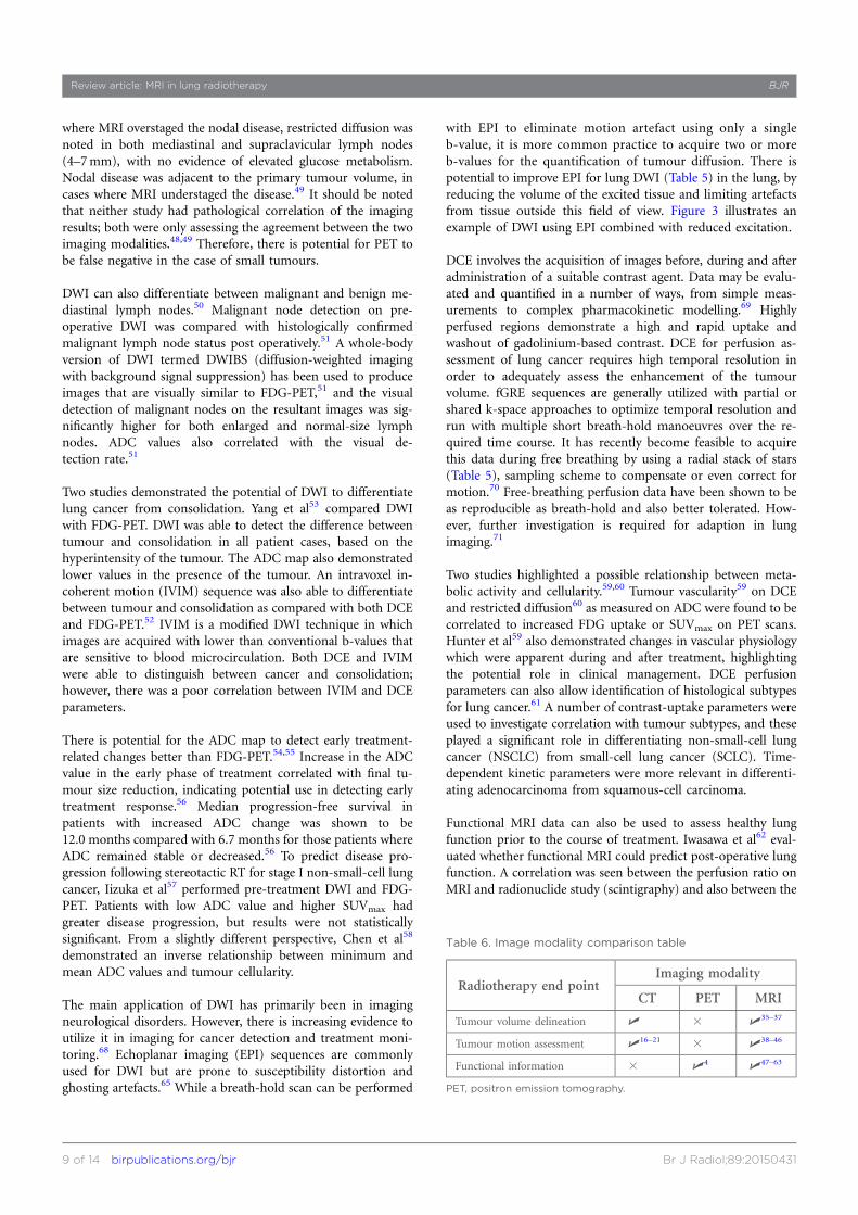
Table 6. Image modality comparison table
Radiotherapy end pointImaging modality
CT PET MRI
Tumour volume delineation U 3 U35–37
Tumour motion assessment U16–21 3 U38–46
Functional information 3 U4 U47–63
PET, positron emission tomography.
Review article: MRI in lung radiotherapy BJR
9 of 14 birpublications.org/bjr Br J Radiol;89:20150431

predicted FEV1 on MRI and the tested FEV1 post operatively.MRI can also be a useful tool in evaluating respiratory me-chanics and volumetry. Using the diaphragm and chest wallmotion as measured on MRI imaging, Plathow et al63 createda volumetric model to calculate vital capacity and compared thiswith spirometry-based vital capacity measurement as lungfunction tests. They were able to show a strong correlation be-tween the vital capacity measurement on MRI and that mea-sured on spirometry. Similarly, Shibata et al64 investigated thedifference in respiratory motion between healthy individuals andpatients with chronic obstructive pulmonary disease, trackingpulmonary vessels using an in-house developed algorithm. MRI-based respiratory motion correlated strongly with spirome-try data.
While not yet in routine clinical use, MRI using hyperpolarizedgases or oxygen enhancement can allow for ventilation studies ofthe lung.34 For hyperpolarized imaging, two noble gases arepredominantly used, 3-helium (3He) and 129-xenon (129Xe).3He has been used to asses ventilation in post-treatmentassessment72,73 or identifying well-ventilated lung for avoid-ance of functional lung at the time of treatment planning.74,75129Xe has the added advantage in that it can be imaged followingventilation perfusion and it has been shown to highlight gasexchange impairment.76 Oxygen-enhanced MRI utilizes pureoxygen as the contrast agent to study ventilation abnormalities.77
DISCUSSIONImprovements in MRI technology continue to enable the im-aging of low-proton-density lung tissue in the presence of re-spiratory and cardiac motion. The main challenges of lung RTare accurate identification of the GTV and nodal volume, par-ticularly in the presence of pathological changes in the sur-rounding lung tissue. Accurate measurement of tumour motionis also necessary to ensure that the RT field encompasses ap-propriate margins. MRI has the potential to help overcome boththese challenges. Table 6 highlights the potential application ofMRI in the RT process of lung cancer.
However, the universal application of MRI in lung RT is cur-rently limited by the ability to generate treatment plans on MRIdata sets and create reference images for treatment verification.Currently, MRI is used for specific lung cancer cases (e.g. Pan-coast tumours) to aid in anatomical delineation; however, theCT data set is still used for treatment planning. The imageregistration process between CT and MRI is prone to errorowing to changes in the position and shape of the organ andtumour, even if the scans are contemporaneous. A MRI-only RTworkflow is being considered for many RT sites and will benecessary for MRI-guided RT systems such as the MRI linearaccelerator. RT treatment-planning systems still require CT orCT-equivalent data for dose calculation. A number of methodshave been proposed to derive CT equivalent data from MRimages.78 Assigning bulk density to the entire patient anatomy isthe simplest solution, and has been shown to give acceptabledosimetric accuracy.79 The previously mentioned UTE sequen-ces may also have a role in improving bone and tissue classifi-cation and direct CT conversion approaches, which makes theirapplication in the lung even more interesting.79
In addition, for lung GTV delineation, detection of a pulmonarynodule or mass is just as important as being able to define theboundary of the nodule or mass. To allow volume definition,tumour infiltration into the chest wall and or mediastinum isrequired along with detection of involved mediastinal lymphnodes. A combination of sequences is required to facilitate GTVdelineation. T2 weighted images such as half-Fourier acquisitionsingle-shot turbo spin echo (HASTE) or short-tau inversionrecovery (STIR) can demonstrate tumour infiltration. T1weighted images are ideal for identifying mediastinal lymphnodes. A limitation of these sequences for anatomical detectionis that they are either breath-hold or respiratory-gated images.The exhale phase in respiratory gating with bellows or naviga-tion is generally used, as this is the longest period of relaxationduring the respiratory cycle. Knowledge regarding the de-formation of tumour volume during the respiratory cycle isimportant. Hence, a more dynamic approach to imaging is re-quired or at the very least anatomical scans at inspiration andexpiration to capture the position and shape of the tumour atthe extremes of the respiratory cycle. Imaging requirements forRT are different from those of diagnostic imaging. Imaging forRT serves the purpose of tumour and associated nodal volumedelineation rather than detection and staging via diagnosticimaging. As such, these anatomical image sequences need to befurther assessed in a radiation oncology setting.
There is evidence supporting the value of MRI in quantifyinglung tumour motion. However, the data reported are pre-dominantly based on sagittal and coronal planes, imaging planesthat in most cases are not compatible with the majority of RT-planning systems. Furthermore, image acquisition is generally intwo dimensions with either a single plane or the given numberof planes through the region of interest. This can potentiallyneglect any out-of-plane tumour motion during respiration andintroduce geometric errors in the planning process. Furtherstudy into 3D registration and incorporation of non-axial datainto treatment-planning systems is needed. While 4DCTremainsthe gold standard for lung tumour motion, it is limited by thenumber of breathing cycles acquired and any irregularity inbreathing. Real-time MRI can be acquired over a greater numberof respiratory cycles to better understand and capture motionover time and is not influenced by irregular breathing patternsas 4DCT currently is.
In terms of functional imaging, FDG-PET remains the goldstandard in defining metabolically active disease, particularly inmediastinal nodes and in the presence of consolidation. There isevidence to support DWI with ADC mapping and DCE MRI asan alternative to FDG-PET for assessing functional tumour ac-tivity. However, studies to date have all been from a diagnosticperspective, where the end points are disease presence or ab-sence. For RT, the definition of the malignant target volume isnecessary. Further research is needed to determine whether MRIwill be a complementary or competing technology for PETimaging in the lung. The availability of hybrid PET-MR systemsalso offers promise for lung imaging.80
While the use of ADC and DWI imaging has been reportedon extensively for lung cancer, it should be noted that
BJR Kumar et al
10 of 14 birpublications.org/bjr Br J Radiol;89:20150431
reproducibility of quoted ADC parameters such as the mini-mum, maximum and mean is a consideration. Kivrak et al81
demonstrated this in their study of ADC values across six dif-ferent MRI scanners with a phantom. However, rather thanlooking at ADC values, the analysis of histogram distributions81
shows potential and may be better for comparing data acquiredon different scanners.82 Conversely, DWI is reproducible be-tween scanners and has shown good interobserver and intra-observer agreement for lung cancer for tumour sizes .2 cm.83
The ability to differentiate histological subtypes is potentiallyuseful for patients in whom biopsy confirmation of lung canceris not possible, usually owing to underlying lung disease. His-tological subtype is important in determining treatment, andMRI parameters could have a potential role in this.61
However, improvement in the standard EPI technique is re-quired to overcome some of the current limitations of suscep-tibility and motion artefacts.
Fundamentally, breath-hold MRI scans are challenging forpatients with lung cancer owing to their already limited re-spiratory function. Further improvements in image technologyand navigation for gating may make breath-hold scans obsoletein these patients.
Lung function tests prior to the start of any treatment to someextent dictate whether a patient is able to receive radical orpalliative RT. This is usually based on spirometry to assess lungfunction. However, two studies63,64 highlighted the potential ofMRI to predict lung function in patients, which could be usefulin patients who cannot undergo spirometry. Hyperpolarized gas-and oxygen-enhanced MRI can potentially allow for the analysis
of lung microstructure and quantify ventilation and perfusion ofthe lung. MRI scans can thus provide an anatomical and functionalrepresentation of pulmonary function, which could potentially beused in RT planning by avoiding areas of a well-functioning lungand assessing treatment response. However, both techniques arecurrently restricted to research settings with limited clinical use.
MRI shows potential for monitoring of early response duringa course of treatment, information which is currently not uti-lized in RT. There is evidence in the literature to suggest a linkbetween early changes as seen on functional MRI and progression-free survival.56 This information can be used to adapt treatmentto an individual patient’s tumour response.
CONCLUSIONUsing a combination of free breathing, breath-hold and gatedscans with parallel imaging techniques, the quality of lung im-aging has improved, with minimal artefacts from respiratory andcardiac motion. There are still challenges in adopting MRI forRT imaging but nevertheless based on the evidence available inthe literature, a potential lung RT-imaging protocol can includefor GTV delineation a T1 and T2 weighted gradient and spin-echo sequence either as breath-hold or respiratory gated. Motionassessment is feasible; however, the incorporation of the motiondata to RT planning needs further investigation. DWI can proveto be an ideal non-invasive imaging technique to assess func-tional information and assist in GTV delineation. A DCE scanhas the potential to provide additional information on the vas-cular nature of the tumour volume and healthy lung perfusion.However, imaging sequences need to be further assessed in theradiation oncology setting to evaluate and further developRT-specific requirements.
REFERENCES
1. Armstrong JG. Target volume definition for
three-dimensional conformal radiation ther-
apy of lung cancer. Br J Radiol 1998; 71:
587–94. doi: http://dx.doi.org/10.1259/
bjr.71.846.9849380
2. Mac Manus MP, Hicks RJ. The role of
positron emission tomography/computed
tomography in radiation therapy planning
for patients with lung cancer. Semin Nucl
Med 2012; 42: 308–19. doi: http://dx.doi.org/
10.1053/j.semnuclmed.2012.04.003
3. Haasbeek CJA, Slotman BJ, Senan S. Radio-
therapy for lung cancer: Clinical impact of
recent technical advances. Lung Cancer 2009;
64: 1–8. doi: http://dx.doi.org/10.1016/j.
lungcan.2008.07.008
4. Caldwell CB, Mah K, Ung YC, Danjoux CE,
Balogh JM, Ganguli SN, et al. Observer
variation in contouring gross tumor volume
in patients with poorly defined non-small-
cell lung tumors on CT: the impact of
18FDG-hybrid PET fusion. Int J Radiat Oncol
Biol Phys 2001; 51: 923–31. doi: http://dx.doi.
org/10.1016/S0360-3016(01)01722-9
5. Ashamalla H, Rafla S, Parikh K, Mokhtar B,
Goswami G, Kambam S, et al. The contri-
bution of integrated PET/CT to the evolving
definition of treatment volumes in radiation
treatment planning in lung cancer. Int J
Radiat Oncol Biol Phys 2005; 63: 1016–23.
doi: http://dx.doi.org/10.1016/j.
ijrobp.2005.04.021
6. Bradley J, Thorstad WL, Mutic S, Miller TR,
Dehdashti F, Siegel BA, et al. Impact of FDG-
PET on radiation therapy volume delineation
in non-small-cell lung cancer. Int J Radiat
Oncol Biol Phys 2004; 59: 78–86. doi: http://
dx.doi.org/10.1016/j.ijrobp.2003.10.044
7. Spratt DE, Diaz R, McElmurray J, Csiki I,
Duggan D, Lu B, et al. Impact of FDG PET/CT
on delineation of the gross tumor volume for
radiation planning in non-small-cell lung cancer.
Clin Nucl Med 2010; 35: 237–43. doi: http://dx.
doi.org/10.1097/RLU.0b013e3181d18eb0
8. Fox JL, Rengan R, O’Meara W, Yorke E, Erdi
Y, Nehmeh S, et al. Does registration of PET
and planning CT images decrease interob-
server and intraobserver variation in de-
lineating tumor volumes for non-small-cell
lung cancer? Int J Radiat Oncol Biol Phys
2005; 62: 70–5. doi: http://dx.doi.org/
10.1016/j.ijrobp.2004.09.020
9. Deniaud-Alexandre E, Touboul E, Lerouge D,
Grahek D, Foulquier JN, Petegnief Y, et al.
Impact of computed tomography and 18F-
deoxyglucose coincidence detection emission
tomography image fusion for optimization of
conformal radiotherapy in non-small-cell
lung cancer. Int J Radiat Oncol Biol Phys
2005; 63: 1432–41. doi: http://dx.doi.org/
10.1016/j.ijrobp.2005.05.016
10. MacManus M, Nestle U, Rosenzweig KE,
Carrio I, Messa C, Belohlavek O, et al. Use
of PET and PET/CT for radiation therapy
planning: IAEA expert report 2006–2007.
Radiother Oncol 2009; 91: 85–94. doi:
Review article: MRI in lung radiotherapy BJR
11 of 14 birpublications.org/bjr Br J Radiol;89:20150431

http://dx.doi.org/10.1016/j.
radonc.2008.11.008
11. Nestle U, Walter K, Schmidt S, Licht N,
Nieder C, Motaref B, et al. 18 F-Deoxyglucose
positron emission tomography (FDG-PET)
for the planning of radiotherapy in lung
cancer: high impact in patients with atelectasis.
Int J Radiat Oncol Biol Phys 1999; 44: 593–7.
doi: http://dx.doi.org/10.1016/S0360-
3016(99)00061-9
12. Senan S, De Ruysscher D. Critical review of
PET-CT for radiotherapy planning in lung
cancer. Crit Rev Oncol Hematol 2005; 56:
345–51. doi: http://dx.doi.org/10.1016/j.
critrevonc.2005.05.001
13. Steenbakkers RJ, Duppen JC, Fitton I,
Deurloo KE, Zijp LJ, Comans EFI, et al.
Reduction of observer variation using
matched CT-PET for lung cancer delineation:
a three-dimensional analysis. Int J Radiat
Oncol Biol Phys 2006; 64: 435–48. doi: http://
dx.doi.org/10.1016/j.ijrobp.2005.06.034
14. De Ruysscher D, Belderbos J, Reymen B, van
Elmpt W, van Baardwijk A, Wanders R, et al.
State of the art radiation therapy for lung
cancer 2012: a glimpse of the future. Clin
Lung Cancer 2013; 14: 89–95. doi: http://dx.
doi.org/10.1016/j.cllc.2012.06.006
15. Nestle U, Kremp S, Schaefer-Schuler A,
Sebastian-Welsch C, Hellwig D, Rube C, et al.
Comparison of different methods for de-
lineation of 18F-FDG PET-Positive tissue for
target volume definition in radiotherapy of
patients with non-small cell lung cancer. J
Nucl Med 2005; 46: 1342–8.
16. Keall PJ, Mageras GS, Balter JM, Emery RS,
Forster KM, Jiang SB, et al. The management
of respiratory motion in radiation oncology
report of AAPM Task Group 76. Med Phys
2006; 33: 3874–900. doi: http://dx.doi.org/
10.1118/1.2349696
17. Ford E, Mageras G, Yorke E, Ling C.
Respiration-correlated spiral CT: a method of
measuring respiratory-induced anatomic
motion for radiation treatment planning.
Med Phys 2003; 30: 88. doi: http://dx.doi.org/
10.1118/1.1531177
18. Balter JM, Ten Haken RK, Lawrence TS,
Lam KL, Robertson JM. Uncertainties in
CT-based radiation therapy treatment
planning associated with patient breathing.
Int J Radiat Oncol Biol Phys 1996; 36:
167–74. doi: http://dx.doi.org/10.1016/
S0360-3016(96)00275-1
19. Keall P. 4-dimensional computed tomogra-
phy imaging and treatment planning. Semin
Radiat Oncol 2004; 14: 81–90. doi: http://dx.
doi.org/10.1053/j.semradonc.2003.10.006
20. Rietzel E, Liu AK, Doppke KP, Wolfgang JA,
Chen AB, Chen GT, et al. Design of 4D
treatment planning target volumes. Int J
Radiat Oncol Biol Phys 2006; 66: 287–95. doi:
http://dx.doi.org/10.1016/j.
ijrobp.2006.05.024
21. Starkschall G, Britton K, McAleer MF, Jeter
MD, Kaus MR, Bzdusek K, et al. Potential
dosimetric benefits of four-dimensional ra-
diation treatment planning. Int J Radiat
Oncol Biol Phys 2009; 73: 1560–5. doi: http://
dx.doi.org/10.1016/j.ijrobp.2008.12.024
22. Sarker J, Chu A, Mui K, Wolfgang JA, Hirsch
AE, Chen GT, et al. Variations in tumor size
and position due to irregular breathing in
4D-CT: a simulation study. Med Phys 2010;
37: 1254. doi: http://dx.doi.org/10.1118/
1.3298007
23. Khoo VS, Joon DL. New developments in
MRI for target volume delineation in radio-
therapy.. Br J Radiol 2006; 79: Spec-15. doi:
http://dx.doi.org/10.1259/bjr/41321492
24. Khoo VS, Dearnaley DP, Finnigan DJ,
Padhani A, Tanner SF, Leach MO. Magnetic
resonance imaging (MRI): considerations
and applications in radiotherapy treatment
planning. Radiother Oncol 1997; 42: 1–15.
doi: http://dx.doi.org/10.1016/S0167-8140
(96)01866-X
25. Dirix P, Haustermans K, Vandecaveye V. The
value of magnetic resonance imaging for
radiotherapy planning. Semin Radiat Oncol
2014; 24: 151–9. doi: http://dx.doi.org/
10.1016/j.semradonc.2014.02.003
26. Liney G, Holloway L. MRI in radiotherapy.
RAD Magazine; 2013. pp. 19–20.
27. Fallone BG. The rotating biplanar linac–
magnetic resonance imaging system. Semin
Radiat Oncol 2014; 24: 200–2. doi: http://dx.
doi.org/10.1016/j.semradonc.2014.02.011
28. Keall PJ, Barton M, Crozier S. The Australian
magnetic resonance imaging–linac program.
Semin Radiat Oncol 2014; 24: 203–6. doi:
http://dx.doi.org/10.1016/j.
semradonc.2014.02.015
29. Lagendijk JJ, Raaymakers BW, van Vulpen M.
The magnetic resonance imaging–linac sys-
tem. Semin Radiat Oncol 2014;; 24: 207–9.
doi: http://dx.doi.org/10.1016/j.
semradonc.2014.02.009
30. Wild J, Marshall H, Bock M, Schad L, Jakob
P, Puderbach M, et al. MRI of the lung (1/3):
methods. Insights Imaging 2012; 3: 345–53.
doi: http://dx.doi.org/10.1007/s13244-012-
0176-x
31. Biederer J, Mirsadraee S, Beer M, Molinari F,
Hintze C, Bauman G, et al. MRI of the lung
(3/3)—current applications and future per-
spectives. Insights Imaging 2012; 3: 373–86.
doi: http://dx.doi.org/10.1007/s13244-011-
0142-z
32. Miller GW, Mugler JP, Sa RC, Altes TA, Prisk
GK, Hopkins SR. Advances in functional and
structural imaging of the human lung using
proton MRI. NMR Biomed 2014; 27:
1542–56. doi: http://dx.doi.org/10.1002/
nbm.3156
33. Biederer J, Beer M, Hirsch W, Wild J, Fabel
M, Puderbach M, et al. MRI of the lung (2/
3). Why… when… how? Insights Into
Imaging 2012; 3: 373–86. doi: http://dx.doi.
org/10.1007/s13244-011-0142-z
34. Wielputz M, Kauczor HU. MRI of the lung:
state of the art. Diagn Interv Radiol 2012; 18:
344–53. doi: http://dx.doi.org/10.4261/1305-
3825.DIR.5365-11.0
35. Biederer J, Both M, Graessner J, Liess C,
Jakob P, Reuter M, et al. Lung morphology:
fast MR imaging assessment with a volumet-
ric interpolated breath-hold technique: initial
experience with patients. Radiology 2003;
226: 242–9. doi: http://dx.doi.org/10.1148/
radiol.2261011974
36. Bruegel M, Gaa J, Woertler K, Ganter C,
Waldt S, Hillerer C, et al. MRI of the lung:
value of different turbo spin-echo, single-
shot turbo spin-echo, and 3D gradient-echo
pulse sequences for the detection of pulmo-
nary metastases. J Magn Reson Imaging 2007;
25: 73–81. doi: http://dx.doi.org/10.1002/
jmri.20824
37. Chin AY, Jeon TY, Lee KS, Lee JH, Seo JB,
Kim YK, et al. 3-T MRI: usefulness for
evaluating primary lung cancer and small
nodules in lobes not containing primary
tumors. AJR Am J Roentgenol 2007;
189: 386–92.
38. Biederer J, Hintze C, Fabel M, Dinkel J.
Magnetic resonance imaging and computed
tomography of respiratory mechanics. J
Magn Reson Imaging 2010; 32: 1388–97. doi:
http://dx.doi.org/10.1002/jmri.22386
39. Cai J, Read PW, Baisden JM, Larner JM,
Benedict SH, Sheng K. Estimation of error
in maximal intensity projection-based internal
target volume of lung tumors: a simulation and
comparison study using dynamic magnetic
resonance imaging. Int J Radiat Oncol Biol Phys
2007; 69: 895–902. doi: http://dx.doi.org/
10.1016/j.ijrobp.2007.07.2322
40. Cai J, Read PW, Larner JM, Jones DR,
Benedict SH, Sheng K. Reproducibility of
interfraction lung motion probability distri-
bution function using dynamic MRI: statis-
tical analysis. Int J Radiat Oncol Biol Phys
2008; 72: 1228–35. doi: http://dx.doi.org/
10.1016/j.ijrobp.2008.07.028
41. Koch N, Liu HH, Olsson LE, Jackson EF.
Assessment of geometrical accuracy of mag-
netic resonance images for radiation therapy
of lung cancers. J Appl Clin Med Phys 2003; 4:
352–64. doi: http://dx.doi.org/10.1120/
1.1617211
42. Liu HH, Koch N, Starkschall G, Jacobson M,
Forster K, Liao Z, et al. Evaluation of internal
BJR Kumar et al
12 of 14 birpublications.org/bjr Br J Radiol;89:20150431
lung motion for respiratory-gated radiother-
apy using MRI: Part II - Margin reduction of
internal target volume. Int J Radiat Oncol Biol
Phys 2004; 60: 1473–83. doi: http://dx.doi.
org/10.1016/j.ijrobp.2004.05.054
43. Plathow C, Hof H, Kuhn S, Puderbach M,
Ley S, Biederer J, et al. Therapy monitoring
using dynamic MRI: analysis of lung motion
and intrathoracic tumor mobility before and
after radiotherapy. Eur Radiol 2006; 16:
1942–50. doi: http://dx.doi.org/10.1007/
s00330-006-0237-y
44. Plathow C, Ley S, Fink C, Puderbach M,
Hosch W, Schmahl A, et al. Analysis of
intrathoracic tumor mobility during whole
breathing cycle by dynamic MRI. Int J Radiat
Oncol Biol Phys 2004; 59: 952–9. doi: http://
dx.doi.org/10.1016/j.ijrobp.2003.12.035
45. Blackall JM, Ahmad S, Miquel ME,
McClelland JR, Landau DB, Hawkes DJ.
MRI-based measurements of respiratory
motion variability and assessment of imaging
strategies for radiotherapy planning. Phys
Med Biol 2006; 51: 4147–69. doi: http://dx.
doi.org/10.1088/0031-9155/51/17/003
46. Koch N, Liu HH, Starkschall G, Jacobson M,
Forster K, Liao Z, et al. Evaluation of internal
lung motion for respiratory-gated radiother-
apy using MRI: part I—correlating internal
lung motion with skin fiducial motion. Int J
Radiat Oncol Biol Phys 2004; 60: 1459–72.
doi: http://dx.doi.org/10.1016/j.
ijrobp.2004.05.055
47. Lichy MPM, Aschoff PMD, Plathow CMD,
Stemmer AM, Horger WM, Mueller-Horvat
CMD, et al. Tumor detection by diffusion-
weighted MRI and ADC-mapping—initial
clinical experiences in comparison to PET-CT.
Invest Radiol 2007; 42: 605–13. doi: http://dx.
doi.org/10.1097/RLI.0b013e31804ffd49
48. Mori T, Nomori H, Ikeda K, Kawanaka K,
Shiraishi S, Katahira K, et al. Diffusion-
weighted magnetic resonance imaging for
diagnosing malignant pulmonary nodules/
masses: comparison with positron emission
tomography. J Thorac Oncol 2008; 3: 358–64.
doi: http://dx.doi.org/10.1097/
JTO.0b013e318168d9ed
49. Pauls S, Schmidt SA, Juchems MS, Klass O,
Luster M, Reske SN, et al. Diffusion-weighted
MR imaging in comparison to integrated
[18F]-FDG PET/CT for N-staging in patients
with lung cancer. Eur J Radiol 2012; 81:
178–82. doi: http://dx.doi.org/10.1016/j.
ejrad.2010.09.001
50. Abdel Razek AA, Elkammary S, Elmorsy AS,
Elshafey M, Elhadedy T. Characterization of
mediastinal lymphadenopathy with
diffusion-weighted imaging. Magn Reson
Imaging 2011; 29: 167–72. doi: http://dx.doi.
org/10.1016/j.mri.2010.08.002
51. Xu L, Tian J, Liu Y, Li C. Accuracy of
diffusion-weighted (DW) MRI with back-
ground signal suppression (MR-DWIBS)
in diagnosis of mediastinal lymph node
metastasis of nonsmall-cell lung cancer
(NSCLC). J Magn Reson Imaging 2013; 40:
200–5. doi: http://dx.doi.org/10.1002/
jmri.24343
52. Wang LL, Lin J, Liu K, Chen CZ, Liu H, Lv P,
et al. Intravoxel incoherent motion diffusion-
weighted MR imaging in differentiation of
lung cancer from obstructive lung consoli-
dation: comparison and correlation with
pharmacokinetic analysis from dynamic
contrast-enhanced MR imaging. Eur Radiol
2014; 24: 1914–22. doi: http://dx.doi.org/
10.1007/s00330-014-3176-z
53. Yang RM, Li L, Wei XH, Guo YM, Huang
YH, Lai LS, et al. Differentiation of central
lung cancer from atelectasis: comparison of
diffusion-weighted MRI with PET/CT. PLoS
ONE 2013; 8: e60279. doi: http://dx.doi.org/
10.1371/journal.pone.0060279
54. Ohno Y, Koyama H, Yoshikawa T, Matsu-
moto K, Aoyama N, Onishi Y, et al.
Diffusion-weighted MRI versus 18F-FDG
PET/CT: performance as predictors of tumor
treatment response and patient survival in
patients with non–small cell lung cancer
receiving chemoradiotherapy. AJR Am J
Roentgenol 2012; 198: 75–82. doi: http://dx.
doi.org/10.2214/AJR.11.6525
55. Chang Q, Wu N, Ouyang H, Huang Y.
Diffusion-weighted magnetic resonance im-
aging of lung cancer at 3.0 T: a preliminary
study on monitoring diffusion changes
during chemoradiation therapy. Clin Imaging
2012; 36: 98–103. doi: http://dx.doi.org/
10.1016/j.clinimag.2011.07.002
56. Yabuuchi H, Hatakenaka M, Takayama K,
Matsuo Y, Sunami S, Kamitani T, et al.
Non–small cell lung cancer: detection of
early response to chemotherapy by using
contrast-enhanced dynamic and diffusion-
weighted MR Imaging. Radiology 2011; 261:
598–604. doi: http://dx.doi.org/10.1148/
radiol.11101503
57. Iizuka Y, Matsuo Y, Umeoka S, Nakamoto
Y, Ueki N, Mizowaki T, et al. Prediction of
clinical outcome after stereotactic body
radiotherapy for non-small cell lung can-
cer using diffusion-weighted MRI and 18F-
FDG PET. Eur J Radiol 2014; 83: 2087–92.
doi: http://dx.doi.org/10.1016/j.
ejrad.2014.07.018
58. Chen L, Zhang J, Chen Y, Wang W, Zhou X,
Yan X, et al. Relationship between apparent
diffusion coefficient and tumour cellularity
in lung cancer. PLoS ONE 2014; 9: e99865.
doi: http://dx.doi.org/10.1371/journal.
pone.0099865
59. Hunter GJ, Hamberg LM, Choi N, Jain RK,
McCloud T, Fischman AJ. Dynamic T1-
weighted magnetic resonance imaging and
positron emission tomography in patients
with lung cancer: correlating vascular phys-
iology with glucose metabolism. Clin Cancer
Res 1998; 4: 949–55.
60. Regier M, Derlin T, Schwarz D, Laqmani A,
Henes FO, Groth M, et al. Diffusion weighted
MRI and 18F-FDG PET/CT in non-small cell
lung cancer (NSCLC): does the apparent
diffusion coefficient (ADC) correlate with
tracer uptake (SUV)? Eur J Radiol 2012; 81:
2913–8. doi: http://dx.doi.org/10.1016/j.
ejrad.2011.11.050
61. Pauls S, Breining T, Muche R, Schmidt SA,
Wunderlich A, Kruger S, et al. The role of
dynamic, contrast-enhanced MRI in differ-
entiating lung tumor subtypes. Clin Imaging
2011; 35: 259–65. doi: http://dx.doi.org/
10.1016/j.clinimag.2010.07.002
62. Iwasawa T, Saito K, Ogawa N, Ishiwa N,
Kurihara H. Prediction of postoperative
pulmonary function using perfusion mag-
netic resonance imaging of the lung. J Magn
Reson Imaging 2002; 15: 685–92. doi: http://
dx.doi.org/10.1002/jmri.10121
63. Plathow C, Ley S, Fink C, Puderbach M,
Heilmann M, Zuna I, et al. Evaluation of
chest motion and volumetry during the
breathing cycle by dynamic MRI in healthy
subjects: comparison with pulmonary func-
tion tests. Invest Radiol 2004; 39: 202–9. doi:
http://dx.doi.org/10.1097/01.
rli.0000113795.93565.c3
64. Shibata H, Iwasawa T, Gotoh T, Kagei S,
Shinohara T, Ogura T, et al. Automatic
tracking of the respiratory motion of lung
parenchyma on dynamic magnetic resonance
imaging: comparison with pulmonary func-
tion tests in patients with chronic obstructive
pulmonary disease. J Thorac Imaging 2011;
27: 387–92. doi: http://dx.doi.org/10.1097/
RTI.0b013e3182242b11
65. Bernstein MA, King KF, Zhou XJ. Handbook
of MRI pulse sequences. Burlington, MA:
Elsevier; 2004.
66. Hashemi R, Bradley W, Lisanti C. MRI: the
basics. Philadelphia, PA: Lippincott Williams
& Wilkins; 2004. pp. 82.
67. Tetzlaff R. Respiratory mechanics and pul-
monary motion. In: Kauczor HU, editor.
MRI of the lung. Medical radiology. Heidel-
berg, Berlin: Springer 2009. pp. 91–103.
68. Bozgeyik Z, Onur MR, Poyraz AK. The role
of diffusion weighted magnetic resonance
imaging in oncologic settings. Quant Imaging
Med Surg 2013; 3: 269–78. doi: http://dx.doi.
org/10.3978/j.issn.2223-4292.2013.10.07
69. Henzler T, Schmid-Bindert G, Schoenberg
SO, Fink C. Diffusion and perfusion MRI of
Review article: MRI in lung radiotherapy BJR
13 of 14 birpublications.org/bjr Br J Radiol;89:20150431
the lung and mediastinum. Eur J Radiol 2010;
76: 329–36. doi: http://dx.doi.org/10.1016/j.
ejrad.2010.05.005
70. Block KT, Chandarana H, Milla S, Bruno M,
Mulholland T, Fatterpekar G, et al. Towards
routine clinical use of radial stack-of-stars 3D
gradient-echo sequences for reducing motion
sensitivity. J Korean Soc Magn Reson Med
2014; 18: 87–106. doi: http://dx.doi.org/
10.13104/jksmrm.2014.18.2.87
71. Ingrisch M, Maxien D, Schwab F, Reiser MF,
Nikolaou K, Dietrich O. Assessment of
pulmonary perfusion with breath-hold and
free-breathing dynamic contrast-enhanced
magnetic resonance imaging: quantification
and reproducibility. Invest Radiol 2014; 49:
382–9. doi: http://dx.doi.org/10.1097/
RLI.0000000000000020
72. Allen AM, Albert M, Caglar HB,
Zygmanski P, Soto R, Killoran J, et al.
Can Hyperpolarized Helium MRI add to
radiation planning and follow-up in lung
cancer? J Appl Clin Med Phys 2011; 12: 3357.
73. Ireland RH, Din OS, Swinscoe JA, Wood-
house N, van Beek EJ, Wild JM, et al.
Detection of radiation-induced lung injury in
non-small cell lung cancer patients using
hyperpolarized helium-3 magnetic resonance
imaging. Radiother Oncol 2010; 97: 244–8.
doi: http://dx.doi.org/10.1016/j.
radonc.2010.07.013
74. Bates EL, Bragg CM, Wild JM, Hatton MQF,
Ireland RH. Functional image-based radio-
therapy planning for non-small cell lung
cancer: a simulation study. Radiother Oncol
2009; 93: 32–6. doi: http://dx.doi.org/
10.1016/j.radonc.2009.05.018
75. Ireland RH, Bragg CM, McJury M,
Woodhouse N, Fichele S, Van Beek EJ,
et al. Feasibility of image registration and
intensity-modulated radiotherapy planning
with hyperpolarized helium-3 magnetic res-
onance imaging for non–small-cell lung
cancer. Int J Radiat Oncol Biol Phys 2007; 68:
273–81. doi: http://dx.doi.org/10.1016/j.
ijrobp.2006.12.068
76. Kauczor HU, Kreitner KF. Contrast-
enhanced MRI of the lung. Eur J Radiol 2000;
34: 196–207. doi: http://dx.doi.org/10.1016/
S0720-048X(00)00199-6
77. Ohno Y, Sugimura K, Hatabu H. Clinical
oxygen-enhanced magnetic resonance
imaging of the lung. Top Magn Reson
Imaging 2003; 14: 237–43. doi: http://dx.
doi.org/10.1097/00002142-
200306000-00004
78. Nyholm T, Jonsson J. Counterpoint: oppor-
tunities and challenges of a magnetic reso-
nance imaging–only radiotherapy work flow.
Semin Radiat Oncol 2014; 24: 175–80. doi:
http://dx.doi.org/10.1016/j.
semradonc.2014.02.005
79. Jonsson JH, Karlsson MG, Karlsson M,
Nyholm T. Treatment planning using MRI
data: an analysis of the dose calculation
accuracy for different treatment regions.
Radiat Oncol 2010; 5: 62. doi: http://dx.doi.
org/10.1186/1748-717X-5-62
80. Partovi S, Kohan A, Rubbert C, Vercher-
Conejero JL, Gaeta C, Yuh R, et al. Clinical
oncologic applications of PET/MRI: a new
horizon. Am J Nucl Med Mol Imaging 2014;
4: 202–12.
81. Kivrak AS, Paksoy Y, Erol C, Koplay M,
Ozbek S, Kara F. Comparison of apparent
diffusion coefficient values among differ-
ent MRI platforms: a multicenter phantom
study. Diagn Interv Radiol 2013; 19: 433–7.
doi: http://dx.doi.org/10.5152/
dir.2013.13034
82. Tsien C, Cao Y, Chenevert T. Clinical
applications for diffusion magnetic resonance
imaging in radiotherapy. Semin Radiat Oncol
2014; 24: 218–26. doi: http://dx.doi.org/
10.1016/j.semradonc.2014.02.004
83. Bernardin L, Douglas N, Collins D, Giles S,
O’Flynn E, Orton M. Diffusion-weighted
magnetic resonance imaging for assess-
ment of lung lesions: repeatability of the
apparent diffusion coefficient measure-
ment. Eur Radiology 2014; 24: 502–11. doi:
http://dx.doi.org/10.1007/s00330-013-
3048-y
BJR Kumar et al
14 of 14 birpublications.org/bjr Br J Radiol;89:20150431