Magnetic properties of bioactive glass-ceramics containing nanocrystalline zinc ferrite
4
Magnetic properties of bioactive glass-ceramics containing nanocrystalline zinc ferrite Rajendra Kumar Singh, A. Srinivasan n Department of Physics, Indian Institute of Technology Guwahati, Guwahati 781039, India article info Article history: Received 5 July 2010 Available online 7 October 2010 Keywords: Glass-ceramic X-ray diffraction Ferrimagnetism Nanocrystalline magnetic material Biomaterial abstract Glass-ceramics with finely dispersed zinc ferrite (ZnFe 2 O 4 ) nanocrystallites were obtained by heat treatment of x(ZnO,Fe 2 O 3 )(65 x)SiO 2 20(CaO,P 2 O 5 )15Na 2 O (6 rx r21 mole%) glasses. X-ray diffraction patterns of the glass-ceramic samples revealed the presence of calcium sodium phosphate [NaCaPO 4 ] and zinc ferrite [ZnFe 2 O 4 ] as major crystalline phases. Zinc ferrite present in nanocrystalline form contributes to the magnetic properties of the glass-ceramic samples. Magnetic hysteresis cycles of the glass-ceramic samples were obtained with applied magnetic field sweeps of 720 kOe and 7500 Oe, in order to evaluate the potential of these glass-ceramics for hyperthermia treatment of cancer. The evolution of magnetic properties in these samples, viz., from a partially paramagnetic to fully ferrimagnetic nature has been explored using magnetometry and X-ray diffraction studies. & 2010 Elsevier B.V. All rights reserved. 1. Introduction Glass-ceramics with ferrimagnetic, ferromagnetic or super- paramagnetic particles dispersed in a bioactive glass matrix are being developed for use as thermo-seeds in hyperthermia treatment of deep seated cancerous tissues such as bone tumours [1–4]. Magnetic induction hyperthermia, which is one of the therapies for cancer treatment, involves localized heating of infected parts of the body by the heat generated by magnetic field cycling of an implanted magnetic material. If the heat treatment is selective (i.e., up to 43 1C) and the application time is controlled, it is possible to destroy the cancerous cells without damaging the healthy cells [5–7]. Since the inclusion of magnetic aggregates in bioglasses or bioglass-ceramics could be a solution for this application, magnetic glass-ceramics are potential candidates [1–4,8] for use as thermo-seeds in magnetic induction hyperthermia treatment. Ferrimagnetic glass-ceramics with zinc–iron ferrite crystallites in CaO–SiO 2 glass [9], Fe 3 O 4 crystallites in MgO–CaO–SiO 2 –P 2 O 5 –CaF 2 [10], Fe 3 O 4 crystallites in Na 2 O–CaO–SiO 2 –P 2 O 5 glass [4,11–14], Fe 3 O 4 crystallites in CaO–SiO 2 –P 2 O 5 glass [15] and Fe 3 O 4 crystallites in CaO–SiO 2 – B 2 O 3 –P 2 O 5 glass [2,16–19] are some of the materials that have been investigated with this objective. These studies have stimulated interest in the development of ferrimagnetic thermo- seeds with higher heat generation ability. ZnFe 2 O 4 and Fe 3 O 4 form a complete series of solid solutions with the chemical formula of Zn x Fe 1 x [Fe 1 x Fe 1+ x ]O 4 or Zn x Fe 3 x O 4 . The solid solution Zn 0.4 Fe 2.6 O 4 , which forms for x ¼ 0.4, exhibits higher saturation magnetization than magnetite (Fe 3 O 4 ) [20–22]. Therefore, glass- ceramics containing Zn 0.4 Fe 2.6 O 4 are expected to generate higher amount of heat for the same applied magnetic field as compared with other systems [4,10]. This higher magnetization of Zn 0.4 Fe 2.6 O 4 is attributed to the occupancy of the tetrahedral sites by Fe 3+ ions accompanied with Zn 2+ ions in the octahedral sites in the surface region of the ultrafine particle [23–25]. A series of glass-ceramics with compositions x(ZnO,Fe 2 O 3 ) (65 x)SiO 2 20(CaO,P 2 O 5 )15Na 2 O mole% have been prepared with the Ca/P and Fe/Zn ratios maintained at 1.67 and 6.5, respectively. While Ca/P ¼ 1.67 corresponds the Ca/P value in the bone mineral hydroxyapatite and hence optimized bioactivity, the latter ratio ensures that optimized amount of the Zn 0.4 Fe 2.6 O 4 phase is formed in all the compositions. In vitro studies performed by immersion of these glass-ceramics in a simulated body fluid showed apatite formation on the surface these glass-ceramics [26]. Evolution of magnetic properties of these glasses as a function of composition has been investigated in order to assess the applicability for these glass-ceramics for hyperthermia application and the results obtained are presented in this paper. 2. Materials and methods Bulk glass samples with composition x(ZnO,Fe 2 O 3 )(65 x)- SiO 2 20(CaO, P 2 O 5 )15Na 2 O(x ¼ 6, 9, 12, 15, 18 and 21 mole%) were first prepared by melt quenching appropriately weighed quan- tities of analytical grade SiO 2 , Na 2 CO 3 , ZnO, Fe 2 O 3 , CaCO 3 and Contents lists available at ScienceDirect journal homepage: www.elsevier.com/locate/jmmm Journal of Magnetism and Magnetic Materials 0304-8853/$ - see front matter & 2010 Elsevier B.V. All rights reserved. doi:10.1016/j.jmmm.2010.09.029 n Corresponding author. Tel.: + 91 361 2582712; fax: + 91 361 2690762. E-mail addresses: [email protected] (R.K. Singh), [email protected] (A. Srinivasan). Journal of Magnetism and Magnetic Materials 323 (2011) 330–333
-
Upload
rajendra-kumar-singh -
Category
Documents
-
view
214 -
download
0
Transcript of Magnetic properties of bioactive glass-ceramics containing nanocrystalline zinc ferrite

Journal of Magnetism and Magnetic Materials 323 (2011) 330–333
Contents lists available at ScienceDirect
Journal of Magnetism and Magnetic Materials
0304-88
doi:10.1
n Corr
E-m
asrini@
journal homepage: www.elsevier.com/locate/jmmm
Magnetic properties of bioactive glass-ceramics containing nanocrystallinezinc ferrite
Rajendra Kumar Singh, A. Srinivasan n
Department of Physics, Indian Institute of Technology Guwahati, Guwahati 781039, India
a r t i c l e i n f o
Article history:
Received 5 July 2010Available online 7 October 2010
Keywords:
Glass-ceramic
X-ray diffraction
Ferrimagnetism
Nanocrystalline magnetic material
Biomaterial
53/$ - see front matter & 2010 Elsevier B.V. A
016/j.jmmm.2010.09.029
esponding author. Tel.: +91 361 2582712; fa
ail addresses: [email protected] (R.K. Sin
iitg.ernet.in (A. Srinivasan).
a b s t r a c t
Glass-ceramics with finely dispersed zinc ferrite (ZnFe2O4) nanocrystallites were obtained by heat
treatment of x(ZnO,Fe2O3)(65�x)SiO220(CaO,P2O5)15Na2O (6rxr21 mole%) glasses. X-ray diffraction
patterns of the glass-ceramic samples revealed the presence of calcium sodium phosphate [NaCaPO4]
and zinc ferrite [ZnFe2O4] as major crystalline phases. Zinc ferrite present in nanocrystalline form
contributes to the magnetic properties of the glass-ceramic samples. Magnetic hysteresis cycles of the
glass-ceramic samples were obtained with applied magnetic field sweeps of 720 kOe and 7500 Oe, in
order to evaluate the potential of these glass-ceramics for hyperthermia treatment of cancer. The
evolution of magnetic properties in these samples, viz., from a partially paramagnetic to fully
ferrimagnetic nature has been explored using magnetometry and X-ray diffraction studies.
& 2010 Elsevier B.V. All rights reserved.
1. Introduction
Glass-ceramics with ferrimagnetic, ferromagnetic or super-paramagnetic particles dispersed in a bioactive glass matrix arebeing developed for use as thermo-seeds in hyperthermiatreatment of deep seated cancerous tissues such as bone tumours[1–4]. Magnetic induction hyperthermia, which is one of thetherapies for cancer treatment, involves localized heating ofinfected parts of the body by the heat generated by magnetic fieldcycling of an implanted magnetic material. If the heat treatment isselective (i.e., up to 43 1C) and the application time is controlled, itis possible to destroy the cancerous cells without damaging thehealthy cells [5–7]. Since the inclusion of magnetic aggregates inbioglasses or bioglass-ceramics could be a solution for thisapplication, magnetic glass-ceramics are potential candidates[1–4,8] for use as thermo-seeds in magnetic inductionhyperthermia treatment. Ferrimagnetic glass-ceramics withzinc–iron ferrite crystallites in CaO–SiO2 glass [9], Fe3O4
crystallites in MgO–CaO–SiO2–P2O5–CaF2 [10], Fe3O4 crystallitesin Na2O–CaO–SiO2–P2O5 glass [4,11–14], Fe3O4 crystallites inCaO–SiO2–P2O5 glass [15] and Fe3O4 crystallites in CaO–SiO2–B2O3–P2O5 glass [2,16–19] are some of the materials that havebeen investigated with this objective. These studies havestimulated interest in the development of ferrimagnetic thermo-seeds with higher heat generation ability. ZnFe2O4 and Fe3O4 forma complete series of solid solutions with the chemical formula of
ll rights reserved.
x: +91 361 2690762.
gh),
ZnxFe1�x[Fe1�xFe1 +x]O4 or ZnxFe3�xO4. The solid solutionZn0.4Fe2.6O4, which forms for x¼0.4, exhibits higher saturationmagnetization than magnetite (Fe3O4) [20–22]. Therefore, glass-ceramics containing Zn0.4Fe2.6O4 are expected to generate higheramount of heat for the same applied magnetic field as comparedwith other systems [4,10]. This higher magnetization ofZn0.4Fe2.6O4 is attributed to the occupancy of the tetrahedralsites by Fe3 + ions accompanied with Zn2 + ions in the octahedralsites in the surface region of the ultrafine particle [23–25].A series of glass-ceramics with compositions x(ZnO,Fe2O3)(65�x)SiO220(CaO,P2O5)15Na2O mole% have been prepared withthe Ca/P and Fe/Zn ratios maintained at 1.67 and 6.5, respectively.While Ca/P¼1.67 corresponds the Ca/P value in the bone mineralhydroxyapatite and hence optimized bioactivity, the latter ratioensures that optimized amount of the Zn0.4Fe2.6O4 phase isformed in all the compositions. In vitro studies performed byimmersion of these glass-ceramics in a simulated body fluidshowed apatite formation on the surface these glass-ceramics[26]. Evolution of magnetic properties of these glasses as afunction of composition has been investigated in order to assessthe applicability for these glass-ceramics for hyperthermiaapplication and the results obtained are presented in this paper.
2. Materials and methods
Bulk glass samples with composition x(ZnO,Fe2O3)(65�x)-SiO220(CaO, P2O5)15Na2O (x¼6, 9, 12, 15, 18 and 21 mole%) werefirst prepared by melt quenching appropriately weighed quan-tities of analytical grade SiO2, Na2CO3, ZnO, Fe2O3, CaCO3 and

R.K. Singh, A. Srinivasan / Journal of Magnetism and Magnetic Materials 323 (2011) 330–333 331
NH4(H2PO4). Proper calcination of the charge was carried out at1073 K for 24 h prior to melting it in a platinum crucible at1623 K. The homogenized melt was subsequently quenched bypouring into pre-heated graphite mould to form glass. The as-quenched glasses were then heat treated at 1073 K for 1 h to formglass-ceramics and then slowly cooled to room temperature. AnX-ray diffractometer (Bruker D8 Advance) was used to confirmthe amorphous nature of the as-quenched glasses and to identifythe crystalline phases in the heat treated glasses. The data bankfrom the International Center for Diffraction Data (ICDD) wasused for phase identification. The average crystallite size of zincferrite [ZnFe2O4] was estimated from the broadening of theprimary (3 1 1) X-ray reflection using Scherrer’s formula [27].Room temperature magnetic hysteresis loop of the samples wasrecorded using a vibrating sample magnetometer (VSM,Lakeshore 7410) operated under magnetic field sweeps of720 kOe and 7500 Oe. The lower field sweep was performedto evaluate the material at clinically amenable field conditions.Temperature dependent magnetization measurements wereperformed using the VSM with a high temperature attachmentand the magnetic susceptibility was obtained from the data.
3. Results and discussion
The X-ray diffraction (XRD) patterns of the glass-ceramicssamples are presented in Fig. 1. Calcium sodium phosphate(NaCaPO4) [PDF#76-1456] and zinc ferrite (ZnFe2O4) [PDF#82-1042] are the two major crystalline phases present in all the glass-ceramic samples. Calcium sodium phosphate is a bone mineraland its presence indicates the biocompatible nature of the glass-ceramic samples. The amount of calcium sodium phosphate phasedecreased as the zinc-iron oxide concentration increased. The
Fig. 1. X-ray diffraction patterns of glass-ceramic samples derived from glasses
with composition x(ZnO, Fe2O3)(65�x)SiO220(CaO, P2O5)15Na2O (6rxr21
mole% zinc–iron oxide). The two major crystalline phases present are calcium
sodium phosphate [NaCaPO4] (stars) and zinc ferrite [ZnFe2O4] (filled circle).
average crystallite size of ZnFe2O4 (d) increased from 10 nm forthe x¼6 sample to and 25 nm for the x¼21 sample (Table 1).
Fig. 2 depicts the room temperature magnetic hysteresis(M–H) loops of heat treated x(ZnO,Fe2O3)(65�x)SiO220(CaO,P2O5)15Na2O glasses with different zinc–iron oxide concen-tration for magnetic field strength of 720 kOe. The coercivity andremanence are individuated in the expanded view shown in theinset (a) of Fig. 2. The magnetic field necessary to saturate thesamples increases with increase in zinc–iron oxide concentration.It is apparent from Fig. 2 that the samples with x¼6 and 9 mole%zinc–iron oxide do not show a tendency to saturate even at720 kOe. The initial magnetization curves of these samplesshown as inset (b) in Fig. 2 exemplify this aspect. It can be inferredfrom the nature of these two curves that the samples with x¼6and 9 mole% zinc–iron oxide exhibit a combination of linear(paramagnetic) and non-linear (spontaneous magnetization)behavior. In order to establish the ferro/ferri magnetic nature ofthe glass-ceramics samples with x49 mole% zinc–iron oxide,temperature dependent magnetization measurements were per-formed at an applied field of 1 kOe to obtain the temperaturedependent magnetic susceptibility. Variation of inverse magneticsusceptibility (w�1) with temperature over the range 600–900 K isshown in Fig. 3. The w�1 versus T plots, which are nearly linearabove 775 K show a sharp decrease at low temperatures. Thedeviation from linearity and the systematic downward drop ofw�1–T curves indicate the onset of short-range order justabove the ferrimagnetic Neel temperature (TN). Completelyferrimagentic samples exhibit w�1-0 at T¼TN. Samples withx412 are entirely ferrimagnetic as depicted by the nature of thecurves w(T) shown in Fig. 3. TN of the glass-ceramic samplesincreases from 693 K to 713 K as x is varied from 12 to 21 mole%.Electron paramagnetic resonance (EPR) spectra of samples withx¼6 and 9 show two absorption lines, one centred at gE2.1 andanother at gE4.3 [26]. But, samples with x49 show only oneabsorption centred at gE2.1. The disappearance of the gE4.3peak in the EPR spectra of samples with x49 is attributed toexistence of ferrimagnetically coupled clusters in these samples.These EPR studies support the existence of ferrimagnetic orderingin these glass-ceramics and the gradual improvement from partialto fully ferrimagnetic behavior in these samples as the zinc–ironoxide concentration is increased.
Fig. 4 displays the relevant magnetic parameters [saturationmagnetization (MS), coercivity (HC), remanence (Mr) and areaunder the hysteresis loop] obtained from M–H loops shown inFig. 2. MS (Fig. 4a) increases with increase in zinc–iron oxideconcentration from x¼6 to 21 mole% and shows a trend tosaturate for x¼21 with the maximum value of about 19.6 emu/g.The increase in saturation magnetization with increase in zinc–iron oxide concentration could be attributed to the developmentof ZnFe2O4 phase in the samples, as observed in Fig. 1. Magneticanisotropy, shape and dimension of the crystals, residual stressand crystal imperfections influence the coercivity and remanencemagnetization [28]. The coercivity of the samples (Fig. 4b)decreases with increase in zinc–iron oxide concentration. Thelinear variation of HC versus 1/d plot shown in Fig. 5 confirms theinfluence of crystalline size on the coercive field. The coercivefield slowly decreases with increase in average crystallite size ofZnFe2O4 as shown in Table 1. ZnxFe3�xO4 has a magnetite-likestructure in which some of the iron ions in the tetrahedral site ofthe magnetite crystal are substituted by zinc ions. In the case ofFe3O4 nanocrystals, the degree of ordering of the magneticmoment in the individual crystallites increases with increase incrystallite size until a well ordered single domain structure isreached at 40 nm. Further increase in crystallite size increases thenumber of magnetic domains in the individual crystallites,thereby leading to a decrease in coercivity [29]. The remanence

Table 1Magnetic and structural parameters of glass-ceramics derived from glasses with composition x(ZnO, Fe2O3)(65�x)SiO220(CaO, P2O5)15Na2O.
Magnetic and structural parameters Sample (x) (mole% zinc–iron oxide)
x¼6 x¼9 x¼12 x¼15 x¼18 x¼21
Average crystallite size of ZnFe2O4 d (nm) 10 14 16 21 23 25
Saturation magnetization Ms (emu/g) 0.41 1.10 3.33 8.89 13.33 19.60
Coercive force, HC (Oe) 204 149 120 75 52 44
Remanence magnetization Mr (10�2 emu/g) 1.37 1.94 18.16 33.82 58.92 61.81
Interpolated hysteresis area720 kOe (erg/g) 446 1063 2195 5414 7347 9631
Interpolated hysteresis area7500 Oe (erg/g) 6.4 13.6 127.0 287.0 501.0 656.0
Fig. 2. Room temperature magnetic hysteresis loops of glass-ceramics with
different zinc-iron oxide concentration under 720 kOe field sweep. Inset
(a) shows an expanded view of hysteresis loops close to the origin. Inset
(b) displays the initial magnetization curves of the sample with x¼6 and 9 mole%
zinc–iron oxide.
Fig. 3. w�1 versus T plots for glass-ceramic samples x49 mole% zinc–iron oxide.
Fig. 4. Variation of (a) saturation magnetization, (b) coercivity, (c) remanent
magnetization, and (d) area under the hystersis loop under 720 kOe of glass-
ceramics as a function of zinc–iron oxide content.
R.K. Singh, A. Srinivasan / Journal of Magnetism and Magnetic Materials 323 (2011) 330–333332
signifies the ability of a magnetic material to be spontaneouslymagnetized, even in the absence of external magnetic field. Areaof the hysteresis loop (Fig. 4d), increases with increase in zinc–iron oxide content. The largest area is obtained for the samples
with x¼21 mole% sample, which also exhibits the highestsaturation magnetization and lowest coercivity among thesamples studied. Since the area under the hysteresis loop isproportional to the energy loss and hence the heat generated by asample under an alternating field, samples with higher zinc ironoxide concentration are capable of generating more heat.The large variation in the area under the loops for samples withx¼6–21 mole% provides a means for controlled heat generationby appropriate choice of sample. Since high magnetic fields(20 kOe) are difficult to realize in clinical conditions, hysteresiscurves were also obtained using a magnetic field 40 times smaller(i.e., 500 Oe). The corresponding M–H loops are shown in Fig.6.The calculated values of the hysteresis loops area for an appliedfield of 20 kOe and 500 Oe are listed in Table 1. It can be seen that
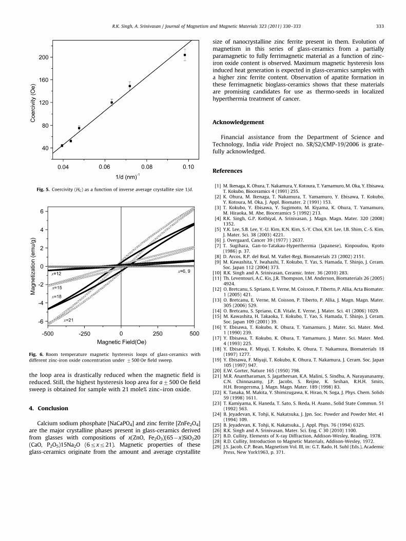
Fig. 5. Coercivity (HC) as a function of inverse average crystallite size 1/d.
Fig. 6. Room temperature magnetic hysteresis loops of glass-ceramics with
different zinc-iron oxide concentration under 7500 Oe field sweep.
R.K. Singh, A. Srinivasan / Journal of Magnetism and Magnetic Materials 323 (2011) 330–333 333
the loop area is drastically reduced when the magnetic field isreduced. Still, the highest hysteresis loop area for a7500 Oe fieldsweep is obtained for sample with 21 mole% zinc–iron oxide.
4. Conclusion
Calcium sodium phosphate [NaCaPO4] and zinc ferrite [ZnFe2O4]are the major crystalline phases present in glass-ceramics derivedfrom glasses with compositions of x(ZnO, Fe2O3)(65�x)SiO220(CaO, P2O5)15Na2O (6rxr21). Magnetic properties of theseglass-ceramics originate from the amount and average crystallite
size of nanocrystalline zinc ferrite present in them. Evolution ofmagnetism in this series of glass-ceramics from a partiallyparamagnetic to fully ferrimagnetic material as a function of zinc-iron oxide content is observed. Maximum magnetic hysteresis lossinduced heat generation is expected in glass-ceramics samples witha higher zinc ferrite content. Observation of apatite formation inthese ferrimagnetic bioglass-ceramics shows that these materialsare promising candidates for use as thermo-seeds in localizedhyperthermia treatment of cancer.
Acknowledgement
Financial assistance from the Department of Science andTechnology, India vide Project no. SR/S2/CMP-19/2006 is grate-fully acknowledged.
References
[1] M. Ikenaga, K. Ohura, T. Nakamura, Y. Kotoura, T. Yamamuro, M. Oka, Y. Ebisawa,T. Kokubo, Bioceramics 4 (1991) 255.
[2] K. Ohura, M. Ikenaga, T. Nakamura, T. Yamamuro, Y. Ebisawa, T. Kokubo,Y. Kotoura, M. Oka, J. Appl. Biomater. 2 (1991) 153.
[3] T. Kokubo, Y. Ebisawa, Y. Sugimoto, M. Kiyama, K. Ohura, T. Yamamuro,M. Hiraoka, M. Abe, Bioceramics 5 (1992) 213.
[4] R.K. Singh, G.P. Kothiyal, A. Srinivasan, J. Magn. Magn. Mater. 320 (2008)1352.
[5] Y.K. Lee, S.B. Lee, Y.-U. Kim, K.N. Kim, S.-Y. Choi, K.H. Lee, I.B. Shim, C.-S. Kim,J. Mater. Sci. 38 (2003) 4221.
[6] J. Overgaard, Cancer 39 (1977) ) 2637.[7] T. Sugihara, Gan-to-Tatakau-Hyperthermia (Japanese), Kinpoudou, Kyoto
(1986) p. 37.[8] D. Arcos, R.P. del Real, M. Vallet-Regi, Biomaterials 23 (2002) 2151.[9] M. Kawashita, Y. Iwahashi, T. Kokubo, T. Yas, S. Hamada, T. Shinjo, J. Ceram.
Soc. Japan 112 (2004) 373.[10] R.K. Singh and A. Srinivasan, Ceramic. Inter. 36 (2010) 283.[11] Th. Leventouri, A.C. Kis, J.R. Thompson, I.M. Anderson, Biomaterials 26 (2005)
4924.[12] O. Bretcanu, S. Spriano, E. Verne, M. Coisson, P. Tiberto, P. Allia, Acta Biomater.
1 (2005) 421.[13] O. Bretcanu, E. Verne, M. Coisson, P. Tiberto, P. Allia, J. Magn. Magn. Mater.
305 (2006) 529.[14] O. Bretcanu, S. Spriano, C.B. Vitale, E. Verne, J. Mater. Sci. 41 (2006) 1029.[15] M. Kawashita, H. Takaoka, T. Kokubo, T. Yao, S. Hamada, T. Shinjo, J. Ceram.
Soc. Japan 109 (2001) 39.[16] Y. Ebisawa, T. Kokubo, K. Ohura, T. Yamamuro, J. Mater. Sci. Mater. Med.
1 (1990) 239.[17] Y. Ebisawa, T. Kokubo, K. Ohura, T. Yamamuro, J. Mater. Sci. Mater. Med.
4 (1993) 225.[18] Y. Ebisawa, F. Miyaji, T. Kokubo, K. Ohura, T. Nakamura, Biomaterials 18
(1997) 1277.[19] Y. Ebisawa, F. Miyaji, T. Kokubo, K. Ohura, T. Nakamura, J. Ceram. Soc. Japan
105 (1997) 947.[20] E.W. Gorter, Nature 165 (1950) 798.[21] M.R. Anantharaman, S. Jagatheesan, K.A. Malini, S. Sindhu, A. Narayanasamy,
C.N. Chinnasamy, J.P. Jacobs, S. Reijne, K. Seshan, R.H.H. Smits,H.H. Brongersma, J. Magn. Magn. Mater. 189 (1998) 83.
[22] K. Tanaka, M. Makita, Y. Shimizugawa, K. Hirao, N. Soga, J. Phys. Chem. Solids59 (1998) 1611.
[23] T. Kamiyama, K. Haneda, T. Sato, S. Ikeda, H. Asano., Solid State Commun. 51(1992) 563.
[24] B. Jeyadevan, K. Tohji, K. Nakatsuka, J. Jpn. Soc. Powder and Powder Met. 41(1994) 109.
[25] B. Jeyadevan, K. Tohji, K. Nakatsuka., J. Appl. Phys. 76 (1994) 6325.[26] R.K. Singh and A. Srinivasan, Mater. Sci. Eng. C 30 (2010) 1100.[27] B.D. Cullity, Elements of X-ray Diffraction, Addison-Wesley, Reading, 1978.[28] R.D. Cullity, Introduction to Magnetic Materials, Addison-Wesley, 1972.[29] J.S. Jacob, C.P. Bean, Magnetism Vol. III, in: G.T. Rado, H. Suhl (Eds.), Academic
Press, New York1963, p. 371.