MAGNESIUM ALLOYS FOR USE AS AN INTRALUMINAL TRACHEAL...
66
MAGNESIUM ALLOYS FOR USE AS AN INTRALUMINAL TRACHEAL STENT by Sarah Anne Luffy Bachelor of Biomedical Engineering, The Catholic University of America, 2009 Submitted to the Graduate Faculty of Swanson School of Engineering in partial fulfillment of the requirements for the degree of Master of Science University of Pittsburgh 2013
Transcript of MAGNESIUM ALLOYS FOR USE AS AN INTRALUMINAL TRACHEAL...
MAGNESIUM ALLOYS FOR USE AS AN INTRALUMINAL TRACHEAL STENT
by
Sarah Anne Luffy
Bachelor of Biomedical Engineering, The Catholic University of America, 2009
Submitted to the Graduate Faculty of
Swanson School of Engineering in partial fulfillment
of the requirements for the degree of
Master of Science
University of Pittsburgh
2013

ii
UNIVERSITY OF PITTSBURGH
SWANSON SCHOOL OF ENGINEERING
This thesis was presented
by
Sarah Anne Luffy
It was defended on
March 12, 2013
and approved by
Prashant Kumta, PhD Edward R. Weidlein Chair Professor, Departments of Bioengineering, Chemical and
Petroleum Engineering, Mechanical Engineering and Materials Science
Jenora Waterman, PhD Assistant Professor, Department of Animal Sciences, NCA&T
Peter Wearden MD, PhD Assistant Professor, Department of Cardiothoracic Surgery
Thesis Advisor: Thomas Gilbert, PhD Adjunct Professor, Department of Bioengineering
iii
Copyright © by Sarah Anne Luffy
2013
iv
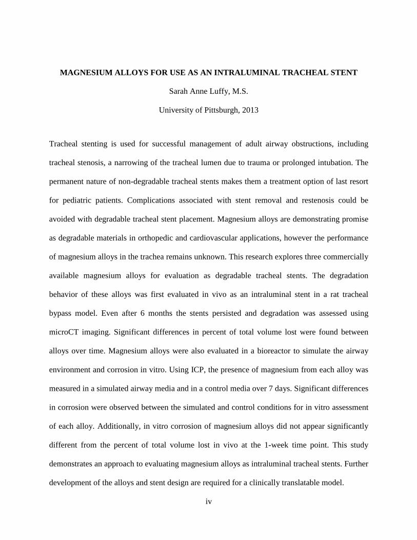
Tracheal stenting is used for successful management of adult airway obstructions, including
tracheal stenosis, a narrowing of the tracheal lumen due to trauma or prolonged intubation. The
permanent nature of non-degradable tracheal stents makes them a treatment option of last resort
for pediatric patients. Complications associated with stent removal and restenosis could be
avoided with degradable tracheal stent placement. Magnesium alloys are demonstrating promise
as degradable materials in orthopedic and cardiovascular applications, however the performance
of magnesium alloys in the trachea remains unknown. This research explores three commercially
available magnesium alloys for evaluation as degradable tracheal stents. The degradation
behavior of these alloys was first evaluated in vivo as an intraluminal stent in a rat tracheal
bypass model. Even after 6 months the stents persisted and degradation was assessed using
microCT imaging. Significant differences in percent of total volume lost were found between
alloys over time. Magnesium alloys were also evaluated in a bioreactor to simulate the airway
environment and corrosion in vitro. Using ICP, the presence of magnesium from each alloy was
measured in a simulated airway media and in a control media over 7 days. Significant differences
in corrosion were observed between the simulated and control conditions for in vitro assessment
of each alloy. Additionally, in vitro corrosion of magnesium alloys did not appear significantly
different from the percent of total volume lost in vivo at the 1-week time point. This study
demonstrates an approach to evaluating magnesium alloys as intraluminal tracheal stents. Further
development of the alloys and stent design are required for a clinically translatable model.
MAGNESIUM ALLOYS FOR USE AS AN INTRALUMINAL TRACHEAL STENT
Sarah Anne Luffy, M.S.
University of Pittsburgh, 2013
v
TABLE OF CONTENTS
NOMENCLATURE ..................................................................................................................... X
PREFACE .................................................................................................................................... XI
1.0 INTRODUCTION ........................................................................................................ 1
1.1 ANATOMY OF THE TRACHEA ..................................................................... 1
1.1.1 Management of Tracheal Stenosis ................................................................. 3
1.1.2 Need for a Degradable Tracheal Stent .......................................................... 4
1.1.3 Historical Applications of Magnesium .......................................................... 7
1.1.4 Magnesium Alloys for Tracheal Stents .......................................................... 9
1.2 RESEARCH OBJECTIVES ............................................................................. 10
1.2.1 Aims and Hypotheses .................................................................................... 10
1.2.1.1 Specific Aim 1: To determine the in vivo degradation patterns of
magnesium alloys in a tracheal location. ......................................................... 10
1.2.1.2 Specific Aim 2: To determine the in vitro degradation of
magnesium alloys. .............................................................................................. 11
2.0 EVALUATION OF MAGNESIUM ALLOYS IN VIVO ....................................... 12
2.1 IN VIVO STUDY DESIGN AND METHODS ............................................... 12
2.1.1 In vivo study design ....................................................................................... 12
2.1.1 Methods .......................................................................................................... 13
vi
2.1.1.1 Materials Preparation......................................................................... 13
2.1.1.2 Surgical Procedure .............................................................................. 14
2.1.1.3 Post-Operative Care ........................................................................... 15
2.1.1.4 Assessment of Corrosion In Vivo ....................................................... 15
2.1.1.5 Histological Evaluation ....................................................................... 16
2.1.1.6 Statistical Analysis .............................................................................. 17
2.2 RESULTS ........................................................................................................... 17
2.2.1 Pure Mg .......................................................................................................... 17
2.2.1.1 Stent Volume Lost ............................................................................... 17
2.2.1.2 Histological Evaluation ....................................................................... 19
2.2.2 AZ31 ................................................................................................................ 23
2.2.2.1 Stent Volume Lost ............................................................................... 23
2.2.2.2 Histological Evaluation ....................................................................... 24
2.2.3 MgY................................................................................................................. 28
2.2.3.1 Stent Volume Lost ............................................................................... 28
2.2.3.2 Histological Evaluation ....................................................................... 29
2.3 SUMMARY OF IN VIVO RESULTS ............................................................. 33
3.0 EVALUATION OF MAGNESIUM ALLOYS IN VITRO .................................... 35
3.1 IN VITRO STUDY DESIGN AND METHODS ............................................. 35
3.1.1 Assessment of Corrosion in vitro ................................................................. 36
3.1.1.1 ICP Analysis ........................................................................................ 36
3.1.1.2 MicroCT Analysis ............................................................................... 37
3.1.1.3 Statistical Analysis .............................................................................. 37
vii
3.2 RESULTS ........................................................................................................... 38
3.2.1 ICP Results ..................................................................................................... 38
3.2.1.1 Pure Mg ................................................................................................ 38
3.2.1.2 AZ31 ..................................................................................................... 39
3.2.1.3 MgY ...................................................................................................... 41
3.2.2 MicroCT Analyses ......................................................................................... 42
3.2.2.1 Pure Mg ................................................................................................ 43
3.2.2.2 AZ31 ..................................................................................................... 44
3.2.2.3 MgY ...................................................................................................... 45
3.2.3 Statistical Analysis ......................................................................................... 45
3.3 SUMMARY OF IN VITRO RESULTS ........................................................... 46
4.0 DISCUSSION ............................................................................................................. 47
5.0 CONCLUSIONS ........................................................................................................ 51
BIBLIOGRAPHY ....................................................................................................................... 52

viii
LIST OF FIGURES
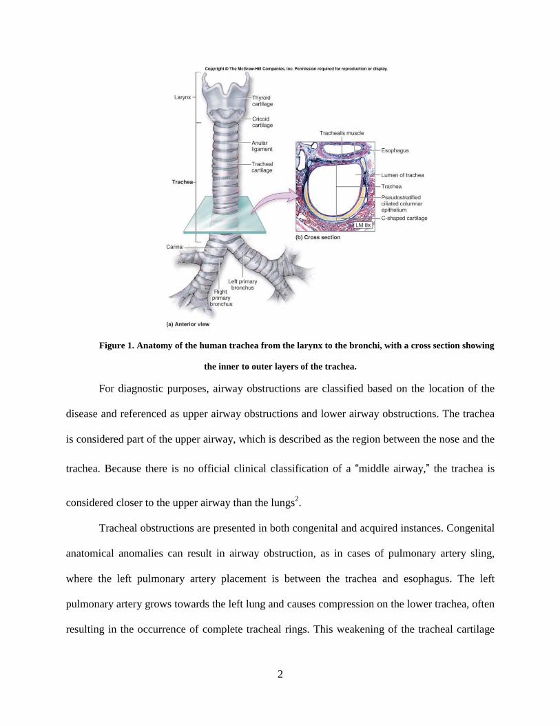
Figure 1. Anatomy of the human trachea from the larynx to the bronchi, with a cross section showing the inner to outer layers of the trachea. ............................................................................ 2
Figure 2. AZ31 tracheal stent is represented in this image, but all stents were machined with outer diameter of 2.25mm, inner diameter of 1.25mm and length of 5mm. ................................. 13
Figure 3. Surgical placement of an intraluminal tracheal stent in a Lewis rat trachea. A tracheal bypass model was used to evaluate the stent in vivo. ................................................................... 15
Figure 4. MicroCT image with 3-dimmensional reconstruction using Mimics software ............. 16
Figure 5. Percent of total volume lost during in vivo testing for each alloy at 1, 8, 16 and 24 weeks. Significant differences among alloys and between time points are indicated by the v, *, and #. ............................................................................................................................................. 18
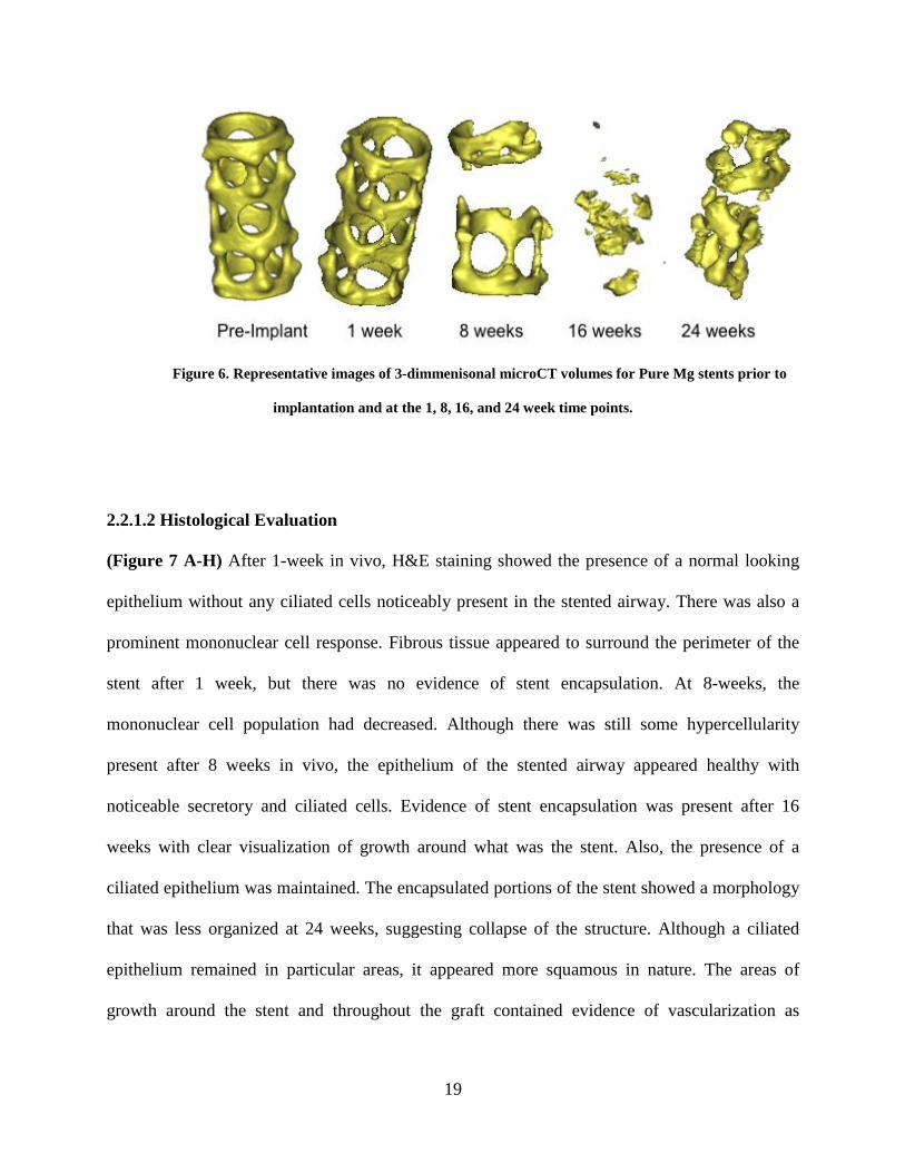
Figure 6. Representative images of 3-dimmenisonal microCT volumes for Pure Mg stents prior to implantation and at the 1, 8, 16, and 24 week time points........................................................ 19
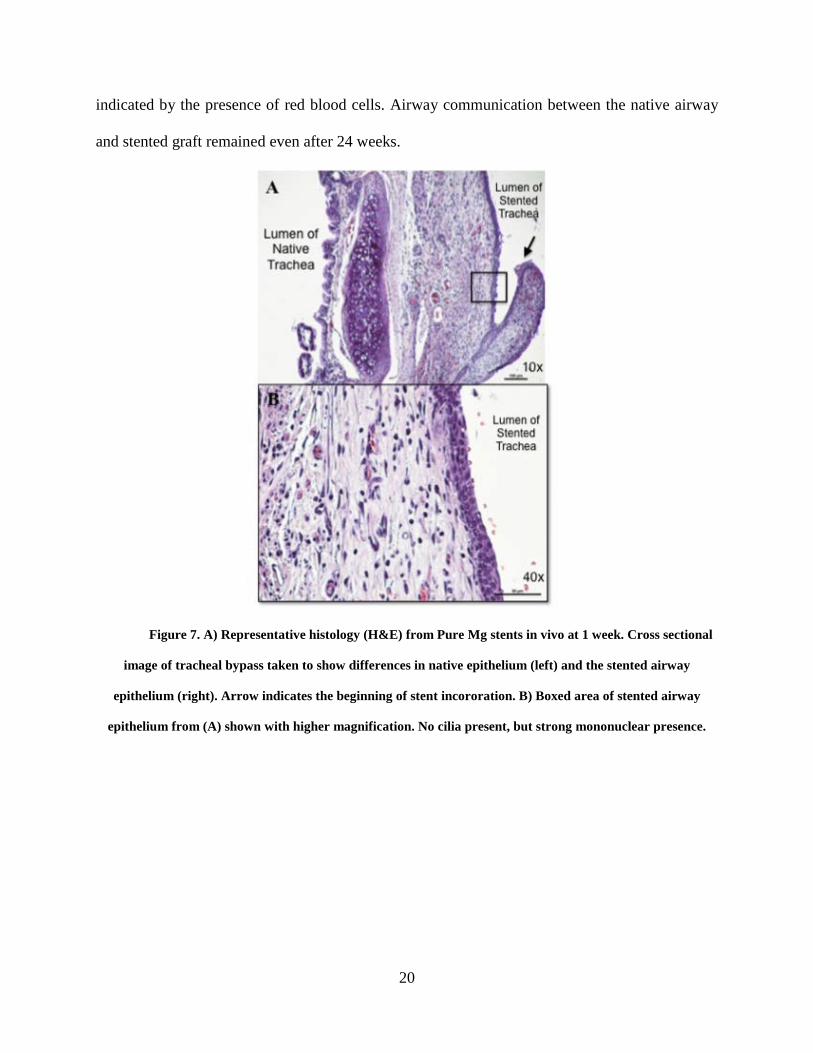
Figure 7. A) Representative histology (H&E) from Pure Mg stents in vivo at 1 week. Cross sectional image of tracheal bypass taken to show differences in native epithelium (left) and the stented airway epithelium (right). Arrow indicates the beginning of stent incororation. B) Boxed area of stented airway epithelium from (A) shown with higher magnification. No cilia present, but strong mononuclear presence.................................................................................................. 20
Figure 8. Representative images of 3-dimmenisonal microCT volumes for AZ31 stents prior to implantation and at the 1, 8, 16, and 24 week time points. ........................................................... 24

ix
Figure 9. A) Representative histology (H&E) from AZ31 stents in vivo at 1 week. Cross sectional image of stented trachea showing mononuclear cell population. B) Boxed area from (A) shown with higher magnification to display disruption of epithelium, most likely due to surgical placement of the stent. .................................................................................................................. 25
Figure 10. Representative images of 3-dimmenisonal microCT volumes for MgY stents prior to implantation and at the 1, 8, 16, and 24 week time points. ........................................................... 29
Figure 11. A) Representative histology (H&E) from MgY stents after 1 week in vivo. Hypercellularity in the native tracheal epithelium with the presence of possible giant cells. B) Boxed area from (A) shown with higher magnification. Epithelium is disrupted and arrow indicates possible giant cell. ......................................................................................................... 30
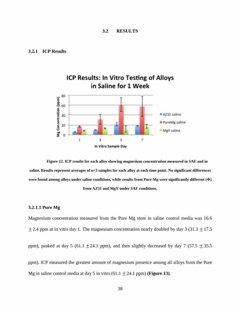
Figure 12. ICP results for each alloy showing magnesium concentration measured in SAF and in saline. Results represent averages of n=3 samples for each alloy at each time point. No significant differences were found among alloys under saline conditions, while results from Pure Mg were significantly different () from AZ31 and MgY under SAF conditions. ..................... 38
Figure 13. ICP results for Pure Mg tracheal stents, displaying average magnesium concentrations (n=3) at 1, 3, 5, and 7 days in vitro for both SAF and saline conditions. ..................................... 39
Figure 14. ICP results for AZ31 tracheal stents, displaying average magnesium concentrations (n=3) at 1, 3, 5, and 7 days in vitro for both SAF and saline conditions. ..................................... 41
Figure 15. ICP results for MgY tracheal stents, displaying average magnesium concentrations (n=3) at 1, 3, 5, and 7 days in vitro for both SAF and saline conditions. ..................................... 42
Figure 16. Bar graph summarizing the comparison of corrosion after 1 week in vivo to 1 week in vitro under both SAF and saline conditions. The (#) indicates significant differences were observed between all alloys under the saline in vitro control conditions, while the (*) indicates only Pure Mg experienced significantly different results under SAF in vitro conditions. ........... 43
Figure 17. Representative 3-dimensional microCT images of Pure Mg, AZ31, and MgY stents after 1 week of in vitro testing in SAF media and saline control conditions. ............................... 44
x
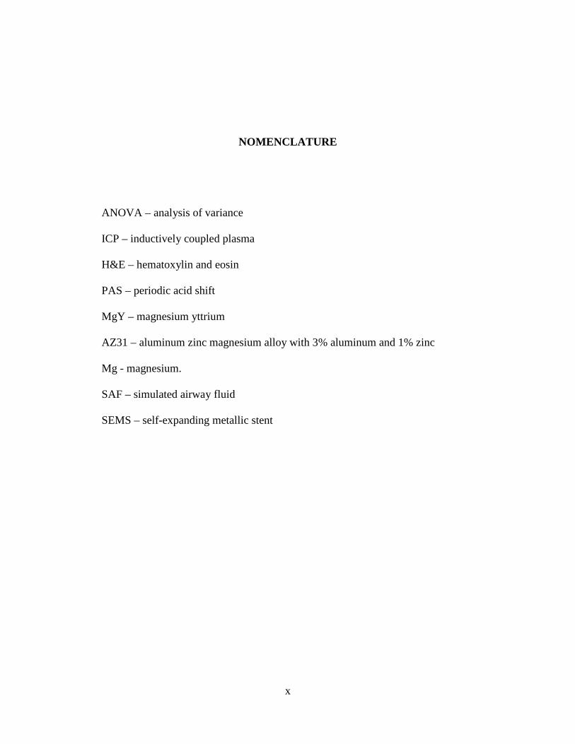
NOMENCLATURE
ANOVA – analysis of variance
ICP – inductively coupled plasma
H&E – hematoxylin and eosin
PAS – periodic acid shift
MgY – magnesium yttrium
AZ31 – aluminum zinc magnesium alloy with 3% aluminum and 1% zinc
Mg - magnesium.
SAF – simulated airway fluid
SEMS – self-expanding metallic stent
xi
PREFACE
Thank you to the members of my committee for your guidance, encouragement, and support. A
special thank you to my thesis adviser and graduate mentor, Dr. Thomas Gilbert. This work was
part of the National Science Foundation Engineering Research Center for Revolutionizing
Metallic Biomaterials (NSF ERC-RMB). The magnesium ingots containing yttrium were
obtained from Helmholtz-Zentrum Geesthacht, Institute of Materials Research, Geesthacht,
Germany through a gracious gift from Dr. Norbert Hort and Dr. Frank Witte. The machining
expertise for the manufacturing of the tracheal stents was provided by Mr. Andrew Holmes at the
University of Pittsburgh Swanson Center for Product Innovation (SCPI).
1
1.0 INTRODUCTION
1.1 ANATOMY OF THE TRACHEA
Characterized as a hollow, single lumen tube, the trachea allows for air passage from the larynx
to the bronchi. C-shaped cartilage rings provide the rigid structure necessary to support the
trachea and prevent collapse during the breathing process. The posterior wall of the trachea
contains smooth muscle, which offers flexibility in addition to preventing over distension of the
airway during expiration. Mechanical ventilation is the primary function of the trachea, but the
intraluminal mucosal lining of the trachea provides clearance pathways for bronchial secretions.
A heterogeneous cell layer including pseudostratified columnar epithelial cells, including ciliated
cells, secretory and goblet cells, and basal cells create the inner most layer of the tracheal wall.
The basement membrane is subjacent to the epithelial cell layer, which lies against the
connective tissue and smooth muscle cells followed by the cartilage rings1, (Figure 1).
2
Figure 1. Anatomy of the human trachea from the larynx to the bronchi, with a cross section showing
the inner to outer layers of the trachea.
For diagnostic purposes, airway obstructions are classified based on the location of the
disease and referenced as upper airway obstructions and lower airway obstructions. The trachea
is considered part of the upper airway, which is described as the region between the nose and the
trachea. Because there is no official clinical classification of a “middle airway,” the trachea is
considered closer to the upper airway than the lungs2.
Tracheal obstructions are presented in both congenital and acquired instances. Congenital
anatomical anomalies can result in airway obstruction, as in cases of pulmonary artery sling,
where the left pulmonary artery placement is between the trachea and esophagus. The left
pulmonary artery grows towards the left lung and causes compression on the lower trachea, often
resulting in the occurrence of complete tracheal rings. This weakening of the tracheal cartilage

3
leading to collapse of the airway is described as tracheomalacia3,4. Tracheal obstructions are rare,
but acquired tracheal obstructions are more common in children than congenital cases and are
often the result of long-term intubation5,6. Severe cases of tracheal obstruction resulting from
acquired stenosis, often require multiple surgical and endoscopic procedures to achieve long-
term goals of a desirable outcome7.
1.1.1 Management of Tracheal Stenosis
Injury to the lumen of the trachea typically leads to the formation of scar tissue that can cause
tracheal obstruction through a narrowing of the tracheal lumen. The condition is called tracheal
stenosis8. Tracheal stenosis can be the result of a congenital disorder, but more often it is
acquired from prolonged intubation, trauma, a tumor, or infection9. Although tracheal
obstructions are rare among the pediatric patient population, pediatric tracheal stenosis is
associated with severe morbidity and mortality, and the management of stenosis consistently
challenges clinicians10,11.
Endoscopic management of tracheal stenosis by balloon dilation is used as a method of
treatment when attempting to avoid open surgical procedures in both children and adults12.
However, the effects of balloon dilation on the stenotic trachea are poorly understood, and
repeated treatments are often necessary. Surgical management of tracheal stenosis in adults is
commonly practiced by resection of the compromised trachea with primary reconstruction by
end-to-end anastomosis. Reconstruction is a primary treatment option for pediatric patients,
however because of the small size of the pediatric airway, children do not experience the same
success with resection and end-to-end anastomosis that adults can achieve13. Ischemia and
4
anastomotic tension resulting from reconstruction can cause restenosis. Slide tracheoplasty has
become the treatment of choice when surgery is necessary. While alternative temporary or
definitive methods are being developed for those who are not surgical candidates, surgical
reconstruction is still considered the ideal approach for management of tracheal stenosis14. In
order to avoid a reoccurrence of tracheal stenosis following surgical repair, tracheal stents have
been used with reasonable success. Minimally invasive deployment options for intraluminal
balloon expandable stents have become an attractive option for treatment of tracheal stenosis.
Complications associated with tracheal stenting include encapsulation and incorporation into the
mucosal tissue, migration, perforation, and compromised mucociliary clearance of the airway9,15.
1.1.2 Need for a Degradable Tracheal Stent
Tracheal stents are classified as equivalent to tracheal prostheses under FDA guidelines for
medical device approval. Intentions for use of a tracheal stent must include the maintenance of
airway patency. Stenosis from prolonged intubation, tracheal burn or trauma,
tracheobronchomalacia, and external compression of the trachea are the most common
conditions responsive to stent placement16. Stents are more broadly used in the context of
vascular medicine. A stent is defined by the National Institutes of Health (NIH) in the context of
a coronary stent as a mesh tube used to support weak arteries41. Coronary stents vary in size,
geometry, function, and deployment method. Following angioplasty, a procedure using a balloon
to dilate the blood vessels compromised by disease or plaque, stents are placed in about 60
percent of cases to support the vessel and prevent restenosis17. Restenosis is a reduction of the
vessel lumen by greater than 50 percent at the area of previous treatment18,19. In-stent restenosis
(ISR) still persists in about 25 percent of stents placed following angioplasty17. Depending on the

5
diagnosis, a variety of stents may be used for treatment of a compromised vessel. Percutaneous
intervention allows for placement of self-expanding, balloon expandable, and drug-eluting stents
ranging in material composition.
Tracheal stents currently available vary in material composition and physical geometry,
as well as application purpose. Stent selection typically depends on whether the patient has a
benign or malignant disease, the need for the stent is temporary or permanent, the physician’s
experience with stent insertion, and finally, the cost of the procedure20. For a pediatric case
involving the onset of long segment stenosis following tracheostomy, surgical resection was used
to remove the stenosis and release the trachea, followed by reconstruction and placement of a
silicone Dumon stent21. In 2007, one retrospective study focused on Dumon stent use in 35
patients. The Japanese study found the Dumon stent the most effective tracheal stent for
treatment of both tracheal stenosis and airway fistulas, based on cost and safety20. Although the
stent is made from radiopaque silicone and the design features include external struts to prevent
migration, it is a permanent silicone implant. Even with surface treatments designed to maximize
mucociliary clearance and minimize the risk of obstruction, the described study still included
multiple patients who experienced migration, obstruction, and restenosis20.
Self-expanding metallic stents, or SEMS, offer properties consistent with the ideal design
requirements for a tracheal stent – easy insertion, biocompatible, non-obstructive, does not
inhibit mucociliary clearance, and adapts to the varying dimensions of the airway during
respiration and coughing22. The consistent problem with metallic stents, as with all stents, is their
permanent nature and difficult removal despite these positive features. In addition, the very
nature of SEMS can be problematic over time as the stent continually applies an outward
6
pressure on the trachea that can lead to perforation. Problems and complications associated with
stent removal make pediatric tracheal stenting a treatment option of last resort14,15. When surgical
intervention is limited, SEMS have shown reasonable success in the airway, but SEMS use
should be avoided in the treatment of benign airway obstructions, as in the case of
laryngotracheal airway obstruction23. In fact, stent related complications are often more difficult
to treat than the original indication for initial stent placement24. In the pediatric patient
population, even the most widely accepted tracheal stents succumb to the complications of
granulation tissue, obstruction, and fracture after just 2 weeks11.
Tracheal stenting is used for management of adult airway obstructions, but because non-
degradable stents are considered permanent implants after encapsulation, they are considered a
treatment option of last resort for children. The pediatric population requires growth potential
over time, and the permanent nature of a non-degradable stent increases the likelihood of
complications associated with stent removal and restenosis. In fact, the pediatric population
experiences an estimated stent related mortality of 12.9%15. A degradable stent would allow for
growth potential while supporting an open tracheal lumen, making it an ideal treatment option
for pediatric tracheal stenosis. Biodegradable stents have been developed for use in esophageal,
urethral, and intestinal stenosis, while biodegradable stents for treatment of tracheal stenosis has
been used experimentally25. One study presents the use of a polydioxanone tracheobronchial
degradable stent as an alternative to metallic or silicone stents for the treatment of stenosis25.
Permanent stent alternatives are also being investigated for cardiovascular stent design
using degradable metallic materials. As with the trachea, the need for a coronary stent is
temporary and a degradable material could decrease the risk of complications associated with
permanent implants. Coronary stents made from stainless steel, nitinol, and cobalt-chromium
7
alloys have shown success, but more widespread use is limited by their permanent nature,
thrombogenicity, inflammatory response, potential to cause perforation, and inability to provide
for growth potential17. Iron based biodegradable stents are believed to be a suitable material for
coronary stents because of the ability of iron to bind oxygen molecules, and especially as a larger
size stent because of its robust nature and strong mechanical properties throughout the
degradation period17. Biodegradable stents have demonstrated feasibility in early studies26,27, and
while magnesium alloys have historically been used in orthopedic fixation devices28,29, they are
showing promise in cardiovascular applications. These applications include a multi-center
clinical trial of magnesium stents30-32. The multi-center human clinical trial described the use of
a balloon-expandable magnesium based coronary stent for treatment of coronary artery lesions.
1.1.3 Historical Applications of Magnesium
Magnesium is a common biometal, naturally occurring in the human body and essential for life.
Critical in many enzymatic reactions, magnesium and calcium also work together to regulate
neuromuscular transmission33. Additionally, magnesium promotes relaxation of the airway by
inhibiting the influx of calcium ions and blocking the voltage-dependent calcium channels33. The
presence of magnesium and magnesium sulfates in the airway has lead to positive outcomes in
the treatment of pediatric asthma by increasing lung function and reducing hospitalization34.
Sir Humphrey Davy discovered elemental magnesium in 1808, and magnesium has been
investigated as a degradable biomaterial for over 100 years29. Magnesium wires were first
implanted in humans as ligatures to control the bleeding of human vessels, where it was
discovered that corrosion of Mg was slower in vivo and that the time for degradation
corresponded directly to the size of the Mg wire17,29. Pure Mg wire was too brittle to serve as a

8
suture material, which prompted the development of magnesium alloys. Absorbable magnesium
staples and clips were used to ensure hemostasis in many applications such as intestinal
anastomosis and deep wound closure29,35. When used as a screw for bone fixation, the first
human clinical study reported completed resorption of magnesium after 1 year with
physiological bone healing and benign gas evolution that disappeared a few weeks after
implantation, according to x-ray29. The combination of these early clinical studies found
magnesium non-toxic and a non-irritant, but emphasized the importance of separating
magnesium implants from other metals due to the risk of electrolytic corrosion29. The corrosion
product of magnesium oxide was believed early on to promote bone and callus formation.
Investigators believed that inserting magnesium into inflammatory tissue of pseudarthosis cases
allowed for the neutralization of the acidic environment, which lead to the formation of callus
bone and healing17,29,36. Inflammation following magnesium implantation appears consistent
throughout historical studies, leading to fibrous encapsulation of the corroding magnesium,
especially in bone tissue29. Early use of magnesium in vessels and subcutaneous tissue resulted
in vascularized granulation tissue surrounding the magnesium implant, as well as localized
foreign body giant cells and leukocytes. Interestingly, no infections were reported during the
early investigational uses of magnesium implants29. Important insights into magnesium corrosion
were observed during these initial studies. Total absorption of magnesium, for example, was
found to depend more on the exposed surface of the metal than on the total weight of the metal,
as indicated by comparing magnesium wires and sheets to thicker pieces with the same surface
area29,36. Although consistencies in corrosion rate of magnesium among tissue type and species
remain unpredictable, the corrosion rate does appear to depend on the size of the magnesium
implant and the tissue type in which it was imbedded29,35. Even early investigators postulated the
9
corrosion of magnesium to be dependent on an oxidation process that was directly related to
oxygen content in the blood, as well as the water content of the surrounding tissue and the local
hydrogen carbonic acid content. Regardless of the magnesium metals used throughout the early
history of magnesium implants, a non-toxic, non-irritant, and non-carcinogenic result of
magnesium oxides and magnesium phosphates were found17,29. Advancements in science and
medicine throughout recent history are only now beginning to produce magnesium implants for
use in clinical trials.
1.1.4 Magnesium Alloys for Tracheal Stents
The history of magnesium showed its potential for use as a degradable biomaterial. Magnesium
is also known to have low thrombogenicity and strong biocompatibility due to its naturally
occurring presence in the human body17. Because magnesium can experience fast degradation
and loss of mechanical properties, it is alloyed with other elements, like aluminum, to decrease
the rate of degradation37. Magnesium alloys degrade in aqueous environments and are resorbed
by the body through oxidation making them attractive materials for the development of a
degradable tracheal stent. Yttrium has been used as an alloying element in magnesium
cardiovascular stents with <5% yttrium and has showed results of endothelialization, minimal
inflammation, and stent degradation within approximately 90 days38. Alloys containing
aluminum and zinc have been shown to increase resistance to corrosion in vitro39.
The performance of magnesium alloys in the trachea remains unknown. The response of
the tracheal tissue to the presence of magnesium cannot be predicted without adequate models.
Furthermore, standard in vitro corrosion assays inaccurately predict in vivo behavior of
magnesium alloys4021. The objective of this research is to evaluate the degradation behavior of
10
three commercially available magnesium alloys: Mg3Y, AZ31, and 99.9% Pure Mg. First, a
specially developed rat tracheal bypass model was used to allow for evaluation of the stent
materials in the airway without risk of death due to airway obstruction. In vitro analysis was then
conducted using a bioreactor with simulated airway fluid containing mucins to simulate the
airway environment. The in vitro results were then compared to the in vivo degradation kinetics
to determine a correlation between degradation of magnesium alloys in the airway. MgY was
evaluated in a previous study as an extraluminal tracheal stent for the treatment of
tracheaomalacia (appendix). This study showed that the body could tolerate the presence of an
extraluminal stent with a mild host response, without negatively impacting the native cartilage
after 8 weeks.
1.2 RESEARCH OBJECTIVES
1.2.1 Aims and Hypotheses
1.2.1.1 Specific Aim 1: To determine the in vivo degradation patterns of magnesium alloys
in a tracheal location.
Hypothesis: Each of the alloys are expected to have different corrosion behaviors, with the AZ31
and MgY stents experiencing slower degradation than the Pure Mg at both early and late time
points. It is hypothesized that histological evaluation will reveal minimal host response to the
stent with the trachea maintaining patency and a healthy airway epithelium.
11
1.2.1.2 Specific Aim 2: To determine the in vitro degradation of magnesium alloys.
Hypothesis: The amount of magnesium present in the media after 1 week is expected to
correspond to the total volume loss of the stent in vivo at the 1-week time point.
12
2.0 EVALUATION OF MAGNESIUM ALLOYS IN VIVO
2.1 IN VIVO STUDY DESIGN AND METHODS
2.1.1 In vivo study design
All animal experiments were reviewed and approved by the University of Pittsburgh Institutional
Animal Care and Use Committee and were performed in compliance with the Guide for the Care
and Use of Laboratory Animals (NIH Publication #85-23 Rev. 1985, Lab Animal 23(8):28-
29,1994). Female Lewis rats, ranging in weight from 150 to 350g (Charles River), were used in
this study for the evaluation of three different magnesium alloys as degradable intraluminal
tracheal stents. A machinable, non-balloon expandable stent geometry was designed to evaluate
the degradable alloys in vivo through the design of a tracheal bypass model. The alloys were
evaluated at 1, 8, 16, and 24-week time points. Upon explant the stent-tissue complex was
imaged using microCT and processed to analyze volume loss and stent geometry over time.
Explants were prepared for histological analysis using paraffin processing.
13
2.1.1 Methods
2.1.1.1 Materials Preparation
Commercially available 99.9% pure magnesium was obtained from Goodfellow
(Coraopolis, PA, USA) and used as the first material for evaluation as an intraluminal tracheal
stent. Magnesium containing 3% aluminum and 1% zinc, also obtained from Goodfellow, was
considered as a second material for stent evaluation in this study. The third material for an
intraluminal tracheal stent was magnesium containing 3 wt% Y (W3) obtained from Helmholtz
Zentrum Geesthacht Institute of Materials Research, Geesthacht, Germany, provided as a gift
from Dr. Norbert Hort and Dr. Frank Witte. All three materials were T4 heat treated at 525 °C
under a protective environment of Ar + 0.1% SF6 for 8 h to homogenize intermetallics and
alloying elements in the alloy matrix. The magnesium alloys were machined into stents at the
University of Pittsburgh Swanson School for Product Innovation (SCPI), with an outer diameter
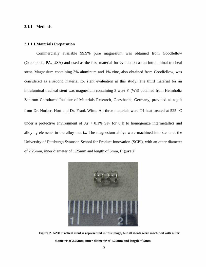
of 2.25mm, inner diameter of 1.25mm and length of 5mm, Figure 2.
Figure 2. AZ31 tracheal stent is represented in this image, but all stents were machined with outer
diameter of 2.25mm, inner diameter of 1.25mm and length of 5mm.
14
To remove stresses imparted by the machining process, the stents were then subjected to
a second heat treatment at 205°C for 1.5h in ultra high purity argon (UHP-Ar). All stents were
sonicated in isopropanol for 5 minutes and dried in air (3x), and then terminally sterilized with
2MRad γ-irradiation.
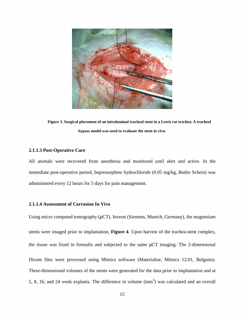
2.1.1.2 Surgical Procedure
Donor female Lewis rats were anesthetized by intraperitoneal injection of ketamine (80 mg/kg)
xylazine (8 mg/kg) to maintain the surgical plane of anesthesia, and were euthanized via
exsanguination. The tracheas were harvested to provide a bypass surgical model for stent
evaluation, Figure 3. Recipient animals were anesthetized in a similar way. Aseptic technique
was used to expose the proximal cervical trachea through a midline neck incision. Small
proximal and distal defects separated by 5 cartilage rings were created in the recipient trachea.
The stent was placed intraluminally in the donor trachea, which was anastomosed to the distal
and then proximal defects using 7-0 prolene suture (Ethicon, Somerville, NJ). The bypass model
allowed for airway communication and evaluation of the non-optimized stent geometries in vivo
without compromising respiration. The skin incision was closed using an interrupted suturing
technique with 5-0 PDS suture (Ethicon, Somerville, NJ). Proper anastomosis was determined by
visual inspection and by the absence of any sub-cutaneous air upon closure.
15
Figure 3. Surgical placement of an intraluminal tracheal stent in a Lewis rat trachea. A tracheal
bypass model was used to evaluate the stent in vivo.
2.1.1.3 Post-Operative Care
All animals were recovered from anesthesia and monitored until alert and active. In the
immediate post-operative period, buprenorphine hydrochloride (0.05 mg/kg, Butler Schein) was
administered every 12 hours for 5 days for pain management.
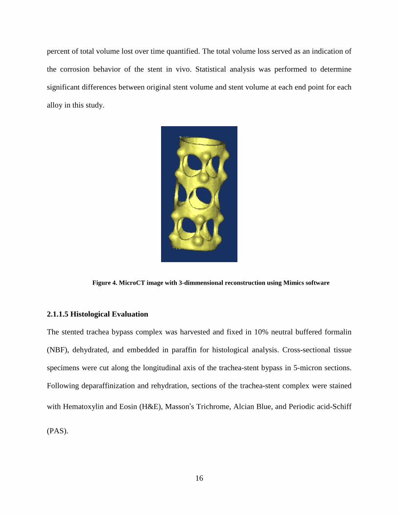
2.1.1.4 Assessment of Corrosion In Vivo
Using micro computed tomography (μCT), Inveon (Siemens, Munich, Germany), the magnesium
stents were imaged prior to implantation, Figure 4. Upon harvest of the trachea-stent complex,
the tissue was fixed in formalin and subjected to the same μCT imaging. The 2-dimensional
Dicom files were processed using Mimics software (Materialise, Mimics 12.01, Belguim).
Three-dimensional volumes of the stents were generated for the data prior to implantation and at
1, 8, 16, and 24 week explants. The difference in volume (mm3) was calculated and an overall
16
percent of total volume lost over time quantified. The total volume loss served as an indication of
the corrosion behavior of the stent in vivo. Statistical analysis was performed to determine
significant differences between original stent volume and stent volume at each end point for each
alloy in this study.
Figure 4. MicroCT image with 3-dimmensional reconstruction using Mimics software
2.1.1.5 Histological Evaluation
The stented trachea bypass complex was harvested and fixed in 10% neutral buffered formalin
(NBF), dehydrated, and embedded in paraffin for histological analysis. Cross-sectional tissue
specimens were cut along the longitudinal axis of the trachea-stent bypass in 5-micron sections.
Following deparaffinization and rehydration, sections of the trachea-stent complex were stained
with Hematoxylin and Eosin (H&E), Masson’s Trichrome, Alcian Blue, and Periodic acid-Schiff
(PAS).
17
2.1.1.6 Statistical Analysis
All statistical analyses were performed using the SPSS package (version 20.0; IBM SPSS, Inc,
Chicago, Ill). Statistical significance was set at p < 0.05.
To compare the differences of in vivo corrosion within and between the alloy groups over
time, a two-way independent ANOVA with Tukey’s Post Hoc analysis was used to analyze the
percent of total stent volume lost in each animal. Percent of total stent volume lost over time is
reported as mean standard deviation.
2.2 RESULTS
2.2.1 Pure Mg
2.2.1.1 Stent Volume Lost
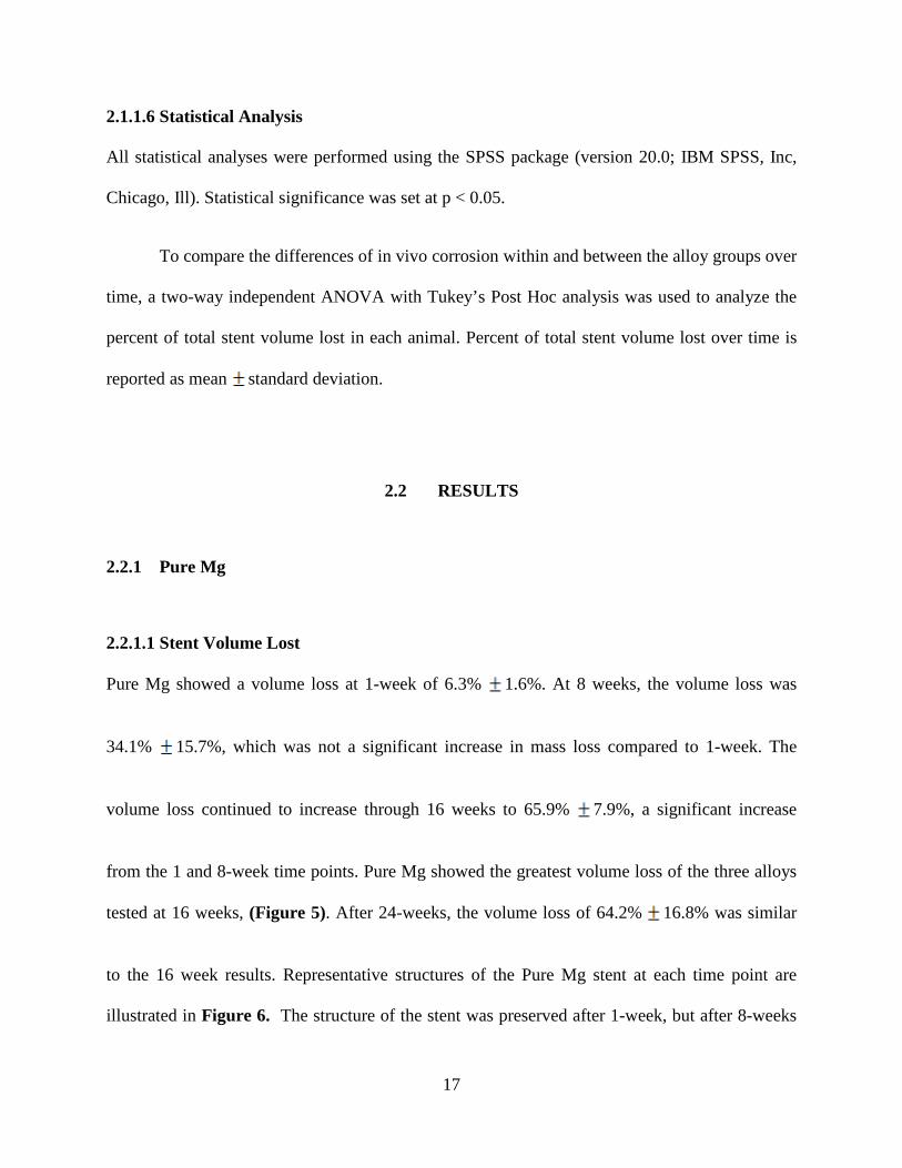
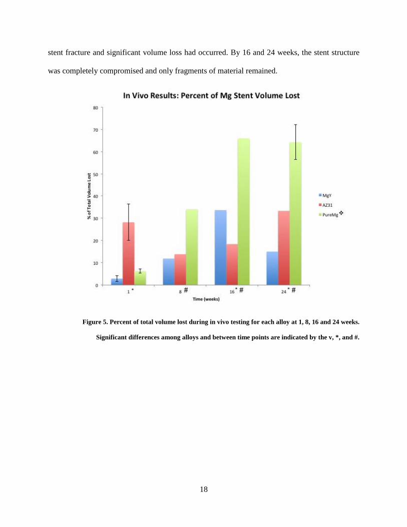
Pure Mg showed a volume loss at 1-week of 6.3% 1.6%. At 8 weeks, the volume loss was
34.1% 15.7%, which was not a significant increase in mass loss compared to 1-week. The
volume loss continued to increase through 16 weeks to 65.9% 7.9%, a significant increase
from the 1 and 8-week time points. Pure Mg showed the greatest volume loss of the three alloys
tested at 16 weeks, (Figure 5). After 24-weeks, the volume loss of 64.2% 16.8% was similar
to the 16 week results. Representative structures of the Pure Mg stent at each time point are
illustrated in Figure 6. The structure of the stent was preserved after 1-week, but after 8-weeks
18
stent fracture and significant volume loss had occurred. By 16 and 24 weeks, the stent structure
was completely compromised and only fragments of material remained.
Figure 5. Percent of total volume lost during in vivo testing for each alloy at 1, 8, 16 and 24 weeks.
Significant differences among alloys and between time points are indicated by the v, *, and #.
19
Figure 6. Representative images of 3-dimmenisonal microCT volumes for Pure Mg stents prior to
implantation and at the 1, 8, 16, and 24 week time points.
2.2.1.2 Histological Evaluation
(Figure 7 A-H) After 1-week in vivo, H&E staining showed the presence of a normal looking
epithelium without any ciliated cells noticeably present in the stented airway. There was also a
prominent mononuclear cell response. Fibrous tissue appeared to surround the perimeter of the
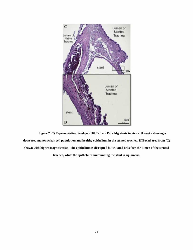
stent after 1 week, but there was no evidence of stent encapsulation. At 8-weeks, the
mononuclear cell population had decreased. Although there was still some hypercellularity
present after 8 weeks in vivo, the epithelium of the stented airway appeared healthy with
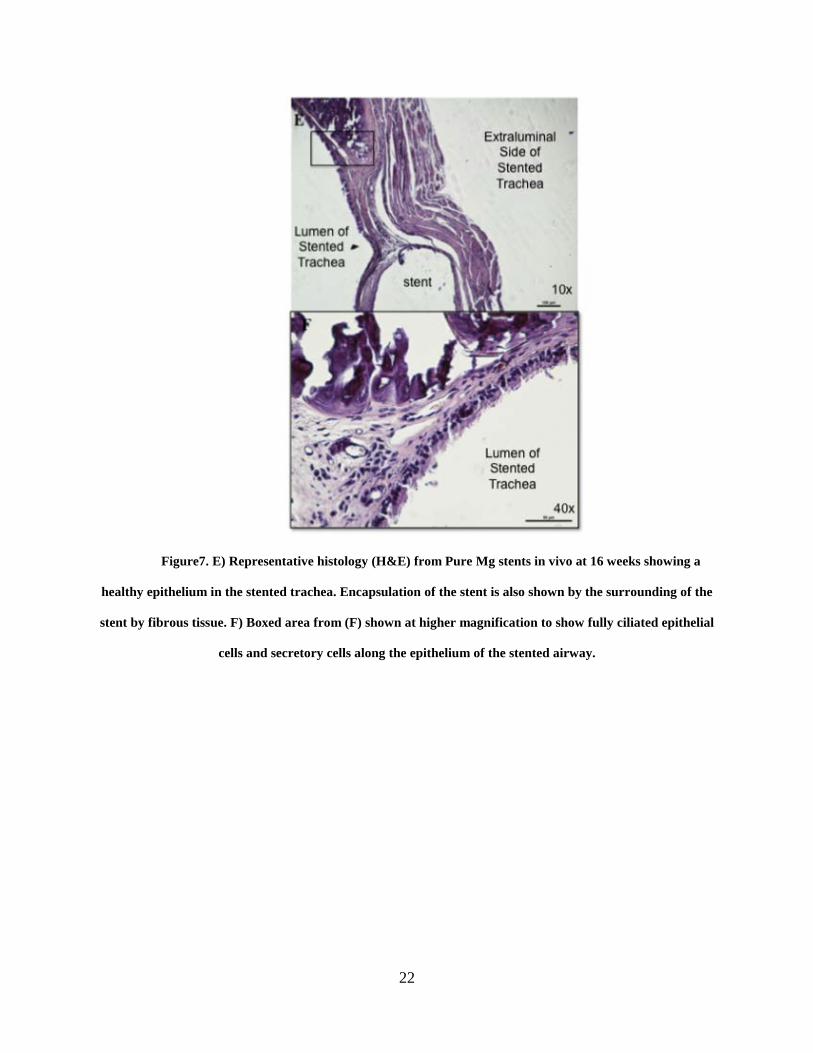
noticeable secretory and ciliated cells. Evidence of stent encapsulation was present after 16
weeks with clear visualization of growth around what was the stent. Also, the presence of a
ciliated epithelium was maintained. The encapsulated portions of the stent showed a morphology
that was less organized at 24 weeks, suggesting collapse of the structure. Although a ciliated
epithelium remained in particular areas, it appeared more squamous in nature. The areas of
growth around the stent and throughout the graft contained evidence of vascularization as
20
indicated by the presence of red blood cells. Airway communication between the native airway
and stented graft remained even after 24 weeks.
Figure 7. A) Representative histology (H&E) from Pure Mg stents in vivo at 1 week. Cross sectional
image of tracheal bypass taken to show differences in native epithelium (left) and the stented airway
epithelium (right). Arrow indicates the beginning of stent incororation. B) Boxed area of stented airway
epithelium from (A) shown with higher magnification. No cilia present, but strong mononuclear presence.
21
Figure 7. C) Representative histology (H&E) from Pure Mg stents in vivo at 8 weeks showing a
decreased mononuclear cell population and healthy epithelium in the stented trachea. D)Boxed area from (C)
shown with higher magnification. The epithelium is disrupted but ciliated cells face the lumen of the stented
trachea, while the epithelium surrounding the stent is squamous.
22
Figure7. E) Representative histology (H&E) from Pure Mg stents in vivo at 16 weeks showing a
healthy epithelium in the stented trachea. Encapsulation of the stent is also shown by the surrounding of the
stent by fibrous tissue. F) Boxed area from (F) shown at higher magnification to show fully ciliated epithelial
cells and secretory cells along the epithelium of the stented airway.
23
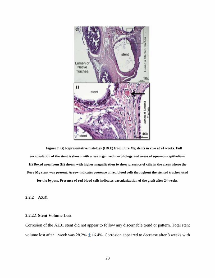
Figure 7. G) Representative histology (H&E) from Pure Mg stents in vivo at 24 weeks. Full
encapsulation of the stent is shown with a less organized morphology and areas of squamous epithelium.
H) Boxed area from (H) shown with higher magnification to show presence of cilia in the areas where the
Pure Mg stent was present. Arrow indicates presence of red blood cells throughout the stented trachea used
for the bypass. Presence of red blood cells indicates vascularization of the graft after 24 weeks.
2.2.2 AZ31
2.2.2.1 Stent Volume Lost
Corrosion of the AZ31 stent did not appear to follow any discernable trend or pattern. Total stent
volume lost after 1 week was 28.2% 16.4%. Corrosion appeared to decrease after 8 weeks with

24
a volume loss of 13.85% 11.1%. After 16 weeks, total AZ31 stent volume loss increased to
18.4% 15.62%, and continued to a maximum stent volume loss of 33.3% 18.4% after 24
weeks. These findings are not consistent with the morphology of the stent as depicted by
microCT imaging in Figure 8. No corrosion is evident after 1-week, but the structure began to
change at 8 weeks. By 16 weeks, the stent structure is compromised by corrosion and fracture,
which continued at the 24-week time point. The variability among AZ31 stent volume loss was
so large, no patterns in corrosion were observed.
Figure 8. Representative images of 3-dimmenisonal microCT volumes for AZ31 stents prior to
implantation and at the 1, 8, 16, and 24 week time points.
2.2.2.2 Histological Evaluation
(Figure 9A-H) One week after AZ31 stent placement in vivo, H&E staining of the stented
trachea revealed a squamous epithelium with a large presence of mononuclear cells. At the 8-
week time point, histology revealed both columnar and squamous epithelium with some stent
encapsulation and cell growth. The columnar epithelium had progressed to a ciliated epithelium
after 16 weeks, with reduced presence of mononuclear cell response. After 24 weeks, an open
lumen was maintained with the presence of ciliated cells. Overall, a columnar ciliated epithelium
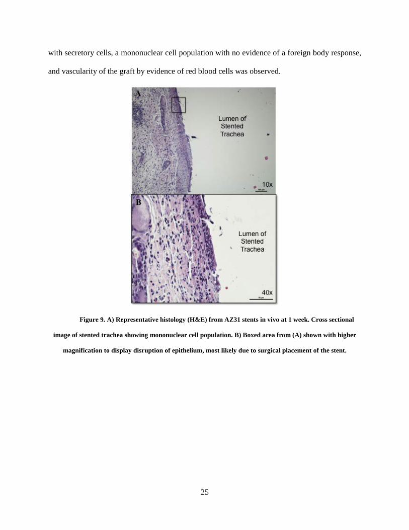
25
with secretory cells, a mononuclear cell population with no evidence of a foreign body response,
and vascularity of the graft by evidence of red blood cells was observed.
Figure 9. A) Representative histology (H&E) from AZ31 stents in vivo at 1 week. Cross sectional
image of stented trachea showing mononuclear cell population. B) Boxed area from (A) shown with higher
magnification to display disruption of epithelium, most likely due to surgical placement of the stent.
26
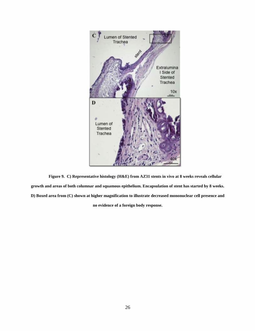
Figure 9. C) Representative histology (H&E) from AZ31 stents in vivo at 8 weeks reveals cellular
growth and areas of both columnar and squamous epithelium. Encapsulation of stent has started by 8 weeks.
D) Boxed area from (C) shown at higher magnification to illustrate decreased mononuclear cell presence and
no evidence of a foreign body response.

27
Figure 9. E) Representative histology (H&E) from AZ31 stents in vivo at 16 weeks. F) Boxed area
from (E) shown with higher magnification to demonstrate progression to a columnar, ciliated epithelium with
the presence of red blood cells to indicate vascularization of the stented graft.
28
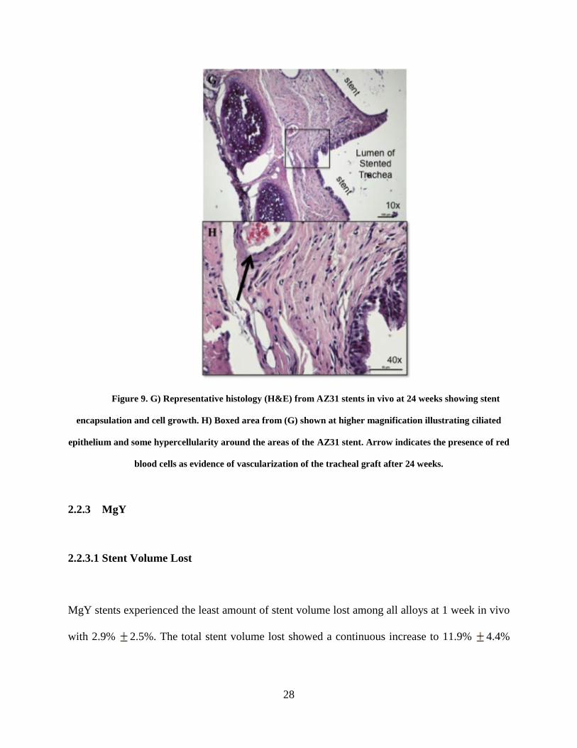
Figure 9. G) Representative histology (H&E) from AZ31 stents in vivo at 24 weeks showing stent
encapsulation and cell growth. H) Boxed area from (G) shown at higher magnification illustrating ciliated
epithelium and some hypercellularity around the areas of the AZ31 stent. Arrow indicates the presence of red
blood cells as evidence of vascularization of the tracheal graft after 24 weeks.
2.2.3 MgY
2.2.3.1 Stent Volume Lost
MgY stents experienced the least amount of stent volume lost among all alloys at 1 week in vivo
with 2.9% 2.5%. The total stent volume lost showed a continuous increase to 11.9% 4.4%
29
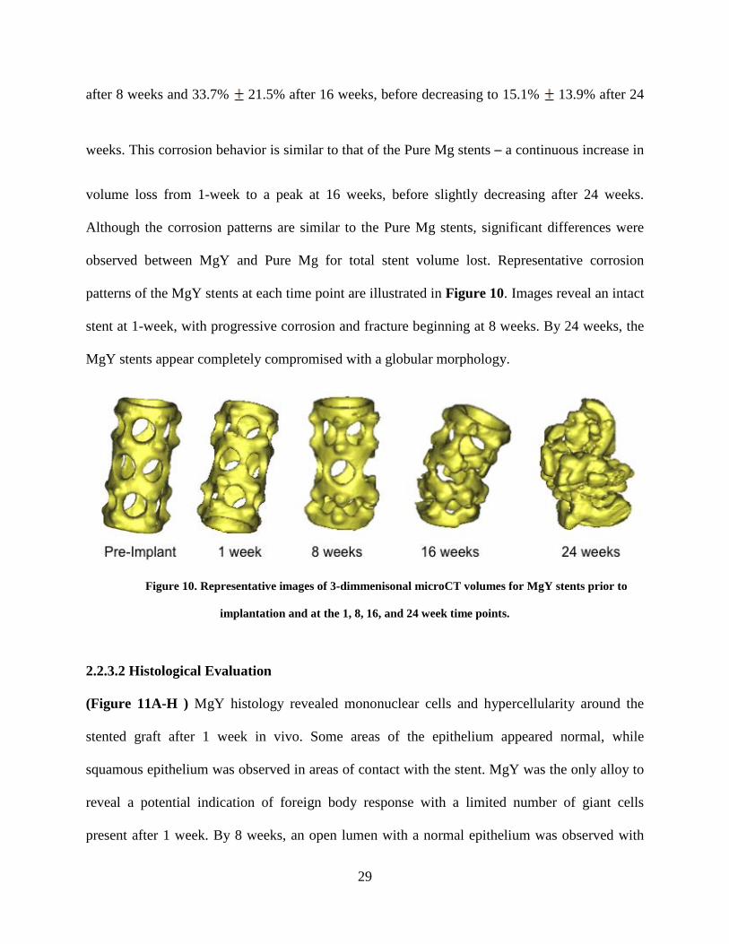
after 8 weeks and 33.7% 21.5% after 16 weeks, before decreasing to 15.1% 13.9% after 24
weeks. This corrosion behavior is similar to that of the Pure Mg stents – a continuous increase in
volume loss from 1-week to a peak at 16 weeks, before slightly decreasing after 24 weeks.
Although the corrosion patterns are similar to the Pure Mg stents, significant differences were
observed between MgY and Pure Mg for total stent volume lost. Representative corrosion
patterns of the MgY stents at each time point are illustrated in Figure 10. Images reveal an intact
stent at 1-week, with progressive corrosion and fracture beginning at 8 weeks. By 24 weeks, the
MgY stents appear completely compromised with a globular morphology.
Figure 10. Representative images of 3-dimmenisonal microCT volumes for MgY stents prior to
implantation and at the 1, 8, 16, and 24 week time points.
2.2.3.2 Histological Evaluation
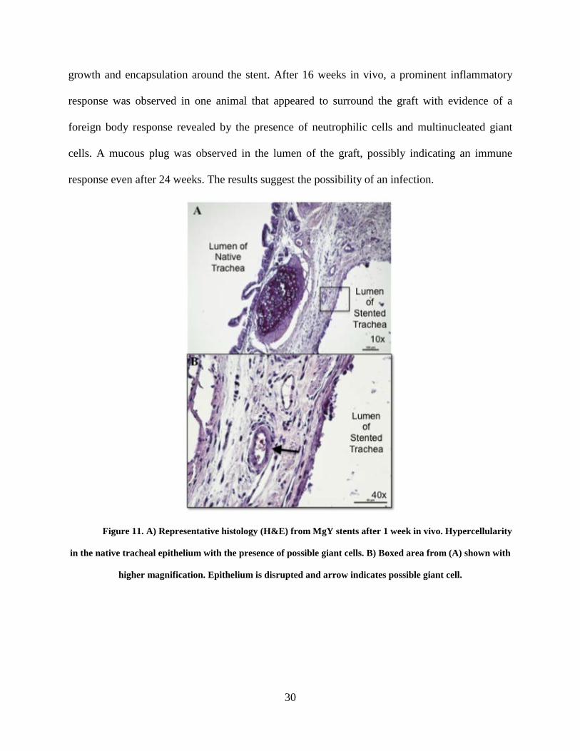
(Figure 11A-H ) MgY histology revealed mononuclear cells and hypercellularity around the
stented graft after 1 week in vivo. Some areas of the epithelium appeared normal, while
squamous epithelium was observed in areas of contact with the stent. MgY was the only alloy to
reveal a potential indication of foreign body response with a limited number of giant cells
present after 1 week. By 8 weeks, an open lumen with a normal epithelium was observed with
30
growth and encapsulation around the stent. After 16 weeks in vivo, a prominent inflammatory
response was observed in one animal that appeared to surround the graft with evidence of a
foreign body response revealed by the presence of neutrophilic cells and multinucleated giant
cells. A mucous plug was observed in the lumen of the graft, possibly indicating an immune
response even after 24 weeks. The results suggest the possibility of an infection.
Figure 11. A) Representative histology (H&E) from MgY stents after 1 week in vivo. Hypercellularity
in the native tracheal epithelium with the presence of possible giant cells. B) Boxed area from (A) shown with
higher magnification. Epithelium is disrupted and arrow indicates possible giant cell.
31
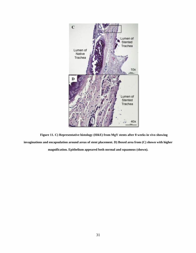
Figure 11. C) Representative histology (H&E) from MgY stents after 8 weeks in vivo showing
invaginations and encapsulation around areas of stent placement. D) Boxed area from (C) shown with higher
magnification. Epithelium appeared both normal and squamous (shown).
32
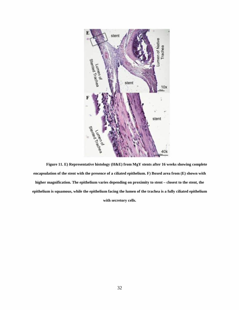
Figure 11. E) Representative histology (H&E) from MgY stents after 16 weeks showing complete
encapsulation of the stent with the presence of a ciliated epithelium. F) Boxed area from (E) shown with
higher magnification. The epithelium varies depending on proximity to stent – closest to the stent, the
epithelium is squamous, while the epithelium facing the lumen of the trachea is a fully ciliated epithelium
with secretory cells.

33
Figure 11. G) Representative histology (H&E) from MgY stents after 24 weeks showing the mucous
plug found in the stented airway of n=1 animal. Arrow indicates area of mucous plug. H) Boxed area from
(G) with higher magnification to show presence of a ciliated epithelium after 24 weeks. Although they are not
healthy, the cilia are present even in the presence of a mucous plug.
2.3 SUMMARY OF IN VIVO RESULTS
Statistical analysis indicated significantly more stent corrosion for Pure Mg than for AZ31 and
MgY (p=0.003), while no significant differences were observed in stent corrosion between the
magnesium alloys AZ31 and MgY (p=0.376), as indicated in Figure 5. Post-hoc analysis

34
showed that MgY corroded the least at the 1-week time point. Pure Mg alloys showed the most
corrosion with the greatest amount of total volume lost at the 16-week time point.
Corrosion results compared at early time points of 1 and 8 weeks did not have significant
differences (p=0.633), while the same is true for results compared at late time points of 16 and
24 weeks (p=0.999). Significant differences in stent corrosion were observed between the 1-
week and both the 16 and 24-week time points (p=0.002). Comparisons of the 8-week time point
with both the 16 and 24-week time points revealed significant differences in stent corrosion
(p=0.030 and p=0.034, respectively). Even with differences in stent corrosion at the 1-week time
point, the structure of the stents remained intact. Changes in stent structure began to occur after 8
weeks for all alloys with corrosion at the thinner areas of the stent. Stent fractures lead to a
compromise of the structure of the stent, usually occurring at the later time points. It is unclear
whether the fractures resulted from corrosion or mechanical stresses, and results vary among
alloys and time points.
In general, histology revealed similar results among all three magnesium stents. Airway
communication between the stented graft and the native airway was maintained throughout the
study with the tracheal bypass model. A ciliated epithelium was present after 8 weeks and
persisted until 24 weeks. The MgY stents were the only stents to indicate a slight inflammatory
or foreign body response, possibly due to an infection. All alloys experienced tissue growth
around the stent leading to encapsulation around 16 weeks and increasing by 24 weeks. At the
24-week time point, the stented graft was vascularized by the native trachea as demonstrated by
the presence of red blood cells.
35
3.0 EVALUATION OF MAGNESIUM ALLOYS IN VITRO
3.1 IN VITRO STUDY DESIGN AND METHODS
Magnesium alloys as tracheal stents were assessed using an in vitro model in order to the
compare in vivo degradation results to degradation of the stent in vitro. The stent was placed in a
decellularized lewis rat trachea and mounted in a bioreactor chamber containing extraluminal
media and intraluminal media. All three alloys were evaluated in both simulated airway fluid
media (Dissolution Technologies, Marques et al.) and 0.9% saline (Fisher Scientific) for a
control comparison. The simulated airway fluid media contains a mixture of mucins and salts to
mimic the environment of the trachea. All of the components of the SAF media are added in the
following order to allow for complete dissolution: magnesium chloride (0.095 g/L), sodium
chloride (6.019 g/L), potassium chloride (0.298 g/L), disodium hydrogen phosphate (0.126 g/L),
sodium sulfate (0.063 g/L), calcium chloride dihydrate (0.368 g//L), sodium acetate (0.574 g/L),
sodium hydrogen carbonate (2.604 g/L), sodium citrate dihydrate (0.097 g/L), and finally porcine
stomach mucin (0.6 g/L). More or less mucin is added to the media in order to vary the viscosity
of the SAF. A total of n=3 stents were used for each alloy and each condition. The stented
decellularized trachea persisted in the bioreactor chamber for 1 week. A continuous flow pump
(Ismatec) was used to circulate media at a constant rate of 0.5 mL/hour for the duration of the
experiment. Polycarbonate square chambers held the stented trachea on hollow, steel rods that
36
allowed for internal media flow through the trachea and pump tubing. Side ports on the chambers
allowed for samples to be taken from the external media of the chamber to measure the amount
of magnesium present in the media. Samples of the media (0.5mL) were taken every 48 hours. In
vitro degradation kinetics were assessed using inductively coupled plasma (ICP). After one week
in vitro, the stents were imaged using microCT and processed for quantification of volume loss.
Data from in vivo and in vitro stent degradation were compared and analyzed for statistical
significance.
3.1.1 Assessment of Corrosion in vitro
3.1.1.1 ICP Analysis
Inductively coupled plasma (ICP) was used to measure the amount of magnesium present in the
media at 1, 3, 5, and 7 days in vitro. Standard calibrations were made to achieve baseline
magnesium concentrations in parts per million (ppm). Every sample taken from the bioreactor
with SAF conditions was diluted 1:20 in saline, and by diluting only SAF in saline by 1:20 a
blank was also created. The blank was used to subtract the mucins and salts found in SAF and
gain a more accurate measurement of magnesium at each time point. Each sample taken from the
bioreactor with control conditions was also diluted 1:20 in fresh saline, and a saline blank was
created.
Upon completion of ICP, the SAF and saline blanks were subtracted from the SAF and
control measurements, respectively. The difference was then multiplied by 20 to achieve the
amount of magnesium measured in the media at 1, 3, 5 and 7 days in vitro for both the simulated
and control conditions for each magnesium stent.
37
3.1.1.2 MicroCT Analysis
As with the in vivo assessment, μCT was used to image the tracheal stents after 1 week of in
vitro testing. The stents were manually removed from the bioreactor chambers and gently
removed from the decellularized trachea before being subjected to μCT imaging. The 2-D dicom
files of the in vitro stents were processed using Mimics software. Three-dimensional volumes of
the stents were generated for each of the alloys tested in vitro for 1 week and compared to 3-D
volumes of the original stents prior to undergoing testing. The difference in volume was
calculated and an overall percent of volume lost over time was quantified. Statistical analysis
was performed to determine significant differences between original stent volume and stent
volume at each end point for each alloy in this study.
3.1.1.3 Statistical Analysis
A one-way ANOVA with Tukey’s Post Hoc analysis was performed on the percent of total stent
volume lost over 7 days in vitro for both the simulated and control conditions. Percent of total
stent volume lost in vitro is reported as a mean standard deviation.
38
3.2 RESULTS
3.2.1 ICP Results
Figure 12. ICP results for each alloy showing magnesium concentration measured in SAF and in
saline. Results represent averages of n=3 samples for each alloy at each time point. No significant differences
were found among alloys under saline conditions, while results from Pure Mg were significantly different ()
from AZ31 and MgY under SAF conditions.
3.2.1.1 Pure Mg
Magnesium concentration measured from the Pure Mg stent in saline control media was 16.6
2.4 ppm at in vitro day 1. The magnesium concentration nearly doubled by day 3 (31.3 17.5
ppm), peaked at day 5 (61.1 24.1 ppm), and then slightly decreased by day 7 (57.5 35.5
ppm). ICP measured the greatest amount of magnesium presence among all alloys from the Pure
Mg in saline control media at day 5 in vitro (61.1 24.1 ppm) (Figure 13).
39
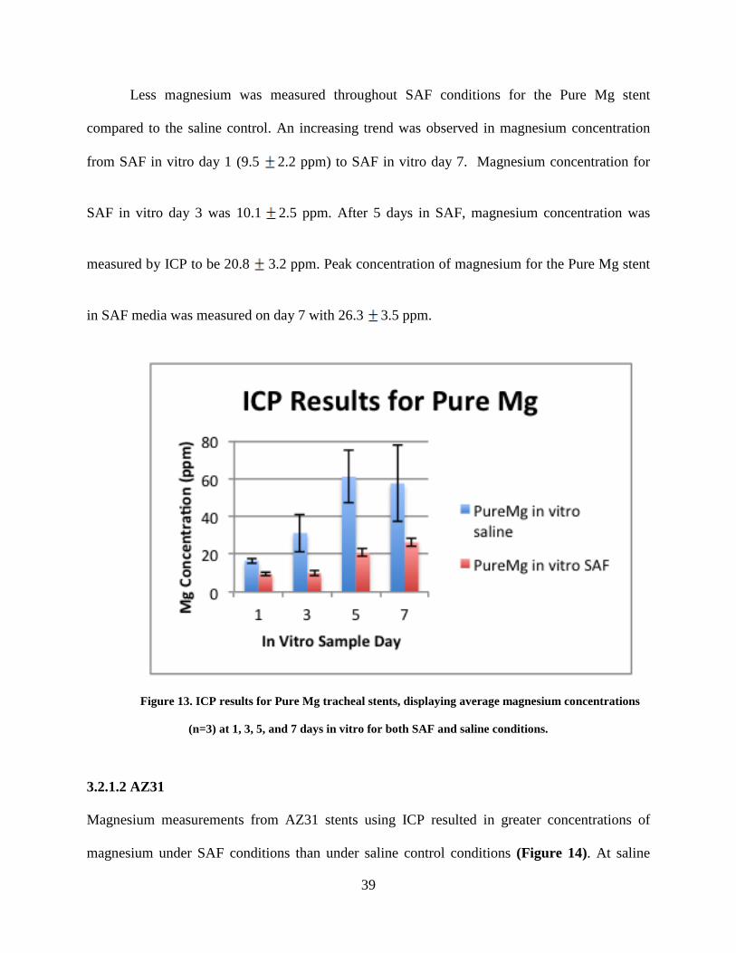
Less magnesium was measured throughout SAF conditions for the Pure Mg stent
compared to the saline control. An increasing trend was observed in magnesium concentration
from SAF in vitro day 1 (9.5 2.2 ppm) to SAF in vitro day 7. Magnesium concentration for
SAF in vitro day 3 was 10.1 2.5 ppm. After 5 days in SAF, magnesium concentration was
measured by ICP to be 20.8 3.2 ppm. Peak concentration of magnesium for the Pure Mg stent
in SAF media was measured on day 7 with 26.3 3.5 ppm.
Figure 13. ICP results for Pure Mg tracheal stents, displaying average magnesium concentrations
(n=3) at 1, 3, 5, and 7 days in vitro for both SAF and saline conditions.
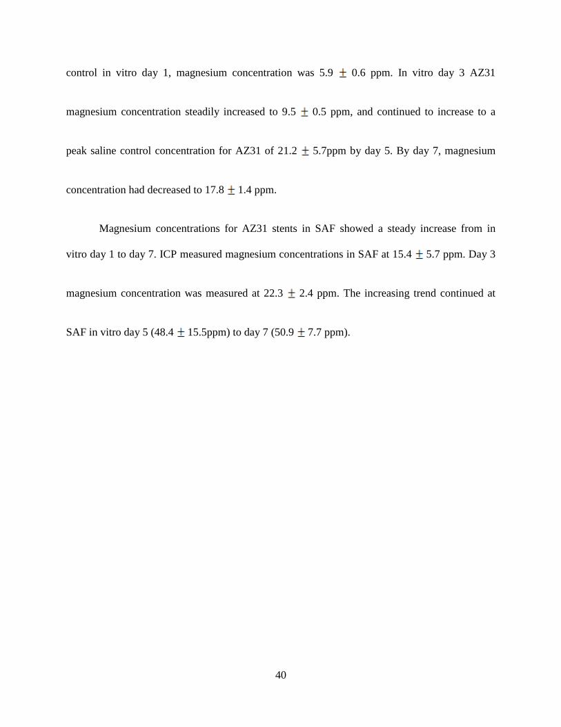
3.2.1.2 AZ31
Magnesium measurements from AZ31 stents using ICP resulted in greater concentrations of
magnesium under SAF conditions than under saline control conditions (Figure 14). At saline
40
control in vitro day 1, magnesium concentration was 5.9 0.6 ppm. In vitro day 3 AZ31
magnesium concentration steadily increased to 9.5 0.5 ppm, and continued to increase to a
peak saline control concentration for AZ31 of 21.2 5.7ppm by day 5. By day 7, magnesium
concentration had decreased to 17.8 1.4 ppm.
Magnesium concentrations for AZ31 stents in SAF showed a steady increase from in
vitro day 1 to day 7. ICP measured magnesium concentrations in SAF at 15.4 5.7 ppm. Day 3
magnesium concentration was measured at 22.3 2.4 ppm. The increasing trend continued at
SAF in vitro day 5 (48.4 15.5ppm) to day 7 (50.9 7.7 ppm).
41
Figure 14. ICP results for AZ31 tracheal stents, displaying average magnesium concentrations (n=3)
at 1, 3, 5, and 7 days in vitro for both SAF and saline conditions.
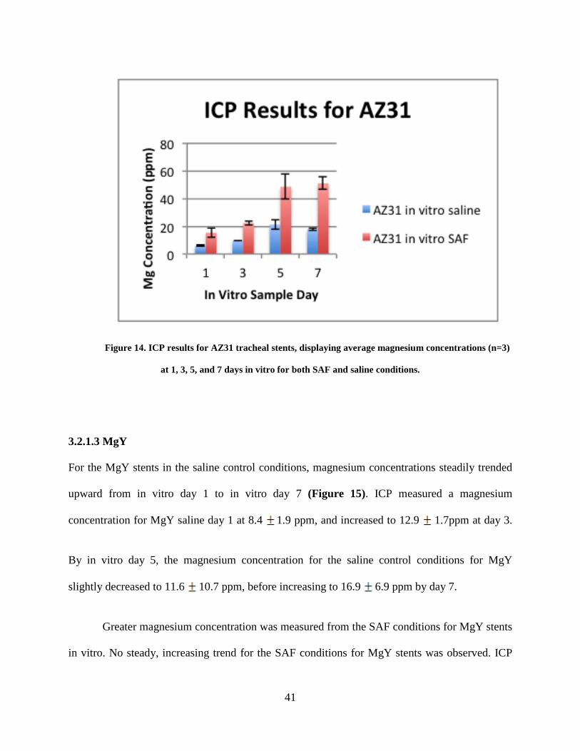
3.2.1.3 MgY
For the MgY stents in the saline control conditions, magnesium concentrations steadily trended
upward from in vitro day 1 to in vitro day 7 (Figure 15). ICP measured a magnesium
concentration for MgY saline day 1 at 8.4 1.9 ppm, and increased to 12.9 1.7ppm at day 3.
By in vitro day 5, the magnesium concentration for the saline control conditions for MgY
slightly decreased to 11.6 10.7 ppm, before increasing to 16.9 6.9 ppm by day 7.
Greater magnesium concentration was measured from the SAF conditions for MgY stents
in vitro. No steady, increasing trend for the SAF conditions for MgY stents was observed. ICP
42
measurements resulted in similar day 1 and day 3 magnesium concentration results (7.1 4.3
ppm and 7.3 4.7 ppm), and increased but similar measurements at day 5 and day 7 (30.6
10.6 ppm and 33.5 9.7 ppm).
Figure 15. ICP results for MgY tracheal stents, displaying average magnesium concentrations (n=3)
at 1, 3, 5, and 7 days in vitro for both SAF and saline conditions.
3.2.2 MicroCT Analyses
After the stents were tested for 1 week in vitro under both SAF and control conditions, microCT
images were obtained and the volume loss was calculated. The in vitro stent volumes were
compared to the in vivo stent volumes at the 1-week time point.
43
3.2.2.1 Pure Mg
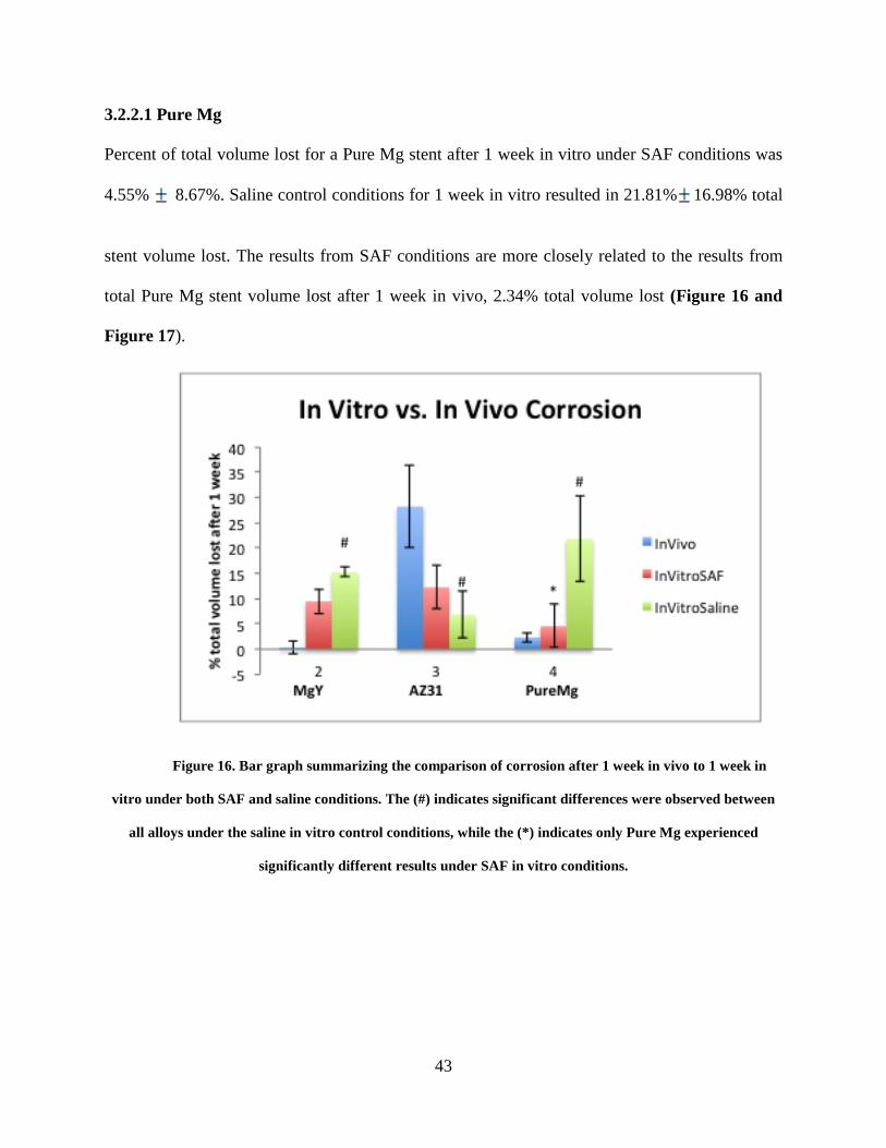
Percent of total volume lost for a Pure Mg stent after 1 week in vitro under SAF conditions was
4.55% 8.67%. Saline control conditions for 1 week in vitro resulted in 21.81% 16.98% total
stent volume lost. The results from SAF conditions are more closely related to the results from
total Pure Mg stent volume lost after 1 week in vivo, 2.34% total volume lost (Figure 16 and
Figure 17).
Figure 16. Bar graph summarizing the comparison of corrosion after 1 week in vivo to 1 week in
vitro under both SAF and saline conditions. The (#) indicates significant differences were observed between
all alloys under the saline in vitro control conditions, while the (*) indicates only Pure Mg experienced
significantly different results under SAF in vitro conditions.
44
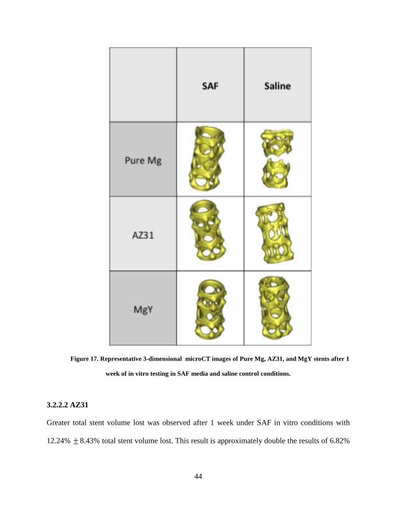
Figure 17. Representative 3-dimensional microCT images of Pure Mg, AZ31, and MgY stents after 1
week of in vitro testing in SAF media and saline control conditions.
3.2.2.2 AZ31
Greater total stent volume lost was observed after 1 week under SAF in vitro conditions with
12.24% 8.43% total stent volume lost. This result is approximately double the results of 6.82%
45
9.12% total stent volume lost after 1 week in vitro under control conditions. Percent of total
stent volume lost after 1 week in vivo appears much greater than the in vitro results with 28.19%
7.71% total stent volume lost, (Figure 16 and Figure 17 ).
3.2.2.3 MgY
While MgY stents experienced very little (0.35% 2.53%) total volume loss after 1 week in
vivo, the results from 1 week in vitro demonstrated greater volume loss under saline control
conditions with 15.22% 1.99% total volume lost. The MgY stents under SAF conditions for 1
week in vitro experienced 9.46% 4.95% total stent volume lost, (Figure 16 and Figure 17).
3.2.3 Statistical Analysis
Significant differences in ICP results were observed between the magnesium stents under SAF
conditions (p=0.019), while no significant differences between magnesium stents were observed
for the saline control conditions (p=0.224), (Figure 16). Magnesium content measured from
AZ31 and MgY stents were not significantly different under SAF conditions, however the
magnesium content measured for Pure Mg under SAF conditions was significantly different from
AZ31 (p=0.035) and MgY (p=0.029). No significant differences in magnesium concentration
were observed between any of the magnesium stents under saline control conditions.
46
When comparing the 1, 3, 5 and 7 day time points under SAF conditions, no significant
differences between magnesium stents were observed (p=0.478). Under saline control
conditions, however, significant differences were observed between magnesium stents over time
(p=0.031), but no significant differences were found among ICP results at 1, 3, 5, and 7 days.
3.3 SUMMARY OF IN VITRO RESULTS
ICP allowed for the measurement of magnesium concentration in media at 48 hour time points
throughout 1 week. The measurement of magnesium in the media is representative of the amount
of magnesium lost from the stent. Measurements from ICP revealed the most magnesium was
lost on day 5 of the Pure Mg in saline control media. Pure Mg stents also experienced the
greatest volume lost among the 3 stents under saline control conditions after 1 week in vitro. The
AZ31 alloy experienced the most magnesium loss on in vitro day 7 under SAF conditions. This
is also consistent with the greatest amount of stent volume lost according to microCT analsysis
among the alloys under SAF conditions after 1 week in vitro.
All 3 of the magnesium stents experienced greater correlation between volume loss after
1 week in vivo and volume loss after 1 week in vitro under SAF conditions. Statistical analysis
suggests no differences among magnesium stents under saline control conditions, while
significant differences are found under simulated conditions. These results suggest the simulated
conditions are more accurately predicting the behavior of magnesium stents after 1 week in vivo.
47
4.0 DISCUSSION
Both in vitro and in vivo testing of magnesium alloys as tracheal stents are necessary to predict
and understand the degradation process of the metal. The results of this study show preliminary
evidence supporting further investigation for use of magnesium alloys as a potential material for
a degradable tracheal stent.
Mild evidence of a foreign body response to the MgY stents was observed in vivo with
the isolated presence of a possible foreign body giant cell after 1 week and hypercellularity with
neutrophils and multinucleated giant cells after 16 and 24 weeks, although the later result may
have been caused by infection. Both AZ31 and Pure Mg stents did not display any signs of an
inflammatory or host response throughout the in vivo study. Histological analysis of all stents
revealed the presence of a normal, ciliated epithelium in the stented airway by 8 weeks and
persisting after 24 weeks. Although present, the epithelium of the stented airway appeared more
squamous in nature by the 24 week time point. After 1 week in vivo, the stented airway also
appeared squamous in some areas, but this may have been the result of surgical manipulation
since it takes time for the graft be become revascularized after transplantation. By 8 weeks, a
ciliated epithelium was observed for all stents. Encapsulation of the stents began between the 8
and 16-week time points.
Little is known about the effects of magnesium on the airway, and evidence of a normal
and functional airway epithelium following magnesium stent placement are encouraging results.
48
Because this study was limited by magnesium stents with a geometry that was not optimized, the
tracheal bypass model was used for tracheal stent evaluation. It is unknown whether similar
results could be achieved without the bypass model and optimized stent geometry. It would also
be interesting to compare an in vivo study using a tracheal bypass model, but place the stent in
the native trachea and allow for the donor trachea to truly bypass the stented native airway.
Because of limitations in achieving a machinable and appropriately sized rat tracheal stent, we
did not try this alternative surgical model.
In using the tracheal bypass model for the evaluation of the magnesium stents, we did not
include a control group. The control group would have had the tracheal bypass but without a
stent present in the donor tracheal graft. The lack of the control group leads to the question of
what would be seen in the progression of the airway epithelium. Presumably the initial disruption
of the airway epithelium would not occur because there would be no stent placement. Having a
control group for comparison would allow for understanding into how the magnesium truly
affected the health, morphology, and persistence of the airway epithelium.
MicroCT imaging provided insight into the corrosion patterns of the magnesium stents
over time. By 16 or even 8 weeks, the stents appeared compromised with total stent volume
losses between 18 and 66% after 16 weeks. As expected, the Pure Mg stent did experience
greater corrosion in vivo than the magnesium alloys. MgY and AZ31 both experienced a
maximum of 33% total stent volume lost, while MgY peaked after 16weeks and AZ31 did not
reach maximum volume loss until 24 weeks. Visually, the compromised structure of the MgY
stents can be qualitatively observed as more compromised than that of the AZ31 stent, even
though the stents experienced similar amounts of total volume lost. This could be the result of
the alloy composition, or even the tensions experienced in vivo.
49
The hypothesis from specific aim 1 was successful in predicting less degradation for the
AZ31 and MgY alloys compared to the Pure Mg stents. Statistical analysis also supported the
hypothesis that differences in volume lost were observed between the early and late time points,
suggesting a pattern for rate of degradation.
Results from ICP also partially supported the hypothesis from specific aim 2, as Pure Mg
experienced the greatest volume lost in vivo and also registered the highest amount of
magnesium measured in vitro. MicroCT results also revealed the closest correlation between
magnesium stent volume lost at 1 week in vivo and after 1 week in vitro under SAF conditions.
Differences between the in vitro results of saline control conditions and the in vivo results from 1
week, as compared to the SAF in vitro results, suggest the SAF media is contributing to creating
an environment in vitro that is more closely related to an in vivo airway environment. Because
the bioreactors used to evaluate the magnesium stents in vitro were continuous flow, the varying
pressures associated with respiration were not applied to the stents in vitro. The tracheal bypass
model also places the stented airway in a partially passive trachea environment. As a result, the
continuous flow system may be a good model for comparing in vitro results to the in vivo bypass
model. Similarities between in vitro and in vivo results are very encouraging for the evaluation
of magnesium alloys. Historically, in vitro evaluation of magnesium could not be depended on to
accurately predicted in vivo behavior. To further address the limitations of the in vitro study,
magnesium alloys should be evaluated at later time points in vitro and compared to later time
points in vivo.
An important limitation of the in vivo study was that the trachea stent intended for
treatment of tracheal stenosis was applied to a normal, healthy trachea in a bypass model.
Because the stent geometry was not yet optimized and little is known about the use of
50
magnesium in the trachea, this study aimed to evaluate the effects of magnesium on the airway
environment. Histological analysis demonstrated minimal effects of magnesium on the function
of the airway environment at early time points (1 week) and up to 24 weeks. Another study
evaluating the effects of magnesium on the trachea at later time points of 6 months to 1 year,
could provide further understanding on the longer term effects of magnesium on the airway
environment.
51
5.0 CONCLUSIONS
Tracheal stents successfully manage adult airway obstructions, including tracheal stenosis, but
are used as a treatment option of last resort for pediatric patients because of their permanent
nature. A degradable tracheal stent would provide pediatric patients the opportunity for growth
potential while maintaining an open lumen. The magnesium alloys evaluated in this study
demonstrate potential for use in the tracheal environment. More studies will be necessary to
further develop the stent design and optimize mechanical properties in order to support a
pediatric airway with tracheal stenosis. This study illustrated the trachea’s tolerance and
acceptance of magnesium after 6 months with minimal foreign body response and without any
negative effects on airway function. Initial in vitro studies using SAF provide support for
additional in vitro testing to more closely mimic the response of magnesium in vivo.
52
BIBLIOGRAPHY
1. Jungebluth, P., Moll, G., Baiguera, S. & Macchiarini, P. Tissue-engineered airway: a regenerative solution. Clin. Pharmacol. Ther. 91, 81–93 (2012).
2. Bardin, P. G., Johnston, S. L. & Hamilton, G. Middle airway obstruction--it may be happening under our noses. Thorax 68, 396–398 (2013).
3. Yong, M. S. et al. Surgical management of pulmonary artery sling in children. J. Thorac. Cardiovasc. Surg. 145, 1033–1039 (2013).
4. Tatekawa, Y. & Muraji, T. Surgical strategy for acquired tracheomalacia due to innominate artery compression of the trachea. Eur J Cardiothorac Surg 39, 412–413 (2011).
5. Maksoud-Filho, J. G., Gonçalves, M. E. P., Cardoso, S. R. & Tannuri, U. Early diagnostic and endoscopic dilatation for the treatment of acquired upper airway stenosis after intubation in children. J. Pediatr. Surg. 43, 1254–1258 (2008).
6. Gallagher, T. Q. & Hartnick, C. J. Tracheal resection and reanastomosis. Adv. Otorhinolaryngol. 73, 50–57 (2012).
7. Zaima, A. et al. Long-term T-tube stenting as definitive treatment of severe acquired subglottic stenosis in children. J. Pediatr. Surg. 45, 996–999 (2010).
8. Xu, X. et al. Treatment of congenital tracheal stenosis by balloon-expandable metallic stents in paediatric intensive care unit. Interact Cardiovasc Thorac Surg 14, 548–550 (2012).
53
9. Ng, A. H. C., Ng, N. S. P., Zhu, G. H., Lim, L. H. Y. & Venkatraman, S. S. A fully degradable tracheal stent: in vitro and in vivo characterization of material degradation. J. Biomed. Mater. Res. Part B Appl. Biomater. 100, 693–699 (2012).
10. Antón-Pacheco, J. L. et al. The role of airway stenting in pediatric tracheobronchial obstruction. Eur J Cardiothorac Surg 33, 1069–1075 (2008).
11. Preciado, D. & Zalzal, G. Laryngeal and tracheal stents in children. Curr Opin Otolaryngol Head Neck Surg 16, 83–85 (2008).
12. Brigger, M. T. & Boseley, M. E. Management of tracheal stenosis. Curr Opin Otolaryngol Head Neck Surg (2012).doi:10.1097/MOO.0b013e328358566d
13. Mainwaring, R. D. et al. Surgical reconstruction of tracheal stenosis in conjunction with congenital heart defects. Ann. Thorac. Surg. 93, 1266–72– discussion 1272–3 (2012).
14. Charokopos, N. et al. The management of post-intubation tracheal stenoses with self-expandable stents: early and long-term results in 11 cases. Eur J Cardiothorac Surg 40, 919–924 (2011).
15. Nicolai, T. Airway stents in children. Pediatr. Pulmonol. 43, 330–344 (2008).
16. Noppen, M., Stratakos, G., D'Haese, J., Meysman, M. & Vinken, W. Removal of covered self-expandable metallic airway stents in benign disorders: indications, technique, and outcomes. Chest 127, 482–487 (2005).
17. Moravej, M. & Mantovani, D. Biodegradable metals for cardiovascular stent application: interests and new opportunities. Int J Mol Sci 12, 4250–4270 (2011).
18. Serruys, P. W., de Jaegere, P. & Kiemeneij, F. A comparison of balloon-expandable-stent implantation with balloon angioplasty in patients with coronary artery disease. … England Journal of … (1994).
19. Rassaf, T., Steiner, S. & Kelm, M. Postoperative care and follow-up after coronary stenting. Dtsch Arztebl Int 110, 72–82 (2013).

54
20. Mitsuoka, M., Sakuragi, T. & Itoh, T. Clinical benefits and complications of Dumon stent insertion for the treatment of severe central airway stenosis or airway fistula. Gen Thorac Cardiovasc Surg 55, 275–280 (2007).
21. Lawton, B. & Gungor, A. Airway salvation after failed anterior graft in a child with long segment stenosis. Am J Otolaryngol (2013).doi:10.1016/j.amjoto.2013.02.009
22. Mroz, R. M. et al. Severe respiratory distress caused by central airway obstruction treated with self-expandable metallic stents. J. Physiol. Pharmacol. 59 Suppl 6, 491–497 (2008).
23. Coordes, A., Todt, I., Ernst, A. & Seidl, R. O. Multi-stage surgery for airway patency after metallic stent removal in benign laryngotracheal airway disease in two adolescents. Int. J. Pediatr. Otorhinolaryngol. (2013).doi:10.1016/j.ijporl.2013.02.012
24. Murthy, S. C., Gildea, T. R. & Mehta, A. C. Removal of self-expandable metallic stents: is it possible? Semin Respir Crit Care Med 25, 381–385 (2004).
25. Vondrys, D., Elliott, M. J., McLaren, C. A., Noctor, C. & Roebuck, D. J. First experience with biodegradable airway stents in children. Ann. Thorac. Surg. 92, 1870–1874 (2011).
26. Peuster, M. et al. A novel approach to temporary stenting: degradable cardiovascular stents produced from corrodible metal-results 6-18 months after implantation into New Zealand white rabbits. Heart 86, 563–569 (2001).
27. Heublein, B. et al. Biocorrosion of magnesium alloys: a new principle in cardiovascular implant technology? Heart 89, 651–656 (2003).
28. Chen, D. et al. Biocompatibility of magnesium-zinc alloy in biodegradable orthopedic implants. Int. J. Mol. Med. 28, 343–348 (2011).
29. Witte, F. The history of biodegradable magnesium implants: a review. Acta Biomater 6, 1680–1692 (2010).
30. Zartner, P., Cesnjevar, R., Singer, H. & Weyand, M. First successful implantation of a biodegradable metal stent into the left pulmonary artery of a preterm baby. Catheter Cardiovasc Interv 66, 590–594 (2005).
55
31. Schranz, D., Zartner, P., Michel-Behnke, I. & Akintürk, H. Bioabsorbable metal stents for percutaneous treatment of critical recoarctation of the aorta in a newborn. Catheter Cardiovasc Interv 67, 671–673 (2006).
32. Erbel, R. et al. Temporary scaffolding of coronary arteries with bioabsorbable magnesium stents: a prospective, non-randomised multicentre trial. Lancet 369, 1869–1875 (2007).
33. Gourgoulianis, K. I., Chatziparasidis, G., Chatziefthimiou, A. & Molyvdas, P. A. Magnesium as a relaxing factor of airway smooth muscles. J Aerosol Med 14, 301–307 (2001).
34. Pollart, S. M., Compton, R. M. & Elward, K. S. Management of acute asthma exacerbations. Am Fam Physician 84, 40–47 (2011).
35. SEELIG, M. G. A STUDY OF MAGNESIUM WIRE AS AN ABSORBABLE SUTURE AND LIGATURE MATERIAL. Arch Surg 8, 669–680 (1924).
36. Witte, F. et al. In vitro and in vivo corrosion measurements of magnesium alloys. Biomaterials 27, 1013–1018 (2006).
37. Hänzi, A. C., Gerber, I., Schinhammer, M., Löffler, J. F. & Uggowitzer, P. J. On the in vitro and in vivo degradation performance and biological response of new biodegradable Mg-Y-Zn alloys. Acta Biomater 6, 1824–1833 (2010).
38. Di Mario, C. et al. Drug-eluting bioabsorbable magnesium stent. J Interv Cardiol 17, 391–395 (2004).
39. Kannan, M. B. & Raman, R. K. S. In vitro degradation and mechanical integrity of calcium-containing magnesium alloys in modified-simulated body fluid. Biomaterials 29, 2306–2314 (2008).
40. Scheideler, L. et al. Comparison of different in vitro tests for biocompatibility screening of Mg alloys. Acta Biomater (2013).doi:10.1016/j.actbio.2013.02.020
41. United States Food and Drug Administration, FDA.