Magne™ Protein A Beads and Magne™ Protein G …/media/Files/Resources/Protocols...Revised 7/15...
12
Revised 7/15 TM371 TECHNICAL MANUAL Magne™ Protein A Beads and Magne™ Protein G Beads for Antibody Purification Instrucons for Use of Products G7471, G7472, G7473, G8781, G8782 and G8783
Transcript of Magne™ Protein A Beads and Magne™ Protein G …/media/Files/Resources/Protocols...Revised 7/15...
Revised 7/15 TM371
T E C H N I C A L M A N U A L
Magne™ Protein A Beads and Magne™ Protein G Beads for Antibody PurificationInstructions for Use of Products G7471, G7472, G7473, G8781, G8782 and G8783
Promega Corporation · 2800 Woods Hollow Road · Madison, WI 53711-5399 USA · Toll Free in USA 800-356-9526 · 608-274-4330 · Fax 608-277-2516 1www.promega.com TM371 · Revised 7/15
All technical literature is available at: www.promega.com/protocols/ Visit the web site to verify that you are using the most current version of this Technical Manual.
E-mail Promega Technical Services if you have questions on use of this system: [email protected]
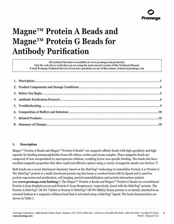
Magne™ Protein A Beads and Magne™ Protein G Beads for Antibody Purification
1. Description
Magne™ Protein A Beads and Magne™ Protein G Beads(a) are magnetic affinity beads with high specificity and high capacity for binding immunoglobulins from cell culture, ascites and serum samples. These magnetic beads are composed of iron encapsulated by macroporous cellulose, resulting in low non-specific binding. The beads also have excellent magnetic properties that allow rapid and efficient capture using a variety of magnetic stands (see Section 7).
Both beads use a novel attachment chemistry based on the HaloTag® technology to immobilize Protein A or Protein G. The HaloTag® protein is a multi-functional protein tag that forms a covalent bond with its ligand and is used for protein expression and purification, cell imaging, protein immobilization and protein-interaction analysis (see www.promega.com/halotag/). The Magne™ Protein A Beads and Magne™ Protein G Beads use recombinant Protein A from Staphylococcus and Protein G from Streptococci, respectively, fused with the HaloTag® protein. The Protein A-HaloTag® (M.Wt 71kDa) or Protein G-HaloTag® (M.Wt 58kDa) fusion protein is covalently attached in an oriented fashion to a magnetic cellulose bead that is activated using a HaloTag® ligand. The bead characteristics are shown in Table 1.
1. Description .........................................................................................................................................1
2. Product Components and Storage Conditions ........................................................................................4
3. Before You Begin .................................................................................................................................5
4. Antibody Purification Protocol .............................................................................................................6
5. Troubleshooting..................................................................................................................................9
6. Composition of Buffers and Solutions ................................................................................................. 10
7. Related Products ............................................................................................................................... 10
8. Summary of Changes ......................................................................................................................... 10
2 Promega Corporation · 2800 Woods Hollow Road · Madison, WI 53711-5399 USA · Toll Free in USA 800-356-9526 · 608-274-4330 · Fax 608-277-2516TM371 · Revised 7/15 www.promega.com
Table 1. Characteristics of the Magne™ Protein A and Magne™ Protein G Beads.
Composition Magnetic bead based on macroporous cellulose
Chemistry Oriented and covalent attachment of Protein A or Protein G using HaloTag® technology
Particle Size 30–80µm.
Antibody-Binding Capacity ≥18mg of human IgG/1ml of settled beads
Formulation 20% slurry in 20% ethanol
Storage 2–10°C
The choice between selecting Magne™ Protein A Beads or Magne™ Protein G Beads depends on the difference in the binding affinities of Protein A or Protein G for antibodies from different species and for different antibody isotypes. See Table 2.
Advantages of Magne™ Protein A Beads and Magne™ Protein G Beads• Simple and easy to use• High purity and recovery of antibodies• No expensive instrumentation required• Processes 1–96 samples in parallel • Easily handles sample volumes of 20µl to 50ml• Purification can be automated
Magne™ Protein A Beads and Magne™ Protein G Beads are not recommended for use in immunoprecipitation (IP) or co-IP applications.
Magne™ Protein G Beads are optimized for immunoglobulin purification. Do not use the Magne™ HaloTag® Beads (Cat.# G7281) or HaloLink™ Resin (Cat.# G1912) to bind the Protein G-HaloTag® Fusion Protein (Cat.# G7291), as these products are not optimized for immunoglobulin purification.
Special Acknowledgement: The Magne™ Protein G Beads, Protein G HaloTag® Fusion Protein and Magne™ Protein A Beads were developed in collaboration with Kazusa DNA Research Institute (KDRI). We would like to thank KDRI for their technical guidance and early testing.
!
!
Promega Corporation · 2800 Woods Hollow Road · Madison, WI 53711-5399 USA · Toll Free in USA 800-356-9526 · 608-274-4330 · Fax 608-277-2516 3www.promega.com TM371 · Revised 7/15
Table 2. Binding Affinity of Protein A and Protein G for Different Antibodies and Isotypes
Species/Subclass Protein A Protein G
Monoclonal
Human
IgG1 +++ +++
IgG2 +++ +++
IgG3 + +++
IgG4 +++ +++
Mouse
IgG1 + +++
IgG2A +++ +++
IgG2b ++ ++
IgG3 + ++
Rat
IgG1 + +
IgG2a + +++
IgG2b + ++
IgG2c + +
Polyclonal
Rabbit +++ +++
Cow ++ +++
Goat + +++
Chicken + +
Human IgG +++ +++
Human IgM + +
Human IgD + +
Human IgA + +
Key: Strong (+++), Medium (++), Weak (+). Adapted from Akerström, B. et al. (1985) J. Immunol. 135(4), 2589-92.
4 Promega Corporation · 2800 Woods Hollow Road · Madison, WI 53711-5399 USA · Toll Free in USA 800-356-9526 · 608-274-4330 · Fax 608-277-2516TM371 · Revised 7/15 www.promega.com
2. Product Components and Storage Conditions
P R O D U C T S I Z E C AT. #
Magne™ Protein A Beads 1.0ml G8781
5 × 1.0ml G8782
50ml G8783
Magne™ Protein G Beads 1.0ml G7471
5 × 1.0ml G7472
50ml G7473
Storage Conditions: Store the Magne™ Protein A and Magne™ Protein G Beads at 2–10°C. Do not freeze. Do not allow beads to dry during storage or use.
Do not reuse the Magne™ Protein A or Magne™ Protein G Beads.
11208TA
Cell Media
M SM A G M SM A G M SM A G
Mouse Ascites Goat Serum
kDa
AlbuminIgG heavy chain (55kDa)
IgG light chain (25kDa)
250
150
100
75
50
37
25
20
15
10
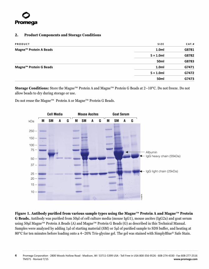
Figure 1. Antibody purified from various sample types using the Magne™ Protein A and Magne™ Protein G Beads. Antibody was purified from 50µl of cell culture media (mouse IgG1), mouse ascites (IgG2a) and goat serum using 50µl Magne™ Protein A Beads (A) and Magne™ Protein G Beads (G) as described in this Technical Manual. Samples were analyzed by adding 1µl of starting material (SM) or 5µl of purified sample to SDS buffer, and heating at 80°C for ten minutes before loading onto a 4–20% Tris-glycine gel. The gel was stained with SimplyBlue® Safe Stain.
Promega Corporation · 2800 Woods Hollow Road · Madison, WI 53711-5399 USA · Toll Free in USA 800-356-9526 · 608-274-4330 · Fax 608-277-2516 5www.promega.com TM371 · Revised 7/15
3. Before You Begin
1. Antibody purification using Magne™ Protein A Beads or Magne™ Protein G Beads requires a magnetic stand. The beads can be used with a variety of magnetic stands available from Promega (see Section 7) to purify antibody from 1–96 samples in parallel, with sample volumes ranging from 20µl to 30ml.
2. Be sure that the Magne™ Protein A Beads and Magne™ Protein G Beads remain in suspension during binding and wash steps for maximal antibody yield and purity. We recommend using a tube shaker or end-over-end mixer.
3. Sample incubation times may need to be optimized. Binding may be performed at 4°C; however, longer incubation times may be necessary for efficient antibody capture.
4. Biological samples may be cleared by filtration through a 0.22µm filter or by a 15-minute centrifugation at 14,000 × g to remove aggregates and insoluble proteins prior to antibody purification.
5. Prior to antibody purification, cell culture medium can be concentrated using a low-molecular-weight-cutoff filter (e.g., molecular weight cutoff of 3,500 Daltons) to reduce the starting sample volume and increase the antibody concentration. Scale the sample, bind/wash buffer and Magne™ Protein A Bead or Magne™ Protein G Bead volumes proportionally. To minimize sample dilution, a 10X bind/wash buffer may be added to the sample to a final concentration of 1X.
6. Magne™ Protein A Beads and Magne™ Protein G Beads are compatible with several bind/wash buffers, including 25mM sodium acetate buffer (pH 6.0), phosphate-buffered saline (pH 7.4) and Tris-buffered saline (pH 7.5). We recommend an elution buffer of 100mM glycine-HCl (pH 2.7) and a neutralization buffer of 2M Tris buffer (pH 7.5).
Note: Purification of different antibody isotypes or antibodies from different species may require protocol optimization for maximal recovery and compatibility with downstream applications.!
6 Promega Corporation · 2800 Woods Hollow Road · Madison, WI 53711-5399 USA · Toll Free in USA 800-356-9526 · 608-274-4330 · Fax 608-277-2516TM371 · Revised 7/15 www.promega.com
4. Antibody Purification Protocol
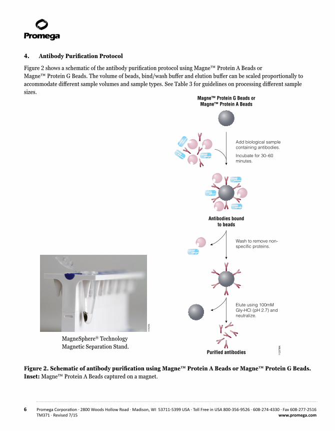
Figure 2 shows a schematic of the antibody purification protocol using Magne™ Protein A Beads or Magne™ Protein G Beads. The volume of beads, bind/wash buffer and elution buffer can be scaled proportionally to accommodate different sample volumes and sample types. See Table 3 for guidelines on processing different sample sizes.
Figure 2. Schematic of antibody purification using Magne™ Protein A Beads or Magne™ Protein G Beads. Inset: Magne™ Protein A Beads captured on a magnet.
11207M
A
Magne™ Protein G Beads orMagne™ Protein A Beads
Antibodies boundto beads
Purified antibodies
Add biological samplecontaining antibodies.
Incubate for 30–60 minutes.
Wash to remove non-specific proteins.
Elute using 100mMGly-HCl (pH 2.7) andneutralize.
11150TA
MagneSphere® Technology Magnetic Separation Stand.
Promega Corporation · 2800 Woods Hollow Road · Madison, WI 53711-5399 USA · Toll Free in USA 800-356-9526 · 608-274-4330 · Fax 608-277-2516 7www.promega.com TM371 · Revised 7/15
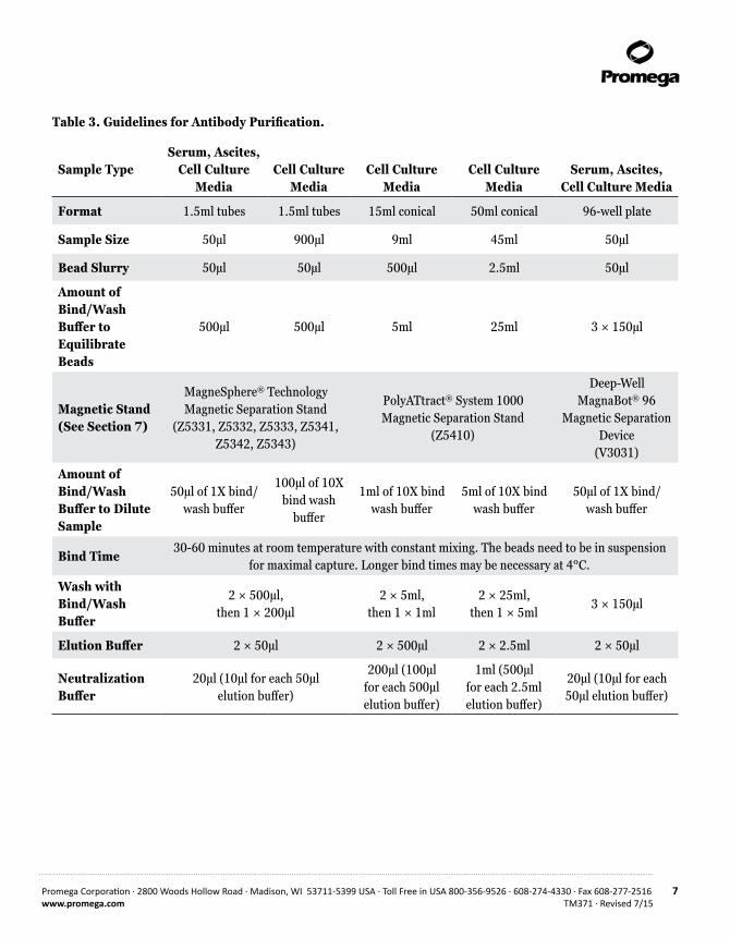
Table 3. Guidelines for Antibody Purification.
Sample TypeSerum, Ascites,
Cell Culture Media
Cell Culture Media
Cell Culture Media
Cell Culture Media
Serum, Ascites, Cell Culture Media
Format 1.5ml tubes 1.5ml tubes 15ml conical 50ml conical 96-well plate
Sample Size 50µl 900µl 9ml 45ml 50µl
Bead Slurry 50µl 50µl 500µl 2.5ml 50µl
Amount of Bind/Wash Buffer to Equilibrate Beads
500µl 500µl 5ml 25ml 3 × 150µl
Magnetic Stand (See Section 7)
MagneSphere® Technology Magnetic Separation Stand
(Z5331, Z5332, Z5333, Z5341, Z5342, Z5343)
PolyATtract® System 1000 Magnetic Separation Stand
(Z5410)
Deep-Well MagnaBot® 96
Magnetic Separation Device
(V3031)
Amount of Bind/Wash Buffer to Dilute Sample
50µl of 1X bind/wash buffer
100µl of 10X bind wash
buffer
1ml of 10X bind wash buffer
5ml of 10X bind wash buffer
50µl of 1X bind/wash buffer
Bind Time30-60 minutes at room temperature with constant mixing. The beads need to be in suspension
for maximal capture. Longer bind times may be necessary at 4°C.
Wash with Bind/Wash Buffer
2 × 500µl, then 1 × 200µl
2 × 5ml, then 1 × 1ml
2 × 25ml, then 1 × 5ml
3 × 150µl
Elution Buffer 2 × 50µl 2 × 500µl 2 × 2.5ml 2 × 50µl
Neutralization Buffer
20µl (10µl for each 50µl elution buffer)
200µl (100µl for each 500µl elution buffer)
1ml (500µl for each 2.5ml elution buffer)
20µl (10µl for each 50µl elution buffer)
8 Promega Corporation · 2800 Woods Hollow Road · Madison, WI 53711-5399 USA · Toll Free in USA 800-356-9526 · 608-274-4330 · Fax 608-277-2516TM371 · Revised 7/15 www.promega.com
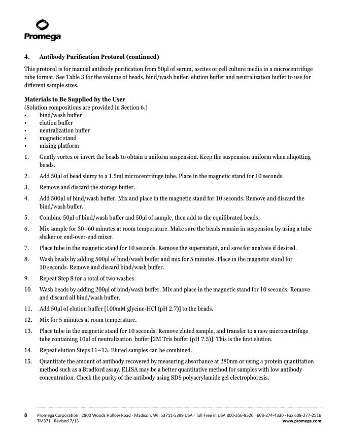
4. Antibody Purification Protocol (continued)
This protocol is for manual antibody purification from 50µl of serum, ascites or cell culture media in a microcentrifuge tube format. See Table 3 for the volume of beads, bind/wash buffer, elution buffer and neutralization buffer to use for different sample sizes.
Materials to Be Supplied by the User(Solution compositions are provided in Section 6.)• bind/wash buffer • elution buffer • neutralization buffer • magnetic stand • mixing platform
1. Gently vortex or invert the beads to obtain a uniform suspension. Keep the suspension uniform when aliqotting beads.
2. Add 50µl of bead slurry to a 1.5ml microcentrifuge tube. Place in the magnetic stand for 10 seconds.
3. Remove and discard the storage buffer.
4. Add 500µl of bind/wash buffer. Mix and place in the magnetic stand for 10 seconds. Remove and discard the bind/wash buffer.
5. Combine 50µl of bind/wash buffer and 50µl of sample, then add to the equilibrated beads.
6. Mix sample for 30–60 minutes at room temperature. Make sure the beads remain in suspension by using a tube shaker or end-over-end mixer.
7. Place tube in the magnetic stand for 10 seconds. Remove the supernatant, and save for analysis if desired.
8. Wash beads by adding 500µl of bind/wash buffer and mix for 5 minutes. Place in the magnetic stand for 10 seconds. Remove and discard bind/wash buffer.
9. Repeat Step 8 for a total of two washes.
10. Wash beads by adding 200µl of bind/wash buffer. Mix and place in the magnetic stand for 10 seconds. Remove and discard all bind/wash buffer.
11. Add 50µl of elution buffer [100mM glycine-HCl (pH 2.7)] to the beads.
12. Mix for 5 minutes at room temperature.
13. Place tube in the magnetic stand for 10 seconds. Remove eluted sample, and transfer to a new microcentrifuge tube containing 10µl of neutralization buffer [2M Tris buffer (pH 7.5)]. This is the first elution.
14. Repeat elution Steps 11–13. Eluted samples can be combined.
15. Quantitate the amount of antibody recovered by measuring absorbance at 280nm or using a protein quantitation method such as a Bradford assay. ELISA may be a better quantitative method for samples with low antibody concentration. Check the purity of the antibody using SDS polyacrylamide gel electrophoresis.
Promega Corporation · 2800 Woods Hollow Road · Madison, WI 53711-5399 USA · Toll Free in USA 800-356-9526 · 608-274-4330 · Fax 608-277-2516 9www.promega.com TM371 · Revised 7/15
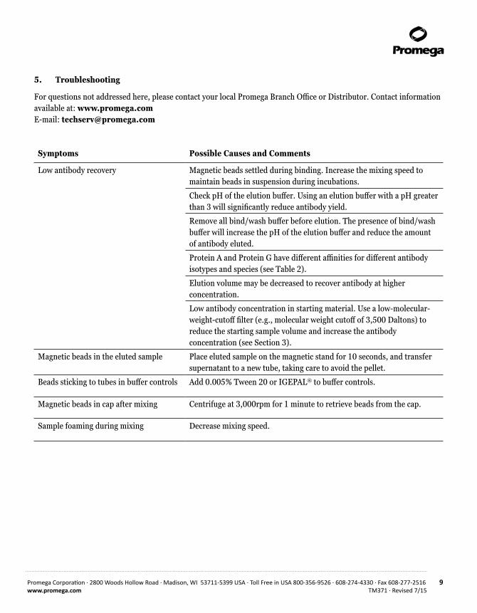
5. Troubleshooting
For questions not addressed here, please contact your local Promega Branch Office or Distributor. Contact information available at: www.promega.com E-mail: [email protected]
Symptoms Possible Causes and Comments
Low antibody recovery Magnetic beads settled during binding. Increase the mixing speed to maintain beads in suspension during incubations.
Check pH of the elution buffer. Using an elution buffer with a pH greater than 3 will significantly reduce antibody yield.
Remove all bind/wash buffer before elution. The presence of bind/wash buffer will increase the pH of the elution buffer and reduce the amount of antibody eluted.
Protein A and Protein G have different affinities for different antibody isotypes and species (see Table 2).
Elution volume may be decreased to recover antibody at higher concentration.
Low antibody concentration in starting material. Use a low-molecular-weight-cutoff filter (e.g., molecular weight cutoff of 3,500 Daltons) to reduce the starting sample volume and increase the antibody concentration (see Section 3).
Magnetic beads in the eluted sample Place eluted sample on the magnetic stand for 10 seconds, and transfer supernatant to a new tube, taking care to avoid the pellet.
Beads sticking to tubes in buffer controls Add 0.005% Tween 20 or IGEPAL® to buffer controls.
Magnetic beads in cap after mixing Centrifuge at 3,000rpm for 1 minute to retrieve beads from the cap.
Sample foaming during mixing Decrease mixing speed.
10 Promega Corporation · 2800 Woods Hollow Road · Madison, WI 53711-5399 USA · Toll Free in USA 800-356-9526 · 608-274-4330 · Fax 608-277-2516TM371 · Revised 7/15 www.promega.com
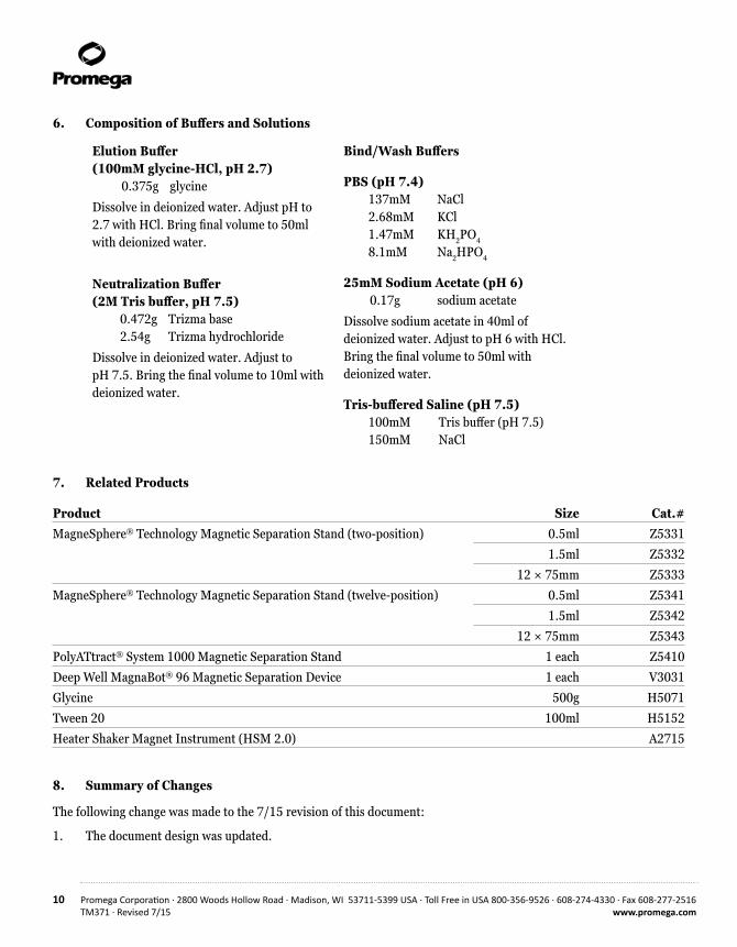
Elution Buffer(100mM glycine-HCl, pH 2.7) 0.375g glycine
Dissolve in deionized water. Adjust pH to 2.7 with HCl. Bring final volume to 50ml with deionized water.
Neutralization Buffer (2M Tris buffer, pH 7.5) 0.472g Trizma base 2.54g Trizma hydrochloride
Dissolve in deionized water. Adjust to pH 7.5. Bring the final volume to 10ml with deionized water.
Bind/Wash Buffers
PBS (pH 7.4) 137mM NaCl 2.68mM KCl 1.47mM KH2PO4
8.1mM Na2HPO4
25mM Sodium Acetate (pH 6) 0.17g sodium acetate
Dissolve sodium acetate in 40ml of deionized water. Adjust to pH 6 with HCl. Bring the final volume to 50ml with deionized water.
Tris-buffered Saline (pH 7.5) 100mM Tris buffer (pH 7.5) 150mM NaCl
6. Composition of Buffers and Solutions
7. Related Products
Product Size Cat.#MagneSphere® Technology Magnetic Separation Stand (two-position) 0.5ml Z5331
1.5ml Z5332
12 × 75mm Z5333
MagneSphere® Technology Magnetic Separation Stand (twelve-position) 0.5ml Z5341
1.5ml Z5342
12 × 75mm Z5343
PolyATtract® System 1000 Magnetic Separation Stand 1 each Z5410
Deep Well MagnaBot® 96 Magnetic Separation Device 1 each V3031
Glycine 500g H5071
Tween 20 100ml H5152
Heater Shaker Magnet Instrument (HSM 2.0) A2715
8. Summary of Changes
The following change was made to the 7/15 revision of this document:
1. The document design was updated.
Promega Corporation · 2800 Woods Hollow Road · Madison, WI 53711-5399 USA · Toll Free in USA 800-356-9526 · 608-274-4330 · Fax 608-277-2516 11www.promega.com TM371 · Revised 7/15
(a)Patent Pending
© 2012–2015 Promega Corporation. All Rights Reserved.
HaloTag, MagnaBot, MagneSphere and PolyATtract are registered trademarks of Promega Corporation. HaloLink,Magne and ReliaPrep are trademarks of Promega Corporation.
IGEPAL is a registered trademark of Rhone-Poulenc AG Co. SimplyBlue is a trademark of Invitrogen Corporation. Tween is a registered trademark of Uniqema Americas LLC.
Products may be covered by pending or issued patents or may have certain limitations. Please visit our Web site for more information.
All prices and specifications are subject to change without prior notice.
Product claims are subject to change. Please contact Promega Technical Services or access the Promega online catalog for the most up-to-date information on Promega products.