MACROSCOPICAL AND ANATOMICAL CHARACTERS OF THE …
18
MACROSCOPICAL AND ANATOMICAL CHARACTERS OF THE WOOD OF EUCALYPTUS GLOBULUS LABILL. AND E. ROSTRATA SCHL. BY CORNELIA A. GOUWENTAK WAGENINGEN Mededeelingenvan de Landbouwhoogedchool Deel 39 .— Verhandeling 3 H. VEENMAN & ZONEN — WAGENINGEN — 1935 -z&'&zy
Transcript of MACROSCOPICAL AND ANATOMICAL CHARACTERS OF THE …
MACROSCOPICAL AND ANATOMICAL CHARACTERS OF THE WOOD OF EUCALYPTUS GLOBULUS LABILL.
AND E. ROSTRATA SCHL. BY
CORNELIA A. GOUWENTAK WAGENINGEN
Mededeelingen van de Landbouwhoogedchool
Deel 39 .— Verhandeling 3
H. VEENMAN & ZONEN — WAGENINGEN — 1935
-z&'&zy

MACROSCOPICAL AND ANATOMICAL CHARACTERS OF THE WOOD OF
EUCALYPTUS GLOBULUS LABILL. AND E. ROSTRATA SCHL.
b y Corne l i a A . Cyouwentak
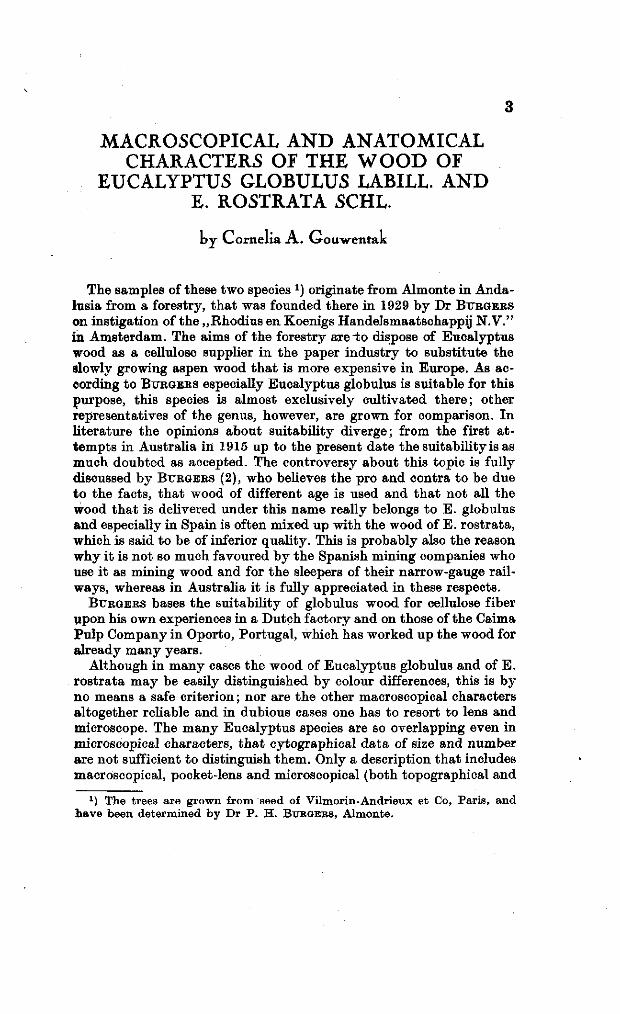
The samples of these two species J) originate from Almonte in Andalusia from a forestry, t ha t was founded there in 1929 by Dr BTTRGERS
on instigation of the „Rhodius en Koenigs Handelsmaatschappij N.V." in Amsterdam. The aims of the forestry arei:o dispose of Eucalyptus wood as a cellulose supplier in the paper industry to substi tute the slowly growing aspen wood t ha t is more expensive in Europe. As according to BURGEES especially Eucalyptus globulus is suitable for this purpose, this species is almost exclusively cultivated there ; other representatives of the genus, however, are grown for comparison. I n l i terature the opinions about suitability diverge; from the first a ttempts in Australia in 1915 up to the present date the suitability is as much doubted as accepted. The controversy about this topic is fully discussed by BURGERS (2), who believes the pro and contra to be due to the facts, t ha t wood of different age is used and t ha t not all the wood t ha t is delivered under this name really belongs to E . globulus and especially in Spain is often mixed up with the wood of E . rostrata, which is said to be of inferior quality. This is probably also the reason why it is not so much favoured by the Spanish mining companies who use i t as mining wood and for the sleepers of their narrow-gauge railways, whereas in Australia it is fully appreciated in these respects.
BURGERS bases the suitability of globulus wood for cellulose fiber upon his own experiences in a Dutch factory and on those of the Caima Pulp Company in Oporto, Portugal, which has worked up the wood for already many years.
Although in many cases the wood of Eucalyptus globulus and of E . ros t ra ta may be easily distinguished by colour differences, this is by no means a safe criterion ; nor are the other macroscopical characters altogether reliable and in dubious cases one has to resort to lens and microscope. The many Eucalyptus species are so overlapping even in microscopical characters, t h a t cytographical da ta of size and number are not sufficient to distinguish them. Only a description tha t includes macroscopical, pocket-lens and microscopical (both topographical and
1) The trees are grown from seed of Vilmorin-Andrieux et Co, Paris, and have been determined by Dr P . H. BUBGEBS, Almonte.
cytographical) characters may be able to represent the separate species of a genus whose species are so numerous (about 200) and alike.
In l i terature I found none such descriptions of Eucalyptus. The most extensive are those of W E L C H (9, 10, 11) who gives only cytographical characters on about 16 species and does not deal with globulus nor with rostrata. W E L C H himself calls „ the results ra ther disappointing from the point of view of identification" on account of the variations in length, width etc. of the elements. BAKER 'S (1) descriptions (with microphotographs) are very short and incomplete; thus he refers for a description of E . rostrata to the descriptions of E . margi-na ta SM. and E. resinifera SM.. Equally short is the characterization in STONE (8), who has compiled from older l i terature (see I.e.). DADS-
WELL and BTXRNELL (3) and DADS WELL, BTTRNELL and ECKERSLEY (4)
have lately published a key to Eucalyptus species. They investigated only such features as had proved to be of diagnostic value ; this may be sufficient when Eucalyptus samples of many species are present for comparison, bu t if this condition cannot be fulfilled I th ink an elaborate description of anatomical s tructure to be indispensable.
Before turning to the descriptions I will mention the name of Prof-fessor A. TE W E C H E L , t he director of t he laboratory of „Boschexploi-tat ie en Boschhuishoudkunde" a t Wageningen, Netherlands, who thinking detailed descriptions of the species mentioned to be of practical value, called my at tention to this work.
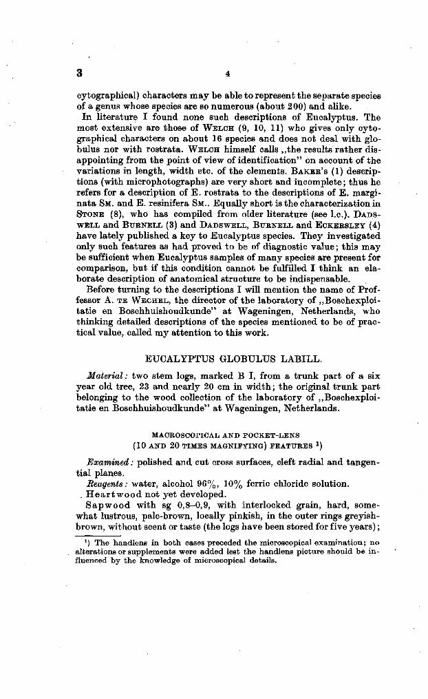
EUCALYPTUS GLOBULUS LABILL.
Material : two stem logs, marked B I, from a t runk par t of a six year old tree, 23 and nearly 20 cm in width; the original t runk pa r t belonging to the wood collection of the laboratory of „Boschexploi-tat ie en Boschhuishoudkunde" a t Wageningen, Netherlands.
MACROSCOPICAL AND POCKET-LENS
( 10 AND 2 0 TIMES MAGNIFYING) FEATURES *)
Examined : polished and cut cross surfaces, cleft radial and tangential planes.
Reagents: water, alcohol 96%, 10% ferric chloride solution. . H e a r t w o o d not yet developed.
S a p w o o d with sg 0,8-0,9, with interlocked grain, hard, somewhat lustrous, pale-brown, locally pinkish, in the outer rings greyish-brown, without scent or taste (the logs have been stored for five years) ;
x) The handlens in both cases preceded the microscopical examination; no alterations or supplements were added lest the handlens picture should be influenced by the knowledge of microscopical details.
wood curls stained blue with 10% ferric chloride;1) aqueous extract pale-brown, alcoholic extract yellow-brown, both extracts stained olive-green with 10% ferric chloride solution; wood burns readily without characteristic scent, and smoulders to a greyish ash.
G r o w t h r i n g s in polished cross section generally pronounced, e.g. 8|-, 9, 4, 5, 9, 5£ mm in thickness, in concentrical, locally deviating layers ; number more than three times as much as corresponds with the age. Growth rings often alternately containing numerous and few vessels ; never more t han two richly pored rings between two scantily pored ones, although still separated by a small layer of fibers; the lat ter with or without very small pores. The layers t ha t contain few pores locally totally devoid of these. Growth rings in radial cleaving plane now and then conspicuous (by colour difference), for t he rest invisible in consequence of cross grain. I n some of the outer layers about twenty splits regularly distributed over one half of t he s tem outline. G r o w t h r i n g b o u n d a r i e s differently pronounced, due t o a tangential linear layer with very small pores, or to the sudden beginning or ending of darker or brighter layers without pores or especially in the outer growth rings with evidently fewer pores.
V e s s e l s on cross sections easily seen with the naked eye, solitary, either separately situated often in somewhat oblique radial rows, each pore embedded in parenchymatous tissue, or connected by some parenchymatous tissue in oblique chains of 2-13, in which most pores radially arranged, sometimes 2 or 3 of them tangentially. The chains frequently bending in the vicinity of a growth ring boundary and forming with i t an angle up to 45°; in the next growth ring suddenly bending back 90°. Number in the richer zones 9-17, mean number 12 per mm 2 ; in the poorer ones 2-9, mean number 5 per mm2 . Pores radially bordering on medullary rays or on wood parenchyma, practically always on both radial sides, rarely on one side 2), tangentially almost always on parenchyma. 2) In tangential direction never more than one vessel between two rays. The pores ra ther uniformly 200 /u, in width, the foresaid very small ones 30 fi. End walls (perforation plates) transverse with simple circular perforations. Damaged tyloses often present, on cross sections evident as thin threads.
F i b e r s forming the bulk of the wood. W o o d p a r e n c h y m a paratracheal, in layers of few cells; a t t he
tangential sides of the vessels in the above mentioned oblique radial pore chains some more cells, t ha t connect the pores together.
M e d u l l a r y r a y s on polished cross planes now and then visible t o
*) SANIO (5) identified tannin in the medullary rays of Eucalyptus cordata with potassium dichromate.
a) Here a difference from the microscopical structure, see page 7.
3 6
the naked eye, forming on radial face little indistinct mirrors, measuring up to x/3 mm in height, not yet 30^ in width, bending strikingly to avoid the pores. All cells procumbent ; the cells of the outermost marginal rows often somewhat higher. 9-14, average number 12 per mm.
MICROGRAPHY
See also pocket-lens characters. Examined: Transverse, radial and tangential sections, macerated
material. Reagents. Phloroglucin and hydrochloric acid, iodine and sulphuric
acid 66%, Sudan III , Jeffreys maceration fluid, hydrochloric acid, phenol and clove oil, concentrated sulphuric acid, chromic acid 20%, ferric chloride solution 10%.
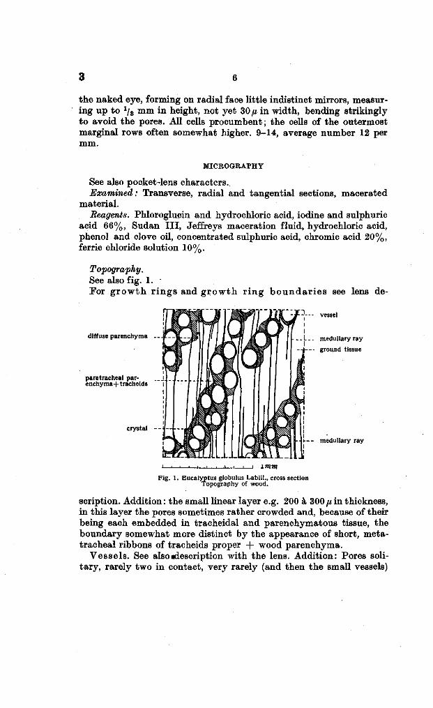
Topography. See also fig. 1. For g rowth r ings and g rowth r ing boundar i e s see lens de-
diffuse parenchyma
paratracheal parenchyma +tracheids
crystal
vessel
medullary ray
ground tissue
-• t--- medullary ray
Fig. 1. Eucalyptus globulus Labill., cross section Topography of wood.
scription. Addition : the small linear layer e.g. 200 à 300 fi in thickness, in this layer the pores sometimes rather crowded and, because of their being each embedded in tracheidal and parenchymatous tissue, the boundary somewhat more distinct by the appearance of short, meta-tracheal ribbons of tracheids proper + wood parenchyma.
Vessels. See also «description with the lens. Addition: Pores solitary, rarely two in contact, very rarely (and then the small vessels)
bordering on wood fibers, if thus apparent1), then in most cases the end projections of tracheids or wood parenchyma cells are touched, or a very wide bending and thus much narrowed ray cell ; on the radial sides either in contact with ray cells, tracheids proper (vascular tracheids) or wood parenchyma. The tissue bordering on the tangential sides of the pores appears to be wood parenchyma or (and) tracheids or {and) fiber tracheids.
T rache ids p roper (vascular tracheids) alone or with wood parenchyma in small layers of different depth in close proximity to the pores; these layers on the radial sides of the vessels 0-2 cells deep, on the tangential sides usually somewhat more, sometimes mixed with wood parenchyma forming short borders, measuring up to nine cells in length, or short metatracheal ribbons in which one or more pores are embedded ; these ribbons in contact with more than one ray and ending trapezoidly or triangularly. Tracheids in contact with vessels but rarely conjugated (disjunctive) and if so always with a wood-parenchyma cell.
F iber t r ache ids 2 ) locally forming the ground tissue in zones, poorly provided with pores and also in such cases, where they are bordering on the tangential side of the above mentioned metatracheal ribbons; for the rest occurring: lstly alone or with wood parenchyma in rows of 4 to many cells, measuring one cell in width, directly along or in some distance of a ray; 2ndly in the junctions between pores, which will be mentioned later on (see wood parenchyma).
L ib r i fo rm 2 ) forming the ground tissue, except in the above mentioned cases; radial arrangement often disturbed by the interference of the end projections of the fibers.
Wood p a r enchyma paratracheal, diffuse and metatracheal. (See also under tracheids proper). The paratracheal alone or with
1) See page 5. 2) For the distinction in fiber tracheids and libriform I tried to conform to
the definitions of SANIO (6, 7); I quote here fully from page H 6 SANIO (6): Es gibt aber Pflanzen bei denen der Grössen- und Formunterschied so gering ist, dass die Trennung schwierig werden kann. Die Familie der Myrtaceae ist in dieser Beziehung sehr lehrreich. Bei Melaleuca imbricata ist der Unterschied zwischen Tracheiden und Libriform durch die beträchtliche Grösse der Tra-cheidentüpfel deutlieh ausgesprochen; bei Tristania neriifolia und Eucalyptus cbrdata nähern sich die Tüpfel des Libriforms denen der Tracheiden schon sehr in der Grösse, bei Callistemon lanuginosus ist der Unterschied kaum noch wahrzunehmen, und bei Myrtus endlich haben sämtliche Holzfasern dieselbe behöfte Tüpfelung wie die Gefässe und müssen deshalb als Tracheiden auf-gefasst werden. Darnach erscheint allerdings der Unterschied zwischen Tracheiden und dem behöft getüpfeltem Libriform mehr als ein gradueller denn als ein absoluter, muss aber fest gehalten werden, da in der überwiegenden Mehrzahl der Fälle beide Bildungen so scharf und deutlich von einander verschieden sind dass an eine Vereinigung nicht gedacht werden kann.
8
tracheids in the foresaid layers ; the diffuse parenchyma locally wanting, but for the rest between the wood fibers as well as between the fiber tracheids, especially rather abundant, where it connects two radial pore rows in cooperation with fiber tracheids. Everywhere readily forming radial or tangential ribbons of 2-5 cells, one cell deep. All the parenchyma appearing as fibers; the fibers composed of 4-9 cells, mostly of 4 or of 7 cells. Chambered crystal fibers regularly distributed among the other wood parenchyma fibers, composed of 4-12 cells; most cells containing 1-4 crystals, mostly one simple ; the latter sometimes in a crystal sac that is connected with the cell wall. The paren
chyma cells that are in direct contact with vessels, often conjugated (disjunctive).
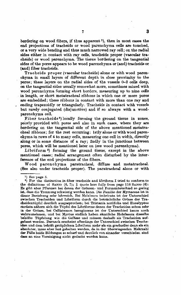
Medul lary r ays sideways separated bij 1-10 generally 4-7 rows of libriform or by 3-7 rows of fiber tracheids. Homogeneous, 2-19 cells in height ; most of them uniseriate, many of the higher ones biseriate in 1-6, mostly in 1-2 tiers (fig. 2, a) ; these tiers differently situated with
regard to another: the biseriate mostly separated by uniseriate, sometimes some biseriate tiers bordering on one another.
For course, structure, number, sizes, see lens characters. Between the procumbent cells of the upmost or undermost tiers sometimes some square cells.
h
Cytography. I. Vessels. Rad. 45-220 jx, mostly 175 à
200/*, tg 30-145,«; segments (vessel members) 200-500 fi in length (average length from 16 measurements 310/t). Oval with radial long axis or circular in section; flattened, when vessels are bordering on one another. End walls (perforation plates) transverse or somewhat oblique. Perforations simple, circular or oval ; annular ridges (perforation rims) smooth, generally small, sometimes rather large.
Walls 4-6(i in thickness; i.e. 6 ft when vessels are bordering on one another, 5fi when on parenchyma; lignified, sometimes the thin tertiary wall layer stained blue by iodine and sulphuric acid 66% ; — with rather numerous bordered pits to congeneric elements or to vascular tracheids ; border 5 fi in width ; inner aperture elongated, but not or seldom extending beyond the limit of the cavity; pit membrane sometimes sieve-like. Conten t s none or irregular thin-walled, colourless or slightly yellow tyloses, often torn while sectioned ;
Fig. 2 a. A locally (**) biseriate medullary ray of Eucalyptus globulus,
ft. A medullary ray of Eucalyptus rostrata with a biseriate part.
9 3
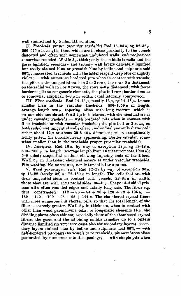
wall stained red by Sudan I I I solution. I I . Tracheids proper (vascular tracheids) Rad 16-24 /i, tg 24-32 fi,
250-675 (i in length ; those which are in close proximity to the vessels distorted and often with somewhat undulated walls; end projections somewhat rounded. Walls 3 fi thick; only the middle lamella and the gores lignified, secondary and tertiary wall layers delicately lignified but easily stained blue or greenish blue by iodine and sulphuric acid 66% ; macerated tracheids with the latter reagent deep blue or slightly violet ; — with numerous bordered pits when in contact with vessels ; the pits on the tangential walls in 2 or 3 rows, the rows 5 fi distanced, on the radial walls in 1 or 2 rows, the rows 4-6 fx distanced ; with fewer bordered pits to congeneric elements, the pits in 1 row ; border circular or somewhat elliptical, 5-8 /j, in width, canal laterally compressed.
I I I . Fiber tracheids. Rad 14-16 ii, mostly 16/t, tg 14-18/*. Lumen smaller than in the vascular tracheids. 550-1050 fi in length, average length 820 it, tapering, often with long rostrum which is on one side undulated. Wall 4 (i in thickness, with chemical nature as under vascular tracheids — with bordered pits when in contact with fiber tracheids or with vascular tracheids ; the pits in 1 or 2 rows, on both radial and tangential walls of each individual unevenly distanced ; either about 12^ or about 30 à 40/t distanced; when exceptionally richly pitted, the borders nearly approaching. Border circular, somewhat smaller than in the tracheids proper (vascular tracheids).
IV. Libriform. Rad 16 /e, by way of exception 18 fi, tg 12-18/«, 600-1700 fi in length (average length from 16 measurements 1000 fi); 5-6 sided; tangential sections showing tapering ends of the fibers. Wal l 5 n in thickness; chemical nature as under vascular tracheids. Pits wanting. No contents , nor i n te rce l lu la r spaces.
V. Wood parenchyma cells. Rad 12-28 by way of exception 36 ii, tg 16-22 (rarely 32) ii; 72-180/« in length. The cells that are with their tangential sides in contact with vessels: 22-34/« in width, those that are with their radial sides : 36-48/«. Shape: 4-6 sided prismas with often rounded edges and axially long axis. The fibers e.g. thus constructed: 112 + 80 + 84 + 96 + 128 + 72 + 136 ii, — 180 + 140 + 100 + 96 + 98 + 144 /«. The chambered crystal fibers with more numerous but shorter cells, so that the total length of the fiber is scarcely greater. Wall 2/« in thickness, when in contact with Other than wood parenchyma cells; to congeneric elements l^ii; the dividing plates often thinner, especially those of the chambered crystal fibers; the gores and the adjoining middle lamellae up to a certain distance lignified (in very rare cases also the secondary layers) ; secondary layers stained blue by iodine and sulphuric acid 66% — with half-bordered pit(-pairs) to vessels or to tracheids, pit membrane often perforated by numerous minute openings-; — with simple pits when
3 10
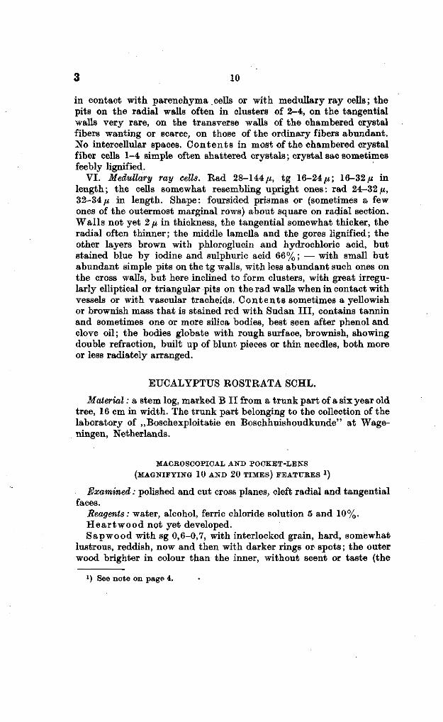
in contact with parenchyma cells or with medullary ray cells ; the pits on the radial walls often in clusters of 2-4, on the tangential walls very rare, on the transverse walls of the chambered crystal fibers wanting or scarce, on those of the ordinary fibers abundant. No intercellular spaces. Conten t s in most of the chambered crystal fiber cells 1-4 simple often shattered crystals ; crystal sac sometimes feebly lignified.
VI. Medullary ray cells. Rad 28-144 p, tg 16-24^; 16-32/* in length; the cells somewhat resembling upright ones: rad 24-32fi, 32-34^ in length. Shape: foursided prismas or (sometimes a few ones of the outermost marginal rows) about square on radial section. Walls not yet 2 ^ in thickness, the tangential somewhat thicker, the radial often thinner; the middle lamella and the gores lignified; the other layers brown with phloroglucin and hydrochloric acid, but stained blue by iodine and sulphuric acid 66% ; — with small but abundant simple pits on the tg walls, with less abundant such ones on the cross walls, but here inclined to form clusters, with great irregularly elliptical or triangular pits on the rad walls when in contact with vessels or with vascular tracheids. Con ten t s sometimes a yellowish or brownish mass that is stained red with Sudan III , contains tannin and sometimes one or more silica bodies, best seen after phenol and clove oil ; the bodies globate with rough surface, brownish, showing double refraction, built up of blunt pieces or thin needles, both more or less radiately arranged.
EUCALYPTUS ROSTRATA SCHL.
Material : a stem log, marked B I I from a trunk part of a six year old tree, 16 cm in width. The trunk part belonging to the collection of the laboratory of „Boschexploitatie en Boschhuishoudkunde" at Wageningen, Netherlands.
MACROSCOPICAL AND POCKET-LENS
(MAGNIFYING 10 AND 2 0 TIMES) FEATURES x )
Examined : polished and cut cross planes, cleft radial and tangential faces.
Beagents : water, alcohol, ferric chloride solution 5 and 10%. Hear twood not yet developed. Sap wo od with sg 0,6-0,7, with interlocked grain, hard, somewhat
lustrous, reddish, now and then with darker rings or spots ; the outer wood brighter in colour than the inner, without scent or taste (the
x) See note on page 4.
i l 3
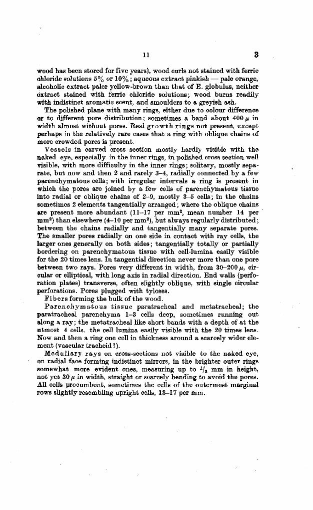
wood has been stored for five years), wood curls not stained with ferric chloride solutions 5% or 10% ; aqueous extract pinkish — pale orange, alcoholic extract paler yellow-brown than that of E. globulus, neither extract stained with ferric chloride solutions; wood burns readily with indistinct aromatic scent, and smoulders to a greyish ash.
The polished plane with many rings, either due to colour difference or to different pore distribution; sometimes a band about 400 fi in width almost without pores. Real g rowth r ings not present, except perhaps in the relatively rare cases that a ring with oblique chains of more crowded pores is present.
Vessels in carved cross section mostly hardly visible with the naked eye, especially in the inner rings, in polished cross section well visible, with more difficulty in the inner rings; solitary, mostly separate, but now and then 2 and rarely 3^4, radially connected by a few parenchymatous cells; with irregular intervals a ring is present in which the pores are joined by a few cells of parenchymatous tissue into radial or oblique chains of 2-9, mostly 3-5 cells; in the chains sometimes 2 elements tangentially arranged ; where the oblique chains are present more abundant (11-17 per mm2, mean number 14 per mm2) than elsewhere (4-10 per mm2), but always regularly distributed; between the chains radially and tangentially many separate pores. The smaller pores radially on one side in contact with ray cells, the larger ones generally on both sides ; tangentially totally or partially bordering on parenchymatous tissue with cell-lumina easily visible for the 20 times lens. In tangential direction never more than one pore between two rays. Pores very different in width, from 30-200 /j,, circular or elliptical, with long axis in radial direction. End walls (perforation plates) transverse, often slightly oblique, with single circular perforations. Pores plugged with tyloses.
F ibers forming the bulk of the wood. P a r enchyma tou s t i s sue paratracheal and metatracheal; the
paratracheal parenchyma 1-3 cells deep, sometimes running out along a ray ; the metatracheal like short bands with a depth of at the utmost 4 cells, the cell lumina easily visible with the 20 times lens. Now and then a ring one cell in thickness around a scarcely wider element (vascular tracheid ?).
Medul lary r ays on cross-sections not visible to the naked eye, on radial face forming indistinct mirrors, in the brighter outer rings somewhat more evident ones, measuring up to 1/3 mm in height, not yet 30 /i in width, straight or scarcely bending to avoid the pores. All cells procumbent, sometimes the cells of the outermost marginal rows slightly resembling upright cells, 13-17 per mm.
12
MICROGRAPHY
See also pocket lens characters. Examined: Transverse, radial and tangential sections, macerated
material. Beagents : Phlorogluzin and hydrochloric acid, iodine and sulphuric
acid 66%, Sudan I I I , Jeffreys maceration fluid, hydrochloric acid, ferric chloride solution 10%, phenol and clove oil.
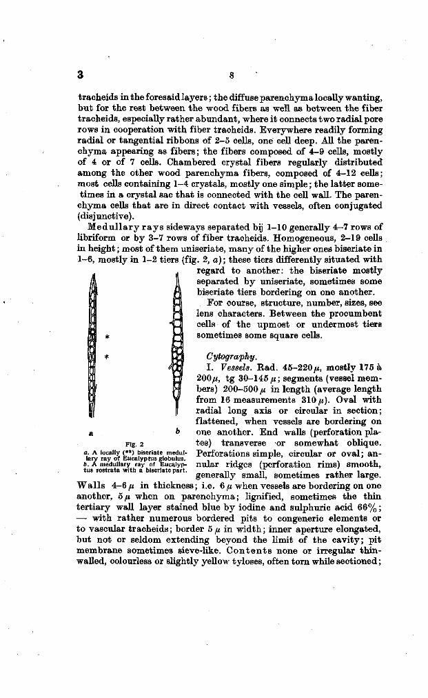
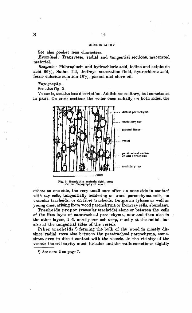
Topography. See also fig. 3. Vessels, see also lens description. Additions: solitary, but sometimes
in pairs. On cross sections the wider ones radially on both sides, the
. diffuse parenchyma
_• medullary ray
ground tissue
. vessel
paratracheal paren-chyma+tracheids
-_ medullary ray
Fig. 3. Eucalyptus rostrata Schl., cross section. Topography of wood.
others on one side, the very small ones often on none side in contact with ray cells, tangentially bordering on wood parenchyma cells, on vascular tracheids, or on fiber tracheids. Outgrown tyloses as well as young ones, arising from wood parenchyma or from ray cells, abundant.
T racheids p roper (vascular tracheids) alone or between the cells of the first layer of paratracheal parenchyma, now and then also in the other layers, 1-3, mostly one cell deep, mostly at the radial, but also at the tangential sides of the vessels.
Tiber t r a che id s 1 ) forming the bulk of the wood in mostly distinct radial rows also between the paratracheal parenchyma, sometimes even in direct contact with the vessels. In the vicinity of the vessels the cell cavity much broader and the walls sometimes slightly
l ) See note 2 on page 7.
13 3
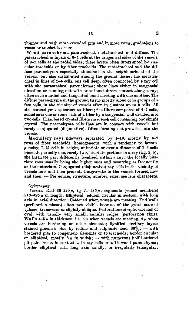
thinner and with more crowded pits and in more rows ; gradations to vascular tracheids occur. Wood p a r enchyma paratracheal, metatracheal and diffuse. The
paratracheal in layers of 0-4 cells at the tangential sides of the vessels, of 0-2 cells at the radial sides ; these layers often interrupted by vascular tracheids or by fiber tracheids. The metatracheal and the diffuse parenchyma especially abundant in the neighbourhood of the vessels, but also distributed among the ground tissue; the metatracheal in Unes of 2-4 cells, one cell deep, often connected by a ray cell with the paratracheal parenchyma; these lines either in tangential direction or running out with or without direct contact along a ray; often such a radial and tangential band meeting with one another. The diffuse parenchyma in the ground tissue mostly alone or in groups of a few cells, in the vicinity of vessels often in clusters up to 9 cells. All the parenchyma apparent as fibers ; the fibers composed of 4-7 cells, sometimes one or some cells of a fiber by a tangential wall divided into two cells. Chambered crystal fibers rare, each cell containing one simple crystal. The parenchyma cells that are in contact with vessels but rarely conjugated (disjunctive). Often forming out-growths into the vessels.
Medul la ry r ays sideways separated by 1-10, mostly by 6-7 rows of fiber tracheids, homogeneous, with a tendency to heterogeneity, 1-21 cells in height, uniseriate or over a distance of 1-5 cells biseriate ; usually one, rarely two, biseriate portions in a ray (fig. 2, b) ; the biseriate part differently localised within a ray; the locally biseriate rays usually being the higher ones and occurring as frequently as the uniseriate. Conjugated (disjunctive) ray cells in the vicinity of vessels now and then present. Outgrowths in the vessels formed now and then. — For course, structure, number, sizes, see lens characters.
Cytography. Vessels. Rad 36-220^, tg 24-135^; segments (vessel members)
210-425 ƒ« in length. Elliptical, seldom circular in section, with long axis in axial direction; flattened when vessels are meeting. End walls (perforation plates) often not visible because of the great mass of tyloses, transverse or slightly oblique. Perforations simple, circular or oval with usually very small, annular ridges (perforation rims). Walls 4-5/a, in thickness, i.e. 5 fi when vessels are meeting, 4/j when vessels are bordering on other elements; lignified, tertiary layers stained greenish blue by iodine and sulphuric acid 66% ; — with bordered pits to congeneric elements or to tracheids ; border circular or elliptical, mostly 8 ̂ in width ; — with numerous half bordered pit-pairs when in contact with ray cells or with wood parenchyma; border elliptical with long axis axially, or irregularly triangular;
3 14
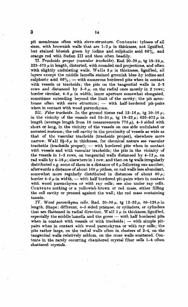
pit membrane often with sieve-structure. Con ten t s : tyloses of all sizes, with brownish walls that are 1-2 /* in thickness, not lignified, but stained blueish green by iodine and sulphuric acid 66%, and orange red with Sudan I I I and then often beadily.
I I . Tracheids proper (vascular tracheitis). Rad 20-28/*, tg 18-24/t, 325-575 /* in length, distorted, with rounded end projections, and often with slightly undulating walls. Walls 3/* in thickness, lignified, all layers except the middle lamella stained greenish blue by iodine and sulphuric acid 66% ; — with numerous bordered pits when in contact with vessels or tracheids; the pits on the tangential walls in 2-3 rows and distanced by 3-8 /*, on the radial ones mostly in 2 rows ; border circular, 4-8/* in width; inner aperture somewhat elongated, sometimes extending beyond the limit of the cavity; the pit membrane often with sieve structure; — with half-bordered pit-pairs when in contact with wood parenchyma.
I I I . Fiber tracheids. In the ground tissue rad 12-16/*, tg 10-16/*; in the vicinity of the vessels rad 16-20 /*, tg 18-23/*; 625-975/* in length (average length from 16 measurements 775/*), 4-5 sided with short or long, in the vicinity of the vessels on one side undulated or serrated rostrum ; the cell cavity in the proximity of vessels as wide as that of the vascular tracheids (tracheids proper), elsewhere more narrow. Wall 2£-5 fi in thickness, for chemical nature see vascular tracheids (tracheids proper); — with bordered pits when in contact with vessels and with vascular tracheids; the pits in the vicinity of the vessels in 1-2 rows, on tangential walls distanced by 4-12 /*, on rad walls by 4-16 /* ; elsewhere in 1 row, and then on tg walls irregularly distributed e.g. some of them in a distance of 6 /* following one another, afterwards a distance of about 100 /* pitless, on rad walls less abundant, somewhat more regularly distributed in distances of about 80/*; border 4-5 /* in width, — with half bordered pit-pairs when in contact with wood parenchyma or with ray cells; see also under ray cells. Conten ts nothing or a yellowish-brown or red mass, either filling the cell cavity or pressed against the wall; the red mass containing tannin.
IV. Wood parenchyma cells. Rad. 20-56/*, tg 12-32/*, 50-128 /*iu length. Shape: different, 4-5 sided prismas, or cylinders, or cylinders that are flattened in radial direction. Wall 1 /* in thickness, lignified, especially the middle lamella and the gores — with half bordered pits when in contact with vessels or with tracheids; — with simple pit-pairs when in contact with wood parenchyma or with ray cells; the pits rather large, on the radial walls often in clusters of 2-4, on the tangential walls relatively seldom, on the cross walls scattered. Contents in the rarely occurring chambered crystal fiber cells 1-4 often shattered crystals.

15 3
V. Medullary ray cells. Procumbent cells rad. 48-212 fi, tg8-28/*, 16-20,« in length. Shape 4-sided prismas, often with oblique tangential walls. Wal ls : cross wall not yet 1 /J, in thickness, tg wall not yet 2 (i, rad. wall between the two cells of a biseriate tier 2 p, elsewhere about 1 /x ; not lignified, except the middle lamella of a biseriate layer, readily stained greenish blue by iodine and sulphuric acid 66% ; — with small abundant simple pits on the tangential and on the cross walls; on the latter the pits often in clusters; — on the radial walls with large irregular pits when in contact with vessels and with tracheids in the vicinity of the vessels ; pit membrane often with sieve-structure; — with small pits when in contact with fiber tracheids in some distance of the vessels or the pits wanting ; with small pits on the uniting wall of a biseriate layer and when in contact with wood parenchyma cells. Con ten t s : often a yellow-brownish or red mass, either filling the cell cavity or pressed against the wall, not or rarely red with Sudan I I I solution; now and then a little simple rectangular or pyramidal crystal.
Upright cells. Rad 36-52 [i, 28-32 n in length. Shape on radial section about square. For the rest see procumbent cells.
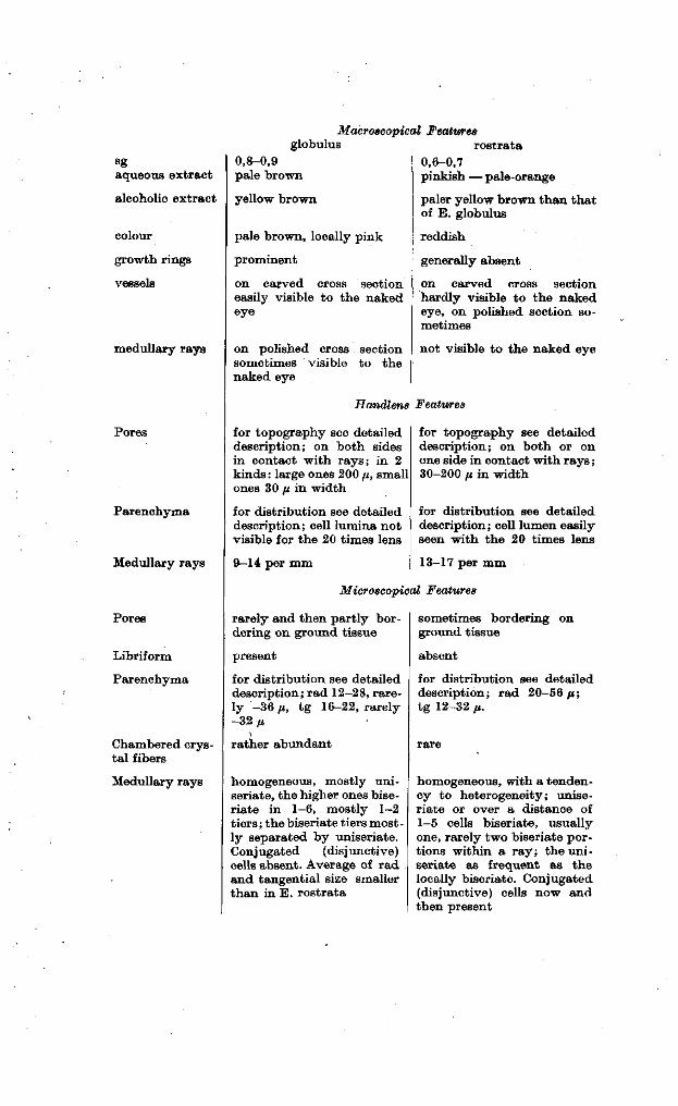
SUMMARY
The principal characterizations are given below. There are, however, but few details that can be summarized in a table, and especially the macroscopical differences are only reliable, if it is certain that no other but these two species are present. Thus I also omitted on purpose to mention in this table most of the cytographical details, as they are known to vary greatly within a species. As also some other species of the genus are grown in Spain, samples of which have not yet been available to me, it will in most cases be necessary to compare carefully the detailed descriptions.
At first sight the staining of globulus wood curls versus the non staining of rostrata wood curls with ferric chloride seems to be highly •characteristic. I am at a loss, however, to say whether this rather physiological feature holds true in case of more samples taken in •different seasons and places.
aqueous extract
alcoholic extract
colour
growth rings
vessels
medullary rays
Pores
Parenchyma
Medullary rays
Pores
Libriform
Parenchyma
Chambered crystal fibers
Medullary rays
globulus 0,8-0,9 pale brown
yellow brown
pale brown, locally pink
prominent
on carved cross section easily visible to the naked eye
on polished cross section sometimes visible to the naked eye
Macroacopical Features rostrata
0,6-0,7 pinkish — pale-orange
paler yellow brown than tha t of E. globulus
reddish
generally absent
on carved cross section hardly visible to the naked eye, on polished section sometimes
not visible to the naked eye
Handlens Features
for topography see detailed description; on both sides in contact with rays; in 2 kinds: large ones 200 fi, small ones 30 yt in width
for distribution see detailed description ; cell lumina not visible for the 20 times lens
9—14 per mm
for topography see detailed description; on both or on one side in contact with rays ; 30-200 fi in width
for distribution see detailed description; cell lumen easily seen with the 20 times lens
13-17 per mm
Microscopical Features
rarely and then partly bordering on ground tissue
present
for distribution see detailed description; rad 12-28, rarely -36 (i, t g 16-22, rarely -32/4
rather abundant
homogeneous, mostly uni-seriate, the higher ones bise-riate in 1-6, mostly 1-2 tiers ; the biseriate tiers mostly separated by uniseriate. Conjugated (disjunctive) cells absent. Average of rad and tangential size smaller than in E. rostrata
sometimes bordering on ground tissue
absent
for distribution see detailed description; rad 20-56 /i; tg 12-32 p.
homogeneous, with a tendency to heterogeneity; uniseriate or over a distance of 1-5 cells biseriate, usually one, rarely two biseriate portions within a r ay ; the uniseriate as frequent as the locally biseriate. Conjugated (disjunctive) cells now and then present
17 3
LITERATURE
1. BAKER, R. T., Hardwoods of Australia and their economics. Govt. Printer, Sydney 1919.
2. BURGEES, P . H. De stichting van een boschbedrijf in Andalusië. Proefschrift Wageningen 1931, 166 p .
3. DADSWELL, H. E . and M. BTJRNELL, Identification of the coloured woods of the genus Eucalyptus. Bull. Counc. for Sei. and Industrial Research 67, 1932, 50 p .
4. DADSWELL, H. E. , M. BURNELL and A. M. ECKERSLEY, Identification of the light-coloured woods of the genus Eucalyptus. Bull. Counc. for Sei. and Industrial Research nr 78, 1934, 60 p .
5. SANIO, C. Einige Bemerkungen über den Gerbstoff und seine Verbreitung bei den Holzpflanzen. Bot. Zeitung 21, 1863, 17-23.
6. SANIO, C. Vergleichende Untersuchungen über die Elementarorgane des Holzkörpers. Bot. Ztg. 21, 1863, p . 85, 93, 101, 113, 121.
7. SANIO, C. Vergleichende Untersuchungen über die Zusammensetzung des Holzkörpers. Bot. Zeit., 21, 1863, p. 357, 369, 377, 389, 401.
8. STONE, H. The timbers of commerce and their identification. London 1905, p . 125.
9. WELCH, M. B. Notes on the structure of some Eucalyptus woods. Journ. Roy. Soc. N. S. W., 58, 1924, 169-177.
10. WELCH, M. B. The identification of the principal ironbarks and allied woods. Journ. Roy. Soc. N. S. W., 59, 1925, 329-346.
11. WELCH, M. B. The wood structure of certain Eucalypts belonging chiefly to the „ash" group. Journ. Roy. Soc. of N. S. W., 60, 1926, 147-166.
Laboratorium voor Plantkunde.
Wageningen, J anuary 1935.