Macropinocytotic uptake and infection of human epithelial ... · Macropinocytotic uptake and...
44
1 Macropinocytotic uptake and infection of human epithelial cells with species B2 adenovirus type 35 Stefan Kälin 1,2 , Beat Amstutz 1 , Michele Gastaldelli 1 , Nina Wolfrum 1 , Karin Boucke 1 , Menzo Havenga 3, +) , Fabienne DiGennaro 1 , Nicole Liska 1 , Silvio Hemmi 1 & Urs F.Greber 1, *) 1 Institute of Molecular Life Sciences, University of Zurich, Zurich, Switzerland 2 Zurich PhD Program in Molecular Life Sciences 3 Crucell Holland BV, Leiden, The Netherlands +) present address: TNO Biosciences, Zernikedreef 9, 2333CK Leiden, The Netherlands 4 Institute of Molecular Life Sciences, University of Zurich, Zurich, Switzerland *) corresponding author: [email protected] , Institute of Molecular Life Sciences, University of Zurich, Winterthurerstrasse 190, 8057 Zurich, Switzerland Word counts: abstract 237 Text: 5352 words, 36942 characters (incl spaces, excluding references, table footnotes, and figure legends) Short title: Infectious macropinocytosis of adenovirus type 35 in human epithelial cells Keywords: macropinocytosis, infectious entry, human epithelial cells, CD46, integrin
Transcript of Macropinocytotic uptake and infection of human epithelial ... · Macropinocytotic uptake and...

1
Macropinocytotic uptake and infection of human
epithelial cells with species B2 adenovirus type 35
Stefan Kälin1,2, Beat Amstutz1, Michele Gastaldelli1, Nina Wolfrum1, Karin Boucke1,
Menzo Havenga3, +), Fabienne DiGennaro1, Nicole Liska1, Silvio Hemmi1 & Urs
F.Greber1, *)
1 Institute of Molecular Life Sciences, University of Zurich, Zurich, Switzerland
2 Zurich PhD Program in Molecular Life Sciences
3 Crucell Holland BV, Leiden, The Netherlands
+) present address: TNO Biosciences, Zernikedreef 9, 2333CK Leiden, The Netherlands
4 Institute of Molecular Life Sciences, University of Zurich, Zurich, Switzerland
*) corresponding author: [email protected], Institute of Molecular Life Sciences,
University of Zurich, Winterthurerstrasse 190, 8057 Zurich, Switzerland
Word counts: abstract 237
Text: 5352 words, 36942 characters (incl spaces, excluding references, table footnotes,
and figure legends)
Short title: Infectious macropinocytosis of adenovirus type 35 in human epithelial cells
Keywords: macropinocytosis, infectious entry, human epithelial cells, CD46, integrin

2
ABSTRACT
The human adenovirus serotype 35 (HAdV-35, short Ad35) causes kidney and urinary
tract infections, and infects respiratory organs of immunocompromised individuals.
Unlike other adenoviruses, Ad35 has a low seroprevalence which makes Ad35-based
vectors promising candidates for gene therapy. Ad35 utilizes CD46 and integrins as
receptors for infection of epithelial and hematopoietic cells. Here, we show that
infectious entry of Ad35 into HeLa, human kidney HK-2 cells and normal human lung
fibroblasts strongly depended on CD46 and integrins but not heparan sulfate, and
variably required the large GTPase dynamin. Ad35 infections were independent of
expression of the carboxy-terminal domain of AP180 which effectively blocks clathrin-
mediated uptake. Ad35 infections were inhibited by small chemicals against the
serine/threonine kinase Pak1 (p21-activated kinase), protein kinase C (PKC), sodium-
proton exchangers, actin and acidic organelles. Remarkably, the F-actin inhibitor
jasplakinolide, the Pak1 inhibitor IPA-3 or the sodium-proton exchange inhibitor EIPA
blocked the endocytic uptake of Ad35. Dominant-negative proteins or small interfering
RNAs against factors driving macropinocytosis, including the small GTPase Rac1, Pak1
or the Pak1 effector C-terminal binding protein 1 (CtBP1) potently inhibited Ad35
infection. Confocal laser scanning microscopy, electron microscopy and live cell
imaging showed that Ad35 colocalized with fluid phase markers in large endocytic
structures that were positive for CD46, alpha v integrins and also CtBP1. Our results
extend earlier observations with HAdV-3 (Ad3), and establish macropinocytosis as an
infectious pathway for species B human adenoviruses in epithelial and hematopoietic
cells.

3
INTRODUCTION
Adenoviruses circulate widely in the human population, and most adults have been
exposed to adenoviruses (13). Currently, there are more than 54 HAdV serotypes
known, and they have partly divergent entry pathways (for taxonomic details, see
http://www.vmri.hu/~harrach/AdVtaxlong.htm). HAdVs are classified into six species A
to F. Clinical manifestations of HAdV vary considerably, but commonly include cold
symptoms, pharyngitis, tonsilitis, otitis, and pharyngoconjunctival fever. Less common
are severe pneumonia, conjunctivitis, cystitis, encephalitis and meningitis (50, 81). In
immunocompromised patients and young military recruits, HAdVs cause life-threatening
infections (49).
The entry of the species C HAdV-2 (Ad2) and HAdV-5 (Ad5) is best characterized.
Ad2/5 bind to the coxsackie B virus-Ad receptor CAR (8), and use αν-integrins as
secondary receptors to induce receptor-mediated endocytosis involving clathrin, clathrin
adaptors and the large GTPase dynamin (31, 34, 63, 102, 105). These viruses also
trigger accessory dynamin-independent macropinocytosis, which is not used to
internalize Ad2/5 into cultured cells (63). Macropinocytosis is, however, an important
infectious pathway into epithelial cells for the species B1 HAdV-3 (Ad3, 3). It is also an
entry pathway for an increasing number of viruses from other families, such as vaccinia
virus (67), echovirus 1 (43), Kaposi sarcoma herpes simplex virus (83) and Ebola virus
(82). Macropinocytosis has been associated with human immunodeficiency virus 1
(HIV) infections in different cell types (27, 58, 60), but dynamin-dependent endocytosis
of HIV has also been reported (70).
Macropinocytosis is a form of endocytosis occurring at large scale. It leads to the
formation of large vacuoles, predominantly at the cellular periphery (97). It often but not
always involves ruffling protrusions from the plasma membrane that either fuse with

4
themselves or with the cell membrane and thereby engulf extracellular material (21).
Macropinocytosis significantly contributes to antigen presentation in immune cells (66,
104), and is used by viral and bacterial pathogens to reduce immune responses (68,
97).
The small GTPase Rac1 and dynamic actin filaments invariably control
macropinocytosis. Macropinocytosis also strongly requires p21-activated kinase Pak1
(20), which binds and activates Rac1 (46), and variably depends on
phosphatidylinositol-3-kinase (PI3K), Ras, and Src activities downstream of activated
receptors. In addition, macropinocytosis requires the C-terminal binding protein 1
(CtBP1), which is phosphorylated by Pak1 and supports membrane fission or
stabilization of the emerging macropinosomal vesicle (35, 56). It is strongly blocked by
inibitors of sodium/proton exchangers (103), such as amiloride or an amiloride analogue
EIPA (37), which decrease cytosolic pH and thereby inhibit the activation of Rac1 and
Cdc42 GTPases in submembranous zones (48). Another hallmark of macropinocytosis
is the dependency on protein kinase C (PKC) in both macrophages and epithelial cells
(4, 5, 63).
Ad35 is a member of the species B2 adenoviruses, which use CD46 as a primary
receptor in epithelial and hematopoietic cells (26, 30), and integrins as coreceptors for
infection of hematopoietic cells (72). This virus was initially isolated from kidney of an
immunocompromised individual (73). Species B2 adenoviruses naturally infect the
kidney and urinary tracts, and are sometimes fatal in immunocompromised individuals,
possibly due to reemergence from a latent state (38, 53). In addition, Ad35 is a
promising vector for clinical gene transfer and vaccination, in part due to the low
seroprevalence of neutralizing antibodies in the population (101). For example, an
Ad35-based malaria vaccine was shown to protect mice from Plasmodium falciparum
sporozoites (78), and induced potent T-cell immunity (84). In addition, Ad35 vectors

5
injected into tissues of nonhuman primates gave rise to specific gene expressions at
sites of injection in most organs indicating the high versatility of Ad35 (87, 88).
Here we examined the infectious entry pathway for Ad35 into human epithelial cells and
human kidney 2 (HK-2) cells, and compared the results with host requirements for
infectious entry of the species C Ad2/5. Unlike Ad2/5, Ad35 uses macropinocytosis as
an infectious uptake pathway. It requires CD46, integrins, PKC, sodium/proton
exchanger, actin, Rac1, Pak1 and CtBP1 but not heparan sulfate, which had been
suggested to be involved in virus attachment to Chinese hamster ovary cells (99).
These results mirror the infectious pathway into epithelial cells for the species B1 Ad3
(3). This is remarkable since Ad3 had been suggested to use other receptors besides
CD46 (61, 95). Notably, it was recently shown that Ad3 binds to the same region of the
terminal short consensus repeat 1 (SCR1) and SCR2 of CD46 as Ad35, albeit with
lower affinity than Ad35 (25, 26), and this leads to gene expression in high CD46-
expressing baby hamster kidney (BHK) cells, CHO cells or malignant glioma cells (36,
95, 100). Our results show that HAdVs of the species B1 and B2 use conserved entry
pathways into epithelial cells which depend on CD46.
MATERIALS AND METHODS
Cells and viruses
Normal diploid human embryonic lung-derived fibroblasts Wi-38 (CCL-75) were
purchased from American Type Cell Culture Collection (ATCC) and grown in DMEM
(Sigma) containing 10% FCS (Invitrogen). HeLa-ATCC (CCL-2), HeLa-K, a variant of
6
HeLa-ATCC with particularly high transfection efficiencies was obtained from Dr. U.
Kutay (Institute of Biochemistry, ETH Zurich, Switzerland). Human melanoma M21,
M21L and M21L4 cells were grown in DMEM (Sigma) containing 10 % FCS (Invitrogen)
at low passage number as described (63). M21 (positive for surface-expressed αν-
integrins), M21L cells (negative for αν-integrins) and M21L4 transfected with αν-integrin
cDNA (24) were obtained from Dr. D. Cheresh (Scripps Research Institute, La Jolla, CA,
USA). Human kidney HK-2 cells originally isolated from kidney proximal tubular cells
and immortalized with human papilloma virus 16 E6 and E7 proteins (86) was obtained
from Dr. F. Verrey (Institute of Physiology, University of Zurich, Zurich), and propagated
in K1-medium. Human hematopoietic K562 cells were used as described (3). CHO
cells stably expressing GFP-CD46 were generated by transfection of CHO-15B6 cells
with a pcDNA3.1-neo vector containing the CD46-BC1 sequence, and single clones
were selected in G418 (1mg/ml) containing medium. Ad35 was isolated from human
bronchial epithelial A549 cells. Fluorescent tagging of Ad35 with Texas-Red (Molecular
Probes, Leiden, The Netherlands) was performed as described (96). Ad35-eGFP, Ad5-
eGFP and Ad2-ts1 were used as described (31, 74, 101). HSV-1-eGFP was a kind gift
from Dr. C. Fraefel (Institute of Veterinary Virology, University of Zurich,76). 3H-Ad35
was generated as described in (33).
Viral eGFP transduction experiments
For infection analyses by wide-field microscopy or confocal laser scanning microscopy
(CLSM) analyses cells were seeded onto glass coverslips in 24 well dishes.
Alternatively, infection measurements were carried out in a Safire monochromator-
based microplate detection system (Tecan Group Ltd. Switzerland) using 96 well plates.
Cells were infected with the indicated eGFP-expressing virus at a MOI 5 in warm
DMEM-0.2% BSA (Sigma A9418) for 60 min, washed twice with warm DMEM-0.2%
BSA and further incubated for 7 h, fixed in 3% paraformaldehyde for 25 min, quenched
7
with ammonium chloride and prepared for analysis as indicated above. Chemical
interference with infection was performed by addition of compounds 30 min prior to
infection in DMEM-0.2% BSA at 37°C. Cells were infected in the presence of the
inhibitors in DMEM-0.2% BSA for 60 min, washed twice and further incubated for 7 h.
cDNAs, antibodies and chemicals
Human K44A-Dyn2 and Dyn2 expression constructs were obtained from Dr. C. Lamaze
(Pasteur Institute, Paris, France). Plasmids expressing human Rac1 and T17N-Rac1
were from Dr. A. Hall (University College, London, UK). Expression plasmids encoding
CtBP1-S (obtained from Dr. A. Colanzi and Dr. A. Luini, Department of Cell Biology and
Oncology, Sta Maria Imbaro, Italy) were used to construct myc-tagged CtBP1 and
S147A-CtBP1 (3). Pak1 and Pak1 autoinhibitory domain (AID) constructs were
obtained from Dr. J. Chernoff (Fox Chase Cancer Center, Philadelphia, PA, USA). The
following antibodies and chemicals were purchased as follows: anti-CtBP1 (BD-
Transduction Laboratories, Switzerland), anti-CD46 E4.3 (monoclonal, BD-Pharmingen,
used for immunofluorescence analyses), anti-CD46 (polyclonal H-294, Santa Cruz, for
Western blots), anti-CD46 (monoclonal MEM-258, used for inhibition
experiments, Serotec Ltd, Oxford, United Kingdom), anti-αν β5 integrin (P1F6,
monoclonal, Chemicon). Secondary antibodies were goat anti-mouse or anti-rabbit
coupled to Cy5 (Pierce). The dynamin inhibitor dynasore (59) was purchased from
Sigma and dissolved in DMSO. Latrunculin was from Enzo Life Sciences, and cyclic
tripeptide arginine-glycine-aspartate (single amino acid code cRGD) and cRAD (alanine
substitution for glycine) peptides were obtained from Peptides International. The PKC
inhibitor Gö6976, the sodium/proton exchanger inhibitor EIPA, cytochalasin D and
jasplakinolide were from Calbiochem. The Pak1 inhibitor IPA-3 and its inactive

8
analogon PIR3.5 were obtained from Dr. J. Peterson (Basic Science Division, Fox
Chase Cancer Center, Philadelphia, USA) (19).
siRNA transfections
HeLa-ATCC, HK-2 and A549 cells were transfected with 20 or 50 nM siRNA for 72 h or
as indicated using Lipofectamine 2000 (Invitrogen) and the following siRNA. Validated
siRNAs were used against CtBP1 (GGAUAGAGACCACGCCAGUUU, Qiagen, (9)) and
Pak1 (Qiagen, cat. no. SI00605696), against eGFP (Qiagen, No:1022064), CD46
(Qiagen, as described by 30), and non-silencing scrambled siRNA
(AGGUAGUGUAAUCGCCUUG dTdT, Microsynth, Switzerland). Clathrin heavy-chain
(AACCUGCGGUCUGGAGUCAAC, Qiagen, (63). Dynamin2
(GACAUGAUCCUGCAGUUCA, Qiagen). K562 cells were transfected with the indicated
siRNA using Nucleofector I (Amaxa, Germany).
Microscopy
Spinning disc live cell microscopy images were recorded with a UplanApo100X
objective (NA 1.35) on an Olympus IX81 inverted microscope (Olympus) equipped with
a Yokogawa scanning head QLC100 (Visitron Systems GmbH, Puchheim, Germany),
containing a triple bandpass excitation filter (488 nm/565 nm/647 nm, Chroma), and a
NV 40/1CL piezo stepper for objective positioning (Piezosystem Jena). Images were
recorded onto a Cascade 512 electron multiplying charge coupled device camera
(Photometrics) with 16 × 16 µm2 pixel size. Image acquisition was controlled
MetaMorph software (Molecular Devices, Visitron Systems Germany). Confocal laser

9
scanning microscopy was conducted with an inverted Leica SP5 microscope (Leica
Microsystems, Switzerland) equipped with a 63x (oil immersion, NA 1.4) objective, a
diode laser (405 nm excitation), an argon laser (458/476/488/496/514 nm excitation), a
helium laser (561/594/633 nm excitation). Wide-field microscopy was carried out with
an Olympus IX81 equipped with an 40x UPlanApo 1.00 oil objective. Cell shapes were
drawn by hand and mean intensities of eGFP-fluorescence per cell was measured from
16 bit images using NIH ImageJ (http://rsbweb.nih.gov/ij/). Samples for analysis by
electron microscopy (EM) were fixed in 1.5% glutaraldehyde–2% formaldehyde in 0.1 m
cacodylate buffer (pH 7.4) for 60 min and processed for quantitative transmission EM
(TEM) analysis as described (31).
Data representation and statistical analyses
Images were batch-processed and contrast enhanced using Adobe-Photoshop. Data
were represented as the mean values from triplicates of a representative experiment
with the indicated number of cells or viruses and the standard deviation. In EM studies
error bars indicate the standard error of the mean. P-values were derived from
Students-t-tests.

10
RESULTS
CD46 and integrin but not HSPG dependent Ad35 infection of HeLa-ATCC and HK-
2 cells
We first tested the receptor requirements for Ad35 for infection of HeLa-ATCC and
human kidney (HK) 2 cells (86) by small interfering RNA (siRNA) mediated CD46
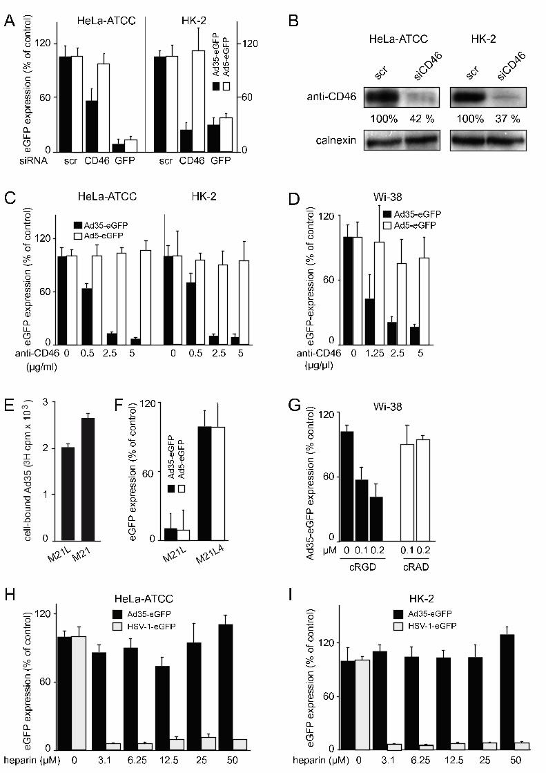
knock-down. A 45% or 72% reduction of the CD46 levels in HeLa-ATCC and HK2 cells
inhibited Ad35-mediated transduction of eGFP by 50% and 78%, respectively (Fig. 1A,
B). This result was confirmed with an Ad35 infection-neutralizing monoclonal antibody
directed against the short consensus repeat region (SCR) 1 of CD46 (26), which gave a
robust dose-dependent inhibition of Ad35 but not Ad5-mediated eGFP transduction in
HeLa-ATCC, HK-2 and Wi-38 normal lung fibroblasts (Fig. 1C,D). This provided
confirmatory evidence that CD46 has a major role for infection of both HeLa-ATCC and
HK-2 cells, in agreement with earlier results from HeLa cells, human bronchial epithelial
A549 cells and hematopoietic cells (25, 26, 30).
All sequenced HAdV serotypes, except for Ad40 and Ad41 that infect the digestive tract
(2) contain an arginine-glycine-aspartate (single amino acid code RGD) motif which
binds integrin and is contained in extracellular matrix proteins (85). Therefore, we
investigated the involvement of integrins in Ad35 infection. The RGD motif in the Ad2
penton base had been shown to bind soluble alpha v beta 5 integrin heterodimers (12).
The attachment of 3H-radiolabelled Ad35 to human melanoma M21L cells lacking αν
integrins (24) was not significantly inhibited compared to αν positive M21 cells (Fig. 1E).
Both Ad35- and Ad5-mediated eGFP transduction of M21L cells was, however, strongly
reduced compared to M21L4 cells transfected with αν integrin cDNA or native M21 cells

11
(not shown), indicating that αν integrins support Ad35 transduction of epithelial cells
(Fig. 1F). A role for integrins was further strengthened by the finding that Ad35-eGFP
expression in Wi-38 cells was significantly reduced by the soluble integrin ligand cRGD
but not cRAD peptides (Fig. 1G). These results were in agreement with an earlier
report showing that integrins are involved in Ad35 transduction of human hematopoietic
cells (72).
It was recently suggested that Ad35 uses heparan sulfate proteoglycans (HSPGs) for
attachment to CHO cells in the absence of CD46 receptors, as concluded from
competition experiments with soluble heparin (99). To address if HSPGs were involved
in Ad35 infection of HeLa-ATCC or HK-2 cells, we preincubated Ad35-eGFP with
increasing concentrations of heparin for 30 min added the mixture to HeLa-ATCC or
HK-2 cells for 8 hours at MOI 5 and measured eGFP transgene expression.
Unexpectedly, we did not observe any significant inhibition or stimulation of eGFP
expression with Ad35 in both cell types (Fig. 1H, I). We did however find a strong block
of herpes simplex virus 1 (HSV1) mediated eGFP transduction, as reported earlier
(107). We concluded that HSPGs are not involved in Ad35 infection of the CD46-
positive HeLa and HK-2 cells.
Cell type-dependent requirement of dynamin for Ad35 infection
Ligation of CD46 by antibodies or Ad3 has been shown to trigger clathrin-mediated
endocytosis or dynamin-independent macropinocytosis, depending on the degree of
CD46 crosslinking (3, 16, 95). By transfecting cells with dominant-negative K44A-dyn2
tagged with mRFP for 24 hours followed by inoculation of the cells with Ad35-eGFP, we
found that Ad35 transduction of HeLa-ATTC cells was independent of K44A-dyn2, in
contrast to Ad5-eGFP (Fig. 2A, B). Ad35 infection of HeLa-K cells, however, was

12
dynamin-dependent (Fig. 2C), exactly as reported for Ad3 (3). To test if dynamin-
dependent Ad35 infection of HeLa-K cells required a clathrin related pathway, we
overexpressed the carboxy-terminal clathrin heavy chain binding domain of AP180 (aa
530-915), which blocks clathrin recruitment to the plasma membrane (28), and inhibits
clathrin-mediated endocytosis (71). It also inhibits infectious clathrin and dynamin-
dpendent endocytosis of Ad2 (31). We found that Ad35 infection of HeLa-K cells was
not inhibited by the C-terminal domain of AP180, unlike Ad5-eGFP transduction (Fig.
2C). In support of this, the dynamin inhibitor dynasore (59) had no significant effects on
Ad35 transduction but affected Ad5 transduction of Wi-38 cells (Fig. 2D). This
suggested that Ad35 entry into HeLa-K cells was at least partly dependent on dynamin
and most likely clathrin, and Ad35 entry into HeLa-ATCC and Wi-38 cells was dynamin-
independent.
Macropinocytosis inhibitors block infectious Ad35 uptake into epithelial cells
To analyze the dynamin-independent infection pathway of Ad35 we treated HeLa-
ATCC, HK-2 or Wi-38 cells with pharmacological inhibitors of macropinocytosis. The
classical inhibitors of macropinocytosis amiloride and its derivative 5-(N-ethyl-N-
isopropyl) amiloride (EIPA) block the sodium/proton exchangers, lead to mild
acidification of the cytosol, alter the subcellular localization of early and late endosomes,
and possibly raise the lumenal pH of mildly acidic organelles (29). Likewise, millimolar
concentrations of NH4Cl inhibit macropinocytosis (15) possibly by acidification of the
cytosol and effects on actin dynamics (39). We found that low micromolar
concentrations of EIPA inhibited Ad35-eGFP transduction of HeLa-ATCC, HK-2 and Wi-
38 cells (Fig. 3A, H), similar as millimolar concentrations of NH4Cl in HeLa-ATCC, HK2
or Wi-38 cells (Fig. 3B, and not shown). In contrast, the proton ATPase inhibitor
bafilomycin A1 (Baf) did not affect Ad35-eGFP transduction of HeLa or HK-2 cells,
except at the very highest concentration of 100 nM where it decreased Ad35-eGFP

13
transduction by 10 to 25% (Fig. 3C). It did not affect Ad35-eGFP transduction of Wi-38
cells (not shown). In contrast, Baf (50 nM) reduced human rhinovirus serotype 16
infection of HeLa cells at least 20-fold compared to control cells, and blocked
acidification of endosomal organelles (Neugebauer, Jurgeit, Greber, unpublished
observations), suggesting that vacuolar ATPases are not required for infectious Ad35
entry.
Another class of commonly used inhibitors of macropinocytosis are actin-directed
compounds. Macropinosome formation involves filamentous (F)-actin, and is sensitive
against pharmacological inhibitors (65, 68), including jasplakinolide (Jas), which
stabilizes actin polymers, cytochalasin D (CytD), which blocks actin polymerisation at
the barbed ends of F-actin, and the G-actin binding marine macrolide latrunculin B
(LatB) (10, 18, 80). Both Jas and CytD inhibited Ad35-eGFP transduction of HeLa-
ATTC and HK-2 cells in a dose-dependent manner (Fig. 3D), and LatB inhibited Ad35-
eGFP transduction of Wi-38 cells (Fig. 3I).
A further requirement for macropinocytosis is the activation of calcium and
diacylglycerol-dependent protein kinase C (PKC) family members (4, 69). PKC
activators induce ruffling and fluid uptake by various mechanisms involving signal
transduction at the plasma membrane, which can be inhibited for example by the
calcium-dependent PKC inhibitor Gö6976 (17, 63). The treatment of HeLa-ATCC or
HK-2 cells with Gö6976 lead to a dose-dependent inhibition of Ad35-eGFP transduction
(Fig. 3E), similar to Ad3 which uses PKC-dependent macropinocytosis for infectious
entry into epithelial cells (3). Interestingly, Gö6976 had no effect on Ad35-eGFP
transduction of Wi-38 cells suggesting a cell type-variable involvement of PKC in Ad35
infection (data not shown).

14
An important regulator of macropinocytosis is the serine/threonine kinase p21-activated
kinase (Pak) (20, 68). We treated cells with the Pak inhibitor IPA-3, which allosterically
blocks autoinhibited Pak1 and related isoforms of this kinase (19). Ad35 transduction of
HeLa-ATCC, HK-2 or Wi-38 cells was inhibited by IPA-3 in a dose-dependent manner,
but not by an inactive IPA-3 related compound PIR3.5 (Fig. 3F, J). In HeLa-ATCC or
HK-2 cells, IPA-3 treatment specifically inhibited Ad35-eGFP without affecting Ad5-
eGFP transduction (Fig. 3G), although Ad5 transduction of Wi-38 cells was sensitive to
IPA-3 at low micromolar concentrations (Fig. 3K). It is possible that Paks are
overexpressed and/or hyperactivated in HeLa cells, consistent with the notion that
signalling pathways in human tumor cells differ from those in normal tissue (23). Ad5
could thus circumvent Pak-1 inhibition in cancer cells by using other signalling
pathways, which are not upregulated or absent in normal human diploid fibroblasts Wi-
38 cells. Noteably, our earlier data showed that in cancer cells Ad2/5 depends on
dynamin and clathrin-mediated pathways (31), activates Pak1 and triggers accessory
Pak1-dependent macropinocytosis as indicated by RNA interference (3, 63). We
conclude that Ad35 transduction is sensitive to inhibition of macropinocytosis.
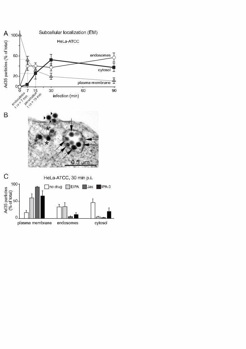
We next used electron microscopy (EM) to determine the subcellular localization of
Ad35 in epithelial HeLa-ATCC cells. Time course analyses showed that Ad35 particles
were rapidly and efficiently cleared from the plasma membrane, and more than 50% of
the virus particles localized to the cytosol at 30 min pi (Fig. 4A, for a representative
image, see Fig. 4B). This indicated rapid viral uptake and endosomal escape with
estimated half maximal times of 7 and 15 min, respectively. EIPA, Jas and IPA-3
delayed viral uptake into cells, and strongly reduced Ad35 localization in the cytosol 30
min pi (Fig. 4C). This suggested that infectious Ad35 endocytosis required sodium-
proton exchangers, dynamic F-actin and Pak1.
We next analyzed if actin inhibition by Jas, or Pak1 inhibition by IPA-3 affected the
dynamics of the plasma membrane during entry of Texas-Red-labeled Ad35 (Ad35-TR).
15
For this, we monitored the localization of eGFP-tagged human CD46 BC1 splice form in
stably transfected Chinese Hamster ovary cells (CHO-GFP-CD46) in warm infected
cells 5 to 15 min pi. CHO cells are CD46-negative and resistant to Ad35 infection,
whereas GFP-CD46 expression in these cells (CHO-CD46) mediates Ad35 transduction
similar to CD46-expression (data not shown, and 25, 26, 30). Control infected or
noninfected cells showed extensive dynamics of the plasma membrane marker GFP-
CD46, including membrane ruffling and protrusions, which we call here collectively
‘wobbling ruffles’, and frequent events of virus and CD46 colocalizations were observed
(Fig. 5A, Suppl. Mov. 1, and Fig. 5D , Suppl. Mov. 4). In contrast, the wobbling ruffles of
GFP-CD46 at the plasma membrane were strongly suppressed in cells treated with Jas,
although Ad35-TR particles readily associated with the cell periphery, and remained
confined there throughout the observation period (Fig. 5B, and Suppl. Mov. 2).
Likewise, the Pak1 inhibitor IPA-3 suppressed to a large extent the peripheral wobbling
of GFP-CD46 (Fig. 5C and 5F, Suppl. Mov. 3). Significantly, the virus particles in the
cell periphey remained confined and were largely immobile, suggesting that IPA-3
inhibited viral uptake into cells (see Fig. 4B). Interestingly, viruses that were attached to
protruding filopodia-like extensions of control or IPA-3 treated cells moved towards the
cell body. Such movements were not observed in Jas-treated cells where filopodia-
associated viruses remained stationary. This is consistent with earlier results showing
that filopodial dynamics can be blocked by Jas which freezes F-actin (10, 93). Pak1
apparently does not contribute to filipodial surfing of Ad35. Whether filopodial surfing of
Ad35 contributes to infection remains to be analyzed.
Pak1 and CtBP1 facilitate infectious Ad35 macropinocytosis
Pak1 is activated by the small GTPase Rac1 (Ras-related C3 botulinum toxin substrate
1) during growth factor-stimulated macropinocytosis, cell adhesion and motility (41), and
is implicated in endocytosis of interleukin-2 receptor (32), and infectious
16
macropinocytosis of Ad3 and vaccinia virus (3, 67). The expression of dominant-
negative Rac1 (mutated threonine 17 to asparagine, T17N) inhibited the expression of
Ad35-eGFP by about 50% (Fig. 6A, B). Likewise, the autoinhibitory domain of Pak1
reduced Ad35-eGFP levels by about 70% but did not affect Ad5-eGFP (Fig. 6C, D)
supporting the notion that Pak1 is involved in species B but not species C adenovirus
infections (3).
Activated Pak1 phosphorylates the transcriptional repressor C-terminal binding protein 1
(CtBP1) at serine 147 in the nucleus, and recruits CtBP1 into the cytoplasm (7). There
are two splice forms of CtBP1, a short form truncated by 11 amino acids (also called
CtBP3/BARS) and a long form (CtBP1). Both forms control dynamin-independent
endocytosis (9), membrane fission and endocytic cup formation (3, 43, 56). We found
that the expression of dominant-negative, phosphorylation-defective CtBP1 (serine 147
to alanine mutation, S147A), but not the phosphomimetic mutant S147D reduced Ad35-
eGFP by about 35% without affecting Ad5-eGFP (Fig. 6E, F). These results were
corroborated by siRNA experiments against Pak1 and CtBP1 in HeLa-ATCC and
human alveolar epithelial A549 cells. Knock-down of Pak1 by 65% and CtBP1 by 35%
(measured by Western blotting in HeLa cells) reduced Ad35 transduction between 40
and 60%, depending on the cell type, but without affecting Ad5-eGFP expression (Fig.
6G, H, data not shown). Control siRNAs against eGFP inhibited both Ad35- and Ad5-
eGFP expression (Fig. 6G). Similar to epithelial cells, Ad35-eGFP transduction of
hematopoietic K562 cells was inhibited by CtBP1 siRNA but not clathrin siRNA,
analogous to an earlier report for Ad3 (3). We concluded that CtBP1 supports Ad35
infection.
Both Pak1 and CtBP1 are involved in macropinocytosis, which engulfs a large amount
of fluids into cells. We tested if Ad35 induced the endocytic uptake of fluid phase into
HeLa-ATCC cells. Ad35 was bound to HeLa-ATCC in the cold, free virus washed off,
and cells incubated in warm medium and pulsed with dextran-FITC for 5 min prior to

17
analyses of acid-washed cells for intracellular dextran by flow cytometry. We found a
peak of fluid-phase uptake 10 min post warming indicating that Ad35 transiently induced
dextran uptake (Fig. 7A), similar to Ad2/5 and Ad3 (3, 40, 63, 64). Multichannel
confocal immunofluorescence analyses revealed that dextran and Ad35-TR positive
endosomes contained CD46, alpha v integrins and to a lower degree also CtBP1 at 10
min pi (Fig. 7B). Noninfected cells did not contain detectable dextran-FITC positive
endosomes under these pulse labeling conditions, indicating that the dextran-positive
endosomal structures described in Fig. 7B were virus induced (Fig. S1). Quantitative
analyses using wide field fluorescence microscopy showed that 91% of the dextran-
filled vesicles in Ad35 infected cells were positive for CD46, essentially all were alpha v
integrin positive, and 77% were positive for CtBP1 (Fig. S2).
Transmission electron microscopy showed that Ad35 particles were in large endosomes
10 min pi several micrometers in diameter (Fig. 7C). These endosomes contained
cointernalized BSA-gold. To test if macropinocytosis contributed to the levels of BSA-
gold in Ad35 positive endosomes, we compared the amounts of BSA-gold in
endosomes containing Ad2-ts1. Ad2-ts1 enters cells by dynamin and clathrin-mediated
endocytosis in the absence of macropinocytic stimulation (31, 40). More than 90% of
the endosomal Ad35 particles were in BSA-gold positive vesicles, whereas about 65%
of the Ad2-ts1 particles were in gold-positive endosomes (Fig. 7D). We found that
about 60% of the total BSA-gold particles were in Ad35-positive endosomes, whereas
about 15% of the total BSA-gold were in Ad2-ts1 positive endosomes. This strongly
supported the conclusion that Ad35 was predominantly in fluid-enriched endosomes 10
min pi. Together the data indicate that Ad35 induces CD46 and integrin-dependent
macropinocytosis for infectious uptake into epithelial HeLa-ATCC and kidney HK-2 cells
as well as hematopoietic cells (see Fig. 8 for a schematic overview).

18
DISCUSSION
Adenoviruses cause infectious disease and significant health problems across the
world. Therapeutic forms of adenoviruses are increasingly developed to treat human
diseases, including cancer and immune disorders (92, 98). Research in the past years
has shown that both virulent and therapeutic viruses strongly depend on host factors for
eliciting therapeutic or disease phenotypes (for overviews, see 47, 92, 94). Here, we
analyzed the early interactions of Ad35 with cultured human cells, and show that they
involve macropinocytosis of CD46-associated Ad35 particles. Unlike an earlier study in
CHO cells (99), we found no requirement of heparan sulfate proteoglycans for Ad35
infection of HeLa or human kidney HK-2 cells, which emphasizes the importance of
CD46 for infectious entry of Ad35 (30). This is in line with earlier findings showing that
the species B1 Ad3 uses macropinocytosis for infectious uptake into epithelial and
hematopoietic cells (3). Noteably, Ad35 is one of several species B human
adenoviruses which have been shown to bind with their fiber knobs to CD46 (25, 26, 30,
79). Other CD46-tropic adenoviruses include Ad3 (95), Ad11 (91), Ad14, Ad16, Ad21
and Ad50 (30), and also the species D Ad37 (106) and Ad49 (54). It is possible that
members of the species B HAdVs use CD46 with different affinities or by unknown
mechanisms, or bind to additional receptors.
In addition to binding to the species B HAdVs, CD46 also binds the Edmonston strain of
measles virus (22, 75), human herpes virus 6 (89), bovine viral diarrhea virus (62), and
various bacteria including uropathogenic E. coli (55, 57). The selection of CD46 as a
receptor for numerous pathogens suggests that there are advantages for pathogens to
bind to CD46, e.g. dampening or suppression of innate immune responses (44, 77).
The ubiquitously expressed human CD46 is involved in the control of complement lysis
and inhibition of T cell effector functions (6, 45). The cytoplasmic splice variant 1 of

19
CD46 controls the induction of autophagic degradation (42), which can lead to
enhanced presentation of antigenic peptides to major histocompatibility complexes of
the class II and promote adaptive immune responses and inflammation (90). Although
both cytoplasmic splice variants 1 and 2 of CD46 have been found to bind Ad3 and
Ad35 (30, 95), it is unknown if binding of HAdVs to CD46 induces autophagy.
Our data here indicate that Ad35 induced infectious CD46-dependent macropinocytosis,
similar to Ad3 in epithelial cells (3), and this could be related to integrin-dependent Ad35
infection of hematopoietic cells (72). The pathway that we delineate for Ad35 infection
involves alpha v integrins, independent of clathrin-mediated endocytosis, as concluded
from insensitivity against dynamin inhibition by dynasore, dominant-negative constructs
or siRNA in HeLa-ATCC, Wi-38 human lung fibroblasts, A549 cells or hematopoietic
K562 cells. Interestingly, K44A-dynamin inhibited the transduction of Ad35 in HeLa-K
cells, a variant of HeLa-ATCC, but infection of these cells was not affected by the C-
terminal fragment of AP180, which binds to clathrin and blocks clathrin-mediated
endocytosis (28). This is similar to Ad3 transduction reported earlier (3). Since
dynamin interference inhibited the uptake of transferrin, a classical ligand for clathrin-
mediated endocytosis, to a similar extent in both HeLa-ATCC and HeLa-K cells, we
speculate that in certain cell types, such as HeLa-K, dynamin supports Ad3 and Ad35
transduction at the level of endosomal trafficking (51, 52). This speculation could be
supported by an earlier finding that the clathrin binding protein CALM (clathrin assembly
lymphoid myeloid) is required for infection with wild type Ad2 but not the endosomal
escape-defective mutant Ad2-ts1 (40).
The requirements for infectious endocytosis of Ad35 into human epithelial cells closely
reflect those for macropinocytosis, as indicated by drug sensitivity and morphological
analyses at the level of light microscopy and EM. We found a strong but cell-type
dependent requirement of Ad35 transduction for PKC, and a strong cell-type
independent requirement for the sodium/proton exchanger, actin and Pak1 in HeLa-
20
ATCC, HK-2 and Wi-38 cells, as well as the actin modulator Rac1 and the Pak1 effector
CtBP1 in HeLa-ATCC and HK-2 cells. CtBP1 is commonly acting as a transcriptional
repressor and membrane organizer (11, 14). Ad35 colocalized with its receptors CD46
and αν-integrins in dextran-filled macropinosomes and was associated with dynamic
GFP-CD46 clusters early in infection. These endosomes were positive for CtBP1,
unlike noninfected cells, suggesting that CtBP1 was recruited to Ad35-positive
membrane domains, possibly from the nucleus upon phosphorylation by Pak1 (7). This
was supported by the finding that dominant-negative phosphorylation defective CtBP1
inhibited Ad35 transduction. Since macropinocytosis has been implicated in immune
suppression, for example in the uptake of apoptotic bodies (1), it is possible that
Pak1/CtBP1 dependent macropinocytosis leads to transcriptional depression, and
silences immune responses.
Together, our results provide supportive evidence that crosslinking of CD46 by
multivalent Ad35 particles leads to the formation of macropinocytic vesicles, similar to
crosslinking induced by anti-CD46 antibodies or measles virus which triggered the
formation of pseudopodia and macropinocytic engulfment of CD46 (16). This indicates
that macropinocytosis is an infectious uptake route exploited by an increasing number
of pathogens, and has implications for gene delivery and vaccination strategies.
ACKNOWLEDGEMENTS
We thank Nicola Imelli for expert help in EM analyses and Corinne Wilhelm for excellent
cell culture and support. This project was supported by the Swiss National Science

21
Foundation, and in part by the Swiss SystemsX.ch initiative, grant LipidX-2008/011 to
UFG.

22
FIGURE LEGENDS
Fig. 1: CD46 and alpha-v-integrins are required for Ad35 infection but not HSPG.
(A) HeLa-ATCC (left) or HK-2 (right) were transfected with 20 nM siRNAs against
CD46, GFP or non-silencing scrambled (scr) for 72 h and infected with Ad35-eGFP
(MOI 5) or Ad5-eGFP (MOI 5) for 8 h, followed by Safire fluorescence analysis. (B)
Quantification of knock-down efficiency by Western blots against CD46, normalized
against calnexin. (C) HeLa-ATCC (left) or HK-2 (right) or Wi-38 cells (panel (D)) were
preincubated with an inhibiting antibody against CD46 (MEM-258) in the cold for 30 min,
and infected for 8 h or 15 h, respectively. (E) 3H-Ad35 was bound to human melanoma
M21L cells lacking alpha v integrin or M21 alpha v integrin positive cells. (F) M21L or
M21 cells were infected with Ad35-eGFP or Ad5-eGFP, and analyzed by Safire
fluorescence analyses. (G) Wi-38 cells were preincubated with cRGD or cRAD
peptides in the cold for 30 min and infected with Ad35-eGFP for 15 h. (H, I) Ad35-eGFP
(MOI 5) or HSV-1-eGFP (MOI 5) were preincubated for 30 min with different
concentrations of heparin as indicated followed by inoculation of HeLa-ATCC or HK-2
cells for 8 h, and infection analyses with a Safire2 plate reader after normalization to the
cell numbers determined by DAPI staining (see Materials and Methods). Note that
Ad35-eGFP transduction was not affected, whereas HSV-1-eGFP transduction was
strongly reduced in both cell lines.
Fig. 2: Cell type specific requirements of dynamin.
HeLa-ATCC or HeLa-K cells were transfected with dominant-negative mRFP-Dyn2-
K44A, mRFP-Dyn2-wt or mRFP for 24 h, and infected with Ad35-eGFP or Ad5-eGFP

23
(MOI 5) for 8 h. (A) Representative images of transfected and infected HeLa-ATCC,
fixed and recorded for mRFP and GFP fluorescence in a wide-field microscope. (B)
NIH ImageJ quantification of total fluorescence intensities. (C) HeLa-K cells were
transfected as in (A) and in addition with the carboxy-terminal domain of AP180,
infected and assayed by FACS. Dominant-negative K44A-Dyn2 inhibited both Ad35-
eGFP and Ad5-eGFP, whereas carboxy-terminal domain of AP180 had no effects on
Ad35-eGFP but inhibited Ad5-eGFP by 40%. (D) Dynasore independent transduction of
Wi-38 cells by Ad35-eGFP (left) and dynasore-dependent transduction by Ad5-eGFP
(right panel) in the absence of cell toxicity indicated by cell number measurements
shown with grey line graphs, with 100% representing noninfected non-drug treated
conditions.
Fig. 3: Inhibitors against Pak1, PKC, sodium/proton exchanger and actin reduce
Ad35-eGFP transduction.
HeLa-ATCC and HK-2 were preincubated with indicated concentrations of drugs for 30
min, and infected with Ad35-eGFP for 8 h. Cells were analyzed on a Safire2 plate
reader and eGFP intensities normalized to the DAPI signal of the cell nuclei
representing cell numbers. Results for EIPA are shown in panel (A), NH4Cl in (B),
bafilomycin (Baf) in (C), jasplakinolide (Jas) and cytochalasin D (CytD) in (D), the PKC
inhibitor Gö6976 in (E), and the Pak1 inhibitor IPA-3 and its inactive derivative PIR3.5 in
(F) and (G) for both Ad35-eGFP and Ad5-eGFP, respectively. Panels H-K show results
with macropinocytic interference in Wi-38 cells transduced with Ad35-eGFP or Ad5-
eGFP. Note the absence of significant cell toxicity as indicated by cell number
measurements shown with grey line graphs, with 100% representing noninfected non-
drug treated conditions.

24
Fig. 4: EM analysis of Ad35 entry into HeLa-ATCC cells
(A) Cells on glass coverslips were incubated with double CsCl purified Ad35 particles
for 90 min in the cold at high MOI of 5000, washed extensively, warmed to 37°C in
growth medium containing 0.2% BSA for indicated times, fixed and processed for
quantitative transmission EM analyses of virus particles at the plasma membrane, in
endosomes and in the cytosol. Half maximal time points for endocytic uptake and
penetration into the cytosol were 7 and 15 min, respectively. (B) Electron micrograph of
an Ad35-infected HeLa-ATCC cell 30 p.i.. Stars depict virus particles in the cytosol,
arrows particles in endosomes and arrowheads at the plasma membrane. (C) The
same experiment was repeated with cells treated or not treated with the sodium/proton
inhibitor EIPA (50 µM), the F-actin stabilizer Jas (300 nM) or the Pak1 inhibitor IPA-3
(25 µM), and infected for 30 min.
Fig. 5: Ad35-TR is associated with GFP-CD46 clusters early in infection.
Stably transfected CHO-GFP-CD46 cells were infected with Ad35-TR (1 µg/ml) for 5
min, and recorded in a spinning disc confocal microscope equipped with a warm
chamber at 37° C from 5 min to 15 min p.i. at acquisition frequency of 0.06 Hz. Panel
(A) shows a control cell with extended wobbling of GFP-CD46 membrane domains and
frequently overlapping signals of CD46 and Ad35-TR. Panel (B) shows a cell pretreated
with 300 nM Jasplakinolide (Jas) for 30 min and (C) depicts a cell pretreated with 25 µM
IPA-3. Time stamps are in min (‘). (D) Analyses of GFP-CD46 wobbling ruffles with
schematic drawing shown in (E) of an untreated, infected cell with two zoom-in views
marked * and ** with the corresponding time stamp min (‘) and seconds (‘’) p.i.. (F)

25
Quantitative boxplot analyses of wobbling ruffles in control noninfected cells, infected
cells and infected cells treated with Jas or IPA-3.
Fig. 6: Rac1, Pak1 and CtBP1 are required for Ad35-eGFP transduction of HeLa-
ATCC cells.
Cells were transfected with indicated constructs for 24 h, and infected for 8 h, fixed,
recorded in a wide-field microscope and analyzed with ImageJ. (A) Representative
images of dominant-negative mRFP-T17N-Rac1 or Rac-wt transfected cells. (B)
Quantification of the mean fluorescence intensity per cell with indicated number of cells
(n). (C) Representative images of Pak1-autoinhibitory domain (AID) transfected cells
infected with Ad35-eGFP or Ad5-eGFP, and quantification of the mean fluorescence
intensity per cell (D). (E) Representative images of phosphorylation defective CtBP1-
S147A transfected cells infected with Ad35-eGFP or Ad5-eGFP. (F) Quantification of
the mean fluorescence intensity of GFP expression per cell in wild type (wt) CtBP1,
S147A and S147D CtBP1 mutants. (G) Cells were transfected with 50nM siRNA
against indicated targets for 72 h, infected for 8 h and analyzed with Safire2. Cell
numbers were normalized to DAPI signal. (H) Knock-down quantification of Pak1 and
CtBP1 by Western blot, normalized against calnexin. (I) A549 cells were transfected
with 20 nM siRNA against CtBP1, Pak1 or dynamin2 for 48 h, infected with Ad35-eGFP
(16h) and analyzed by flow cytometry. K562 cells were transfected with 20 nM siRNA
against CtBP1 or clathrin heavy-chain (CHC) as described (3), infected with Ad35-eGFP
(16h) and analyzed by flow cytometry.
Fig. 7: Ad35-TR induced fluid phase uptake and colocalizes with CD46, integrins
and CtBP1 in dextran-filled macropinosomes.

26
(A) Ad35 (2 mg/ml) was cold bound to HeLa-ATCC cells for 1 h. Cells were washed,
pulsed with dextran-FITC (0.5 mg/ml) in warm medium (37°C) containing BSA 5 min
before the indicated time points, and prepared for flow cytometric analysis. (B) Cells
were cold-bound with Ad35-TR (1 µg/ml) for 1 h, washed and incubated at 37°C in the
presence of dextran-FITC (0.5 mg/ml) for 10 min, fixed, stained for the indicated
antigens and analyzed by confocal laser scanning microscopy with corresponding
differentail interference contrast (DIC) images. Fluorescence images represent single
sections, and inlets are magnifications of the white boxed areas. (C) Electron
micrograph of an Ad35 infected HeLa-ATCC cell (cold synchronized infection at MOI
5000), pulsed in the presence of BSA-nano-gold for 10 min. Arrowheads indicate BSA-
gold particles, black arrows show Ad35 in BSA-gold positive endosomes, white arrows
show virus particles in endosomes without BSA-gold, small triangles indicate viruses at
the PM, and stars depict Ad35 particles in the cytosol. (D) Quantification of (C),
including HeLa-ATCC cells inoculated with Ad2-ts1 which is defective in endosomal
escape. N indicates number of virus particles and n number of BSA-gold particles.
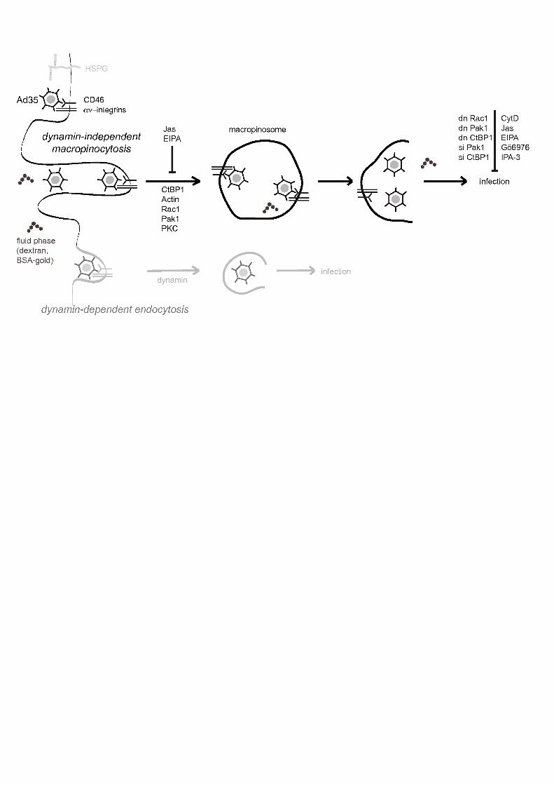
Fig. 8: Schematic model of infectious macropinocytosis of Ad35 in human
epithelial cells.
Ad35 binds to CD46 and alpha v-integrins independently of heparan sulfate
proteoglycans (HSPGs). Ad35 is internalized in an actin, Rac1, Pak1, CtBP1 and PKC
dependent manner and localizes to dextran-filled vesicles with its receptors CD46,
alpha v integrins and also CtBP1. Ad35 escapes to the cytosol by an unknown
mechanism and traffics to the nucleus for infection.

27
Supplemental figures
Fig. S1: Immunofluorescent stainings of CD46, αααανννν-integrins or CtBP1 in non-
infected cells.
HeLa-ATCC were incubated with 0.5 mg/ml Dextran-FITC for 10 min at 37°C in the
absence of virus. Cells were fixed, and processed for immunofluorescence analyses as
indicated in Fig. 7B. Images represent single sections.
Fig. S2: Quantification of Ad35 induced dextran-FITC filled vesicles containing
CD46, αααανννν-integrins and CtBP1.
Dextran-FITC uptake was induced by Ad35 with a 10 min pulse as described in Fig. 7A.
Samples were fixed and analyzed by wide-field microscopy. Panel (A) displays
representative sample images for all three marker proteins. Panel (B) shows the
quantification of dextran-FITC positive vesicles for the presence of CD46 (n=70), αν-
integrins (n=63) or CtBP1 (n=61), displayed as percentage of macropinosomes positive
for CD46, αν-integrins or CtBP1 by boxplot analyses.
Supplemental movies S1-S4: Dynamics of eGFP-CD46 and Ad35-TR in control
cells and cells treated with Jasplakinolide (Jas) or IPA-3.
Stably transfected CHO-GFP-CD46 cells were infected with Ad35-TR (Texas Red, 1
µg/ml) for 5 min and recorded in a spinning disc confocal microscope equipped with a
warm chamber at 37° C from 5 min to 15 min p.i. with an acquisition frequency of 0.06

28
Hz. Supplemental movie 1 shows control cells infected with Ad35-TR. Supplemental
movie 2 shows cells treated with Jas (300 nM), and supplemental movie 3 shows cells
treated with IPA-3 (25 µM). Supplemental movie 4 shows GFP-CD46 wobbling ruffles
of infected control cells 5 to 20 min pi. Still images of these movies are shown in Fig.
5A-D.

29
REFERENCES
1. Albert, M. L. 2004. Death-defying immunity: do apoptotic cells influence antigen processing and presentation? Nat Rev Immunol 4:223-31.
2. Albinsson, B., and A. H. Kidd. 1999. Adenovirus type 41 lacks an RGD alpha(v)-integrin binding motif on the penton base and undergoes delayed uptake in A549 cells. Virus Research. 64:125-136.
3. Amstutz, B., M. Gastaldelli, S. Kälin, N. Imelli, K. Boucke, E. Wandeler, J. Mercer, S. Hemmi, and U. F. Greber. 2008. Subversion of CtBP1 controlled macropinocytosis by human Adenovirus serotype 3. EMBO J. 27:956-966.
4. Amyere, M., B. Payrastre, U. Krause, P. V. Smissen, A. Veithen, and P. J. Courtoy. 2000. Constitutive macropinocytosis in oncogene-transformed fibroblasts depends on sequential permanent activation of phosphoinositide 3-kinase and phospholipase C. Mol Biol Cell 11:3453-67.
5. Araki, N., M. T. Johnson, and J. A. Swanson. 1996. A role for phosphoinositide 3-kinase in the completion of macropinocytosis and phagocytosis by macrophages. J Cell Biol 135:1249-60.
6. Astier, A. L. 2008. T-cell regulation by CD46 and its relevance in multiple sclerosis. Immunology 124:149-54.
7. Barnes, C. J., R. K. Vadlamudi, S. K. Mishra, R. H. Jacobson, F. Li, and R. Kumar. 2003. Functional inactivation of a transcriptional corepressor by a signaling kinase. Nat Struct Biol 10:622-8.
8. Bergelson, J. M., J. A. Cunningham, G. Droguett, E. A. Kurt-Jones, A. Krithivas, J. S. Hong, M. S. Horwitz, R. L. Crowell, and R. W. Finberg. 1997. Isolation of a common receptor for Coxsackie B viruses and adenoviruses 2 and 5. Science 275:1320-1323.
9. Bonazzi, M., S. Spano, G. Turacchio, C. Cericola, C. Valente, A. Colanzi, H. S. Kweon, V. W. Hsu, E. V. Polishchuck, R. S. Polishchuck, M. Sallese, T. Pulvirenti, D. Corda, and A. Luini. 2005. CtBP3/BARS drives membrane fission in dynamin-independent transport pathways. Nat Cell Biol 7:570-80.
10. Burckhardt, C. J., and U. F. Greber. 2009. Virus movements on the plasma membrane support infection and transmission between cells. PLoS Pathogens 5:e1000621. Epub 2009 Nov 26.
11. Chinnadurai, G. 2007. Transcriptional regulation by C-terminal binding proteins. Int J Biochem Cell Biol 39:1593-607.
12. Chiu, C. Y., P. Mathias, G. R. Nemerow, and P. L. Stewart. 1999. Structure of adenovirus complexed with its internalization receptor, alpha(v)beta 5 integrin. J. Virol. 73:6759-6768.

30
13. Cohen, J., and W. Powderly. 2004. Infectious diseases, 2nd edn ed. Mosby, New York.
14. Corda, D., A. Colanzi, and A. Luini. 2006. The multiple activities of CtBP/BARS proteins: the Golgi view. Trends Cell Biol 16:167-73.
15. Cosson, P., I. de Curtis, J. Pouyssegur, G. Griffiths, and J. Davoust. 1989. Low cytoplasmic pH inhibits endocytosis and transport from the trans-Golgi network to the cell surface. J Cell Biol 108:377-87.
16. Crimeen-Irwin, B., S. Ellis, D. Christiansen, M. J. Ludford-Menting, J. Milland, M. Lanteri, B. E. Loveland, D. Gerlier, and S. M. Russell. 2003. Ligand binding determines whether CD46 is internalized by clathrin-coated pits or macropinocytosis. J Biol Chem 278:46927-37.
17. Davies, S. P., H. Reddy, M. Caivano, and P. Cohen. 2000. Specificity and mechanism of action of some commonly used protein kinase inhibitors. Biochem J 351:95-105.
18. Dayel, M. J., and R. D. Mullins. 2004. Activation of Arp2/3 complex: addition of the first subunit of the new filament by a WASP protein triggers rapid ATP hydrolysis on Arp2. PLoS Biol 2:E91.
19. Deacon, S. W., A. Beeser, J. A. Fukui, U. E. Rennefahrt, C. Myers, J. Chernoff, and J. R. Peterson. 2008. An isoform-selective, small-molecule inhibitor targets the autoregulatory mechanism of p21-activated kinase. Chem Biol 15:322-31.
20. Dharmawardhane, S., A. Schurmann, M. A. Sells, J. Chernoff, S. L. Schmid, and G. M. Bokoch. 2000. Regulation of macropinocytosis by p21-activated kinase-1. Mol Biol Cell 11:3341-52.
21. Doherty, G. J., and H. T. McMahon. 2009. Mechanisms of endocytosis. Annu Rev Biochem 78:857-902.
22. Dorig, R. E., A. Marcil, A. Chopra, and C. D. Richardson. 1993. The human CD46 molecule is a receptor for measles virus (Edmonston strain). Cell 75:295-305.
23. Dummler, B., K. Ohshiro, R. Kumar, and J. Field. 2009. Pak protein kinases and their role in cancer. Cancer Metastasis Rev 28:51-63.
24. Felding-Habermann, B., B. M. Mueller, C. A. Romerdahl, and D. A. Cheresh. 1992. Involvement of integrin alpha V gene expression in human melanoma tumorigenicity. J Clin Invest 89:2018-22.
25. Fleischli, C., D. Sirena, G. Lesage, M. J. Havenga, R. Cattaneo, U. F. Greber, and S. Hemmi. 2007. Species B adenovirus serotypes 3, 7, 11 and 35 share similar binding sites on the membrane cofactor protein CD46 receptor. J Gen Virol 88:2925-34.
26. Fleischli, C., S. Verhaagh, M. Havenga, D. Sirena, W. Schaffner, R. Cattaneo, U. F. Greber, and S. Hemmi. 2005. The Distal Short Consensus Repeats 1 and 2 of the Membrane Cofactor Protein CD46 and Their Distance from the Cell Membrane Determine Productive Entry of Species B Adenovirus Serotype 35. J Virol 79:10013-22.

31
27. Fontenot, D. R., P. den Hollander, E. M. Vela, R. Newman, J. K. Sastry, and R. Kumar. 2007. Dynein light chain 1 peptide inhibits human immunodeficiency virus infection in eukaryotic cells. Biochem Biophys Res Commun 363:901-7.
28. Ford, M. G., B. M. Pearse, M. K. Higgins, Y. Vallis, D. J. Owen, A. Gibson, C. R. Hopkins, P. R. Evans, and H. T. McMahon. 2001. Simultaneous binding of PtdIns(4,5)P2 and clathrin by AP180 in the nucleation of clathrin lattices on membranes. Science 291:1051-5.
29. Fretz, M., J. Jin, R. Conibere, N. A. Penning, S. Al-Taei, G. Storm, S. Futaki, T. Takeuchi, I. Nakase, and A. T. Jones. 2006. Effects of Na+/H+ exchanger inhibitors on subcellular localisation of endocytic organelles and intracellular dynamics of protein transduction domains HIV-TAT peptide and octaarginine. J Control Release 116:247-54.
30. Gaggar, A., D. M. Shayakhmetov, and A. Lieber. 2003. CD46 is a cellular receptor for group B adenoviruses. Nat Med 9:1408-12.
31. Gastaldelli, M., N. Imelli, K. Boucke, B. Amstutz, O. Meier, and U. F. Greber. 2008. Infectious adenovirus type 2 transport through early but not late endosomes. Traffic 9:2265-78.
32. Grassart, A., A. Dujeancourt, P. B. Lazarow, A. Dautry-Varsat, and N. Sauvonnet. 2008. Clathrin-independent endocytosis used by the IL-2 receptor is regulated by Rac1, Pak1 and Pak2. EMBO Rep 9:356-62.
33. Greber, U. F., M. Willetts, P. Webster, and A. Helenius. 1993. Stepwise dismantling of adenovirus 2 during entry into cells. Cell 75:477-86.
34. Greber, U. F., M. Willetts, P. Webster, and A. Helenius. 1993. Stepwise dismantling of adenovirus 2 during entry into cells. Cell 75:477-486.
35. Haga, Y., N. Miwa, S. Jahangeer, T. Okada, and S. Nakamura. 2009. CtBP1/BARS is an activator of phospholipase D1 necessary for agonist-induced macropinocytosis. Embo J 28:1197-207.
36. Hall, K., M. E. Blair Zajdel, and G. E. Blair. 2009. Defining the role of CD46, CD80 and CD86 in mediating adenovirus type 3 fiber interactions with host cells. Virology 392:222-9.
37. Harris, C., and L. Fliegel. 1999. Amiloride and the Na(+)/H(+) exchanger protein: mechanism and significance of inhibition of the Na(+)/H(+) exchanger (review). Int J Mol Med 3:315-21.
38. Hierholzer, J. 1992. Adenoviruses in the immunocompromised host. Clin Microbiol Rev. 5:262-74.
39. Huotari, V., J. Vaaraniemi, V. P. Lehto, and S. Eskelinen. 1996. Regulation of the disassembly/assembly of the membrane skeleton in Madin-Darby canine kidney cells. J Cell Physiol 167:121-30.
40. Imelli, N., Z. Ruzsics, D. Puntener, M. Gastaldelli, and U. F. Greber. 2009. Genetic reconstitution of the human adenovirus type 2 temperature-sensitive 1 mutant defective in endosomal escape. Virol J 6:174.
32
41. Jaffer, Z. M., and J. Chernoff. 2002. p21-activated kinases: three more join the Pak. Int J Biochem Cell Biol 34:713-7.
42. Joubert, P. E., G. Meiffren, I. P. Gregoire, G. Pontini, C. Richetta, M. Flacher, O. Azocar, P. O. Vidalain, M. Vidal, V. Lotteau, P. Codogno, C. Rabourdin-Combe, and M. Faure. 2009. Autophagy induction by the pathogen receptor CD46. Cell Host Microbe 6:354-66.
43. Karjalainen, M., E. Kakkonen, P. Upla, H. Paloranta, P. Kankaanpaa, P. Liberali, G. H. Renkema, T. Hyypia, J. Heino, and V. Marjomaki. 2008. A Raft-derived, Pak1-regulated entry participates in alpha2beta1 integrin-dependent sorting to caveosomes. Mol Biol Cell 19:2857-69.
44. Karp, C. L., M. Wysocka, L. M. Wahl, J. M. Ahearn, P. J. Cuomo, B. Sherry, G. Trinchieri, and D. E. Griffin. 1996. Mechanism of suppression of cell-mediated immunity by measles virus. Science 273:228-31.
45. Kemper, C., and J. P. Atkinson. 2007. T-cell regulation: with complements from innate immunity. Nat Rev Immunol 7:9-18.
46. Knaus, U. G., Y. Wang, A. M. Reilly, D. Warnock, and J. H. Jackson. 1998. Structural requirements for PAK activation by Rac GTPases. J Biol Chem 273:21512-8.
47. Knipe, D. M., and P. M. Howley. 2007. Fields Virology, 5 ed, vol. 1 & 2. Lippincott Williams & Wilkins, Philadelphia, PA, USA.
48. Koivusalo, M., C. Welch, H. Hayashi, C. C. Scott, M. Kim, T. Alexander, N. Touret, K. M. Hahn, and S. Grinstein. 2010. Amiloride inhibits macropinocytosis by lowering submembranous pH and preventing Rac1 and Cdc42 signaling. J Cell Biol 188:547-563.
49. Kojaoghlanian, T., P. Flomenberg, and M. S. Horwitz. 2003. The impact of adenovirus infection on the immunocompromised host. Rev Med Virol 13:155-71.
50. Kosulin, K., C. Haberler, J. A. Hainfellner, G. Amann, S. Lang, and T. Lion. 2007. Investigation of adenovirus occurrence in pediatric tumor entities. J Virol 81:7629-35.
51. Kreitzer, G., A. Marmorstein, P. Okamoto, R. Vallee, and E. Rodriguez-Boulan. 2000. Kinesin and dynamin are required for post-Golgi transport of a plasma-membrane protein. Nat Cell Biol 2:125-7.
52. Lajoie, P., and I. R. Nabi. 2007. Regulation of raft-dependent endocytosis. J Cell Mol Med 11:644-53.
53. Leen, A. M., and C. M. Rooney. 2005. Adenovirus as an emerging pathogen in immunocompromised patients. Br J Haematol 128:135-44.
54. Lemckert, A. A., J. Grimbergen, S. Smits, E. Hartkoorn, L. Holterman, B. Berkhout, D. H. Barouch, R. Vogels, P. Quax, J. Goudsmit, and M. J. Havenga. 2006. Generation of a novel replication-incompetent adenoviral vector derived from human adenovirus type 49: manufacture on PER.C6 cells, tropism and immunogenicity. J Gen Virol 87:2891-9.

33
55. Li, K., M. J. Feito, S. H. Sacks, and N. S. Sheerin. 2006. CD46 (membrane cofactor protein) acts as a human epithelial cell receptor for internalization of opsonized uropathogenic Escherichia coli. J Immunol 177:2543-51.
56. Liberali, P., E. Kakkonen, G. Turacchio, C. Valente, A. Spaar, G. Perinetti, R. A. Bockmann, D. Corda, A. Colanzi, V. Marjomaki, and A. Luini. 2008. The closure of Pak1-dependent macropinosomes requires the phosphorylation of CtBP1/BARS. Embo J 27:970-81.
57. Lindahl, G., U. Sjobring, and E. Johnsson. 2000. Human complement regulators: a major target for pathogenic microorganisms. Curr. Opin. Immunol. 12:44-51.
58. Liu, N. Q., A. S. Lossinsky, W. Popik, X. Li, C. Gujuluva, B. Kriederman, J. Roberts, T. Pushkarsky, M. Bukrinsky, M. Witte, M. Weinand, and M. Fiala. 2002. Human immunodeficiency virus type 1 enters brain microvascular endothelia by macropinocytosis dependent on lipid rafts and the mitogen-activated protein kinase signaling pathway. J Virol 76:6689-700.
59. Macia, E., M. Ehrlich, R. Massol, E. Boucrot, C. Brunner, and T. Kirchhausen. 2006. Dynasore, a cell-permeable inhibitor of dynamin. Dev Cell 10:839-50.
60. Marechal, V., M. C. Prevost, C. Petit, E. Perret, J. M. Heard, and O. Schwartz. 2001. Human immunodeficiency virus type 1 entry into macrophages mediated by macropinocytosis. J Virol 75:11166-77.
61. Marttila, M., D. Persson, D. Gustafsson, M. K. Liszewski, J. P. Atkinson, G. Wadell, and N. Arnberg. 2005. CD46 is a cellular receptor for all species B adenoviruses except types 3 and 7. J Virol 79:14429-36.
62. Maurer, K., T. Krey, V. Moennig, H. J. Thiel, and T. Rumenapf. 2004. CD46 is a cellular receptor for bovine viral diarrhea virus. J Virol 78:1792-9.
63. Meier, O., K. Boucke, S. V. Hammer, S. Keller, R. P. Stidwill, S. Hemmi, and U. F. Greber. 2002. Adenovirus triggers macropinocytosis and endosomal leakage together with its clathrin-mediated uptake. J Cell Biol 158:1119-31.
64. Meier, O., M. Gastaldelli, K. Boucke, S. Hemmi, and U. F. Greber. 2005. Early steps of clathrin-mediated endocytosis involved in phagosomal escape of Fcgamma receptor-targeted adenovirus. J Virol 79:2604-13.
65. Meier, O., and U. F. Greber. 2003. Adenovirus endocytosis. J Gene Med 5:451-62.
66. Mellman, I. 2005. Antigen processing and presentation by dendritic cells: cell biological mechanisms. Adv Exp Med Biol 560:63-7.
67. Mercer, J., and A. Helenius. 2008. Vaccinia virus uses macropinocytosis and apoptotic mimicry to enter host cells. Science 320:531-5.
68. Mercer, J., and A. Helenius. 2009. Virus entry by macropinocytosis. Nat Cell Biol 11:510-20.

34
69. Miyata, Y., E. Nishida, S. Koyasu, I. Yahara, and H. Sakai. 1989. Protein kinase C-dependent and -independent pathways in the growth factor-induced cytoskeletal reorganization. J Biol Chem 264:15565-8.
70. Miyauchi, K., Y. Kim, O. Latinovic, V. Morozov, and G. B. Melikyan. 2009. HIV enters cells via endocytosis and dynamin-dependent fusion with endosomes. Cell 137:433-44.
71. Motley, A., N. A. Bright, M. N. Seaman, and M. S. Robinson. 2003. Clathrin-mediated endocytosis in AP-2-depleted cells. Journal of Cell Biology 162:909-18.
72. Murakami, S., F. Sakurai, K. Kawabata, N. Okada, T. Fujita, A. Yamamoto, T. Hayakawa, and H. Mizuguchi. 2007. Interaction of penton base Arg-Gly-Asp motifs with integrins is crucial for adenovirus serotype 35 vector transduction in human hematopoietic cells. Gene Ther 14:1525-33.
73. Myerowitz, R. L., H. Stalder, M. N. Oxman, M. J. Levin, M. Moore, J. D. Leith, N. M. Gantz, J. C. Hierholzer, and J. C. Hierholzer. 1975. Fatal disseminated adenovirus infection in a renal transplant recipient. Am J Med 59:591-8.
74. Nagel, H., S. Maag, A. Tassis, F. O. Nestle, U. F. Greber, and S. Hemmi. 2003. The alphavbeta5 integrin of hematopoietic and nonhematopoietic cells is a transduction receptor of RGD-4C fiber-modified adenoviruses. Gene Ther 10:1643-53.
75. Naniche, D., G. Varior-Krishnan, F. Cervoni, T. F. Wild, B. Rossi, C. Rabourdin-Combe, and D. Gerlier. 1993. Human membrane cofactor protein (CD46) acts as a cellular receptor for measles virus. J Virol 67:6025-32.
76. Nunez, R., M. Ackermann, Y. Saeki, A. Chiocca, and C. Fraefel. 2001. Flow cytometric assessment of transduction efficiency and cytotoxicity of herpes simplex virus type 1-based amplicon vectors. Cytometry 44:93-9.
77. Oliaro, J., A. Pasam, N. J. Waterhouse, K. A. Browne, M. J. Ludford-Menting, J. A. Trapani, and S. M. Russell. 2006. Ligation of the cell surface receptor, CD46, alters T cell polarity and response to antigen presentation. Proc Natl Acad Sci U S A 103:18685-90.
78. Ophorst, O. J., K. Radosevic, M. J. Havenga, M. G. Pau, L. Holterman, B. Berkhout, J. Goudsmit, and M. Tsuji. 2006. Immunogenicity and protection of a recombinant human adenovirus serotype 35-based malaria vaccine against Plasmodium yoelii in mice. Infect Immun 74:313-20.
79. Persson, B. D., S. Muller, D. M. Reiter, B. B. Schmitt, M. Marttila, C. V. Sumowski, S. Schweizer, U. Scheu, C. Ochsenfeld, N. Arnberg, and T. Stehle. 2009. An arginine switch in the species B adenovirus knob determines high-affinity engagement of cellular receptor CD46. J Virol 83:673-86.
80. Peterson, J. R., and T. J. Mitchison. 2002. Small molecules, big impact: a history of chemical inhibitors and the cytoskeleton. Chem Biol 9:1275-85.
81. Pickering, L. 2006. Red book: 2006 report of the Committee on Infectious Diseases. American Academy of Pediatrics, Elk Grove Village, IL, USA.
82. Quinn, K., M. A. Brindley, M. L. Weller, N. Kaludov, A. Kondratowicz, C. L. Hunt, P. L. Sinn, P. B. McCray, Jr., C. S. Stein, B. L. Davidson, R. Flick, R.

35
Mandell, W. Staplin, W. Maury, and J. A. Chiorini. 2009. Rho GTPases modulate entry of Ebola virus and vesicular stomatitis virus pseudotyped vectors. J Virol 83:10176-86.
83. Raghu, H., N. Sharma-Walia, M. V. Veettil, S. Sadagopan, and B. Chandran. 2009. Kaposi's sarcoma-associated herpesvirus utilizes an actin polymerization-dependent macropinocytic pathway to enter human dermal microvascular endothelial and human umbilical vein endothelial cells. J Virol 83:4895-911.
84. Rodriguez, A., J. Goudsmit, A. Companjen, R. Mintardjo, G. Gillissen, D. Tax, J. Sijtsma, G. J. Weverling, L. Holterman, D. E. Lanar, M. J. Havenga, and K. Radosevic. 2008. Impact of recombinant adenovirus serotype 35 priming versus boosting of a Plasmodium falciparum protein: characterization of T- and B-cell responses to liver-stage antigen 1. Infect Immun 76:1709-18.
85. Ruoslahti, E. 1996. RGD and other recognition sequences for integrins. Annu Rev Cell Dev Biol 12:697-715.
86. Ryan, M. J., G. Johnson, J. Kirk, S. M. Fuerstenberg, R. A. Zager, and B. Torok-Storb. 1994. HK-2: an immortalized proximal tubule epithelial cell line from normal adult human kidney. Kidney Int 45:48-57.
87. Sakurai, F., S. Nakamura, K. Akitomo, H. Shibata, K. Terao, K. Kawabata, T. Hayakawa, and H. Mizuguchi. 2008. Transduction properties of adenovirus serotype 35 vectors after intravenous administration into nonhuman primates. Mol Ther 16:726-33.
88. Sakurai, F., S. I. Nakamura, K. Akitomo, H. Shibata, K. Terao, K. Kawabata, T. Hayakawa, and H. Mizuguchi. 2009. Adenovirus serotype 35 vector-mediated transduction following direct administration into organs of nonhuman primates. Gene Ther 16:297-302.
89. Santoro, F., P. E. Kennedy, G. Locatelli, M. S. Malnati, E. A. Berger, and P. Lusso. 1999. CD46 is a cellular receptor for human herpesvirus 6. Cell 99:817-827.
90. Schmid, D., and C. Munz. 2007. Innate and adaptive immunity through autophagy. Immunity 27:11-21.
91. Segerman, A., J. P. Atkinson, M. Marttila, V. Dennerquist, G. Wadell, and N. Arnberg. 2003. Adenovirus type 11 uses CD46 as a cellular receptor. J Virol 77:9183-91.
92. Sharma, A., X. Li, D. S. Bangari, and S. K. Mittal. 2009. Adenovirus receptors and their implications in gene delivery. Virus Res 143:184-94.
93. Sherer, N. M., and W. Mothes. 2008. Cytonemes and tunneling nanotubules in cell-cell communication and viral pathogenesis. Trends Cell Biol 18:414-20.
94. Singh, R., and K. Kostarelos. 2009. Designer adenoviruses for nanomedicine and nanodiagnostics. Trends Biotechnol 27:220-9.
95. Sirena, D., B. Lilienfeld, M. Eisenhut, S. Kaelin, K. Boucke, R. R. Beerli, L. Vogt, C. Ruedl, M. F. Bachmann, U. F. Greber, and S. Hemmi. 2004. The human membrane cofactor CD46 is a receptor for species B Adenovirus serotype 3. J. Virol. 78:4454-62.

36
96. Suomalainen, M., M. Y. Nakano, K. Boucke, S. Keller, R. P. Stidwill, and U. F. Greber. 1999. Microtubule-dependent minus and plus end-directed motilities are competing processes for nuclear targeting of adenovirus. J. Cell Biol. 144:657-672.
97. Swanson, J. A. 2008. Shaping cups into phagosomes and macropinosomes. Nat Rev Mol Cell Biol 9:639-49.
98. Tatsis, N., and H. C. Ertl. 2004. Adenoviruses as vaccine vectors. Mol Ther 10:616-29.
99. Tuve, S., H. Wang, J. D. Jacobs, R. C. Yumul, D. F. Smith, and A. Lieber. 2008. Role of cellular heparan sulfate proteoglycans in infection of human adenovirus serotype 3 and 35. PLoS Pathog 4:e1000189.
100. Ulasov, I. V., M. A. Tyler, S. Zheng, Y. Han, and M. S. Lesniak. 2006. CD46 represents a target for adenoviral gene therapy of malignant glioma. Hum Gene Ther 17:556-64.
101. Vogels, R., D. Zuijdgeest, R. van Rijnsoever, E. Hartkoorn, I. Damen, M. P. de Bethune, S. Kostense, G. Penders, N. Helmus, W. Koudstaal, M. Cecchini, A. Wetterwald, M. Sprangers, A. Lemckert, O. Ophorst, B. Koel, M. van Meerendonk, P. Quax, L. Panitti, J. Grimbergen, A. Bout, J. Goudsmit, and M. Havenga. 2003. Replication-deficient human adenovirus type 35 vectors for gene transfer and vaccination: efficient human cell infection and bypass of preexisting adenovirus immunity. J Virol 77:8263-71.
102. Wang, K., S. Huang, A. Kapoor-Munshi, and G. Nemerow. 1998. Adenovirus internalization and infection require dynamin. J Virol 72:3455-8.
103. West, M. A., M. S. Bretscher, and C. Watts. 1989. Distinct endocytotic pathways in epidermal growth factor-stimulated human carcinoma A431 cells. J Cell Biol 109:2731-9.
104. West, M. A., R. P. Wallin, S. P. Matthews, H. G. Svensson, R. Zaru, H. G. Ljunggren, A. R. Prescott, and C. Watts. 2004. Enhanced dendritic cell antigen capture via toll-like receptor-induced actin remodeling. Science 305:1153-7.
105. Wickham, T. J., P. Mathias, D. A. Cheresh, and G. R. Nemerow. 1993. Integrins alpha v beta 3 and alpha v beta 5 promote adenovirus internalization but not virus attachment. Cell 73:309-319.
106. Wu, E., S. A. Trauger, L. Pache, T. M. Mullen, D. J. von Seggern, G. Siuzdak, and G. R. Nemerow. 2004. Membrane cofactor protein is a receptor for adenoviruses associated with epidemic keratoconjunctivitis. J Virol 78:3897-905.
107. Wudunn, D., and P. G. Spear. 1989. Initial interaction of herpes simplex virus with cells is binding to heparan sulfate. J.Virol. 63:52-58.
A BC
eGFP expressi on(% of cont rol) 6 01 2 00
A d 3 5 � e G F PH S V � 1 � e G F PH e L a * A T C CeGFP expressi on(% of cont rol) 6 01 2 00 0 0 . 5 2 . 5 5a n t i 4 C D 4 6 0 0 . 5 2 . 5 5
H e L a * A T C C H K * 2( ; g / m l )
C D 4 6 G F P6 01 2 00 C D 4 6 G F Ps i R N AH K * 2
eGFP expressi on(% of cont rol) 6 01 2 00H e L a * A T C C Ad35 LeGFP Ad5 LeGFP
M 2 1 L M 2 1cell Xb ound Ad
35
(3
H cpmx10 3)
012 3D
Es c r s c r
eGFP expressi on(% of cont rol) 6 01 2 00H K * 2A d 3 5 � e G F PH S V � 1 � e G F P
A d 3 5 � e G F PA d 5 � e G F P
0 3 . 1 6 . 2 5 1 2 . 5 2 5 5 0h e p a r i n ( ; M ) 0 3 . 1 6 . 2 5 1 2 . 5 2 5 5 0H I
h e p a r i n ( ; M )
06 01 2 0a n t i 4 C D 4 6( ; g / ; l ) 0 1 . 2 5 2 . 5 5eGFP oexpressi on(% of cont rol) A d 3 5 t e G F PA d 5 t e G F P
06 01 2 0Ad35 oeGFP expressi on(% cont rol)
0 0 . 1 0 . 2 0 . 1 0 . 2c R G D c R A D; MF G
W i * 3 8W i * 3 8
a n t i * C D 4 6 H e L a * A T C C H K * 2s c r s c rs i C D 4 6 s i C D 4 6c a l n e x i n 4 2 % 3 7 %1 0 0 %1 0 0 %
M 2 1 L M 2 1 L 46 01 2 00 Ad35 LeGFP Ad5 LeGFPeGFP expressi on(% of cont rol)

05 01 0 0 m R F P D y n 2 w t K 4 4 AA d 3 5 � e G F PA d 5 � e G F PeGFP expressi on(% of cont rol)
t r a n s f e c t i o n i n f e c t i o n ( e G F P )
05 01 0 0 A d 3 5 � e G F PA d 5 � e G F PeGFP expressi on(% of cont rol)
A
B Cm R F P D y n 2 w t K 4 4 A
H e L a @ A T C C H e L a @ KA d 3 5K 4 4 A K D y n 2 K m R F P A d 3 5 A d 5
A d 5D y n 2 K m R F P A d 3 5
n=49 n=55 n=51n=43 n=66 n=563 0 [ m
m R F P K 4 4 A K D y n 2 K m R F PD y n 2 K m R F Pm R F PH e L a K A T C CK 4 4 A K D y n 2 K m R F P A d 5
C bt e r m bA P 1 8 0 06 01 2 0Ad35 peGFP expressi on(% cont rol)0 0 . 1 0 . 2 0 .4 d y n a s o r e ( m M )
t r a n s f e c t i o n i n f e c t i o n ( e G F P )H e L a K A T C C
W i @ 3 806 01 2 0 cell numb er(% cont rol)0 0 . 1 0 . 2 0 .4
D06 01 2 0 cell numb er(% cont rol) 06 01 2 0Ad5 peGFP expressi on(% cont rol) A d 5 � e G F PA d 3 5 � e G F Pd y n a s o r e ( m M )

0 60 12002550EIPA(M) 0 60 1200510250 60 120
0 60 120HK�2 HeLa�ATCCG
NHCl(M)4D
FA
HeLa�ATCCHK�2HK�207515030012.55Jas (nM)CytD(M)
0 60 120 0 60 120 HeLa�ATCCB0 60 1200510252510IPA+3(M)PIR3.5(M)
0 60 120HK�20 60 12000.512Gö6976(M) HeLa�ATCCHK�2
0 60 120c e l ln u m b e r ( %o f co n t ro l )0 60 120c e l l n u m b e r ( %o f co n t ro l )
0 60 120c e l l n u m b e r ( %o f co n t ro l ) 0 60 120c e l l n u m b e r ( % o f co n t ro l )0 60 120c e l l n u m b e r ( % o f co n t ro l )
0 60 120c e l l n u m b e r ( %o f co n t ro l )0 60 120c e l l n u m b e r ( %o f co n t ro l )
0 60 120c e l l n u m b e r ( % o f co n t ro l )0 60 120c e l l n u m b e r ( % o f co n t ro l )c e l ln u m b e r ( %o f co n t ro l ) HeLa�ATCC0 60 120
0 60 1200 60 1200 60 120c e l l n u m b e r ( % o f co n t ro l )
Ad35�eGFPHeLa�ATCCAd5�eGFP0 60 120c e l ln u m b e r ( %o f co n t ro l )
0 60 120HK�20510252510IPA+3(M)PIR3.5(M)
C
A d 3 5N e G F P e x p r e s s io nA d 3 5N e G F P e x p r e s s io n
A d 5 N e G F P e x p r e s s io nA d 3 5 N e G F P e x p r e s s io n
A d 5N e G F P e x p r e s s io nA d 3 5 N e G F P e x p r e s s io n A d 3 5 N e G F P e x p r e s s io nA d 3 5 N e G F P e x p r e s s io n
A d 3 5 N e G F P e x p r e s s io nA d 3 5 N e G F P e x p r e s s io nA d 3 5 N e G F P e x p r e s s io n
A d 3 5 N e G F P e x p r e s s io nAd35�eGFP
0 60 1200 60 1200 60 1200 60 120 A d 3 5 N e G F P e x p r e s s io nA d 3 5 N e G F P e x p r e s s io nc e l l n u m b e r ( %o f co n t ro l ) c e l l n u m b e r ( % o f co n t ro l ) HeLa�ATCCHK�202550100Bafi lomyci n(nM)E0 60 120A d 3 5 N e G F P e x p r e s s io n ( %o f co n t ro l )050EIPA(hM)
0 60 120010251025 0 60 120010251025IPAm3PIR3.5(hM)0 60 12000.51latrunculin(hM)
HJ
KI
A d 3 5 N e G F P e x p r e s s io n ( %o f co n t ro l )A d 5 N e G F P e x p r e s s io n ( % o f co n t ro l )
A d 3 5 N e G F P e x p r e s s io n ( %o f co n t ro l ) Wi�38Ad35�eGFPWi�38Ad35�eGFP
Wi�38Ad5�eGFPWi�38Ad35�eGFPIPAm3PIR3.5(hM)
0 60 120c e l ln u m b e r ( %o f co n t ro l )0 60 120c e l ln u m b e r ( %o f co n t ro l )
0 60 120c e l ln u m b e r ( %o f co n t ro l )0 60 120c e l ln u m b e r ( %o f co n t ro l )
BAd35 parti cl es 05 01 0 0 p l a s m a m e m b r a n e e n d o s o m e s c y t o s o lH e L a # A T C C , 3 0 m i n p . i .
2 06 0Ad35 parti cl es(% oft ot al)i n f e c t i o n ( m i n ) p l a s m a m e m b r a n ee n d o s o m e sc y t o s o l7 1 5 3 0 9 00
A0
S u b c e l l u l a r l o c a l i z a t i o n ( E M )
n o d r u g E I P A J a s I P A f 3(% oft ot al)
H e L a j A T C C1 0 06 0e n d o c y t o s i st 1 / 2 = 7 m i np e n e t r a t i o nt 1 / 2 = 1 5 m i n
0 . 5 � mC

5 ` 1 4 `A G F P �C D 4 6A d 3 5 �T R n o d r u gJ a s p l a k i n o l i d eA d 3 5 �T R I P A $ 3A d 3 5 �T R
BC
6 ` 7 ` 8 ` 9 ` 1 0 ` 1 1 ` 1 2 ` 1 3 `5 ` 1 4 `6 ` 7 ` 8 ` 9 ` 1 0 ` 1 1 ` 1 2 ` 1 3 `
5 ` 1 4 `6 ` 7 ` 8 ` 9 ` 1 0 ` 1 1 ` 1 2 ` 1 3 `G F P �C D 4 6G F P �C D 4 6
m e r g em e r g e
m e r g e 1 5 5 mtp l a s m am e m b r a n e w o b b l i n g r u f f l e
DE
F051 01 52 0
A d 3 5 I P A S 3J a sn = 1 0 n = 1 0 n = 1 0n = 2 0numb erof wobbli ngruffl espercell1 2 ` 1 5 ` ` 1 4 ` 3 0 ` `1 3 ` 1 3 ` 4 5 ` 1 5 `1 4 ` 1 5 ` `1 3 ` 3 0 `1 2 ` 4 5 ` `* ** * * * d r u g jj j+ + +G F P j C D 4 61 0 s m
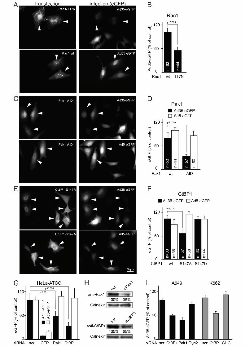
P a k 1 � A I D
A d 5 � e G F P
A d 3 5 � e G F P
C t B P 1 � S 1 4 7 AA d 3 5 � e G F PA d 5 � e G F P
C t B P 1 � S 1 4 7 A
AAd35 �eGFP(% of cont rol) 06 01 2 0
R a c 1 w t T 1 7 NB
C06 01 2 0
w t A I DP a k 1eGFP(% of cont rol)D
06 01 2 0eGFP(% of cont rol)s i R N A s c r G F P P a k 1 C t B P 1GE
06 01 2 0C t B P 1 w t S 1 4 7 A S 1 4 7 DeGFP(% of cont rol)FP a k 1 � A I DC t B P 1 � S 1 4 7 AC t B P 1 � S 1 4 7 A A d 3 5 � e G F PA d 5 � e G F P
A d 3 5 K e G F PA d 5 K e G F PA d 3 5 K e G F P A d 5 K e G F P
Ad35 QeGFP Ad5 QeGFP
R a c 1 � w t A d 3 5 � e G F P n=62 n=44n=53 n=57
n=83 n=57 n=63n=44 n=50
n=56 n=58 n=44
p < 0 . 0 0 5p < 0 . 0 0 5
p < 0 . 0 0 5p < 0 . 0 5 p < 0 . 0 0 5 s i P a k 1s c ra n t i K P a k 1C a l n e x i n s i C tB P 1s c ra n t i K C t B P 1C a l n e x i n
H
R a c 1 � T 1 7 N
3 0 x m1 0 0 % 3 5 %1 0 0 % 6 5 %
i n f e c t i o n ( e G F P )t r a n s f e c t i o n R a c 1P a k 1
C t B P 1
06 01 2 0Ad35 �eGFP(% of cont rol)s i R N A s c r C t B P 1 P a k 1 D y n 2IH e L a � A T C C A 5 4 9 K 5 6 2
s c r C t B P 1 C H C
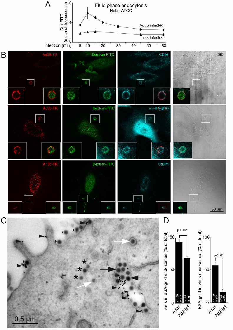
BA d 3 5 � T R D e x t r a n � F I T C
ƒ[i n t e g r i n s
C t B P 1A d 3 5 � T R D e x t r a n � F I T C C D 4 6
D e x t r a n � F I T CA d 3 5 � T RD I C
3 0 � m
A A d 3 5 i n f e c t e dn o t i n f e c t e dD ex -FITC 048 F l u i d p h a s e e n d o c y t o s i si n f e c t i o n ( m i n )( meanoffl uorescence) 5 1 0 2 0 3 0 4 0 6 05 0H e L a a A T C C
C D 1 0 05 001 0 05 00A d 3 5 A d 3 5A d 2 o t s 1 A d 2 o t s 1vi rusi nBSA |gold end osomes(% oft ot al)
BSA |goldi nvi rusend osomes(% oft ot al) p < 0 . 0 1N=149 n=117 N=24 n=38 N=149 n=117 N=24 n=38p < 0 . 0 2 5