Macrophage Polarization in Metabolism and Metabolic Disease
10
81 Macrophage Polarization in Metabolism and Metabolic Disease (Meiliana A) Indones Biomed J. 2013; 5(2): 81-90 DOI: 10.18585/inabj.v5i2.56 REVIEW ARTICLE Macrophage Polarization in Metabolism and Metabolic Disease Anna Meiliana 1,2, , Andi Wijaya 1,2 1 Postgraduate Program in Clinical Biochemistry, Hasanuddin University, Jl. Perintis Kemerdekaan Km.10, Makassar, Indonesia 2 Prodia Clinical Laboratory, Jl. Cisangkuy No.2, Bandung, Indonesia Corresponding author. E-mail: [email protected] B ACKGROUND: Obesity is now recognized as the main cause of the worldwide epidemic of type 2 diabetes. Obesity-associated chronic inflammation is a contributing key factor for type 2 diabetes and cardiovascular disease. Numbers of studies have clearly demonstrated that the immune system and metabolism are highly integrated. CONTENT: Macrophages are an essential component of innate immunity and play a central role in inflammation and host defense. Moreover, these cells have homeostatic functions beyond defense, including tissue remodeling in ontogenesis and orchestration of metabolic functions. Diversity and plasticity are hallmarks of cells of the monocyte-macrophage lineage. In response to interferons (IFNs), toll-like receptor (TLR), or interleukin (IL)-4/IL- 13 signals, macrophages undergo M1 (classical) or M2 (alternative) activation. Progress has now been made in defining the signaling pathways, transcriptional networks, and epigenetic mechanisms underlying M1, M2 or M2-like polarized activation. SUMMARY: In response to various signals, macrophages may undergo classical M1 activation (stimulated by TLR ligands and IFN-γ) or alternative M2 activation (stimulated by IL-4/IL-13); these states mirror the T helper (Th)1–Th2 polarization of T cells. Pathology is frequently associated with dynamic changes in macrophage activation, with classically activated M1 cells implicate in initiating and sustaining inflammation, meanwhile M2 or M2-like activated cells associated with resolution or smoldering chronic inflammation. Identification of the mechanisms and molecules that are associated with macrophage plasticity and polarized activation provides a basis for macrophage- centered diagnostic and therapeutic strategies. L ATAR BELAKANG: Obesitas dikenal sebagai penyebab utama epidemik diabetes tipe 2 di dunia. Inflamasi kronis terkait obesitas adalah faktor kunci yang berkontribusi untuk terjadinya diabetes tipe 2 dan penyakit kardiovaskular. Banyak penelitian telah membuktikan adanya integrasi antara sistim imun dan metabolisme. ISI: Makrofag adalah komponen penting pada imunitas bawaan dan berperan penting dalam inflamasi dan pertahanan inang. Makrofag juga berfungsi untuk menjaga homeostasis selain pertahanan tubuh, termasuk remodeling jaringan pada ontogenesis dan pengaturan fungsi metabolik. Diversitas dan plastisitas merupakan penanda perubahan sel dari monosit menjadi makrofag. Sebagai respon terhadap sinyal interferon (IFN), toll-like receptor (TLR) atau interleukin (IL)-4/IL-13, makrofag menjadi aktif melewati aktivasi M1 (klasik) atau M2 (alternatif). Banyak penelitian dilakukan untuk mengerti lebih banyak mengenai jalur persinyalan, jaringan transkripsi, dan mekanisme epigenetic yang mendasari aktifasi polarisasi M1, M2 atau M2-like. RINGKASAN: Sebagai respons dari berbagai signal makrofag dapat teraktivasi melewati M1 klasik (distimulasi oleh ligan TLR dan IFN-γ) atau M2 sebagai alternatif (distimulasi oleh IL-4/IL-13); hal ini serupa dengan polarisasi T helper (Th)1-Th2 pada sel T. Patologi banyak dikaitkan dengan perubahan dinamis dalam aktivasi makrofag, secara klasik sel dengan M1 teraktivasi menginisiasi dan mempertahankan inflamasi, sedangkan sel dengan M2 atau M2-like teraktivasi berhubungan dengan timbulnya inflamasi kronis. Identifikasi mekanisme dan molekul yang terlibat dengan plastisitas dan aktivasi polarisasi makrofag menjadi dasar untuk strategi diagnostik dan terapi berbasis makrofag. Abstract Abstrak
-
Upload
phungkhuong -
Category
Documents
-
view
221 -
download
4
Transcript of Macrophage Polarization in Metabolism and Metabolic Disease
81
Macrophage Polarization in Metabolism and Metabolic Disease (Meiliana A)Indones Biomed J. 2013; 5(2): 81-90DOI: 10.18585/inabj.v5i2.56
R E V I E W A R T I C L E
Macrophage Polarization in Metabolism and Metabolic Disease
Anna Meiliana1,2,, Andi Wijaya1,2
1Postgraduate Program in Clinical Biochemistry, Hasanuddin University, Jl. Perintis Kemerdekaan Km.10, Makassar, Indonesia2Prodia Clinical Laboratory, Jl. Cisangkuy No.2, Bandung, Indonesia
Corresponding author. E-mail: [email protected]
BACKGROUND: Obesity is now recognized as the main cause of the worldwide epidemic of type 2 diabetes. Obesity-associated chronic inflammation
is a contributing key factor for type 2 diabetes and cardiovascular disease. Numbers of studies have clearly demonstrated that the immune system and metabolism are highly integrated.
CONTENT: Macrophages are an essential component of innate immunity and play a central role in inflammation and host defense. Moreover, these cells have homeostatic functions beyond defense, including tissue remodeling in ontogenesis and orchestration of metabolic functions. Diversity and plasticity are hallmarks of cells of the monocyte-macrophage lineage. In response to interferons (IFNs), toll-like receptor (TLR), or interleukin (IL)-4/IL-13 signals, macrophages undergo M1 (classical) or M2 (alternative) activation. Progress has now been made in defining the signaling pathways, transcriptional networks, and epigenetic mechanisms underlying M1, M2 or M2-like polarized activation.
SUMMARY: In response to various signals, macrophages may undergo classical M1 activation (stimulated by TLR ligands and IFN-γ) or alternative M2 activation (stimulated by IL-4/IL-13); these states mirror the T helper (Th)1–Th2 polarization of T cells. Pathology is frequently associated with dynamic changes in macrophage activation, with classically activated M1 cells implicate in initiating and sustaining inflammation, meanwhile M2 or M2-like activated cells associated with resolution or smoldering chronic inflammation. Identification of the mechanisms and molecules that are associated with macrophage plasticity and polarized activation provides a basis for macrophage-centered diagnostic and therapeutic strategies.
LATAR BELAKANG: Obesitas dikenal sebagai penyebab utama epidemik diabetes tipe 2 di dunia. Inflamasi kronis terkait obesitas adalah faktor
kunci yang berkontribusi untuk terjadinya diabetes tipe 2 dan penyakit kardiovaskular. Banyak penelitian telah membuktikan adanya integrasi antara sistim imun dan metabolisme.
ISI: Makrofag adalah komponen penting pada imunitas bawaan dan berperan penting dalam inflamasi dan pertahanan inang. Makrofag juga berfungsi untuk menjaga homeostasis selain pertahanan tubuh, termasuk remodeling jaringan pada ontogenesis dan pengaturan fungsi metabolik. Diversitas dan plastisitas merupakan penanda perubahan sel dari monosit menjadi makrofag. Sebagai respon terhadap sinyal interferon (IFN), toll-like receptor (TLR) atau interleukin (IL)-4/IL-13, makrofag menjadi aktif melewati aktivasi M1 (klasik) atau M2 (alternatif). Banyak penelitian dilakukan untuk mengerti lebih banyak mengenai jalur persinyalan, jaringan transkripsi, dan mekanisme epigenetic yang mendasari aktifasi polarisasi M1, M2 atau M2-like.
RINGKASAN: Sebagai respons dari berbagai signal makrofag dapat teraktivasi melewati M1 klasik (distimulasi oleh ligan TLR dan IFN-γ) atau M2 sebagai alternatif (distimulasi oleh IL-4/IL-13); hal ini serupa dengan polarisasi T helper (Th)1-Th2 pada sel T. Patologi banyak dikaitkan dengan perubahan dinamis dalam aktivasi makrofag, secara klasik sel dengan M1 teraktivasi menginisiasi dan mempertahankan inflamasi, sedangkan sel dengan M2 atau M2-like teraktivasi berhubungan dengan timbulnya inflamasi kronis. Identifikasi mekanisme dan molekul yang terlibat dengan plastisitas dan aktivasi polarisasi makrofag menjadi dasar untuk strategi diagnostik dan terapi berbasis makrofag.
Abstract Abstrak
82
The Indonesian Biomedical Journal, Vol.5, No.2, August 2013, p.81-90 ISSN: 2085-3297
KEYWORDS: obesity, adipose tissue, inflammation, macrophage polarization
Indones Biomed J. 2013; 5(2): 81-90
Adipose tissue inflammation is a characteristic of obesity. Interactions between metabolic and inflammatory response systems play a significant role in the pathogenesis of a cluster of chronic metabolic diseases including type 2 diabetes, fatty liver disease, and atherosclerosis.(1,2) Macrophage infiltration in adipose tissue has recently been described in both mice and humans, especially in the later stages of obesity.(3,4) It has been suggested that expanding adipocytes or neighboring preadipocytes might produce signals leading to macrophage recruitment.(5) Alternatively, death of adipocytes at late stages of obesity has also been proposed as a mechanism of macrophage infiltration into the adipose tissue.(6) In fact, this is a very attractive hypothesis since much of the presence of macrophages in the adipose tissue occurs in a scattered pattern and is found around the dead adipocytes in obesity. These observations have raised the possibility that macrophages themselves might be a critical regulator of metabolism as a result of their inflammatory capacity, perhaps independent of their stromal counterparts, especially adipocytes. Several studies have identified their contributions to the metabolic regulation of pathways that act in the macrophage.(7,8) Classically activated macrophages (CAMs), termed M1, can be induced in vitro by growing bone marrow–derived hematopoietic cells with granulocyte macrophage colony stimulating factor (GM-CSF). Alternatively, activated macrophages (AAMs), termed M2, can be induced by culturing the bone marrow–derived cells with macrophage colony stimulating factor (M-CSF) and interleukin (IL)-4. M1 macrophages secrete a characteristic signature of pro-inflammatory cytokines, whereas M2 macrophages secrete anti-inflammatory cytokines (for example, IL-10 and IL-1 receptor antagonist (IL-1Ra)).(9) in vivo, it is likely that adipose tissue macrophages (ATMs) span a spectrum from the M1-like pro-inflammatory state to the M2-like non-inflammatory state.(10) Many studies have confirmed that the polarization state of an ATM correlates well with insulin resistance. For example, Fujisaka, et al.(11) showed that the number of M1-like ATMs expressing cluster of differentiation (CD)11c correlated with insulin sensitivity. The phenotype of ATMs is not fixed, and
KATA KUNCI: obesitas, jaringan lemak, inflamasi, polarisasi makrofag
Introductionthey can repolarize from one state to another. For example, evidence suggests that switching from a high fat diet (HFD) to a chow diet(12), or treating obese mice with omega-3 fatty acids(13) or thiazolidinediones (TZDs)(14), can drive conversion of the M1 to the M2 type, which is coincident with increased insulin sensitivity. One can propose that in adipose tissue the recruited M1-like CAMs are responsible for the inflammatory component of insulin resistance, whereas the resident M2-like AAMs function in remodeling and tissue homeostasis.(10)
Inflammation is an adaptive response triggered by noxious stimuli and conditions such as infection and tissue injury.(15,16) Inflammation underlies a wide variety of physiological and pathological processes. Although the pathological aspects of many types of inflammation are well appreciated, their physiological functions are mostly unknown.(17) At a basic level, the acute inflammatory response triggered by infection or tissue injury involves the coordinated delivery of blood components (plasma and leukocytes) to the site of infection or injury.(15,16) This response has been characterized best for microbial infections (particularly bacterial infections), in which it is triggered by receptors of the innate immune system, such as Toll-like receptors (TLRs) and nucleotide-binding oligomerization domain protein (NOD)-like receptors (NLRs).(18) This initial recognition of infection is mediated by tissue-resident macrophages and mast cells, leading to the production of a variety of inflammatory mediators, including chemokines, cytokines, vasoactive amines, eicosanoids and products of proteolytic cascades. A successful acute inflammatory response results in the elimination of the infectious agents followed by a resolution and repair phase, which is mediated mainly by tissue-resident and recruited macrophages.(19) The switch in lipid mediators from pro-inflammatory prostaglandins to lipoxins, which are anti-inflammatory, is crucial for the transition from inflammation to resolution. Lipoxins inhibit the recruitment of neutrophils and, instead, promote
Physiological and Pathological Roles of Inflammation

83
Macrophage Polarization in Metabolism and Metabolic Disease (Meiliana A)Indones Biomed J. 2013; 5(2): 81-90DOI: 10.18585/inabj.v5i2.56
the recruitment of monocytes, which remove dead cells and initiate tissue remodeling. Resolvins and protectins, which constitute another class of lipid mediator, as well as transforming growth factor-β (TGF-β) and growth factors produced by macrophages, also have a crucial role in the resolution of inflammation, including the initiation of tissue repair.(19,20) Homeostatic control mechanisms ensure that internal environmental parameters (such as glucose and oxygen concentrations) are maintained within an acceptable range near a certain set point.(21) Whatever the cause of the inflammatory response, its ‘purpose’ is to remove or sequester the source of the disturbance, to allow the host to adapt to the abnormal conditions and, ultimately, to restore functionality and homeostasis to the tissue. If the abnormal conditions are transient, then a successful acute inflammatory response returns the system to the basal homeostatic set points. If, by contrast, the abnormal conditions are sustained, then an ongoing inflammatory state shifts the system to different set points, as occurs during chronic inflammation. An adaptive change often provides short-term benefits; however, in a chronic phase, it can become maladaptive, as exemplified by a sustained decline in insulin sensitivity of the skeletal muscle or by squamous metaplasia of the respiratory epithelium. More specifically, a transient decrease in insulin sensitivity during acute inflammation would allow the redistribution of glucose from one of its major consumers (for example, skeletal muscle) to leukocytes and other cell types that can have an increased energy demand during infection and tissue repair. However, sustained insulin resistance in skeletal muscle can lead to type 2 diabetes.(17)The mechanisms of systemic chronic inflammatory states in general are poorly understood, but it is clear that they do not seem to fit the classic pattern of transition from acute to chronic inflammation. Thus, in basal conditions, the tissues are maintained in a homeostatic state, in many cases with the help of the tissue-resident macrophages. In noxious conditions, tissues undergo stress and can malfunction. If the changes are considerable, then adaptation to the conditions requires the help of tissue-resident or recruited macrophages and might require small-scale delivery of additional leukocytes and plasma proteins, depending on the extent of the problem. This adaptive response has characteristics that are intermediate between basal and inflammatory states. It could be termed para-inflammation (para- the Greek prefix for near). Indeed, many chronic inflammatory diseases that are not caused by infection or injury seem to be associated with conditions not present during the early evolution of humans, including the continuous availability of high-
calorie nutrients, a low level of physical activity, exposure to toxic compounds, and old age. The human diseases that are associated with these conditions-including obesity, type 2 diabetes, atherosclerosis, asthma and neurodegenerative diseases are all characterized by chronic low-grade inflammation (para-inflammation), which might not have any physiological counterparts. Furthermore, the chronic para-inflammation that persists in these conditions can in turn contribute to further disease progression, in part because of changes in homeostatic set points (such as insulin sensitivity or blood pressure).(17) Thus, tissue stress or malfunction similarly induces an adaptive response, which here is referred to para-inflammation. This response relies mainly on tissue-resident macrophages and is intermediate between the basal homeostatic state and a classic inflammatory response. Para-inflammation is probably responsible for the chronic inflammatory conditions that are associated with modern human diseases.
Approximately 20 years ago, Feingold and Grunfeld observed that administration of the proinflammatory cytokine tumor necrosis factor-α (TNF-α) led to increased serum glucose concentrations, which prompted them to suggest that hyperglycemia may be exacerbated by cytokine overproduction.(22,23) However, the first studies that established the concept of obesity-induced adipose tissue inflammation were conducted by Hotamisligil, et al.(24), who found that TNF-α was elevated in obese rodents and that neutralization of TNF-α ameliorated insulin resistance. A mechanistic link between inflammatory processes and insulin resistance was further established by the fact that the signaling pathways leading to activation of inhibitor of κB kinase-β (IKK-β) and nuclear factor-κB (NF-κB) are stimulated in obesity and insulin resistance.(25,26) Chronic low-grade inflammation induced by obesity leads to activation of other protein kinases such as Jun N-terminal kinases (JNKs), and ablation of JNK in mice fed HFD leads to protection from diet-induced obesity and inflammation.(8,27,28) Activation of inflammatory pathways has since been observed in all classical insulin target tissues, including fat(3,4), liver(29) and muscle(30,31), indicating that inflammation has a broad role in driving the pathogenesis of systemic insulin resistance(10). An important finding that helped elucidate the cause of tissue inflammation was that adipose tissue from obese mice and humans is infiltrated with large
Metabolism and Immune System

84
The Indonesian Biomedical Journal, Vol.5, No.2, August 2013, p.81-90 ISSN: 2085-3297
numbers of macrophages.(3,4) These ATMs can comprise up to 40% of the cells in obese adipose tissue.(4) ATMs and adipose tissue inflammation have been extensively studied, and ATMs have been shown to have a key role in systemic insulin resistance, glucose tolerance and the development of metabolic syndrome and type 2 diabetes.(32) The activation of tissue macrophages triggers the release of cytokines, which can induce insulin resistance in various ways. Among the proinflammatory cytokines, TNF-α is the most studied and has consistently been shown to cause insulin resistance. For example, TNF-α can stimulate serine kinases—including IKK(33), JNK, S6 kinase (S6K)(34-36) and mammalian target of rapamycin (mTOR)(35), which causes serine phosphorylation of insulin receptor substrate-1 (IRS-1), attenuating its ability to propagate downstream insulin signaling. IL-1β, which is generated by inflammasome activation, can also exert pro-inflammatory effects(37). The inflammasome is composed of NOD-like receptor family, pyrin domain containing 3 (NLRP3) and the adaptor protein apoptosis–associated speck-like protein (ASC) containing a caspase recruitment domain. Deletion of NLRP3 or ASC or pharmacological inhibition of caspase-1(38) can protect against HFD-induced insulin resistance and glucose intolerance. Other proinflammatory cytokines such as IL-18, C-X-C motif chemokine-5 (CXCL5), angiopoietin-related protein 2 and lipocalin 2 may contribute to inflammation in the context of metabolic disease(9). In addition to the pro-inflammatory cytokines, anti-inflammatory cytokines are elevated in obesity, including
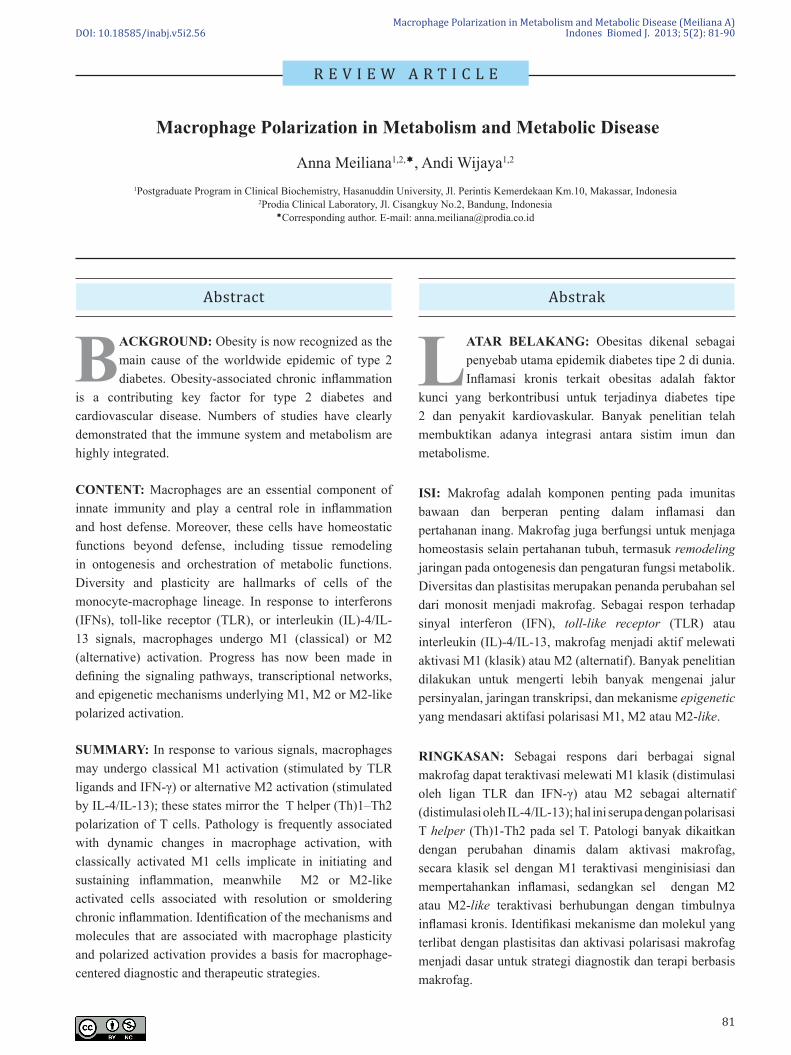
IL-1Ra(39), secreted frizzled-related protein 5 (SFRP5) and IL-10(40). IL-1Ra can block IL-1β signaling, and SFRP5 inhibits the Wnt pathway by sequestering Wnt proteins and preventing them from binding their receptors. IL-10 can inhibit the deleterious effects of pro-inflammatory cytokines on insulin signaling.(41) Furthermore, in vivo administration of IL-10 prevents the development of IL-6– or lipid-induced insulin resistance(42), and muscle-specific transgenic over-expression of IL-10 increases whole-body insulin sensitivity(43,44). In summary, high expression of pro-inflammatory cytokines is associated with insulin resistance, and the homeostatic balance between pro- and anti-inflammatory cytokines defines the profile and magnitude of inflammation and its effects on insulin sensitivity and glucose homeostasis. Systemic insulin resistance in obesity can initiate largely in adipose tissue, and macrophage-mediated tissue inflammation is a core mechanism of this aspect of adipose tissue dysfunction. Adipose tissue can communicate with the liver and muscle through the release of cytokines, adipokines, and fatty acids and possibly through other signals that have yet to be identified, thus causing effects on systemic inflammation and insulin sensitivity. Adipose tissue is often referred to as the master regulator in the development of systemic insulin resistance. The recognition that inflammatory signaling and metabolic signaling are closely linked has given rise to the concept of immunometabolism.(10) Thus far, therapeutic strategies that rely on targeting single cytokines or receptors (for example, TNF-αand IL-1)
Figure 1. Immune cells mediate inflammation in adipose tissue.(10) (Adapted with permission from Nature Publishing Group).

85
Macrophage Polarization in Metabolism and Metabolic Disease (Meiliana A)Indones Biomed J. 2013; 5(2): 81-90DOI: 10.18585/inabj.v5i2.56
Macrophages have long been considered to be important immune effector cells. Élie Metchnikoff, who won the Nobel Prize 100 years ago for his description of phagocytosis, proposed that the key to immunity was to “stimulate the phagocytes”.(50) Since this discovery, immunologists have been occupied by the concept of macrophages as immune effector cells and by the understanding on how these cells participate in host defense. However, by focusing on the immune functions of macrophages, immunologists have ignored their vital homeostatic roles, which are independent of their involvement in immune responses.(51) Macrophages are prodigious phagocytic cells that clear approximately 2×10¹¹ erythrocytes each day; this equates to almost 3 kg of iron and haemoglobin per year that is ‘recycled’ for the host to reuse. This clearance process is a vital metabolic contribution without which the host would not survive. Macrophages are also involved in the removal of cellular debris that is generated during tissue remodeling, and rapidly and efficiently clear cells that have undergone apoptosis. These processes occur independently of immune cell signaling, and the removal of ‘effete’ or apoptotic cells seems to result in little or no production of immune mediators by unstimulated macrophages.(52) Therefore, the primary role of macrophages is not to function as an elite immune effector cell, but instead as a common ‘janitorial’ cell, the main function of which is to clear the interstitial environment of extraneous cellular material.(51) Macrophages detect the endogenous danger signals that are present in the debris of necrotic cells through TLRs(52-54), intracellular pattern-recognition receptors and the IL-1R, most of which signal through the adaptor molecule myeloid differentiation primary-response gene 88 (MyD88)(53). This function makes macrophages one of the primary sensors of danger in the host. The response of macrophages to endogenous danger signals is only one example of the many different stimuli that trigger macrophage activation in tissues. Macrophages have remarkable plasticity that allows them to efficiently respond to environmental signals and change their phenotype, and their physiology can be markedly altered by both innate and adaptive immune responses. Thus, macrophages display
have limited success in humans, suggesting that targeting upstream components rather than single cytokines could provide a more effective therapeutic approach. A more specific approach that selectively targets the proinflammatory M1-like, and not the M2-like, macrophages could provide therapeutic benefits without inhibiting other innate immune functions. A treatment that boosts the number of resident M2-like macrophages could also redirect the ATM balance to a less proinflammatory state and improve inflammation-induced insulin resistance.(10)
Heterogeneity of the macrophage lineage has long been recognized and, in part, is a result of the specialization of tissue macrophages in particular the microenvironments. Circulating monocytes give rise to mature macrophages and are also heterogeneous themselves, although the physiological relevance of this is not yet completely understood.(45) Peripheral-blood monocytes show morphological heterogeneity such as in the variability of size, granularity and nuclear morphology. Monocytes are initially identified by their expression of large amounts of CD14 (which is part of the receptor for lipopolysaccharide). However, the subsequent identification of differential expression of antigenic markers showed that monocytes in human peripheral blood are heterogeneous, and this provided the first clues to the differential physiological activities of monocyte subsets. Differential expression of CD14 and CD16 (also known as Fcγ receptor III (FcγRIII)) allowed monocytes to be divided into two subsets: CD14+CD16–
cells, which are often called classic monocytes, because this phenotype resembles the original description of monocytes; and CD14+ CD16+ cells.(46) It was subsequently shown that the CD14+ CD16+ monocytes expressed higher amounts of major histocompatibility complex (MHC) class II molecules and CD32 (also known as FcγRII), and it was suggested that these cells resemble mature tissue macrophages.(47) Tissue macrophages have a broad role in the maintenance of tissue homeostasis through the clearance of senescent cells and the remodeling and repair of tissues after inflammation.(48,49) They are generally considered to be derived from circulating monocytes and show a high degree of heterogeneity. The heterogeneity reflects the specialization of function that is adopted by macrophages in different anatomical locations. Monocytes migrate from the blood into the tissue to replenish long-lived tissue-specific macrophages of the bone (osteoclasts), alveoli,
Macrophage Heterogeneity
central nervous system (microglial cells), connective tissue (histiocytes), gastrointestinal tract, liver (Kupffer cells), spleen and peritoneum.(45) In addition to macrophage heterogeneity in different organs, macrophage heterogeneity can be observed in a single organ.(45)
Macrophage Plasticity andPolarization
86
The Indonesian Biomedical Journal, Vol.5, No.2, August 2013, p.81-90 ISSN: 2085-3297
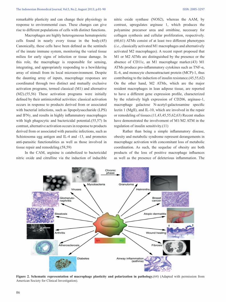
nitric oxide synthase (NOS2), whereas the AAM, by contrast, upregulates arginase 1, which produces the polyamine precursor urea and ornithine, necessary for collagen synthesis and cellular proliferation, respectively.(60,61) ATMs consist of at least two different phenotypes (i.e., classically activated M1 macrophages and alternatively activated M2 macrophages). A recent report proposed that M1 or M2 ATMs are distinguished by the presence or the absence of CD11c, an M1 macrophage marker.(43) M1 ATMs produce pro-inflammatory cytokines such as TNF-α, IL-6, and monocyte chemoattractant protein (MCP)-1, thus contributing to the induction of insulin resistance.(45,55,62) On the other hand, M2 ATMs, which are the major resident macrophages in lean adipose tissue, are reported to have a different gene expression profile, characterized by the relatively high expression of CD206, arginase-1, macrophage galactose N-acetyl-galactosamine specific lectin 1 (MglI), and IL-10, which are involved in the repair or remodeling of tissues.(11,43,45,55,62,63) Recent studies have demonstrated the involvement of M1/M2 ATM in the regulation of insulin sensitivity.(11) Rather than being a simple inflammatory disease, obesity and metabolic syndrome represent derangements in macrophage activation with concomitant loss of metabolic coordination. As such, the sequelae of obesity are both products of the loss of positive macrophage influences as well as the presence of deleterious inflammation. The
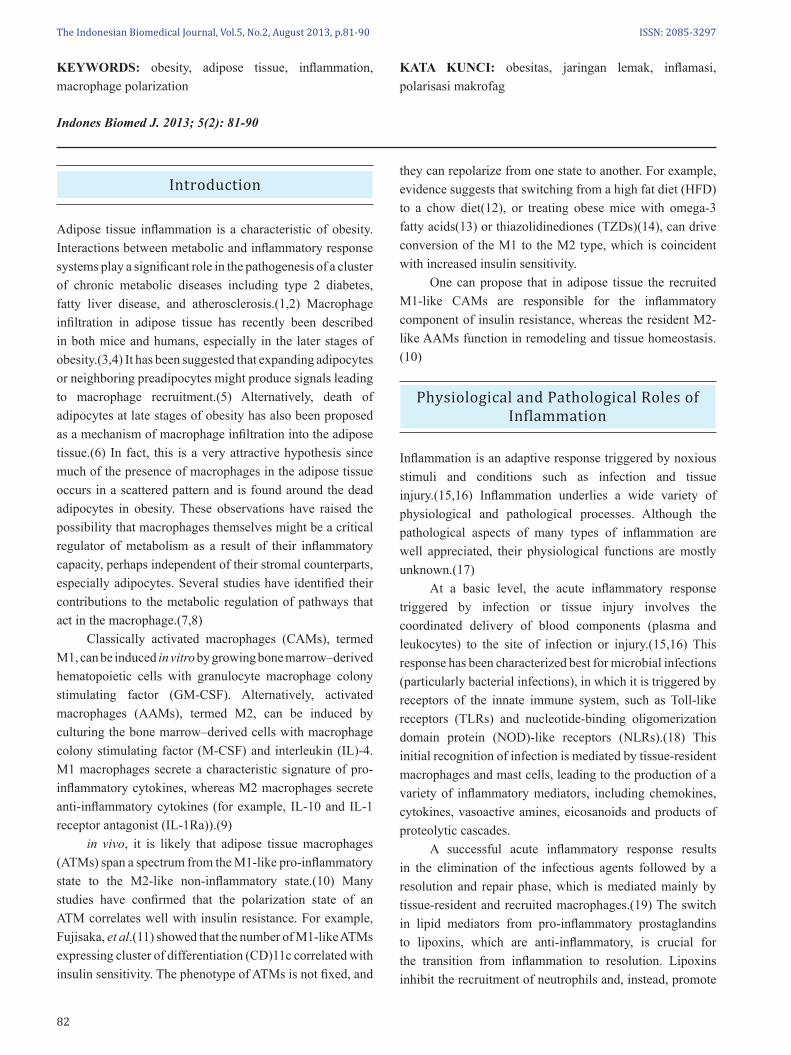
Figure 2. Schematic representation of macrophage plasticity and polarization in pathology.(64) (Adapted with permission from American Society for Clinical Investigation).
remarkable plasticity and can change their physiology in response to environmental cues. These changes can give rise to different populations of cells with distinct functions. Macrophages are highly heterogeneous hematopoietic cells found in nearly every tissue in the body.(45) Canonically, these cells have been defined as the sentinels of the innate immune system, monitoring the varied tissue milieu for early signs of infection or tissue damage. In this role, the macrophage is responsible for sensing, integrating, and appropriately responding to a bewildering array of stimuli from its local microenvironment. Despite the daunting array of inputs, macrophage responses are coordinated through two distinct and mutually exclusive activation programs, termed classical (M1) and alternative (M2).(55,56) These activation programs were initially defined by their antimicrobial activities: classical activation occurs in response to products derived from or associated with bacterial infections, such as lipopolysaccharide (LPS) and IFNγ, and results in highly inflammatory macrophages with high phagocytic and bactericidal potential.(55,57) In contrast, alternative activation occurs in response to products derived from or associated with parasitic infections, such as Schistosoma egg antigen and IL-4 and -13, and promotes anti-parasitic functionalities as well as those involved in tissue repair and remodeling.(58,59) In the CAM, arginine is catabolized to bactericidal nitric oxide and citrulline via the induction of inducible

87
Macrophage Polarization in Metabolism and Metabolic Disease (Meiliana A)Indones Biomed J. 2013; 5(2): 81-90DOI: 10.18585/inabj.v5i2.56
The rise in worldwide obesity has resulted in an explosion of obesity-related health problems, including insulin resistance, type 2 diabetes, coronary artery disease, fatty liver disease, and some cancers and degenerative diseases.(65-67) Since behavioral and dietary approaches have been ineffective in combating obesity(68), a greater emphasis is being placed on understanding the molecular links between obesity and chronic metabolic diseases. In this context, chronic, low-grade inflammation, primarily mediated by innate and adaptive immune cells, has emerged as a key pathogenic link between obesity and its metabolic sequelae (1,32,69-72). Obesity-associated insulin resistance, diabetes, and metabolic syndrome are sustained by chronic subclinical inflammation.(1) ATMs are a major component of adipose tissue and have an important role in the obesity-associated pathology.(64) ATMs from obese mice and humans are polarized toward an M1 phenotype, with upregulation of TNF and iNOS. In contrast, “lean” ATMs express high levels of M2 genes, including IL-10, Ym1, and Arginase 1.(43,73) Weight loss is associated with a back shift to an M2-like phenotype. However, evidence indicated that the ATM population in obese patients is diverse, with a minor F4/80+
CD11b+ CD11c–, IL-10+, M2-like population co-existing with M1 cells.(73) Moreover, analysis of CD11c+ ATMs from obese mice revealed a mixed profile of several M1-M2 gene transcripts.(74) Weight loss is associated with an increase in F4/80+ CD11c– CD301+ M2-like macrophages, which promote lipolysis.(75) Progress has been made in defining the molecular pathways that account for polarization of ATMs in obesity.(59,76-79) Mice with macrophage-selective genetic inactivation of (PPAR)γ, PPARδ, and Klf4 show inhibition of obesity-induced insulin resistance.(59,73,76-78) Molecular determinants of M1 versus M2 polarization include members of the PPAR, Klf, IRF, STAT, NF-κB, and HIF families. Regulation of skewing also involves epigenetic modifications with involvement of histone methylation and acetylation. miRNAs have emerged as regulators of phagocyte activation and function(80,81), but their role in macrophage polarization needs to be further defined.
Nguyen, et al. have recently demonstrated that adaptation to lower temperatures (thermogenesis) is associated with polarization of brown adipose tissue (BAT) and white adipose tissue (WAT) macrophages to the alternative state (M2), with increased expression of thermogenic genes (PPARγc1 and uncoupling protein 1 (Ucp1)).(82) In response to cold, IL-4–driven M2 macrophages release noradrenaline in BAT and WAT, which coordinates fatty acid mobilization and energy expenditure.(82) M2-like cells in non-obese individuals are likely involved in maintaining adipose tissue homeostasis, preventing inflammation, and promoting insulin sensitivity. The role of macrophages in obesity and associated disorders underlines a homeostatic function of macrophages in metabolism as cells capable of reorienting their own metabolic activity and as orchestrators of general metabolism.(64) The wealth of literature on macrophages and other leukocytes in adipose tissue clearly defines functional roles for these cells in adipose tissue biology and establishes them as bonafide tissue constituents rather than immunologic transients. The physiologically normal set points in fat tissue can be shifted by chronic over-nutrition; the pro-inflammatory obese adipose tissue itself, once established, demonstrates temporal stability and resistance to change. As in lean adipose tissue, any perturbation of leukocyte set points, whether numeric or phenotypic, disrupts the tissue’s new functional parameters.(83) Therapeutic macrophage targeting is in its infancy. Selected clinically approved therapeutic strategies, such as use of PPARγ inhibitors, statins, zolendronic acid, and preventive activities such as weight loss may have an impact on the functional status of macrophages; however, the extent to which their effect on macrophages explains their clinical efficacy remains to be defined.(64) The identification of mechanisms and molecules associated with macrophage plasticity and polarized activation provides a basis for macrophage-centered diagnostic and therapeutic strategies.
Metabolic function requires a previously unsuspected level of cooperation between the cells that make up each internal organ and that organ’s resident macrophages. Disruption in the tissue set point results in significant functional consequences for both the tissue and the organism as a whole. If nutrient intake and expenditure are mismatched over a long period of time, however, a new set point is
Macrophage Polarization and Metabolic Disease
Conclusion
therapeutic implications of this conclusion are profound because they suggest that pharmacologic targeting of macrophage activation, rather than purely inflammation, might be efficacious in treating this global epidemic.(59)

88
The Indonesian Biomedical Journal, Vol.5, No.2, August 2013, p.81-90 ISSN: 2085-3297
1. Hotamisligil GS. Inflammation and metabolic disorders. Nature. 2006; 444: 860–7.
2. Furuhashi M, Fucho R, Görgun CZ, Tuncman G, Cao H, Hotamisligil GS. Adipocyte / macrophage fatty acid – binding proteins contribute to metabolic deterioration through actions in both macrophages and adipocytes in mice. J Clin Invest. 2008; 118: 2640–50.
3. Xu H, Barnes GT, Yang Q, Tan G, Yang D, Chou CJ, et al. Chronic inflammation in fat plays a crucial role in the development of obesity-related insulin resistance. J Clin Invest. 2003; 112: 1821-30.
4. Weisberg SP, McCann D, Desai M, Rosenbaum M, Leibel RL, Ferrante AW Jr. Obesity is associated with macrophage accumulation in adipose tissue. J Clin Invest. 2003; 112: 1796–808.
5. Wellen KE, Hotamisligil GS. Inflammation, stress, and diabetes. J Clin Invest. 2005; 115: 1111–9.
6. Cinti S, Mitchell G, Barbatelli G, Murano I, Ceresi E, Faloia E, et al. Adipocyte death defines macrophage localization and function in adipose tissue of obese mice and humans. J Lipid Res. 2005; 46: 2347–55.
7. Lesniewski LA, Hosch SE, Neels JG, de Luca C, Pashmforoush M, Lumeng CN, et al. Bone marrow-specific Cap gene deletion protects against high-fat diet- induced insulin resistance. Nat Med. 2007; 13: 455–62.
8. Solinas G, Vilcu C, Neels JG, Bandyopadhyay GK, Luo JL, Naugler W, et al. JNK1 in hematopoietically derived cells contributes to diet-induced inflammation and insulin resistance without affecting obesity. Cell Metab. 2007; 6: 386–97.
9. Ouchi N, Parker JL, Lugus JJ, Walsh K. Adipokines in inflammation and metabolic disease. Nat Rev Immunol. 2011; 11: 85–97.
10. Osborn O, Olefsky JM. The cellular and signaling networks linking the immune system and metabolism in disease. Nat Med. 2012; 18: 363–74.
11. Fujisaka S, Usui I, Bukhari A, Ikutani M, Oya T, Kanatani Y, et al. Regulatory mechanisms for adipose tissue M1 and M2 macrophages in diet-induced obese mice. Diabetes. 2009; 58: 2574–82.
12. Li P, Lu M, Nguyen MT, Bae EJ, Chapman J, Feng D, et al. Functional heterogeneity of CD11c-positive adipose tissue macrophages in diet-induced obese mice. J Biol Chem. 2010; 285: 15333-45.
13. Oh DY, Talukdar S, Bae EJ, Imamura T, Morinaga H, Fan W, et al. GPR120 is an omega-3 fatty acid receptor mediating potent anti- inflammatory and insulin-sensitizing effects. Cell. 2010; 142: 687–98.
14. Bouhlel MA, Derudas B, Rigamonti E, Dièvart R, Brozek J, Haulon S, et al. PPARγ activation primes human monocytes into alternative M2 macrophages with anti-inflammatory properties. Cell Metab. 2007; 6: 137–43.
15. Majno G, Joris I. Cells, Tissues and Disease. Oxford Univ. Press. 2004. 16. Kumar V, Cotran RS, Robbins SL. Robbins Basic Pathology. Saunders,
2003.
17. Medzhitov R. Origin and physiological roles of inflammation. Nature. 2008; 454: 428-35.
18. Barton GMA. A calculated response: control of inflammation by the innate immune system. J Clin Invest. 2008; 118: 413–20.
19. Serhan CN, Savill J. Resolution of inflammation: the beginning programs the end. Nat Immunol. 2005; 6: 1191–7.
20. Serhan CN. Resolution phase of inflammation: novel endogenous anti-inflammatory and proresolving lipid mediators and pathways. Annu Rev Immunol. 2007; 25: 101–37.
21. Cannon W. Organization for physiological homeostasis. Physiol Rev. 1929; 9: 399–431.
22. Feingold KR, Soued M, Staprans I, Gavin LA, Donahue ME, Huang BJ, et al. Effect of tumor necrosis factor (TNF) on lipid metabolism in the diabetic rat. Evidence that inhibition of adipose tissue lipoprotein lipase activity is not required for TNF-induced hyperlipidemia. J Clin Invest. 1989; 83: 1116–21.
23. Grunfeld C, Feingold KR. The metabolic effects of tumor necrosis factor and other cytokines. Biotherapy. 1991; 3: 143–58.
24. Hotamisligil GS, Shargill NS, Spiegelman BM. Adipose expression of tumor necrosis factor-alpha: direct role in obesity-linked insulin resistance. Science. 1993; 259: 87–91.
25. Yin MJ, Yamamoto Y, Gaynor RB. The anti-inflammatory agents aspirin and salicylate inhibit the activity of I(kappa)B kinase-beta. Nature. 1998; 396: 77–80.
26. Yuan M, Konstantopoulos N, Lee J, Hansen L, Li ZW, Karin M, et al. Reversal of obesity- and diet-induced insulin resistance with salicylates or targeted disruption of Ikkß. Science. 2001; 293: 1673–7.
27. Hirosumi J, Tuncman G, Chang L, Görgun CZ, Uysal KT, Maeda K, et al. A central role for JNK in obesity and insulin resistance. Nature. 2002; 420: 333–6.
28. Tuncman G, Hirosumi J, Solinas G, Chang L, Karin M, Hotamisligil GS. Functional in vivo interactions between JNK1 and JNK2 isoforms in obesity and insulin resistance. Proc Natl Acad Sci USA. 2006; 103: 10741–6.
29. Cai D, Yuan M, Frantz DF, Melendez PA, Hansen L, Lee J, et al. Local and systemic insulin resistance resulting from hepatic activation of IKK-beta and NF-kappaB. Nat Med. 2005; 11: 183–90.
30. Itani SI, Ruderman NB, Schmieder F, Boden G. Lipid-induced insulin resistance in human muscle is associated with changes in diacylglycerol, protein kinase C and IkappaB-alpha. Diabetes. 2002; 51: 2005–11.
31. Bandyopadhyay GK, Yu JG, Ofrecio J, Olefsky JM. Increased p85/55/50 expression and decreased phosphotidylinositol 3-kinase activity in insulin-resistant human skeletal muscle. Diabetes. 2005; 54: 2351–9.
32. Olefsky JM, Glass CK. Macrophages, inflammation and insulin resistance. Annu Rev Physiol. 2010; 72: 219–46.
33. Gao Z, Hwang D, Bataille F, Lefevre M, York D, Quon MJ, et al. Serine phosphorylation of insulin receptor substrate 1 by inhibitor kappaB kinase complex. J Biol Chem. 2002; 277: 48115–21.
34. Gao Z, Zuberi A, Quon MJ, Dong Z, Ye J. Aspirin inhibits serine phosphorylation of insulin receptor substrate 1 in tumor necrosis factor-treated cells through targeting multiple serine kinases. J Biol Chem. 2003; 278: 24944–50.
35. Ozes ON, Akca H, Mayo LD, Gustin JA, Maehama T, Dixon JE, et al. A phosphatidylinositol 3-kinase/Akt/mTOR pathway mediates and PTEN antagonizes tumor necrosis factor inhibition of insulin signaling through insulin receptor substrate-1. Proc Natl Acad Sci USA. 2001; 98: 4640–5.
36. Lee DF, Kuo HP, Chen CT, Hsu JM, Chou CK, Wei Y, et al. IKKbeta suppression of TSC1 links inflammation and tumor angiogenesis via the mTOR pathway. Cell. 2007; 130: 440–55.
References
established in WAT, characterized by a chronic low-grade inflammation (mediated by the classically activated M1 macrophage population) and insulin resistance. The therapeutic implications of an improved understanding are profound because of pharmacologic targeting of macrophage activation, rather than simply of inflammation, might be efficacious in treating the global obesity epidemic.

89
Macrophage Polarization in Metabolism and Metabolic Disease (Meiliana A)Indones Biomed J. 2013; 5(2): 81-90DOI: 10.18585/inabj.v5i2.56
37. Martinon F, Burns K, Tschopp J. The inflammasome: a molecular platform triggering activation of inflammatory caspases and processing of proIL-beta. Mol Cell. 2002; 10: 417–26.
38. Stienstra R, Joosten LA, Koenen T, van Tits B, van Diepen JA, van den Berg SA, et al. The inflammasome-mediated caspase-1 activation controls adipocyte differentiation and insulin sensitivity. Cell Metab. 2010; 12: 593–605.
39. Juge-Aubry CE, Somm E, Giusti V, Pernin A, Chicheportiche R, Verdumo C, et al. Adipose tissue is a major source of interleukin-1 receptor antagonist: upregulation in obesity and inflammation. Diabetes. 2003; 52: 1104–10.
40. Ouchi N, Higuchi A, Ohashi K, Oshima Y, Gokce N, Shibata R, et al. Sfrp5 is an anti-inflammatory adipokine that modulates metabolic dysfunction in obesity. Science. 2010; 329: 454–7.
41. Schottelius AJ, Mayo MW, Sartor RB, Baldwin AS Jr. Interleukin-10 signaling blocks inhibitor of kappaB kinase activity and nuclear factor kappaB DNA binding. J Biol Chem. 1999; 274: 31868–74.
42. Kim HJ, Higashimori T, Park SY, Choi H, Dong J, Kim YJ, et al. Differential effects of interleukin-6 and -10 on skeletal muscle and liver insulin action in vivo. Diabetes. 2004; 53: 1060–7.
43. Lumeng CN, Bodzin JL, Saltiel AR. Obesity induces a phenotypic switch in adipose tissue macrophage polarization. J Clin Invest. 2007; 117: 175–84.
44. Hong EG, Ko HJ, Cho YR, Kim HJ, Ma Z, Yu TY, et al. Interleukin-10 prevents diet-induced insulin resistance by attenuating macrophage and cytokine response in skeletal muscle. Diabetes. 2009; 58: 2525–35.
45. Gordon S, Taylor PR. Monocyte and macrophage heterogeneity. Nat Rev Immunol. 2005; 5: 953-64.
46. Passlick B, Flieger D, Ziegler-Heitbrock HW. Identification and characterization of a novel monocyte subpopulation in human peripheral blood. Blood. 1989; 74: 2527–34.
47. Ziegler-Heitbrock HW, Fingerle G, Ströbel M, Schraut W, Stelter F, Schutt C, et al. The novel subset of CD14+/ CD16+ blood monocytes exhibits features of tissue macrophages. Eur J Immunol. 1993; 23: 2053–8.
48. Gordon S. Biology of the macrophage. J Cell Sci Suppl. 1986; 4: 267–86.
49. Gordon S. The role of the macrophage in immune regulation. Res Immunol. 1998; 149: 685–8.
50. Nathan C. Metchnikoff’s legacy in 2008. Nat Immunol. 2008; 9: 695–8.
51. Mosser DM, Edwards JP. Exploring the full spectrum of macrophage activation. Nat Rev Immunol. 2008; 8: 958-69.
52. Kono H, Rock KL. How dying cells alert the immune system to danger. Nat Rev Immunol. 2008; 8: 279–89.
53. Chen CJ, Kono H, Golenbock D, Reed G, Akira S, Rock KL. Identification of a key pathway required for the sterile inflammatory response triggered by dying cells. Nature Med. 2007; 13: 851–6.
54. Park JS, Svetkauskaite D, He Q, Kim JY, Strassheim D, Ishizaka A, et al. Involvement of toll-like receptors 2 and 4 in cellular activation by high mobility group box 1 protein. J Biol Chem. 2004; 279: 7370–7.
55. Gordon S. Alternative activation of macrophages. Nat Rev Immunol. 2003; 3: 23–35.
56. Martinez FO, Sica A, Mantovani A, Locati M. Macrophage activation and polarization. Front Biosci. 2008; 13: 453–61.
57. Goerdt S, Politz O, Schledzewski K, Birk R, Gratchev A, Guillot P, et al. Alternative versus classical activation of macrophages. Pathobiology. 1999; 67: 222–6.
58. Martinez FO, Helming L, Gordon S. Alternative activation of macrophages: an immunologic functional perspective. Annu Rev Immunol. 2009; 27: 451–83.
59. Odegaard JI, Chawla A. Alternative macrophage activation and metabolism. Annu Rev Pathol. 2011; 6: 275-97.
60. Modolell M, Corraliza IM, Link F, Soler G, Eichmann K. Reciprocal regulation of the nitric oxide synthase/arginase balance in mouse bone marrow-derived macrophages by TH1 and TH2 cytokines. Eur J Immunol. 1995; 25: 1101–4.
61. Munder M, Eichmann K, Morán JM, Centeno F, Soler G, Modolell M. Th1/Th2-regulated expression of arginase isoforms in murine macrophages and dendritic cells. J Immunol. 1999; 163: 3771–7.
62. Mantovani A, Sica A, Sozzani S, Allavena P, Vecchi A, Locati M. The chemokine system in diverse forms of macrophage activation and polarization. Trends Immunol. 2004; 25: 677–86.
63. Lumeng CN, DelProposto JB, Westcott DJ, Saltiel AR. Phenotypic switching of adipose tissue macrophages with obesity is generated by spatiotemporal differences in macrophage subtypes. Diabetes. 2008; 57: 3239–46.
64. Sica A, Mantovani A. Macrophage plasticity and polarization: in vivo veritas. J Clin Invest. 2012; 122: 787-95.
65. Berrington de Gonzalez A, Hartge P, Cerhan JR, Flint AJ, Hannan L, MacInnis RJ, et al. Body-mass index and mortality among 1.46 million white adults. N Engl J Med. 2010; 363: 2211–9.
66. Flegal KM, Graubard BI, Williamson DF, Gail MH. Cause-specific excess deaths associated with underweight, overweight, and obesity. JAMA. 2007; 298: 2028–37.
67. Zheng W, McLerran DF, Rolland B, Zhang X, Inoue M, Matsuo K, et al. Association between body-mass index and risk of death in more than 1 million Asians. N Engl J Med. 2011; 364: 719–29.
68. Leibel RL. Molecular physiology of weight regulation in mice and humans. Int J Obes (Lond). 2008; 32(Suppl 7): S98–108.
69. Shoelson SE, Lee J, Goldfine AB. Inflammation and insulin resistance. J Clin Invest. 2006; 116: 1793–801.
70. Odegaard JI, Chawla A. Mechanisms of macrophage activation in obesity-induced insulin resistance. Nat Clin Pract Endocrinol Metab. 2008; 4: 619–26.
71. Lumeng CN, Saltiel AR. Inflammatory links between obesity and metabolic disease. J Clin Invest. 2011; 121: 2111–7.
72. Ferrante AW Jr. Obesity-induced inflammation: a metabolic dialogue in the language of inflammation. J Intern Med. 2007; 262: 408–14.
73. Hevener AL, Olefsky JM, Reichart D, Nguyen MT, Bandyopadyhay G, Leung HY, et al. Macrophage PPAR gamma is required for normal skeletal muscle and hepatic insulin sensitivity and full antidiabetic effects of thiazolidinediones. J Clin Invest. 2007; 117: 1658–69.
74. Shaul ME, Bennett G, Strissel KJ, Greenberg AS, Obin MS. Dynamic, M2-like remodeling phenotypes of CD11c+ adipose tissue macrophages during high-fat diet-induced obesity in mice. Diabetes. 2010; 59: 1171–81.
75. Kosteli A, Sugaru E, Haemmerle G, Martin JF, Lei J, Zechner R, et al. Weight loss and lipolysis promote a dynamic immune response in murine adipose tissue. J Clin Invest. 2010; 120: 3466–79.
76. Odegaard JI, Ricardo-Gonzalez RR, Goforth MH, Morel CR, Subramanian V, Mukundan L, et al. Macrophage-specific PPAR-gamma controls alternative activation and improves insulin resistance. Nature. 2007; 447: 1116–20.
77. Odegaard JI, Ricardo-Gonzalez RR, Red Eagle A, Vats D, Morel CR, Goforth M, et al. Alternative M2 activation of Kupffer cells by PPARdelta ameliorates obesity-induced insulin resistance. Cell Metab. 2008; 7: 496–507.
78. Liao X, Sharma N, Kapadia F, Zhou G, Lu Y, Hong H, et al. Kruppel-like factor 4 regulates macrophage polarization. J Clin Invest. 2011; 121: 2736–49.
79. Hu X, Chung AY, Wu I, Foldi J, Chen J, Ji JD, et al. Integrated regulation of Toll-like receptor responses by Notch and interferon-gamma

90
The Indonesian Biomedical Journal, Vol.5, No.2, August 2013, p.81-90 ISSN: 2085-3297
pathways. Immunity. 2008; 29: 691–703.80. Quinn SR, O’Neill LA. A trio of microRNAs that control Toll-like
receptor signalling. Int Immunol. 2011; 23: 421–5.81. Bazzoni F, Rossato M, Fabbri M, Gaudiosi D, Mirolo M, Mori L, et al.
Induction and regulatory function of miR-9 in human monocytes and neutrophils exposed to proinflammatory signals. Proc Natl Acad Sci USA. 2010; 106: 5282–7.
82. Nguyen KD, Qiu Y, Cui X, Goh YP, Mwangi J, David T, et al. Alternatively activated macrophages produce catecholamines to sustain adaptive thermogenesis. Nature. 2011; 480: 104–8.
83. Chawla A, Nguyen KD, Goh YP. Macrophage-mediated inflammation in metabolic disease. Nat Rev Immunol. 2011; 11: 738-49.